Article

Mycotoxins and Other Secondary Metabolites Are Produced by Gnomoniopsis smithogilvyi When Confronted with Biological and Chemical Control Agents

1
Higiene y Seguridad Alimentaria, Instituto Universitario de Investigación de Carne y Productos Cárnicos, Facultad de Veterinaria, Universidad de Extremadura, Avda. de las Ciencias s/n, 10003 Cáceres, Spain
2
Centro de Investigação de Montanha (CIMO), Instituto Politécnico de Bragança, Campus de Santa Apolónia, 5300-253 Bragança, Portugal
3
Laboratório Associado para a Sustentabilidade e Tecnologia em Regiões de Montanha (SusTEC), Instituto Politécnico de Bragança, Campus de Santa Apolónia, 5300-253 Bragança, Portugal
4
Campus de Pato Branco, Universidade Tecnológica Federal do Paraná (UTFPR), Pato Branco 85503-390, Brazil
5
Department of Agrobiotechnology (IFA-Tulln), Institute of Bioanalytics and Agro-Metabolomics, University of Natural Resources and Life Sciences, 3430 Vienna, Austria
6
Centre for the Research and Technology of Agro-Environmental and Biological Sciences (CITAB), University of Trás-os-Montes e Alto Douro (UTAD), Quinta de Prados, 5000-801 Vila Real, Portugal
7
Laboratório Associado Instituto para a Inovação, Capacitação e Sustentabilidade da Produção Agroalimentar (Inov4Agro), University of Trás-os-Montes e Alto Douro (UTAD), Quinta de Prados, 5000-801 Vila Real, Portugal
*
Author to whom correspondence should be addressed.
Agriculture 2023, 13(6), 1166; https://doi.org/10.3390/agriculture13061166
Submission received: 7 May 2023 / Revised: 28 May 2023 / Accepted: 29 May 2023 / Published: 30 May 2023

Abstract

:
Gnomoniopsis smithogilvyi (Gs) is a relevant pathogen of chestnut since it provokes significant losses worldwide. The aim of this study was to screen the effect of a new biocontrol agent (BCA) against Gs isolated from chestnut (CIMO-BCA1) on the mould’s growth as well as on the production of secondary metabolites. The chemical fungicide Horizon® (tebuconazole; HOR) and the commercial biofungicide Serenade® ASO (Bacillus amyloliquefaciens QST 713; ASO) were also tested. Three concentrations of each antifungal (HOR, ASO, and CIMO-BCA1) were faced with Gs in the growth study in a chestnut-based medium. The intermediate concentrations were used for the analyses of metabolites by LC-MS/MS. CIMO-BCA1 was also identified as B. amyloliquefaciens. All agents reduced the mould’s growth, and the CIMO-BCA1 treatment with an intermediate concentration was the most effective. The metabolite analysis revealed, for the first time, the production of two mycotoxins by Gs, including 3-nitropropionic acid and diplodiatoxin. Additionally, HOR stimulated the production of diplodiatoxin. In conclusion, Gs could present a health risk for consumers. B. amyloliquefaciens strains effectively decreased the mould’s growth, but they must be applied at effective concentrations or in combination with other strategies to completely reduce the hazard.

1. Introduction

Portugal is the third largest producer of sweet chestnut in Europe, with an annual production of around 37 thousand tons and an orchard area of 50 thousand hectares in 2021 [1]. Trás-os-Montes, in the northeast of the country, was the first Portuguese chestnut producer region and produces more than 80% of the national production. The chestnuts produced in this region are of high quality and have been recognized by the European Union with the Protected Denomination of Origin “Castanha da Terra Fria” (Castanea sativa Mill.). Chestnut is a fruit with major significance in the Portuguese import/export balance [2].
Chestnut fruit is a seasonal product, commercialised as fresh or processed ready-to-use products. The storage of chestnuts is a challenge due to their nutritional richness and high water content which create conditions that are conducive to fungal infections and insect infestations [3,4] and result in major losses in fruit quality. These conditions make it necessary for chestnuts to be controlled at both pre- and post-harvest stages. A major post-harvest problem associated with these fruits is rot, which can be caused by several different fungal agents. Gnomoniopsis spp. have been described as pathogens in chestnuts worldwide. While Gnomonipsis daii and Gnomoniopsis chinensis were demonstrated to be emerging pathogens of Castanea mollissima tissues and fruits in China [5,6], Gnomoniopsis smithogilvyi (Gs; syn. Gnomoniopsis castaneae Tamietti) has led to significant losses in various chestnut species—including C. sativa, C. mollissima, Castanea dentata, Castanea crenata, and their hybrids—in Europe, Asia, North America, and Oceania [7]. Gs was first reported as a chestnut fruit pathogen in 2005 in Oceania and Asia and is currently widespread in Europe and North America [7]. Gs has been reported by Portuguese chestnut producers to cause around 89% of nut rot in Portugal and is currently considered the main nut rot agent of chestnut fruit across Europe.
Several isolates of Gs obtained from these chestnuts were confirmed as the causal agent of chestnut brown rot and proved to be highly aggressive in chestnuts and well adapted to a wide range of temperatures, potentially resulting in extreme losses in the context of climatic changes [8].
Currently, there are no field treatments in Portugal being used to control fruit rots, and chestnut orchards are generally conducted under biological production systems. In contrast, several post-harvest methods are used for chestnut insect and fungal control, such as sterilising hydrothermal baths (48–50 °C) for 45 min, followed by cooling and drying [9], and water curing (‘curatura’) consisting of treatments in cold water (14–18 °C) for 7–9 days with or without the addition of biocontrol agents or their metabolites [10]. Rodrigues et al. reported reduced contamination in chestnuts submitted to the traditional industrial hydrothermal bath (48 °C, 45 min), but the process was still not fully efficient against fungi [9]. For the storage of chestnuts, researchers tested the use of hot air assisted radio frequencies [11], electron-beam radiation [12], and ozonation [13], among others, with limited success regarding reducing contamination. Considering the enormous effect of Gs in chestnut production and its fast and aggressive geographic dissemination throughout European producing countries, there is a major demand to find mitigation strategies to retain the spread of the disease and the intensification of its consequences to the chestnut production chain.
Chemical control in chestnuts is not a current practice, and no fungicides are regulated for Gs control, although some studies have suggested the potential use of pyraclostrobin, difenoconazole, and phosphonate salts [14,15]. However, chemical fungicides generally depend on very limited and directed modes of action and are associated with resistance acquisition by pathogens. As such, biological control agents (BCAs) are currently considered effective and environmentally friendly alternatives to chemicals for the control of several plant diseases [16]. Bacillus spp.—in particular, Bacillus amyloliquefaciens and the closely related species Bacillus velezensis, Bacillus siamensis, and Bacillus methylotrophicus (also called the “operational unit” B. amyloliquefaciens due to their close phylogenomic relationship) [17]—are examples of bacteria that have been successfully used as BCAs against several plant diseases and have been useful not only in greenhouse and field conditions (e.g., [18,19,20]) but also at the post-harvest stage for fruit diseases [21,22]. The commercial product Serenade ASO® (Bayer, Leverkusen, Germany), derived from B. amyloliquefaciens (formerly classified as B. subtilis) strain QST 713 [19,23], has been approved as a spraying biofungicide in strawberries and grapes against Botrytis cinerea, although it has been widely tested and used in many other crops to protect against plant pathogens [18,19,20,21,22].
Additionally, although several studies have reported the morphological, physiological, and molecular aspects of Gs isolates from different geographical origins and possible strategies to eliminate it, there is still a lack of knowledge about the metabolic profile of the fungus. Vinale et al. [24] reported two secondary metabolites—abscisic acid and 1′,4′-diol of abscisic acid—produced by a G. castaneae strain isolated in the Campania region (Italy) from chestnut galls infested with Dryocosmus kuriphilus and demonstrated the phytotoxic activity of the isolated metabolites on chestnut leaves. To our best knowledge, no other secondary metabolites have been reported from Gs or other related species associated with the fruit. Similar to other filamentous fungi, this plant pathogen could produce different secondary metabolites of interest for human health, mostly concerning the potential for the production of toxic metabolites or mycotoxins.
Even though several bacteria (including B. amyloliquefaciens) and fungi (e.g., Trichoderma spp.) have been tested for the control of Gs [25,26], to our knowledge, there is still no information about the effects of chemical or biological treatments on the production of secondary metabolites by the pathogen. The aims of the current study were to: (i) screen the antifungal activity of a BCA isolated from chestnuts compared to the effects of a commercial chemical fungicide (Horizon®) and a commercial biological fungicide (Serenade ASO®) and (ii) determine the effects of these different treatments on the metabolic profile of the fungus, including mycotoxins. For this purpose, the fungal growth and the metabolites produced by Gs in the presence of antifungal agents were evaluated.

2. Materials and Methods

2.1. Isolation and Identification of Gnomoniopsis smithogilvyi Isolates

Chestnuts (n = 120) were collected directly from chestnut (C. sativa) trees, variety Côta, in Carrazedo de Montenegro, Portugal in September 2020. In the laboratory, the chestnuts were washed with tap water, disinfected with bleach (5%), dried with sterile paper towel, sprayed with 70% ethanol, and allowed to dry in a biosafety chamber. After the incision, 4 chestnut squares (1 cm) were cut and inoculated on potato dextrose agar (PDA, Liofilchem, Roseto degli Abruzzi, Italy) for 7 days at 25 °C. From this, 23 isolates of Gs were obtained.
The genomic DNA of the isolates was extracted by the SDS protocol described by Rodrigues et al. [27]. The internal transcribed spacer (ITS) region of the ribosomal RNA and a portion of the translation elongation factor 1α (TEF1-α) gene were amplified by PCR and processed, as described by Possamai et al. [8]. All isolates were deposited in the fungal collection at the Micoteca da Universidade do Minho (MUM) in Braga, Portugal with catalogue numbers MUM 21.76 to MUM 21.98. The sequences were deposited in the GenBank with accession numbers OK326904.1 to OK326925.1 (ITS) and OK323164 to OK323179 (TEF1-α gene). From these, the representative isolate MUM 21.93 [GenBank accessions OK326920.1 (ITS) and OK323174 (TEF1-α gene)] was selected for the following assays.

2.2. Isolation and Genetic Characterisation of the BCA

The bacterial agent CIMO-BCA1 used in this study was isolated from a contaminated culture of G. smithogilvyi growing on a Petri dish with a chestnut-based medium. The bacterium was selected and isolated from this mixed culture due to its apparent strong inhibiting effect over the fungus growth. The bacterium was isolated and established as pure culture on plate count agar (PCA, HiMedia, Maharashtra, India). The isolate was verified for morphology, Gram reaction, and spore production using conventional staining techniques. The isolate was deposited in the microbial culture collection CIMOCC (Centro de Investigação de Montanha Culture Collection, IPB, Portugal) under the accession number CIMO 22PR001.
For comparison purposes only, the strain B. amyloliquefaciens QST 713, commercially formulated to be used as a biocontrol agent for several crops, was isolated from the commercial product Serenade ASO. For this, a loop of liquid product was spreadonto a Petri dish containing PCA. After incubation at 25 °C for 24 h, a well-isolated colony was selected and verified under the same conditions described for CIMO-BCA1.
Both isolates were stored in 30% glycerol at −20 °C. Whenever necessary for analysis, the isolates were grown on PCA for 24 h at 25 °C.
The molecular identification was carried out from the DNA extracted using the SDS extraction protocol, as described by Rodrigues et al. [27]. The isolates were molecularly identified by the 16S rRNA gene (727 bp fragment) using the primers V1F (5′ AGAGTTTGATCCTGGCTCAG 3′) and V4R (5′ TACNVGGGTATCTAATCC 3′), as described by Cai et al. [28], and by the rpoB gene (549 bp fragment) using the primers rpoB-f (5′ AGGTCAACTAGTTCAGTATGGAC 3′) and rpoB-r (5′ AGAACCGTAACCGGCAACTT 3′), as described by De Clerck and De Vos [29]. The PCR products were sequenced and analysed as described for the Gs isolate. The sequences of the isolate CIMO-BCA1 were deposited in the GenBank with accession numbers ON921091 (16S) and ON934319 (rpoB gene).
Given the high genetic similarity between CIMO-BCA1 and the strain B. amyloliquefaciens QST 713 observed for the genetic markers 16S and rpoB, the isolates were further submitted to genomic fingerprinting by enterobacterial repetitive intergenic consensus PCR (ERIC-PCR) using the primers ERIC1 and ERIC2 [30] for strain comparison.

2.3. Preparation of Gnomoniopsis smithogilvyi Inoculum and Control Agents

The fungus was inoculated in PDA and incubated for 7 days at 25 °C in dark conditions. Mycelium agar plugs with a 6 cm diameter were cut with a cork borer and used as inoculum for all treatments and assays.
Serenade® ASO (hereinafter designated as ASO), which included the biocontrol agent B. amyloliquefaciens QST 713 at 1.34% (1 × 10⁹ cfu/g), was tested at three different concentrations to evaluate the effect of the bacteria on the mould’s metabolism as well as BCA (Table 1). Since this agent is not homologated for chestnut treatments, the concentrations used in the study were adjusted based on the recommended concentrations used for other fruit trees (in a recommended range between 550 and 1600 mL/hL), as set by the technical sheet of the product (https://cropscience.bayer.pt/temp/ficha_Serenade-ASO.pdf, accessed on 14 October 2021).
The chemical fungicide Horizon® (oil in water with 250 g/L of tebuconazole, hereinafter designated as HOR) was used as a chemical control at three different concentrations based on the same premiss as the ASO. The recommended concentration of 40 mL/hL (as set by the technical sheet of the product for grape treatments; https://cropscience.bayer.pt/temp/ficha_Horizon.pdf, accessed 14 October 2021) was used as an intermediate concentration, and minimum and maximum concentrations were set at 0.5-fold and 1.5-fold the middle concentration (Table 1).

2.4. Evaluation of Biocontrol Agents on Fungal Growth

Experiments were performed in a chestnut-based medium (further designated as CM) as a model to reflect the chemical and nutritional conditions of chestnuts. For the preparation of CM, fresh and healthy chestnuts of the variety Longal were boiled in a microwave for 15 min. Afterwards, the outer shell was removed, and the cooked endosperm was blended in a proportion of 200 g per 1 L of distilled water using a domestic blender (Moulinex, Paris, France) [8]. Agar was added at 2%, and the medium was autoclaved for 121 °C for 15 min. Twenty mL of CM were distributed in nine cm diameter Petri dishes.
Biocontrol agents and HOR were surface distributed on each Petri dish by spreading 100 µL of each suspension/solution (Table 1) on the top of the CM medium. Water was used as a control batch. Gs was then inoculated in the centre of each Petri dish using a mycelial agar plug (6 cm diameter) from a culture of Gs grown on PDA for 7 days at 25 °C.
Fungal growth was determined daily for an incubation period of 23 days by measurements of the colonies’ diameter. All tests were run in triplicate.

2.5. Analysis of Secondary Metabolites by Multi-Analyte LC-MS/MS

Five batches were used for the analysis of the secondary metabolite profile of the fungus using the confrontation method: one batch with only CM (CM) to discard the metabolites originating from the chestnuts used for CM production; a negative control with an agar plug inoculated in one point of CM medium (Gs) at a distance of 3 cm of a 10 µL of phosphate-buffered saline (PBS); and three different batches with Gs inoculated at a distance of 3 cm of 10 µL of each control agent at the intermediate concentration described in Table 1 (Gs + ASO2; Gs + HOR2; Gs + BCA2). For this assay, the intermediate concentrations were used based on the results obtained in the fungal growth assay (cf. Section 2.4). The plates were incubated for 8 days at 25 °C. The experiment was made in triplicate. After incubation, blocks of agar and mycelium were collected from the edge of the mycelium inhibition zone into 15 mL Falcon tubes. Similar agar areas were extracted from the control CM and from the periphery of the fungal growth in the Gs control. The material was weighted, lyophilised, and analysed.
Fungal secondary metabolites were detected and quantified by the multi-metabolite method of liquid chromatography coupled to tandem mass spectrometry (LC-MS/MS) using a QTrap 5500 MS/MS system (Applied Biosystems, Foster City, CA, USA) attached to a TurboIonSpray electrospray ionisation (ESI) source and a 1290 series UHPLC system (Agilent Technologies, Waldbronn, Germany). The LC-MS/MS protocol was applied as previously described [31], with extended coverage to a total of 710 metabolites. Positive analyte identification was confirmed by the acquisition of two MS/MS transitions per analyte (with the exception of 3-nitropropionic acid which only exhibits one fragment ion), which yields 4.0 identification points, according to the Commission Decision 2002/657/EC. In addition, the LC retention time and the ion ratio had to agree with an authentic standard within 0.03 min and 30% related values, respectively. These in-house criteria are stricter than those in the recent Commission Implementing Regulation (EU) 2021/808.

2.6. Statistical Analysis

The statistical analyses were carried out using SPSS IBM v.22 software (IBM, New York, NY, USA). Non-parametric Kruskal–Wallis and Mann–Whitney tests were used since the data did not follow a normal distribution. The correlation analyses were done using the Spearman rho (ρ) test. Statistical significance was established at p ≤ 0.05.

3. Results and Discussion

3.1. Identification of Gnomoniopsis smithogilvyi Isolates

Gnomoniopsis smithogilvyi isolates were molecularly identified on the basis of the ITS region and the TEF1-α gene. Sequences of G. smithogilvyi isolates with different geographical origins, including previously identified isolates from Portugal retrieved from the GenBank, were used for comparison. A phylogenetic tree of the TEF-α gene is shown in Figure S1 (supplementary material). G. smithogilvyi isolates obtained in this study showed a very close relation with the isolates retrieved from the GenBank, including the type strain, as previously shown [8].

3.2. Isolation and Identification of CIMO-BCA1
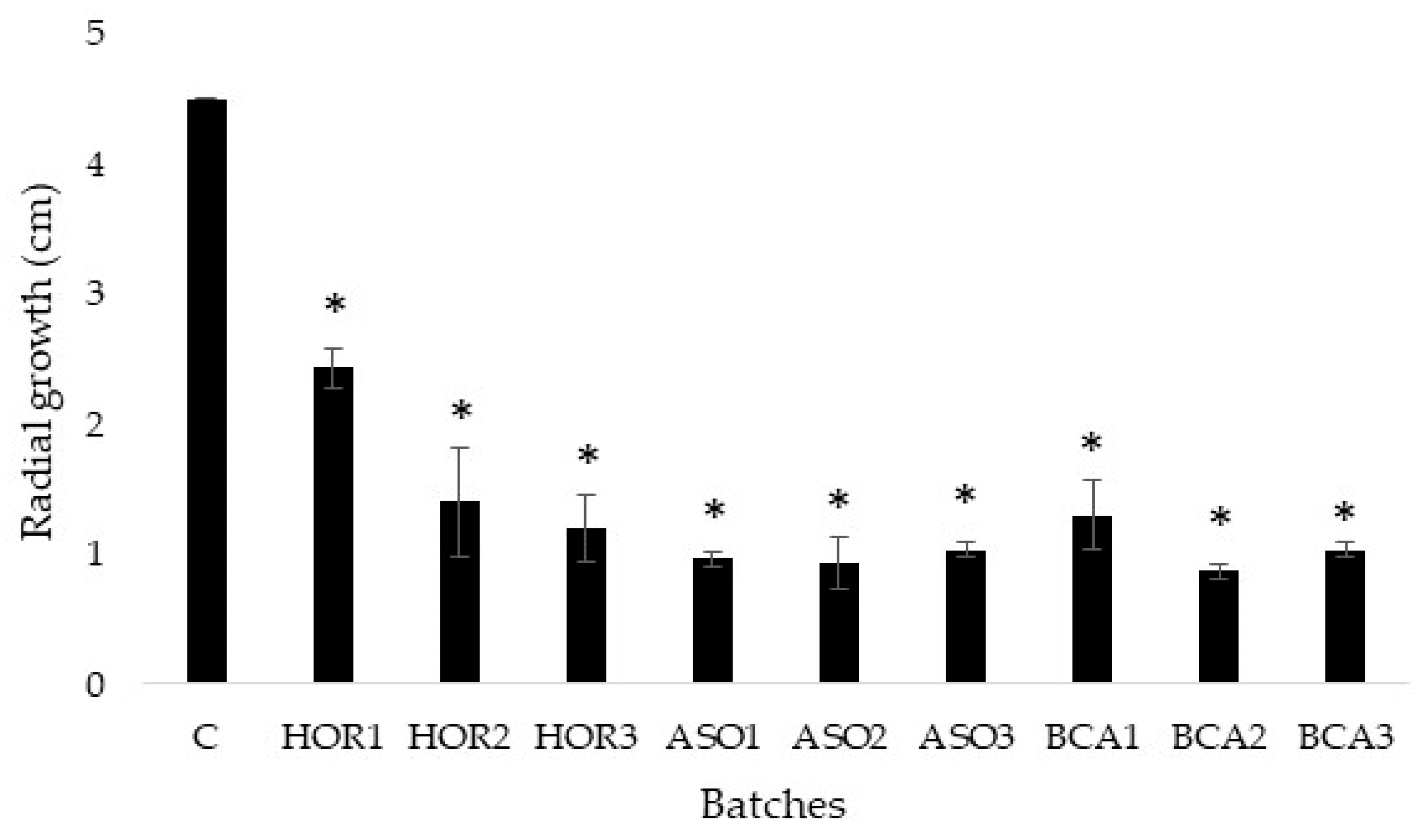
The molecular analyses revealed that the bacterium CIMO-BCA1, which was isolated due to its antifungal effect against Gs, was B. amyloliquefaciens, the same species found in the commercial fungicide ASO. This species has also been isolated from other kinds of chestnuts such as Chinese chestnuts (Castanea mollissima Blume) for biological control application [32]. Although similar to the commercial isolate, CIMO-BCA1 was genotypically different, as revealed by the ERIC-PCR analysis.
It must be noted that the genus Bacillus sp. includes a group of industrially important species, namely B. subtilis, B. amyloliquefaciens, and B. velezensis, which represent a group of phylogenetically and phenetically homogeneous species that are quite hard to distinguish [17]. In particular, the species B. amyloliquefaciens (type strain: DSM7), which includes the ASO strain QST 713, was initially classified as B. subtilis, and this classification is still used by some researchers [20,21]. A group of plant-associated strains of this species, including those used as biocontrol agents, was reclassified in 2005 as B. velezensis (type strain: FZB42) [33]. The close relatedness of representatives of B. amyloliquefaciens and B. velezensis was validated by rpoB gene sequence homology and the analysis of core genomes [17,34]. For that reason, Fan et al. [34] proposed to introduce the term “operational group B. amyloliquefaciens” applied to all “plant-associated B. amyloliquefaciens” strains to underline their close phylogenomic relationship and to reduce inconsistencies due to strain misclassification. For the sake of clarity, the name B. amyloliquefaciens (as in “operational unit B. amyloliquefaciens”) will be used throughout this work to name the isolated strain CIMO-BCA1 as well as the ASO strain QST 713.

3.3. Radial Growth

After the incubation period, Gs was able to grow up to 4.5 cm in CM (Figure 1), while the most effective antifungal agent concentration—CIMO-BCA1 at the intermediate concentration (BCA2)—reduced the mould’s growth to as low as 0.86 cm, indicating a reduction of 80.7% (p ≤ 0.05) when compared to the control (Figure S2, Supplementary Material). It is also worth noting that there was no dose–response effect in the treatments with B. amyloliquefaciens, both for the commercial ASO strain (between ASO1, ASO2, and ASO3; ρ = 0.377, p = 0.317) and the CIMO-BCA1 strain (between BCA1, BCA2, and BCA3; ρ = −0.427, p = 0.252). On the contrary, the chemical fungicide HOR showed a strong and significant negative correlation between dose and fungal growth reduction (ρ = −0.926; p = 0.000). Bacillus amyloliquefaciens previously showed its efficacy against this fungal pathogen when used in grafted scions [26]. Another strain of B. amyloquefaciens also inhibited the growth of pathogenic fungi as Alternaria panax, Botrytis cinerea, or Penicillium digitatum on PDA [35]. Furthermore, the addition of another strain of B. amyloliquefaciens on pepper plants was able to control the invasion of Alternaria sp. by improving the growth of the infected plants [36]. Different biocontrol agents, such as Trichoderma spp. or B. subtilis, have also demonstrated their ability to decrease the growth of other strains of Gs. In contrast, and despite the fact that the commercial antifungal HOR1 limited the radial growth to 2.4 cm (which is a percentage reduction up to 45.92%), this treatment was the least effective. These results indicate that B. amyloliquefaciens is a powerful candidate to be used as BCA against Gs and a good alternative to the chemical antifungal HOR, which presents a long half-life, liver and reproductive toxicity, and endocrine disruption in animals and humans [37].
BCAs act against pathogens via a number of different mechanisms. In particular, Bacillus spp. are known to act by three main mechanisms: colonisation of the host plant and competition for ecological substrate, direct pathogen growth inhibition, and stimulating the plant immune system [16]. These mechanisms mostly rely on the production of a wide range of diverse molecules, such as enzymes, bacteriocins, lantibiotics, non-ribosomal peptides, poliketides, and many others [16]. In our study, the direct effect of the bacterial strains on fungal growth was evident, and this can be explored for post-harvest treatments.

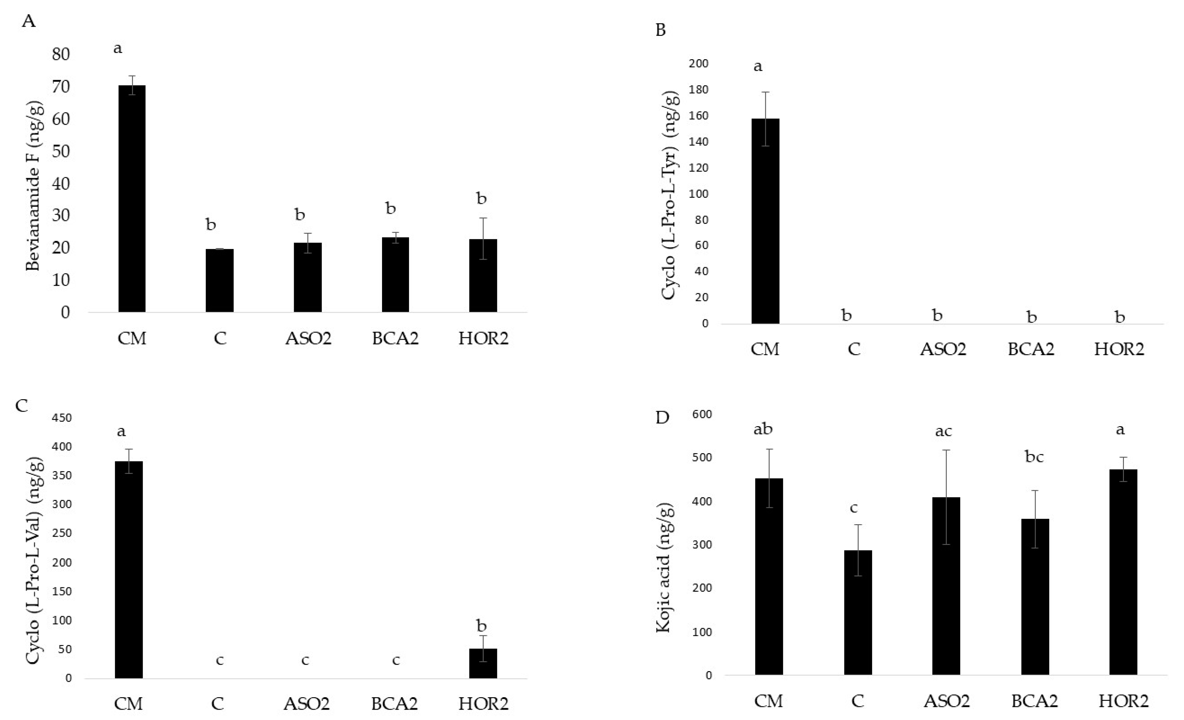
3.4. Fungal Secondary Metabolites

Currently, there are no studies in the literature based on the production of compounds of interest by Gs because the investigations are focused on eliminating this plant pathogen and reducing chestnut losses. The analysis of secondary metabolites produced by the fungus could help with understanding the effects of the different control agents on the metabolism and response of Gs to these stressors. The results showed the detection of nine compounds from the mycelium and culture medium adjacent to the inhibition zone: brevianamide F, cyclo(L-Pro-L-Tyr), cyclo(L-Pro-L-Val), endocrocin, kojic acid, integracin A, integracin B, 3-nitropropionic acid, and diplodiatoxin. Brevianamide F, also known as cyclo-(L-Trp-L-Pro), as well as cyclo-(L-Pro-L-Tyr) and cyclo-(L-Pro-L-Val) are unspecific bioactive cyclic dipeptides produced by some microorganisms, such as Bacillus spp., Streptomyces spp., and Aspergillus fumigatus [38,39,40]. Their concentration (Figure 2A–C) was higher in the CM batch without fungal growth, indicating a possible degradation of the compounds already present in the medium by Gs. The absence of differences between batch C and the other batches with Gs could be due to the effects of antifungal agents on the mould’s metabolism that may be stimulating the degradation of these compounds under stressful conditions. Other diketopiperazines, such as cyclo-(L-Pro-L-Leu), cyclo-(D-Pro-L-Leu), and cyclo-(D-Pro-L-Tyr) showed antifungal effects against other plant pathogenic fungi, including B. cinerea, Phytophthora spp., and Colletotrichum gloeosporioides [41].
Additionally, kojic acid (Figure 2D) is a non-toxic compound commonly synthesised by Aspergillus spp. [42] that showed antifungal activity against the mould Sclerotinia sclerotiorum, which is a common plant pathogen [43], and other fungi, such as Aspergillus niger or Rhizopus oryzae [44]. Furthermore, this metabolite produced by Aspergillus parasiticus can detoxify the mycotoxin citrinin in a glucose yeast agar medium [45]. The presence of kojic acid in all the batches may be due to the contamination of the chestnuts by different moulds before preparing the culture medium.
The integracins A and B (Figure 3A,B) were only detected in the samples with Gs. As the figure shows, the use of HOR decreased their production. These secondary metabolites have shown potential cytotoxicity against the tumour cell lines HepG2 and the repression of human immunodeficiency virus Type 1 (HIV-1) [46], showing a possible industrial use of the mould to produce these compounds.
Figure 3C shows 3-nitropropionic acid, which is a neurotoxic compound for animals synthetised by fungi such as A. flavus and Arthrinium in plants [47,48]. This mycotoxin produces motor disturbances, cognitive deficits, and aggressive behaviours in rats [48]. Human intoxications have also been reported following the ingestion of mouldy plant-derived foods, such as sugarcane and coconut water, resulting in vomiting, nausea, exhaustion, diarrhoea, stomachache and headache, and, rarely, death [49]. The results of our study indicate, for the first time, the ability of Gs to produce this toxin which entails the need to control the presence of Gs, not only due to economic losses but also from the point of view of food safety. The concentrations found in all batches in Figure 3C (around 20,000 to 40,000 ng/g) pose a worrying scenario since 20,000 ng/g of 3-nitropropionic acid intraperitoneal injected in rats for 4 days was able to impair motor coordination [50].
The risk associated with Gs increased due to the detection of diplodiatoxin (Figure 3D), which is a metabolite previously described to be produced by Stenocarpella maydis, which is an ear rot pathogen of maize [51]. Despite few studies existing about this metabolite, it is known that diplodiatoxin produces neurotoxicity, causing diplodiosis in cattle and sheep by the ingestion of infected mouldy feed [52,53]. Moreover, this mycotoxin induces necrosis and apoptosis in the cell lines of Chinese hamster ovary (CHP-K1) and Madin–Bardy bovine kidney (MDBK), which shows a wide range of toxicity [53]. As shown in Figure 3D, the commercial antifungal HOR highly stimulated the production of diplodiatoxin (p ≤ 0.05), increasing the risk in treated chestnuts, while the bacterium CIMO-BCA1 showed a slightly positive stimulation of the compound. The biocontrol agent ASO2 (B. amyloliquefaciens QST 713) exhibited an intermediate response between HOR and CIMO-BCA1 (p ≤ 0.05).
Endocrocin was detected exclusively in the samples of Gs confronted with HOR, even in amounts below the limit of quantification (LOQ = 51 ng/g). Endocrocin is an unspecific anthraquinone that has been isolated from various fungi, insects, and plants [54,55]. In a study on the effects of endocrocin on the pathomechanism of Aspergillus fumigatus, the metabolite was not found to be directly involved in cytotoxicity, but it displayed significant leucocyte recruitment inhibitory properties, negatively interfering with the immune response of the host towards the pathogen attack [55]. The same study also showed that, at least in A. fumigatus, endocrocin is a spore-borne metabolite that is highly dependent on the micro-environmental conditions in which the fungus is developing, particularly the temperature. In our study, endocrocin was exclusively detected in the HOR treatment, which probably reflects a response to stressful conditions that was not observed for the biological treatments. In contrast, the low levels detected may result from the fact that mostly culture medium and mycelium, and not specifically conidia, were collected for secondary metabolite analysis. The level of importance of this metabolite in Gs requires further attention.
To summarise, although all the antifungal agents successfully decreased the growth of Gs, this reduction is not enough to eliminate the presence of the mycotoxins 3-nitropropionic acid and diplodiatoxin produced by Gs, which can even be stimulated in their presence. This lack of relation between growth and mycotoxin production in different moulds has been previously described by numerous researchers and is possibly associated with the stress induced by the antifungal agents [56,57,58]. These results corroborate the importance of using effective doses of antifungals to avoid the presence of unwanted compounds. Additionally, the differences in 3-nitropropionic acid and diplodiatoxin production by Gs facing stressful antifungals (HOR > Bacillus ASO > Bacillus CIMO-BCA1) highlight the importance of using adequate antifungals against Gs. In addition, while both B. amyloliquefaciens strains (ASO and CIMO-BCA1) have similar percentages of inhibition of Gs, their impact on the production of secondary metabolites can be different.

4. Conclusions

We demonstrated, for the first time, the production of the toxins 3-nitropropionic acid and diaplodiatoxin by G. smithogilvyi. Nothing is known yet regarding whether they would be bioavailable via the oral route through the consumption of contaminated chestnuts, and further studies are required. Nonetheless, in addition to the enormous economic losses caused by Gs, this fungus must also be considered based on the public health point of view. However, this mould may also be industrially used to produce integracins A and B that could provide benefits for treatments against human diseases. Finally, the use of biocontrol agents based on B. amyloliquefaciens isolates as alternatives to the toxic HOR are effective strategies against G. smithogilvyi, although other concentrations or combinations with other strategies should be applied to completely inhibit mould growth and mycotoxins production.

Supplementary Materials

The following supporting information can be downloaded at: https://www.mdpi.com/article/10.3390/agriculture13061166/s1, Figure S1: Phylogenetic tree of Gnomoniopsis smithogilvyi isolates for the TEF1-α partial gene; Figure S2: Morphological images after 23 days of incubation of Gnomoniopsis smithogilvyi in a chestnut-based medium in the presence of antifungal agents at three different concentrations of CIMO-BCA1 (Bacillus amyloliquefaciens, BCA), Serenade® (Bacillus amyloliquefaciens QST 713, ASO), and Horizon® (tebuconazole, HOR). References [59,60,61] are cited in the Supplementary Materials.

Author Contributions

Conceptualisation, P.R. and A.S.; methodology, P.R., A.S., I.A., S.S., and M.S.; formal analysis, P.R. and M.Á.; investigation, P.R., I.A., S.S., and M.Á.; resources, P.R.; writing—original draft preparation, P.R. and M.Á.; writing—review and editing, P.R., M.Á., and A.S.; supervision, P.R. and R.D.-G.; project administration, P.R.; funding acquisition, P.R. All authors have read and agreed to the published version of the manuscript.

Funding

This work was funded by the Foundation for Science and Technology (FCT, Portugal) and FEDER under Program PT2020 through financial support to projects GnomoCastRot (CMFPE3; EXPL2021CIMO_02), CIMO (UID/AGR/00690/2020), SusTEC (LA/P/0007/2020), CITAB (UID/AGR/04033/2020), and Inov4Agro (LA/P/0126/2020). M. Álvarez was the recipient of grant BES-2017-081340 funded by MCIN/AEI/10.13039/501100011033 and by “ESF Investing in your future”, and Grant Margarita Salas MS-14 was funded by the Ministerio de Universidades and European Union NextGenerationEU.

Institutional Review Board Statement

Not applicable.

Data Availability Statement

Not applicable.

Conflicts of Interest

The authors declare no conflict of interest.

References

  1. FAOSTAT. Available online: https://www.fao.org/faostat/en/#data/QCL (accessed on 1 February 2023).
  2. INE. Estatísticas Agrícolas 2020; Instituto Nacional de Estatística: Lisboa, Portugal, 2021; ISSN 0079-4139. [Google Scholar]
  3. Rodrigues, P.; Venâncio, A.; Lima, N. Incidence and diversity of the fungal genera Aspergillus and Penicillium in Portuguese almonds and chestnuts. Eur. J. Plant Pathol. 2013, 137, 197–209. [Google Scholar] [CrossRef]
  4. Rodrigues, P.; Venâncio, A.; Lima, N. Mycobiota and mycotoxins of almonds and chestnuts with special reference to aflatoxins. Food Res. Int. 2012, 48, 76–90. [Google Scholar] [CrossRef]
  5. Jiang, N.; Tian, C.M. An emerging pathogen from rotted chestnut in China: Gnomoniopsis daii sp. nov. Forests 2019, 10, 1016. [Google Scholar] [CrossRef]
  6. Jiang, N.; Liang, L.Y.; Tian, C.M. Gnomoniopsis chinensis (Gnomoniaceae, Diaporthales), a new fungus causing canker of Chinese chestnut in Hebei Province, China. MycoKeys 2020, 67, 19–32. [Google Scholar] [CrossRef]
  7. Dobry, E.; Campbell, M. Gnomoniopsis castaneae: An emerging plant pathogen and global threat to chestnut systems. Plant Pathol. 2023, 72, 218–231. [Google Scholar] [CrossRef]
  8. Possamai, G.; Dallemole-Giaretta, R.; Gomes-Laranjo, J.; Sampaio, A.; Rodrigues, P. Chestnut Brown Rot and Gnomoniopsis smithogilvyi: Characterization of the Causal Agent in Portugal. J. Fungi 2023, 9, 401. [Google Scholar] [CrossRef] [PubMed]
  9. Rodrigues, P.; Driss, J.O.; Gomes-Laranjo, J.; Sampaio, A. Impact of Cultivar, Processing and Storage on the Mycobiota of European Chestnut Fruits. Agriculture 2022, 12, 1930. [Google Scholar] [CrossRef]
  10. Ruocco, M.; Lombardi, N.; Lanzuise, S.; Varlese, R.; Aliberti, A.; Carpenito, S.; Woo, S.; Scala, F.; Lorito, M. New tools to improve the shelf life of chestnut fruit during storage. Acta Hortic. 2016, 1144, 309–315. [Google Scholar] [CrossRef]
  11. Hou, L.; Kou, X.; Li, R.; Wang, S. Thermal inactivation of fungi in chestnuts by hot air assisted radio frequency treatments. Food Control 2018, 93, 297–304. [Google Scholar] [CrossRef]
  12. Carocho, M.; Barreira, C.M.; Antonio, A.L.; Bento, A.; Kaluska, I.; Ferreira, I.C.F.R. Effeects of Electron-Beam Radiation on Nutritional Parameters of Portuguese Chestnuts (Castanea sativa Mill.). J. Agric. Food Chem. 2012, 60, 7754–7760. [Google Scholar] [CrossRef]
  13. Vettraino, A.M.; Bianchini, L.; Caradonna, V.; Forniti, R.; Zambelli, M.; Testa, A.; Vinciguerra, V.; Botondi, R.; Lellis, V.S.C. De Ozone gas as a storage treatment to control Gnomoniopsis castanea preserving chestnut quality. J. Sci. Food Agric. 2019, 99, 6060–6065. [Google Scholar] [CrossRef] [PubMed]
  14. Silva-Campos, M.; Islam, M.; Cahill, D.M. Fungicide control of Gnomoniopsis smithogilvyi, casual agent of chestnut rot in Australia. Australas. Plant Pathol. 2022, 51, 483–494. [Google Scholar] [CrossRef]
  15. Bastianelli, G.; Morales-Rodríguez, C.; Caccia, R.; Turco, S.; Rossini, L.; Mazzaglia, A.; Thomidis, T.; Vannini, A. Use of phosphonate salts to control chestnuts ‘Brown Rot’ by Gnomoniopsis castanea in fruit orchards of Castanea sativa. Agronomy 2022, 12, 2434. [Google Scholar] [CrossRef]
  16. Anckaert, A.; Arias, A.A.; Hoff, G.; Calonne-Salmon, M.; Declerck, S.; Ongena, M. The use of Bacillus spp. as bacterial biocontrol agents to control plant diseases. In Microbial Bioprotectants for Plant Disease Management; Köhl, J., Ravensberg, W., Eds.; Burleigh Dodds Science Publishing: Cambridge, UK, 2022; ISBN 978 1 78676 813 1. [Google Scholar] [CrossRef]
  17. Fan, B.; Blom, J.; Klenk, H.P.; Borris, R. Bacillus amyloliquefaciens, Bacillus velezensis, and Bacillus siamensis form and “operational group B. amyloliquefaciens” within the B. subtilis species complex. Front. Microbiol. 2017, 8, 22. [Google Scholar] [CrossRef]
  18. Fousia, S.; Paplomatas, E.J.; Tjamos, S.E. Bacillus subtilis QST 713 confers protection to tomato plants against Pseudomonas syringae pv tomato and induces plant defence-related genes. J. Phytopathol. 2016, 164, 264–270. [Google Scholar] [CrossRef]
  19. Matzen, N.; Heick, T.M.; Jørgensen, L.N. Control of powdery mildew (Blumeria graminis spp.) in cereals by Serenade®ASO (Bacillus amyloliquefaciens (former subtilis) strain QST 713). Biol. Control 2019, 139, 104067. [Google Scholar] [CrossRef]
  20. Cucu, M.A.; Gilardi, G.; Pugliese, M.; Gullino, M.L.; Garibaldi, A. An assessment of the modulation of the population dynamics of pathogenic Fusarium oxysporum f. sp. lycopersici in the tomato rhizosphere by means of the application of Bacillus subtilis QST 713, Trichoderma sp. TW2 and two composts. Biol. Control 2020, 142, 104158. [Google Scholar] [CrossRef]
  21. Punja, Z.K.; Rodriguez, G.; Tirajoh, A. Effects of Bacillus subtilis strain QST 713 and storage temperatures on post-harvest disease development on greenhouse tomatoes. Crop Prot. 2016, 84, 98–104. [Google Scholar] [CrossRef]
  22. Gava, C.A.T.; Alves, Í.L.S.; Duarte, N.C. Timing the application of Bacillus subtilis QST 713 in the integrated management of the postharvest decay of mango fruits. Crop Prot. 2019, 121, 51–56. [Google Scholar] [CrossRef]
  23. EFSA; Anastassiadou, M.; Arena, M.; Auteri, D.; Brancato, A.; Bura, L.; Carrasco Cabrera, L.; Chaideftou, E.; Chiusolo, A.; Crivellente, F.; et al. Peer review of the pesticide risk assessment of the active substance Bacillus amyloliquefaciens strain QST 713 (formerly Bacillus subtilis strain QST 713). EFSA J. 2021, 19, e06381. [Google Scholar] [CrossRef]
  24. Vinale, F.; Ruocco, M.; Manganiello, G.; Guerrieri, E.; Bernardo, U.; Mazzei, P.; Piccolo, A.; Sannino, F.; Caira, S.; Woo, S.L.; et al. Metabolites produced by Gnomoniopsis castanea associated with necrosis of chestnut galls. Chem. Biol. Technol. Agric. 2014, 1, 8. [Google Scholar] [CrossRef]
  25. Silva-Campos, M.; Callahan, D.L.; Cahill, D.M. Metabolites derived from fungi and bacteria suppress in vitro growth of Gnomoniopsis smithogilvyi, a major threat to the global chestnut industry. Metabolomics 2022, 18, 74. [Google Scholar] [CrossRef] [PubMed]
  26. Pasche, S.; Crovadore, J.; Pelleteret, P.; Jermini, M.; Mauch-Mani, B.; Oszako, T.; Lefort, F. Biological control of the latent pathogen Gnomoniopsis smithogylvyi in European chestnut grafting scions using Bacillus amyloliquefaciens and Trichoderma atroviride. Dendrobiology 2016, 75, 113–122. [Google Scholar] [CrossRef]
  27. Rodrigues, P.; Venancio, A.; Lima, N. Toxic reagents and expensive equipment: Are they really necessary for the extraction of good quality fungal DNA? Lett. Appl. Microbiol. 2018, 66, 32–37. [Google Scholar] [CrossRef] [PubMed]
  28. Cai, L.; Ye, L.; Tong, A.H.Y.; Lok, S.; Zhang, T. Biased Diversity Metrics Revealed by Bacterial 16S Pyrotags Derived from Different Primer Sets. PLoS ONE 2013, 8, e53649. [Google Scholar] [CrossRef] [PubMed]
  29. De Clerck, E.; De Vos, P. Genotypic diversity among Bacillus licheniformis strains from various sources. FEMS Microbiol. Lett. 2004, 231, 91–98. [Google Scholar] [CrossRef]
  30. Kumar, A.; Kumar, A.; Pratush, A. Molecular diversity and functional variability of environmental isolates of Bacillus species. Springerplus 2014, 3, 312. [Google Scholar] [CrossRef]
  31. Sulyok, M.; Stadler, D.; Steiner, D.; Krska, R. Validation of an LC-MS/MS-based dilute-and-shoot approach for the quantification of >500 mycotoxins and other secondary metabolites in food crops: Challenges and solutions. Anal. Bioanal. Chem. 2020, 412, 2607–2620. [Google Scholar] [CrossRef]
  32. Zhang, X.; Li, L.; Cheng, S.; Cheng, H. Characterizing CMN1308, a Novel Strain of Bacillus amyloliquefaciens, for Potential Biological Control Application. Not. Bot. Horti. Agrobot. Cluj. Napoca 2016, 44, 60–66. [Google Scholar] [CrossRef]
  33. Ruíz-García, C.; Béjar, V.; Martínez-Checa, F.; Llamas, I.; Quesada, E. Bacillus velezensis sp. nov., a surfactant-producing bacterium isolated from the river Vélez in Málaga, southern Spain. Int. J. Syst. Evol. Microbiol. 2005, 55, 191–195. [Google Scholar] [CrossRef]
  34. Borriss, R.; Chen, X.H.; Rueckert, C.; Blom, J.; Becker, A.; Baumgarth, B.; Fan, B.; Pukall, R.; Schumann, P.; Spröer, C.; et al. Relationship of Bacillus amyloliquefaciens clades associated with strains DSM 7T and FZB42T: A proposal for Bacillus amyloliquefaciens subsp. amyloliquefaciens subsp. nov. and Bacillus amyloliquefaciens subsp. plantarum subsp. nov. based on complete genome sequence comparisons. Int. J. Syst. Evol. Microbiol. 2011, 61, 1786–1801. [Google Scholar] [CrossRef] [PubMed]
  35. Ji, S.H.; Paul, N.C.; Deng, J.X.; Kim, Y.S.; Yun, B.S.; Yu, S.H. Biocontrol activity of Bacillus amyloliquefaciens CNU114001 against fungal plant diseases. Mycobiology 2013, 41, 234–242. [Google Scholar] [CrossRef] [PubMed]
  36. Soliman, S.A.; Khaleil, M.M.; Metwally, R.A. Evaluation of the Antifungal Activity of Bacillus amyloliquefaciens and B. velezensis and Characterization of the Bioactive Secondary Metabolites Produced against Plant Pathogenic Fungi. Biology 2022, 11, 1390. [Google Scholar] [CrossRef] [PubMed]
  37. Ku, T.; Zhou, M.; Hou, Y.; Xie, Y.; Li, G.; Sang, N. Tebuconazole induces liver injury coupled with ROS-mediated hepatic metabolism disorder. Ecotoxicol. Environ. Saf. 2021, 220, 112309. [Google Scholar] [CrossRef] [PubMed]
  38. Danial, A.M.; Medina, A.; Sulyok, M.; Magan, N. Efficacy of metabolites of a Streptomyces strain (AS1) to control growth and mycotoxin production by Penicillium verrucosum, Fusarium verticillioides and Aspergillus fumigatus in culture. Mycotoxin Res. 2020, 36, 225–234. [Google Scholar] [CrossRef]
  39. Szulc, J.; Ruman, T. Laser Ablation Remote-Electrospray Ionisation Mass Spectrometry (LARESI MSI) Imaging- New Method for Detection and Spatial Localization of Metabolites and Mycotoxins Produced by Moulds. Toxins 2020, 12, 720. [Google Scholar] [CrossRef]
  40. Wattana-Amorn, P.; Charoenwongsa, W.; Williams, C.; Crump, M.P.; Apichaisataienchote, B. Antibacterial activity of cyclo(L-Pro-L-Tyr) and cyclo(D-Pro-L-Tyr) from Streptomyces sp. strain 22-4 against phytopathogenic bacteria. Nat. Prod. Res. 2016, 30, 1980–1983. [Google Scholar] [CrossRef]
  41. Kumar, S.N.; Nambisan, B. Antifungal activity of diketopiperazines and stilbenes against plant pathogenic fungi in vitro. Appl. Biochem. Biotechnol. 2014, 172, 741–754. [Google Scholar] [CrossRef]
  42. Ritthibut, N.; Lim, S.T.; Oh, S.J. In vitro cosmeceutical activity of alcoholic extract from chestnut inner shell fermented with Aspergillus sojae. Food Sci. Biotechnol. 2022, 31, 443–450. [Google Scholar] [CrossRef]
  43. Zhu, G.Y.; Shi, X.C.; Wang, S.Y.; Wang, B.; Laborda, P. Antifungal Mechanism and Efficacy of Kojic Acid for the Control of Sclerotinia sclerotiorum in Soybean. Front. Plant Sci. 2022, 13, 845698. [Google Scholar] [CrossRef]
  44. Zilles, J.C.; dos Santos, F.L.; Kulkamp-Guerreiro, I.C.; Contri, R.V. Biological activities and safety data of kojic acid and its derivatives: A review. Exp. Dermatol. 2022, 31, 1500–1521. [Google Scholar] [CrossRef] [PubMed]
  45. Ichinomiya, M.; Kawamoto, A.; Yamaguchi, T.; Iwashita, K.; Nagashima, H.; Hatabayashi, H.; Nakajima, H.; Yabe, K. Detoxication of Citrinin with Kojic Acid by the Formation of the Citrinin-Kojic Acid Adduct, and the Enhancement of Kojic Acid Production by Citrinin via Oxidative Stress in Aspergillus parasiticus. J. Fungi 2022, 9, 51. [Google Scholar] [CrossRef] [PubMed]
  46. Wei, C.; Deng, Q.; Sun, M.; Xu, J. Cytospyrone and Cytospomarin: Two new polyketides isolated from mangrove endophytic fungus, Cytospora sp. Molecules 2020, 25, 4224. [Google Scholar] [CrossRef]
  47. Flores, A.C.; Pamphile, J.A.; Sarragiotto, M.H.; Clemente, E. Production of 3-nitropropionic acid by endophytic fungus Phomopsis longicolla isolated from Trichilia elegans A. JUSS ssp. elegans and evaluation of biological activity. World J. Microbiol. Biotechnol. 2013, 29, 923–932. [Google Scholar] [CrossRef] [PubMed]
  48. Furian, A.F.; Fighera, M.R.; Royes, L.F.F.; Oliveira, M.S. Recent advances in assessing the effects of mycotoxins using animal models. Curr. Opin. Food Sci. 2022, 47, 100874. [Google Scholar] [CrossRef]
  49. Skogvold, H.; Yazdani, M.; Sandås, E.M.; Østeby Vassli, A.; Kristensen, E.; Haarr, D.; Rootwelt, H.; Elgstøen, K.B.P. A pioneer study on human 3-nitropropionic acid intoxication: Contributions from metabolomics. J. Appl. Toxicol. 2022, 42, 818–829. [Google Scholar] [CrossRef]
  50. Salman, M.; Sharma, P.; Alam, M.I.; Tabassum, H.; Parvez, S. Naringenin mitigates behavioral alterations and provides neuroprotection against 3-nitropropinoic acid-induced Huntington’s disease like symptoms in rats. Nutr. Neurosci. 2022, 25, 1898–1908. [Google Scholar] [CrossRef] [PubMed]
  51. Wicklow, D.T.; Rogers, K.D.; Dowd, P.F.; Gloer, J.B. Bioactive metabolites from Stenocarpella maydis, a stalk and ear rot pathogen of maize. Fungal Biol. 2011, 115, 133–142. [Google Scholar] [CrossRef] [PubMed]
  52. Botha, C.J.; Ackerman, L.G.J.; Masango, M.G.; Arnot, L.F. Failure of diplodiatoxin to induce diplodiosis in juvenile goats. Onderstepoort J. Vet. Res. 2020, 87, a1712. [Google Scholar] [CrossRef]
  53. Masango, M.G.; Ellis, C.E.; Botha, C.J. Characterization of cell death caused by diplodiatoxin and dipmatol, toxic metabolites of Stenocarpella maydis. Toxicon 2015, 102, 14–24. [Google Scholar] [CrossRef]
  54. Lim, F.Y.; Hou, Y.; Chen, Y.; Oh, J.H.; Lee, I.; Bugni, T.S.; Keller, N.P. Genome-based cluster delection reveals an endocrocin biosynthetic pathway in Aspergillus fumigatus. Appl. Environ. Microbiol. 2012, 78, 4117–4125. [Google Scholar] [CrossRef]
  55. Berthier, E.; Lim, F.Y.; Deng, Q.; Guo, C.J.; Kontoyiannis, D.P.; Wang, C.C.C.; Rindy, J.; Beebe, D.J.; Huttenlocher, A.; Keller, N.P. Low-volume toolbox for the discoery of immunosuppressive fungal secondary metabolites. PLoS Pathog. 2013, 9, e1003289. [Google Scholar] [CrossRef] [PubMed]
  56. Álvarez, M.; Rodríguez, A.; Núñez, F.; Silva, A.; Andrade, M.J. In vitro antifungal effects of spices on ochratoxin A production and related gene expression in Penicillium nordicum on a dry-cured fermented sausage medium. Food Control 2020, 114, 107222. [Google Scholar] [CrossRef]
  57. da Cruz Cabral, L.; Rodríguez, A.; Andrade, M.J.; Patriarca, A.; Delgado, J. Effect of Debaryomyces hansenii and the antifungal PgAFP protein on Alternaria spp. growth, toxin production, and RHO1 gene expression in a tomato-based medium. Food Microbiol. 2021, 97, 103741. [Google Scholar] [CrossRef] [PubMed]
  58. Schmidt-Heydt, M.; Baxter, E.; Geisen, R.; Magan, N. Physiological relationship between food preservatives, environmental factors, ochratoxin and otapksPV gene expression by Penicillium verrucosum. Int. J. Food Microbiol. 2007, 119, 277–283. [Google Scholar] [CrossRef]
  59. Saitou, N.; Nei, M. The neighbor-joining method: A new method for reconstructing phylogenetic trees. Mol. Biol. Evol. 1987, 4, 406–425. [Google Scholar]
  60. Kimura, M. A simple method for estimating evolutionary rate of base substitutions through comparative studies of nucleotide sequences. J. Mol. Evol. 1980, 16, 111–120. [Google Scholar] [CrossRef]
  61. Tamura, K.; Stecher, G.; Kumar, S. MEGA 11: Molecular Evolutionary Genetics Analysis Version 11. Mol. Biol. Evol. 2021, 33, 1870–1874. [Google Scholar] [CrossRef]
Figure 1. Radial growth after 23 days of incubation of Gnomoniopsis smithogilvyi in the presence of the antifungal agents Horizon® (tebuconazole, HOR), Serenade® (Bacillus amyloliquefaciens QST 713, ASO), and CIMO-BCA1 (Bacillus amyloliquefaciens, BCA) in a chestnut-based medium. C: Gnomoniopsis smithogilvyi. An asterisk indicates statistical differences regarding the control (C) (p ≤ 0.05). The vertical bar represents the standard deviation.
Agriculture 13 01166 g001
Figure 2. Concentrations of brevianamide F, cyclo(L-Pro-L-Tyr), cyclo(L-Pro-L-Val), and kojic acid in the batches studied. CM: chestnut medium; C: Gnomoniopsis smithlogilvyi inoculated in CM; ASO2: C plus the biocontrol agent ASO2 (Bacillus amyloliquefaciens QST 713 (10 µL/mL); BCA2: C plus the biocontrol agent Bacillus amyloliquefaciens CIMO-BCA1 (107 cfu/mL); HOR2: C plus Horizon® (tebuconazole, 0.10 g/L). Different letters indicate statistical differences between batches (p ≤ 0.05). The vertical bar represents the standard deviation.
Agriculture 13 01166 g002
Figure 3. Concentrations of integracin A, integracin B, 3-nitropropionic acid, and diplodiatoxin in the studied batches. CM: chestnut medium; C: Gnomoniopsis smithlogilvyi inoculated in CM; ASO2: C plus the biocontrol agent ASO2 (Bacillus amyloliquefaciens QST 713 (10 µL/mL); BCA2: C plus the biocontrol agent Bacillus amyloliquefaciens CIMO-BCA1 (107 cfu/mL); HOR2: C plus Horizon® (tebuconazole, 0.10 g/L). Different letters indicate statistical differences between batches (p ≤ 0.05). The vertical bar represents the standard deviation.
Agriculture 13 01166 g003
Table 1. Concentrations of the antifungal agents employed in the study: commercial biological fungicide (Serenade ASO), commercial chemical fungicide (Horizon), and BCA (CIMO-BCA1).
Antifungal AgentBatch DenominationConcentration of Antifungal Agent (µL/mL or cfu/mL)Concentration of Active Substance (g/L)
Serenade® ASOASO15.50.074
ASO210.00.134
ASO316.00.214
Horizon®HOR10.20.050
HOR20.40.100
HOR30.60.150
CIMO-BCA1BCA1105-
BCA2107-
BCA3109-
Disclaimer/Publisher’s Note: The statements, opinions and data contained in all publications are solely those of the individual author(s) and contributor(s) and not of MDPI and/or the editor(s). MDPI and/or the editor(s) disclaim responsibility for any injury to people or property resulting from any ideas, methods, instructions or products referred to in the content.

