Article

Eliminating the Pathogen Xanthomonas hortorum pv. carotae from Carrot Seeds Using Different Types of Nanoparticles

1
Mendeleum—Institute of Genetics, Faculty of Horticulture, Mendel University in Brno, Valtická 334, 69144 Lednice, Czech Republic
2
Department of Chemistry and Biochemistry, Faculty of AgriSciences, Mendel University in Brno, Zemědělská 1, 61300 Brno, Czech Republic
*
Author to whom correspondence should be addressed.
Agriculture 2024, 14(3), 498; https://doi.org/10.3390/agriculture14030498
Submission received: 30 January 2024 / Revised: 12 March 2024 / Accepted: 16 March 2024 / Published: 19 March 2024
(This article belongs to the Section Crop Protection, Diseases, Pests and Weeds)

Abstract

:
There exists a wide range of plant pathogens that are commonly referred to as seed-borne pathogens due to their dominant mode of spread. Treating seeds to eliminate such pathogens is therefore very important in contemporary seed production. In the present study, eight types of nanoparticles were evaluated for their effectiveness against Xanthomonas hortorum pv. carotae, a seed-borne pathogen that affects plants of the Apiaceae family. Initially, parameters considering the inhibitory and bactericidal activity of individual nanoparticles were evaluated under in vitro conditions. In this way, three nanoparticles based on copper, silver, and silver/selenium composite were identified as being the most effective. Subsequently, their ability to eliminate Xanthomonas hortorum pv. carotae from artificially infected carrot seeds was tested. This was achieved through the qPCR quantification of the pathogen in 14-day-old plantlets developed from seeds inoculated with Xhc. Based on the obtained results, copper-based nanoparticles were the most effective, resulting in an approximately 10-fold decrease in the occurrence of Xhc in plantlets compared to the untreated control. Taking into account the fact that X. hortorum pathovars also attack other important horticultural crops, the presented results may have a much wider scope than just carrot seeds.

1. Introduction

There are a number of plant pathogens that threaten crop production. In terms of economic loss, it is estimated that 10 bacterial [1] and fungal pathogens [2] cause the greatest economic losses worldwide. According to Mansfield et al. [1], among the 10 most economically harmful bacteria, three species are from the genus Xanthomonas: Xanthomonas oryzae pv. oryzae, Xanthomonas campestris pathovars, and Xanthomonas axonopodis pathovars.
Pathogens of the genus Xanthomonas also include Xanthomonas hortorum pv. carotae (Xhc), which is capable of successfully colonizing the leaves of plants in the Apiaceae family [3]. Hosts from farm crops include common carrot (Daucus carota L.), garden parsley (Petroselinum crispum (Mill.) Fuss, 1886), root celery (Apium graveolens L.), lovage (Levisticum officinale Hill, 1756), and dill (Anethum graveolens L.) [4].
In the these plant species, Xhc causes a disease known as bacterial blight of carrot, characterized by round, water-soaked lesions on the abaxial surface of leaves, which reduces their photosynthetically active area [4]. In the final infection stages, leaves usually harden and dry. However, the leaves may not be the only parts that are damaged by the infection. Xhc can also damage petioles, peduncles, stems, flowers, and leaflets. A comprehensive review of the taxonomy, genomics, and symptoms is presented by Dia et al. [3].
From an epidemiological standpoint, Xhc represents a bacterium capable of transmission through seeds [5]. Therefore, it not only poses challenges for vegetable producers, but also presents serious complications for seed producers of economically important plants of the Apiaceae family [5,6]. Thanks to the global mass distribution of plant products, including seeds, the ease of pathogen spread has become a highly relevant contemporary issue [7].
Regarding the possible approaches for eliminating seed-borne pathogens, there are more or less traditional techniques, ranging from the use of fungicides/bactericides to physical methods such as heat or UV treatment, and include the utilization of natural substances or treatment by biological control agents [8]. From a historical perspective, one of the oldest preventive measures for seed decontamination is hot water treatment. The first deliberate hot water treatment of seeds can be traced back to the late 19th century [9]. This method is still used today, with great significance as a preventive measure, particularly for seed-borne pathogens such as Alternaria spp. [9,10] and Xanthomonas campestris pv. Campestris [10]. The frequently mentioned negative aspects of hot water treatment include its questionable effectiveness in the complete elimination of the pathogen [11] and the reduced germination in seeds treated in this manner, as demonstrated for Xanthomonas campestris pv. vitians by Carrise et al. [12]. There are also approaches that use chemical substances to eliminate seed-borne pathogens, such as 1% sodium hypochlorite, as reported by Carrise et al. [13], and 5% hydrogen peroxide, as reported by Perneczny et al. [14]. A comprehensive review of currently available individual techniques is presented by Moumni et al. [8].
However, some newer approaches are emerging and could theoretically serve as more efficient or environmentally friendly alternatives for seed treatment. One of them is the use of nanoparticles, due to their antimicrobial properties [13]. Their mechanism of action can differ depending on their chemical composition, but the principle of their antimicrobial properties is usually described as: (i) disruption of the peptidoglycan layer of the bacterial cell wall [14], (ii) toxicity by the release of metal ions in cytoplasm that lead to imbalanced nutrient uptake [15], (iii) leakage of intracellular components, ion imbalance, and eventual cell lysis [16], (iv) ROS induction and antioxidant production [17], and (v) interaction with bacterial DNA, leading to strand breaks and consequent disruption of vital cellular functions [18]. Comprehensive overviews of the observed mechanisms of the action of nanoparticles against phytopathogens are presented by Adisa et al. [19] and Ali et al. [20]. Regarding the use of nanoparticles to protect against pathogens within the Xanthomonas hortorum group, there are reports of applying Ag-dsDNA-GO nanoparticles for protection against copper-tolerant strains of X. hortorum pv. gardenia [21]. Additionally, simonkolleite nanoparticles were used for seed treatment to provide protection against tomato bacterial spot caused by Xanthomonas hortorum pv. gardneri [22]. The effects of titanium dioxide-based nanoparticles were also tested for their potential protective properties against Xanthomonas hortorum pv. pelargonii [23] after foliar application.
The prevailing method of employing nanoparticles to manage phytopathogens involves applying the treatment primarily by spraying leaves or entire plants [24]. On the other hand, only a limited number of articles cover the application of nanoparticles to suppress seed-borne pathogens [11,25,26,27,28]. This is perhaps a little surprising, considering that treating seeds with nanomaterials may be more environmentally friendly and safer than spraying whole plants in a field [29]. Thus, treating seeds with nanomaterials to protect them against phytopathogens remains a challenge, especially in the situation where a synergistic effect in terms of improved growth parameters can be observed after seed treatment [24]. However, it remains necessary to fill important gaps in the knowledge before recommending broader application of seed treatment with nanoparticles in practice.
The aim of the present study was to contribute to this field of research by testing eight nanoparticles to identify critical points in this kind of application. Our attention was focused mainly on factors such as the concentration and effective composition of nanoparticles and practical limitations. As an experimental model, we selected a combination of Xhc, a seed-borne pathogen, and carrot seeds artificially inoculated by this pathogen. Based on the obtained results, the most suitable variants were identified, and it was possible to formulate some recommendations. These recommendations will enable more effective application of nanoparticles to eliminate problematic seed-borne pathogens.

2. Materials and Methods

2.1. Xhc Strain and Growth Conditions

Xhc strain NCPPB 4410, obtained from the National Collection of Plant Pathogenic Bacteria (NCPPB, London, UK), was maintained on Luria agar (LA; HiMedia, Mumbai, India) at 28 °C for 24 h and stored in cryotubes at −80 °C for long-term storage. For all experiments, the Xhc strain was cultured in Luria broth (LB; HiMedia, Mumbai, India) at 28 °C for 24 h at 150 rpm on an orbital shaker (Biosan, Riga, Latvia) and the bacterial suspension was adjusted to a concentration of 1 × 10⁸ cfu mL−1, based on optical density at 600 nm (OD600) using a spectrophotometer (SPECTROstar Nano, BMG Labtech, Ortenberg, Germany). The bacterial suspension was serially diluted 10-fold to a concentration of approximately 1 × 10⁶ cfu mL−1 using LB for the in vitro experiment and sterile phosphate-buffered saline (PBS) for the in planta experiment.

2.2. In Vitro Experiment: Evaluation of Nanomaterials for Antibacterial Activity under In Vitro Conditions

Eight nanomaterials with different properties (Table 1) were tested against Xhc strain NCPPB 4410 in in vitro assays. All tested nanomaterials were provided by the Department of Chemistry and Biochemistry, Mendel University in Brno, Czech Republic, and their properties were determined using standard procedures, as presented in the references listed in Table 1.
To evaluate the antibacterial effect of nanomaterials, the minimum inhibitory concentration (MIC) and minimum bactericidal concentration (MBC) were determined, according to da Silva et al. [33]. The MIC was determined using the microdilution method on 96-well microplates. Each well had a total volume of 100 µL and contained a suspension of Xhc strain NCPPB 4410 (approximately 1 × 10⁶ cfu mL−1) and nanomaterials at the concentrations shown in Table 1 diluted with sterile LB. Bacterial suspension without nanomaterials served as a positive control. The microplates were incubated on the shaker (Biosan, Riga, Latvia) for 24 h and OD600 was measured using the spectrophotometer (SPECTROstar Nano, BMG Labtech, Ortenberg, Germany). MBC was determined by subculturing 5 µL of sample from each well on LA without the antimicrobial agent and incubation at 28 °C for 24 h. MBC was considered as the lowest concentration that prevented detectable bacterial growth. All assays were carried out in triplicate.
A time-kill assay was performed to determine the time required for the effective elimination of Xhc from carrot seeds, according to NCCLS [34]. Briefly, 5 mL of nanomaterial solution at the concentration determined as MBC (10 mg L−1 of Ag for AgNPs_29, 397 mg L−1 of Cu for CuNPs_53, and 53 mg L−1 of Ag and 11 mg L−1 of Se for AgSeNPs_8) were mixed at a 1:1 ratio with bacterial suspension in LB at a concentration of approximately 1 × 10⁶ cfu mL−1. The same volume of bacterial suspension without nanomaterials served as a growth control. Samples were incubated at 28 °C and 150 rpm on the orbital shaker (Biosan, Riga, Latvia), and 100 µL of tested suspensions was sampled at 14 points: 0, 10, 20, 30, 40, 50, and 60 min and 2, 3, 4, 5, 6, 12, and 24 h of incubation. Samples were serially diluted 10-fold using sterile phosphate-buffered saline (PBS) and cultivated on LA at 28 °C for 24 h. After incubation, the colonies visible on the medium were counted.

2.3. In Planta Experiment

2.3.1. Artificial Inoculation of Carrot Seeds with Xhc Strain

Prior to artificial inoculation with the Xhc strain, the cv. Galaxy carrot seeds were disinfected with hypochlorite solution at a concentration of 2.5% for 2 min, rinsed with sterile distilled water 5 times, and air-dried on filter paper in a laminar flowbox for 30 min. Disinfected seeds were inoculated with Xhc strain NCPPB 4410 suspended in sterile PBS at a concentration of approximately 1 × 10⁶ cfu mL−1 using a vacuum, according to Roberts et al. [35]. Seeds inoculated with sterile PBS were used for negative control.

2.3.2. Treatment and Germination of Seeds with Nanomaterials

Based on the in vitro experiment, 3 of the most effective nanomaterials were selected: AgNPs_29, AgSeNPs_8, and CuNPs_53. To evaluate the effectiveness of the selected nanomaterials in eliminating Xhc from seeds, the carrot seeds previously inoculated with Xhc were treated as follows: 200 seeds per treatment were soaked in 10 mL of the nanomaterial solution in 25 mL sterile Erlenmeyer flasks at room temperature on an orbital shaker (Biosan, Riga, Latvia) set to 150 rpm. The nanomaterials were diluted with sterile PBS at concentrations determined as MBC values during the in vitro experiment (see Table 1). To verify whether the concentration of nanoparticles correlated with their effectiveness, concentrations higher than the MBC were also tested (see Table 2). The treatment duration was selected based on the time-kill assay results: 24 h for AgNPs_29, 2 h for AgSeNPs_8, and 4 h for CuNPs_53. For the PBS-treated control, an equivalent volume of sterile PBS was used instead of nanomaterials, while the non-treated control consisted of seeds with no treatment applied after inoculation. After the treatment, the solutions were removed and seeds were transferred to sterile filter paper and air-dried in a laminar flowbox for 30 min. Treated and control seeds (50 seeds per treatment in 4 repetitions) were sown on moist sterile filter paper in transparent plastic boxes and germinated at 22 °C for 14 days, with a 16 h light/8 h dark photoperiod. Subsequently, 30 seedlings without testa from each experimental variant and repetition were randomly selected and homogenized with 2 mL sterile PBS using extraction bags (Bioreba, Reinach, Switzerland). DNA was extracted from a 500 µL homogenized sample using a NucleoSpin Tissue kit (Macherey-Nagel, Düren, Germany) according to the manufacturer’s protocol, resulting in a final volume of 50 µL. To evaluate the possible phytotoxic effect of nanoparticles, carrot seeds were treated with the highest tested concentration of nanomaterials (25 mg L−1 of Ag for AgNPs_29, 993 mg L−1 of Cu for CuNPs_53, 74 mg L−1 of Ag and 15 mg L−1 of Se for AgSeNPs_8) and germinated under the same conditions as described above. After 14 days, the number of germinated seeds was counted, and the seedlings were photographed and subsequently checked for possible deficiencies in growth and abnormalities in root and leaf anatomy.

2.4. Detection of Xhc in Carrot Seedlings

The detection of Xhc infection in carrot germinated plants was performed using real-time PCR assays based on SYBR Green dye, targeting the hypersensitive response and pathogenicity-associated phosphatase (hpaP) gene. The assay employed T3S_fwd (5′-CAATTGCCCTCATCTACGCA-3′) and hpaP_rev2 (5′-CTTCATGCAACTGCGACGAC-3′) primers [36]. All reactions were performed in triplicate using Luna qPCR Master Mix (NEB, Ipswich, MA, USA) following the manufacturer’s instructions, and the thermal profile was as follows: 95 °C for 2 min, followed by 40 cycles of 95 °C for 15 s, 60 °C for 45 s, and 70 °C for 10 s. Amplification was immediately followed by a melting analysis with a temperature ramp from 80 to 95 °C in 0.5 °C increments. All analyses were performed on the qTOWER3 instrument (Analytic Jena, Jena, Germany) using qPCRsoft v.4.0 software (Analytic Jena, Jena, Germany). Based on the values obtained during the construction of the standard curve, the detection limit of the assay was determined to be 101 cfu mL−1. Xhc quantification used a cut-off Ct value of 32 and a standard curve with the following characteristics: R2 = 0.99753, slope −3.21, and efficiency 105%. The melting temperature of the targeted amplified product ranged from 89.6 to 90.5 °C.

2.5. Statistical Analysis

Differences in the concentration of Xhc cells detected in germinated plants and in the germination rate of seeds treated with the most effective NPs were identified using variance analysis (Kruskal–Wallis ANOVA) and multiple comparison analysis. A significance level of α = 0.05 was used in the data analysis. All statistical analyses were carried out using the STATISTICA statistical software package (version 12, StatSoft Inc., Tulsa, OK, USA).

3. Results

3.1. Antibacterial Activity of Tested Nanomaterials In Vitro

3.1.1. MIC and MBC Determination

The antibacterial effect of eight nanomaterials against Xhc NCPPB 4410 was first evaluated in vitro by determining the MIC and MBC values (Table 3). The lowest MIC (3 mg L−1) was observed for two silver NPs of different sizes: AgNPs_29 (2 nm) and AgNPs_30 (20 nm), with stock solutions diluted 40×. AgNPs_29 also had the lowest MBC (10 mg L−1), while AgNPs_30 had no bactericidal effect even at the highest tested concentration (stock solution diluted 2×, 50 mg L−1). AgSeNPs_8 also showed high activity against Xhc strain, with an MIC at 19 mg L−1 of Ag and 4 mg L−1 of Se (stock solution diluted 200×). Bactericidal activity of AgSeNPs_8 was observed with the use of 70× diluted stock solution, with 53 mg L−1 of Ag and 11 mg L−1 of Se. Between the two copper NPs, which differed in size, only the larger CuNPs_53 (100 nm) exhibited inhibitory and bactericidal effects. The MIC for CuNPs_53 was 199 mg L−1 and the MBC was 397 mg L−1 (stock solution diluted 20× and 10×, respectively). The MIC was slightly lower for rGO-Cu_25 than CuNPs_53 (169 mg L−1); however, bactericidal activity was observed only at a concentration of 847 mg L−1, approximately two times higher than that of CuNPs_53. The second nanomaterial based on rGO, but without CuNPs decorated on the surface, exhibited no antibacterial effect even at the highest tested concentrations, where the stock solution was mixed with bacterial suspension at a 1:1 ratio (1550 mg L−1 rGO). Based on these results, three of the most effective nanomaterials were selected for subsequent in planta experiments: AgNPs_29, AgSeNPs_8, and CuNPs_53.

3.1.2. Treatment Duration Determined by Time-Kill Assay

To assess the minimal time necessary for eliminating Xhc cells in vitro by the three selected nanomaterials at a concentration equal to MBC, a time-kill assay was performed. The kinetics of killing Xhc strain NCPPB 4410 (Figure 1) shows that AgSeNPs_8 (53 mg L−1 Ag and 11 mg L−1 Se) eliminated bacterial cells after 2 h of treatment, while for CuNPs_53 (397 mg L−1 Cu) it took twice as long (4 h). The longest time to exhibit a bactericidal effect was observed for the nanomaterial with the lowest MBC, AgNPs_29 at concentration 10 mg L−1, which required 24 h of incubation. Based on the killing kinetics, the treatment time for each nanomaterial was assessed, and the data were utilized in the in planta experiment.

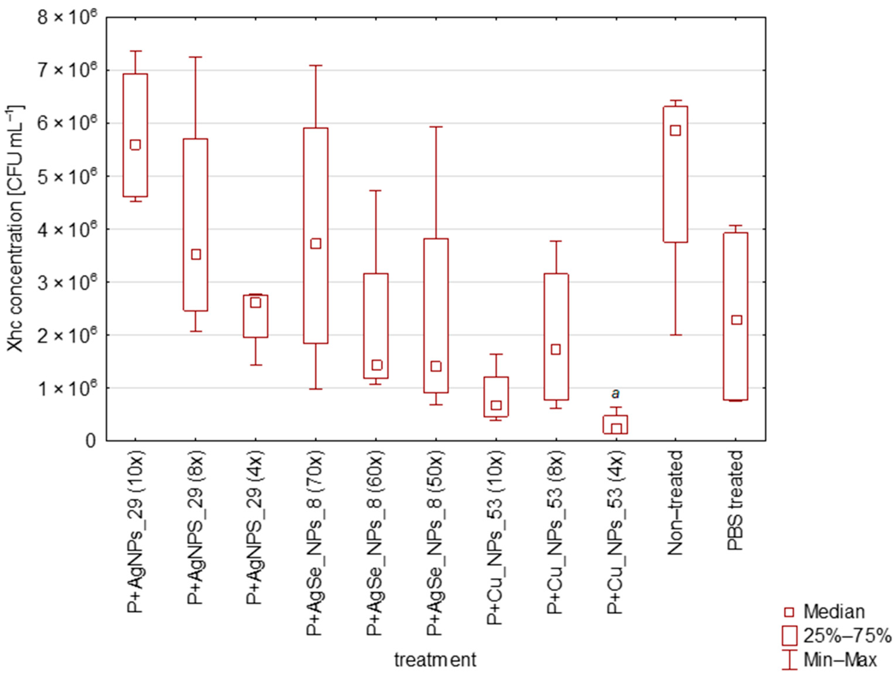
3.2. Effectiviteness of AgNPs_29, CuNPs_53, and AgSeNPs_8 in Elimination of Xhc in Planta

The effectiveness of the NPs in eliminating Xhc infection from germinated plants was tested by real-time PCR (Figure 2). For all the tested treatments on non-inoculated seeds, the absence of Xhc cells was confirmed. In the case of seeds inoculated with Xhc suspension, the results obtained in the in vitro assessment of antibacterial activity were not confirmed in planta. Specifically, complete elimination of Xhc infection from seeds was not achieved at any of the NP concentrations. The highest treatment effect was observed for CuNPs_53 with a 4× diluted stock solution (Cu concentration corresponding to 993 mg L−1), which showed a statistically significant decrease in the concentration of Xhc cells in germinated plants to 3.12 ± 1.63 × 10⁵ cfu mL−1. This value is statistically lower than the amount of Xhc registered in non-treated and PBS-treated controls. In the case of AgNPs_29 and AgSe_NPs_8, Xhc cells were detected in germinated plants at a rate that was not statistically different from the control, even when the highest concentrations of these NPs were used. On the other hand, there was an evident tendency of Xhc decreasing with increasing concentration of nanoparticles used for seed treatment. Worth noting is the reduction of Xhc in the PBS-treated control, which was, however, statistically inconclusive compared to the non-treated control.
The possible negative or positive impact of treatment with individual nanoparticles on the germination of carrot seeds was also evaluated. In comparison to non-treated and PBS-treated seeds, none of the three nanomaterials exhibited a significant positive impact on seed germination percentage (Figure 3). The highest seed germination rate was observed in seeds treated with 10× diluted CuNPs_53 (Cu concentration corresponding to 397 mg mL−1), reaching 92.50 ± 2.75%. Surprisingly, seeds treated with 8× diluted CuNPs_53 showed the lowest germination rate (66.50 ± 3.75%). The germination rates for the remaining nanomaterials were similar to those of the non-treated seeds (85.50 ± 0.75%) and PBS-treated seeds (84.00 ± 1.00%). The significant difference in seed germination was assessed only for CuNPs_53, revealing that the 10× diluted stock solution resulted in higher seed germination compared to the 8× diluted solution. No morphological changes of germinated plants were registered.

4. Discussion

The aim of this study was to assess the possibility of eliminating Xhc, a harmful bacterial pathogen, using an innovative approach based on the application of nanoparticles. Considering that the main source of inoculum for Xhc is typically seeds [3], our work focused on evaluating the effectiveness of treating infected seeds as the key entities responsible for the spread of Xhc. Notably, the overall availability of approaches aimed at eliminating Xhc from seeds is very limited; most studies have tended to focus on foliar applications. For example, the use of copper-based bactericides [37] or isolates of P. syringae pv. syringae has been reported to reduce the colonization of carrot leaves by Xhc [38].
In this study, eight nanoparticles were first tested under in vitro conditions, and silver (Ag), copper (Cu), and Ag/Se composite nanoparticles were identified as the most effective. The identification of just these types of nanoparticles as the most effective is in line with the available literature. Silver nanoparticles are among the historically oldest types of nanoparticles, with their antibacterial effect being one of the first reported properties [39]. In terms of plant protection, using them might seem slightly less logical than, for example, copper nanoparticles, since copper-based products have been used for plant protection for a long time. However, examples can also be found for silver. For instance, Elemawi et al. [40] reduced the incidence of the pathogen Fusarium oxysporum by using biosynthesized Ag-based nanoparticles for treatment of selected crop seeds. Jo et al. [41] significantly reduced the presence of Gibberella fujikuroi, a serious seed-borne fungal pathogen in rice, by treating rice seeds with silver nanoparticles. Pečenka et al. [11] successfully used silver nanoparticles to eliminate Xanthomonas campestris pv. campestris (Xcc) from artificially inoculated seeds, demonstrating significantly higher efficiency than conventional hot water treatment. An overview of the possibilities of using silver nanoparticles in plant disease management is provided in [42].
Based on in vitro tests, copper-based nanoparticles can be recommended being as suitable for protection against plant pathogenic fungi Fusarium solani, Neofusicoccum sp., and Fusarium oxysporum [43]. Copper-based nanoparticles have also been directly applied to tomato plants for protection against Phytophthora infestans. In that study, it was demonstrated that a significantly smaller dose of copper-based nanoparticles was sufficient compared to commercially available copper-based protective products [44]. Copper-based nanoparticles produced through green synthesis were successfully used to eliminate the soil-borne pathogen Ralstonia solanacearum during tobacco plant cultivation [45]. A more comprehensive overview of the sources of copper-based nanoparticles is presented in a review by Banik et al. [46].
The last of the three nanoparticles that was proven to have significant in vitro efficacy against Xhc was silver/selenium composite nanoparticles. Due to their more complex structure and preparation, Ag/Se composite nanoparticles are rarely used in plant protection, and, previously, they were primarily applied in the field of human medicine. An example is a publication demonstrating the effectiveness of silver-selenium nanoparticles for the eradication of dental pathogens [47]. Mittal et al. [48] compared silver and silver-selenium nanoparticles in terms of their anticancer and antimicrobial effects. A more in-depth overview of the antibacterial, fungicidal, and antiviral properties of selenium nanoparticles is provided by Serov et al. [49].
After the in vitro tests, we performed a time-kill assay, which provided important information for subsequent treatment of carrot seed. The observed differences in the efficacy of nanoparticles were surprisingly large, with the AgNPs showing a significantly slower onset than the others. A number of factors could explain these observed differences in antibacterial effect, and these are critically compared in a study by Fan et al. [50], specifically with regard to the combination of AgNPs and CuNPs.
Contrary to the results of in vitro tests, the antibacterial effectiveness of nanoparticles in seeds artificially infected by Xhc did not yield such clearly favorable results. Despite the use of higher concentrations than in the in vitro tests, there was a slight decrease in the Xhc load in treated variants compared to the control group, but this difference was usually not statistically significant. There may be several reasons why such differences between in vitro tests and treatment of inoculated seeds were observed. One is the complex structure of carrot seeds, including their roughness, fibrous spines, and longitudinal projections [51]. These structures may cause pathogen cells to remain wedged, and thus protected from the effects of the applied nanoparticles. Incomplete elimination of Xhc from seeds may subsequently result in surviving Xhc cells contaminating new seedlings, where they can further multiply. Similar trends regarding differences observed between in vitro and in vivo results have also been reported in other studies [52,53].
Even though the results obtained in the seed treatment were not fully conclusive, it is possible to note positive trends in terms of reduced Xhc concentrations in seedlings with increasing nanoparticle concentration. This suggests that the desired effect could be achieved if the concentrations used for seed treatment were increased further. Trends confirming higher efficacy with increasing nanoparticle concentration have also been reported [54,55,56]. On the other hand, it must be considered that increasing the nanoparticle concentration has limitations with regard to a possible negative effect on seed germination. Our results show no negative effect on seed germination in the case of AgNPs. This somewhat disagrees with [57], in which decreased germination in carrot seeds was registered after treatment with silver-based nanoparticles. We also observed a slight decrease in germination with Ag/SeNPs at higher concentrations and, conversely, a statistically significant increase in germination with the lowest concentrations of CuNPs. There are also several examples where Cu treatment increased seed germination, but in species other than carrot [58,59,60]. The results of our study are generally in accordance with the information in review articles summarizing the effect of nanoparticle treatment on seed germination [61,62], which emphasize the influence of the treated plant species and nanoparticle concentration on the final effect.
The only treatment that demonstrated a statistically significant reduction of Xhc in the tested seedlings was copper nanoparticles (CuNPs) at the highest concentration, resulting in approximately 10 times fewer occurrences of Xhc in plantlets. This reduction may also have practical significance, because Xhc infestation is usually associated with large populations of the pathogen on foliage (>106 g−1 leaf tissue) [63,64], and reducing Xhc to one-tenth by CuNP treatment could effectively mitigate economic losses in the production process. This treatment may theoretically be applicable to other pathovars of Xanthomonas hortorum. Additionally, considering that X. hortorum pathovars also attack other important horticultural crops such as lettuce, tomato, artichoke, and pelargonium, the application potential of nanoparticles may be significantly broader than solely carrot seeds.

5. Conclusions

In the present study, three out of eight prepared nanoparticles with the highest in vitro efficacy were selected and tested on artificially infected seeds. Copper nanoparticles (CuNPs) emerged as the most effective. The results further confirm that a responsible assessment of the suitability of nanoparticles for plant protection should not be based solely on in vitro tests of nanoparticle–pathogen interactions. It is essential to test nanoparticles under conditions as close as possible to practical applications, which typically involves monitoring the interactions in the complete triangle of plant material, pathogen, and nanoparticles. Additionally, the results suggest the need to use higher concentrations than those shown to be sufficient in in vitro tests to achieve the desired effect for in planta treatment.

Author Contributions

Conceptualization, E.H., D.T., M.B. and J.Č.; methodology, E.H., D.T., L.R. and Z.B.; validation, E.H., D.T., J.W. and Z.B.; formal analysis, J.W., D.T. and E.H.; investigation, J.W.; resources, E.H., D.T., L.R. and Z.B.; data curation, M.B.; writing—original draft preparation D.T., E.H., M.B. and J.W.; writing—review and editing M.B, L.R., D.T. and E.H.; visualization, D.T. and E.H.; supervision, M.B. and J.Č.; project administration, J.Č. and M.B.; funding acquisition, J.Č. and M.B. All authors have read and agreed to the published version of the manuscript.

Funding

This article was created based on support from the project IGA-ZF/2021-SI2003 (funded by Mendel University in Brno) and the project QK22010031 “Use of innovative potential of nanotechnology to enhance the rentability of selected areas of agricultural production” (funded by the Ministry of Agriculture of the Czech Republic).

Data Availability Statement

The data presented in this study are available on request from the corresponding author.

Conflicts of Interest

The authors declare no conflicts of interest.

References

  1. Mansfield, J.; Genin, S.; Magori, S.; Citovsky, V.; Sriariyanum, M.; Ronald, P.; Dow, M.; Verdier, V.; Beer, S.V.; Machado, M.A. Top 10 plant pathogenic bacteria in molecular plant pathology. Mol. Plant Pathol. 2012, 13, 614–629. [Google Scholar] [CrossRef]
  2. Dean, R.; Van Kan, J.A.; Pretorius, Z.A.; Hammond-Kosack, K.E.; Di Pietro, A.; Spanu, P.D.; Rudd, J.J.; Dickman, M.; Kahmann, R.; Ellis, J. The Top 10 fungal pathogens in molecular plant pathology. Mol. Plant Pathol. 2012, 13, 414–430. [Google Scholar] [CrossRef]
  3. Dia, N.C.; Morinière, L.; Cottyn, B.; Bernal, E.; Jacobs, J.M.; Koebnik, R.; Osdaghi, E.; Potnis, N.; Pothier, J.F. Xanthomonas hortorum–beyond gardens: Current taxonomy, genomics, and virulence repertoires. Mol. Plant Pathol. 2022, 23, 597–621. [Google Scholar] [CrossRef]
  4. Pruvost, O.; Boyer, C.; Robène-Soustrade, I.; Jouen, E.; Saison, A.; Hostachy, B.; Benimadhu, S. First report of Xanthomonas hortorum pv. carotae causing bacterial leaf blight of carrot in Mauritius. Plant Dis. 2010, 94, 1069. [Google Scholar] [CrossRef]
  5. Scott, J.C.; Dung, J.K. Distribution of Xanthomonas hortorum pv. carotae populations in naturally infested carrot seed lots. Plant Dis. 2020, 104, 2144–2148. [Google Scholar] [CrossRef]
  6. Christianson, C.E.; Jones, S.S.; du Toit, L.J. Screening carrot germplasm for resistance to Xanthomonas hortorum pv. carotae. HortScience 2015, 50, 341–350. [Google Scholar] [CrossRef]
  7. Medlock, J.M.; Leach, S.A. Effect of climate change on vector-borne disease risk in the UK. Lancet Infect. Dis. 2015, 15, 721–730. [Google Scholar] [CrossRef]
  8. Moumni, M.; Brodal, G.; Romanazzi, G. Recent innovative seed treatment methods in the management of seedborne pathogens. Food Secur. 2023, 15, 1365–1382. [Google Scholar] [CrossRef]
  9. Nega, E.; Ulrich, R.; Werner, S.; Jahn, M. Hot water treatment of vegetable seed—An alternative seed treatment method to control seed-borne pathogens in organic farming/Heißwasserbehandlung von Gemüsesaatgut—Eine alternative Saatgutbehandlungsmethode zur Bekämpfung samenbürtiger Pathogene im ökologischen Landbau. Zeitschrift für Pflanzenkrankheiten und Pflanzenschutz/J. Plant Dis. Prot. 2003, 110, 220–234. [Google Scholar]
  10. Mandiriza, G.; Kritzinger, Q.; Aveling, T. The evaluation of plant extracts, biocontrol agents and hot water as seed treatments to control black rot of rape in South Africa. Crop. Prot. 2018, 114, 129–136. [Google Scholar] [CrossRef]
  11. Pečenka, J.; Bytešníková, Z.; Kiss, T.; Peňázová, E.; Baránek, M.; Eichmeier, A.; Tekielska, D.; Richtera, L.; Pokluda, R.; Adam, V. Silver nanoparticles eliminate Xanthomonas campestris pv. campestris in cabbage seeds more efficiently than hot water treatment. Mater. Today Commun. 2021, 27, 102284. [Google Scholar] [CrossRef]
  12. Carisse, O.; Ouimet, A.; Toussaint, V.; Philion, V. Evaluation of the effect of seed treatments, bactericides, and cultivars on bacterial leaf spot of lettuce caused by Xanthomonas campestris pv. vitians. Plant Dis. 2000, 84, 295–299. [Google Scholar] [CrossRef]
  13. Kumar, A.; Choudhary, A.; Kaur, H.; Guha, S.; Mehta, S.; Husen, A. Potential applications of engineered nanoparticles in plant disease management: A critical update. Chemosphere 2022, 295, 133798. [Google Scholar] [CrossRef]
  14. Skrzyniarz, K.; Sanchez-Nieves, J.; de la Mata, F.J.; Łysek-Gładysińska, M.; Lach, K.; Ciepluch, K. Mechanistic insight of lysozyme transport through the outer bacteria membrane with dendronized silver nanoparticles for peptidoglycan degradation. Int. J. Biol. Macromol. 2023, 237, 124239. [Google Scholar] [CrossRef]
  15. Soares, E.V.; Soares, H.M. Harmful effects of metal (loid) oxide nanoparticles. Appl. Microbiol. Biotechnol. 2021, 105, 1379–1394. [Google Scholar] [CrossRef] [PubMed]
  16. Gahlawat, G.; Shikha, S.; Chaddha, B.S.; Chaudhuri, S.R.; Mayilraj, S.; Choudhury, A.R. Microbial glycolipoprotein-capped silver nanoparticles as emerging antibacterial agents against cholera. Microb. Cell Factories 2016, 15, 1–14. [Google Scholar] [CrossRef]
  17. Lee, B.; Lee, M.J.; Yun, S.J.; Kim, K.; Choi, I.-H.; Park, S. Silver nanoparticles induce reactive oxygen species-mediated cell cycle delay and synergistic cytotoxicity with 3-bromopyruvate in Candida albicans, but not in Saccharomyces cerevisiae. Int. J. Nanomed. 2019, 14, 4801–4816. [Google Scholar] [CrossRef] [PubMed]
  18. Mikhailova, E.O. Silver nanoparticles: Mechanism of action and probable bio-application. J. Funct. Biomater. 2020, 11, 84. [Google Scholar] [CrossRef] [PubMed]
  19. Adisa, I.O.; Pullagurala, V.L.R.; Peralta-Videa, J.R.; Dimkpa, C.O.; Elmer, W.H.; Gardea-Torresdey, J.L.; White, J.C. Recent advances in nano-enabled fertilizers and pesticides: A critical review of mechanisms of action. Environ. Sci. Nano 2019, 6, 2002–2030. [Google Scholar] [CrossRef]
  20. Ali, M.A.; Ahmed, T.; Wu, W.; Hossain, A.; Hafeez, R.; Islam Masum, M.M.; Wang, Y.; An, Q.; Sun, G.; Li, B. Advancements in plant and microbe-based synthesis of metallic nanoparticles and their antimicrobial activity against plant pathogens. Nanomaterials 2020, 10, 1146. [Google Scholar] [CrossRef] [PubMed]
  21. Strayer, A.; Ocsoy, I.; Tan, W.; Jones, J.; Paret, M. Low concentrations of a silver-based nanocomposite to manage bacterial spot of tomato in the greenhouse. Plant Dis. 2016, 100, 1460–1465. [Google Scholar] [CrossRef] [PubMed]
  22. Oliveira, N.S.; Silva, A.C.A.; Tebaldi, N.D. Simonkolleite nanoparticles for seed treatment and control of tomato bacterial spot caused by Xanthomonas hortorum pv. gardneri. Ciênc. Agrotecnologia 2023, 47, e000623. [Google Scholar] [CrossRef]
  23. Norman, D.J.; Chen, J. Effect of foliar application of titanium dioxide on bacterial blight of geranium and Xanthomonas leaf spot of poinsettia. HortScience 2011, 46, 426–428. [Google Scholar] [CrossRef]
  24. Wohlmuth, J.; Tekielska, D.; Čechová, J.; Baránek, M. Interaction of the nanoparticles and plants in selective growth stages—Usual effects and resulting impact on usage perspectives. Plants 2022, 11, 2405. [Google Scholar] [CrossRef] [PubMed]
  25. Cadena, M.B.; Preston, G.M.; Van der Hoorn, R.A.; Flanagan, N.A.; Townley, H.E.; Thompson, I.P. Enhancing cinnamon essential oil activity by nanoparticle encapsulation to control seed pathogens. Ind. Crop. Prod. 2018, 124, 755–764. [Google Scholar] [CrossRef]
  26. Kaur, P.; Thakur, R.; Choudhary, A. An in vitro study of the antifungal activity of silver/chitosan nanoformulations against important seed borne pathogens. Int. J. Sci. Technol. Res. 2012, 1, 83–86. [Google Scholar]
  27. Prasad, T.; Sudhakar, P.; Sreenivasulu, Y.; Latha, P.; Munaswamy, V.; Reddy, K.R.; Sreeprasad, T.; Sajanlal, P.; Pradeep, T. Effect of nanoscale zinc oxide particles on the germination, growth and yield of peanut. J. Plant Nutr. 2012, 35, 905–927. [Google Scholar] [CrossRef]
  28. Sahab, A.; Waly, A.; Sabbour, M.; Nawar, L.S. Synthesis, antifungal and insecticidal potential of Chitosan (CS)-g-poly (acrylic acid)(PAA) nanoparticles against some seed borne fungi and insects of soybean. Int. J. ChemTech Res. 2015, 8, 589–598. [Google Scholar]
  29. Mawale, K.S.; Nandini, B.; Giridhar, P. Copper and Silver Nanoparticle Seed Priming and Foliar Spray Modulate Plant Growth and Thrips Infestation in Capsicum spp. ACS Omega 2024, 9, 3430–3444. [Google Scholar] [CrossRef]
  30. Štůsková, K.; Pečenka, J.; Tekielska, D.A.; Špetík, M.; Bytešníková, Z.; Švec, P.; Ondreáš, F.; Ridošková, A.; Richtera, L.; Adam, V. The in vitro effects of selected substances and nanomaterials against Diaporthe eres, Diplodia seriata and Eutypa lata. Ann. Appl. Biol. 2023, 182, 226–237. [Google Scholar] [CrossRef]
  31. Bytešníková, Z.; Pečenka, J.; Tekielska, D.; Kiss, T.; Švec, P.; Ridošková, A.; Bezdička, P.; Pekárková, J.; Eichmeier, A.; Pokluda, R. Reduced graphene oxide-based nanometal-composite containing copper and silver nanoparticles protect tomato and pepper against Xanthomonas euvesicatoria infection. Chem. Biol. Technol. Agric. 2022, 9, 1–16. [Google Scholar] [CrossRef]
  32. Bytešníková, Z.; Koláčková, M.; Dobešová, M.; Švec, P.; Ridošková, A.; Pekárková, J.; Přibyl, J.; Cápal, P.; Húska, D.; Adam, V. New insight into the biocompatibility/toxicity of graphene oxides and their reduced forms on Chlamydomonas reinhardtii. NanoImpact 2023, 31, 100468. [Google Scholar] [CrossRef]
  33. da Silva, R.S.; de Oliveira, M.M.G.; de Melo, J.O.; Blank, A.F.; Corrêa, C.B.; Scher, R.; Fernandes, R.P.M. Antimicrobial activity of Lippia gracilis essential oils on the plant pathogen Xanthomonas campestris pv. campestris and their effect on membrane integrity. Pestic. Biochem. Physiol. 2019, 160, 40–48. [Google Scholar] [CrossRef] [PubMed]
  34. Standards, N.C.f.C.L.; Barry, A.L. Methods for Determining Bactericidal Activity of Antimicrobial Agents: Approved Guideline; National Committee for Clinical Laboratory Standards: Wayne, PA, USA, 1999; Volume 19. [Google Scholar]
  35. Roberts, S.; Brough, J.; Hunter, P. Modelling the spread of Xanthomonas campestris pv. campestris in module-raised brassica transplants. Plant Pathol. 2007, 56, 391–401. [Google Scholar] [CrossRef]
  36. Peňázová, E.; Dvořák, M.; Ragasová, L.; Kiss, T.; Pečenka, J.; Čechová, J.; Eichmeier, A. Multiplex real-time PCR for the detection of Clavibacter michiganensis subsp. michiganensis, Pseudomonas syringae pv. tomato and pathogenic Xanthomonas species on tomato plants. PLoS ONE 2020, 15, e0227559. [Google Scholar] [CrossRef]
  37. Du Toft, L.; Crowe, F.; Derie, M.; Simmons, R.; Pelter, G. Bacterial blight of carrot seed crops in the Pacific Northwest. Phytopathology 2004, 94, S26. [Google Scholar] [CrossRef]
  38. Belvoir, T.; Scott, J.C.; Dung, J.K. Identification and screening of bacteria for the biocontrol of Xanthomonas hortorum pv. carotae in carrot seed crops. In Proceedings of the Plant Health 2019, APS Annual Meeting, St Paul, MN, USA, 3–7 August 2019. [Google Scholar]
  39. Clement, J.L.; Jarrett, P.S. Antibacterial silver. Met. Based Drugs 1994, 1, 467–482. [Google Scholar] [CrossRef] [PubMed]
  40. Elamawi, R.M.; Al-Harbi, R.E. Effect of biosynthesized silver nanoparticles on Fusarium oxysporum fungus the cause of seed rot disease of faba bean, tomato and barley. J. Plant Prot. Pathol. 2014, 5, 225–237. [Google Scholar] [CrossRef]
  41. Jo, Y.-K.; Cromwell, W.; Jeong, H.-K.; Thorkelson, J.; Roh, J.-H.; Shin, D.-B. Use of silver nanoparticles for managing Gibberella fujikuroi on rice seedlings. Crop. Prot. 2015, 74, 65–69. [Google Scholar] [CrossRef]
  42. Tariq, M.; Mohammad, K.N.; Ahmed, B.; Siddiqui, M.A.; Lee, J. Biological synthesis of silver nanoparticles and prospects in plant disease management. Molecules 2022, 27, 4754. [Google Scholar] [CrossRef]
  43. Pariona, N.; Mtz-Enriquez, A.I.; Sánchez-Rangel, D.; Carrión, G.; Paraguay-Delgado, F.; Rosas-Saito, G. Green-synthesized copper nanoparticles as a potential antifungal against plant pathogens. RSC Adv. 2019, 9, 18835–18843. [Google Scholar] [CrossRef]
  44. Giannousi, K.; Avramidis, I.; Dendrinou-Samara, C. Synthesis, characterization and evaluation of copper based nanoparticles as agrochemicals against Phytophthora infestans. RSC Adv. 2013, 3, 21743–21752. [Google Scholar] [CrossRef]
  45. Chen, J.; Mao, S.; Xu, Z.; Ding, W. Various antibacterial mechanisms of biosynthesized copper oxide nanoparticles against soilborne Ralstonia solanacearum. RSC Adv. 2019, 9, 3788–3799. [Google Scholar] [CrossRef]
  46. Banik, S.; Pérez-de-Luque, A. In vitro effects of copper nanoparticles on plant pathogens, beneficial microbes and crop plants. Span. J. Agric. Res. 2017, 15, e1005. [Google Scholar] [CrossRef]
  47. Reddy, J.R.S.M.; Kannan, K.P.; Sankaran, K.; Rengasamy, G.; Priya, V.V.; Sathishkumar, P. Eradication of dental pathogens using flavonoid rutin mediated silver-selenium nanoparticles. Inorg. Chem. Commun. 2023, 157, 111391. [Google Scholar] [CrossRef]
  48. Mittal, A.K.; Thanki, K.; Jain, S.; Banerjee, U.C. Comparative studies of anticancer and antimicrobial potential of bioinspired silver and silver-selenium nanoparticles. J. Mater. NanoSci. 2016, 3, 22–27. [Google Scholar]
  49. Serov, D.A.; Khabatova, V.V.; Vodeneev, V.; Li, R.; Gudkov, S.V. A review of the antibacterial, fungicidal and antiviral properties of selenium nanoparticles. Materials 2023, 16, 5363. [Google Scholar] [CrossRef] [PubMed]
  50. Fan, X.; Yahia, L.H.; Sacher, E. Antimicrobial properties of the Ag, Cu nanoparticle system. Biology 2021, 10, 137. [Google Scholar] [CrossRef] [PubMed]
  51. Miranda, R.M.d.; Dias, D.C.F.d.S.; Picoli, E.A.d.T.; Silva, P.P.d.; Nascimento, W.M. Physiological quality, anatomy and histochemistry during the development of carrot seeds (Daucus carota L.). Ciênc. Agrotecnologia 2017, 41, 169–180. [Google Scholar] [CrossRef]
  52. Abdelaziz, A.M.; Salem, S.S.; Khalil, A.M.; El-Wakil, D.A.; Fouda, H.M.; Hashem, A.H. Potential of biosynthesized zinc oxide nanoparticles to control Fusarium wilt disease in eggplant (Solanum melongena) and promote plant growth. BioMetals 2022, 35, 601–616. [Google Scholar] [CrossRef]
  53. Varympopi, A.; Dimopoulou, A.; Papafotis, D.; Avramidis, P.; Sarris, I.; Karamanidou, T.; Kerou, A.K.; Vlachou, A.; Vellis, E.; Giannopoulos, A. Antibacterial activity of copper nanoparticles against Xanthomonas campestris pv. vesicatoria in tomato plants. Int. J. Mol. Sci. 2022, 23, 4080. [Google Scholar] [CrossRef]
  54. Govindan, R.; Chackaravarthi, G.; Ramachandran, G.; Chelliah, C.K.; Muthuchamy, M.; Quero, F.; Mothana, R.A.; Noman, O.M.; Siddiqui, N.A.; Li, W.-J. Effective removal of biofilm formation in Acinetobacter baumannii using chitosan nanoparticles loaded plant essential oils. J. King Saud Univ. Sci. 2022, 34, 101845. [Google Scholar] [CrossRef]
  55. Yang, W.; Fortunati, E.; Gao, D.; Balestra, G.M.; Giovanale, G.; He, X.; Torre, L.; Kenny, J.M.; Puglia, D. Valorization of acid isolated high yield lignin nanoparticles as innovative antioxidant/antimicrobial organic materials. ACS Sustain. Chem. Eng. 2018, 6, 3502–3514. [Google Scholar] [CrossRef]
  56. Bayat, M.; Zargar, M.; Chudinova, E.; Astarkhanova, T.; Pakina, E. In vitro evaluation of antibacterial and antifungal activity of biogenic silver and copper nanoparticles: The first report of applying biogenic nanoparticles against Pilidium concavum and Pestalotia sp. fungi. Molecules 2021, 26, 5402. [Google Scholar] [CrossRef]
  57. Park, S.; Ahn, Y.-J. Multi-walled carbon nanotubes and silver nanoparticles differentially affect seed germination, chlorophyll content, and hydrogen peroxide accumulation in carrot (Daucus carota L.). Biocatal. Agric. Biotechnol. 2016, 8, 257–262. [Google Scholar] [CrossRef]
  58. Bayat, M.; Zargar, M.; Murtazova, K.M.-S.; Nakhaev, M.R.; Shkurkin, S.I. Ameliorating seed germination and seedling growth of nano-primed wheat and flax seeds using seven biogenic metal-based nanoparticles. Agronomy 2022, 12, 811. [Google Scholar] [CrossRef]
  59. Kadri, O.; Karmous, I.; Kharbech, O.; Arfaoui, H.; Chaoui, A. Cu and CuO nanoparticles affected the germination and the growth of barley (Hordeum vulgare L.) seedling. Bull. Environ. Contam. Toxicol. 2022, 108, 585–593. [Google Scholar] [CrossRef] [PubMed]
  60. Essa, H.L.; Abdelfattah, M.S.; Marzouk, A.S.; Shedeed, Z.; Guirguis, H.A.; El-Sayed, M.M. Biogenic copper nanoparticles from Avicennia marina leaves: Impact on seed germination, detoxification enzymes, chlorophyll content and uptake by wheat seedlings. PLoS ONE 2021, 16, e0249764. [Google Scholar] [CrossRef] [PubMed]
  61. Santás-Miguel, V.; Arias-Estévez, M.; Rodríguez-Seijo, A.; Arenas-Lago, D. Use of metal nanoparticles in agriculture. A review on the effects on plant germination. Environ. Pollut. 2023, 334, 122222. [Google Scholar] [CrossRef]
  62. Guo, H.; Liu, Y.; Chen, J.; Zhu, Y.; Zhang, Z. The effects of several metal nanoparticles on seed germination and seedling growth: A meta-analysis. Coatings 2022, 12, 183. [Google Scholar] [CrossRef]
  63. Gilbertson, R. Bacterial Leaf Blight of Carrot; Compendium of Umbelliferous Crop Diseases; American Phytopathological Society: St. Paul, MN, USA, 2002; pp. 11–12. [Google Scholar]
  64. Umesh, K.; Davis, R.; Gilbertson, R. Seed contamination thresholds for development of carrot bacterial blight caused by Xanthomonas campestris pv. carotae. Plant Dis. 1998, 82, 1271–1275. [Google Scholar] [CrossRef] [PubMed]
Figure 1. Kinetics of killing Xhc strain NCPPB 4410 at 14 time points after treatment with nanomaterials at minimum bactericidal concentration: AgNPs_29 (10 mg L−1 Ag), CuNPs_53 (397 mg L−1 Cu), and AgSeNPs_8 (53 mg L−1 Ag and 11 mg L−1 Se).
Agriculture 14 00498 g001
Figure 2. Median concentrations of Xhc cells detected in germinated plants from seeds inoculated with Xhc suspension. Median values ± SD are presented. Lowercase a indicates values statistically different from control (non-treated) variant (Kruskal–Wallis test: H (10, N = 44) = 23.37093, p = 0.0095).
Agriculture 14 00498 g002
Figure 3. Germination of carrot seeds (%) treated with nanomaterials in concentrations used for in planta experiment (Kruskal–Wallis test: H (10, N = 44) = 26.01212, p = 0.0037).
Agriculture 14 00498 g003
Table 1. Nanomaterials tested against Xhc strain NCPPB 4410 in in vitro assays and dilutions used for determination of minimum bactericidal concentration.
NanomaterialSize ShapeConcentration of Stock Solution (mg L−1)Dilution Factor of Stock Solution
AgNPs_29 [11]2 nmSpherical100 Ag10×–100× **
AgNPs_30 [11]22 nmSpherical100 Ag2×–8× *
10×–100×
AgSeNPs_8 [30]60 nmCluster plates3703 Ag
748 Se
10×–100×
200×–1000× ***
CuNPs_50 [30]10 nmSpherical545 Cu2×–8×
CuNPs_53 [30]100 nmSpherical3974 Cu10×–100×
rGO-Cu_25 [31]Flakes, 1×–10 nm thicknessPlate1694 Cu2×–8×
rGO [32]Flakes, 1×–10 nm thickness Plate3100 rGO2×–8×
SeNPs_40 [30]150 nmSpherical517 Se2×–8×
* Stock solutions of nanomaterials were diluted by 2×, 4×, and 8× with sterile Luria broth. ** Stock solutions of nanomaterials were diluted by 10×, 20×, 30×, 40×, 50×, 60×, 70×, 80×, 90×, and 100× with sterile Luria broth medium. *** Stock solutions of nanomaterials were diluted by 200×, 400×, 500×, 600×, 700×, 800×, 900×, and 1000× with sterile Luria broth medium.
Table 2. Nanomaterials tested against Xhc strain NCPPB 4410 in in planta experiment and their concentrations with regard to established minimum bactericidal concentration.
NanomaterialDfConcentration (mg L−1)
AgNPs_2910×10Ag
13
25
CuNPs_5310×397Cu
497
993
AgSeNPs_870×53Ag11Se
60×6213
50×7415
Df: dilution factor used to dissolve stock solution of nanomaterial. Stock solutions were diluted with sterile phosphate-buffered saline. Concentrations of nanomaterials determined as minimum bactericidal concentration are highlighted in bold.
Table 3. Antibacterial efficacy of tested nanomaterials against Xhc strain NCPPB 4410 in vitro based on measured minimum inhibition concentration and minimum bactericidal concentration.
NanomaterialMIC (mg L−1)MBC (mg L−1)
DfAgCuSeDfAgCuSe
AgNPs_2940×3--10×10--
AgNPs_3040×3--NE---
AgSeNPs_8200×19-470×53-11
CuNPs_50NE---NE---
CuNPs_5320×-199-10×-397-
rGO-Cu_2510×-169--847-
rGONE---NE---
SeNPs_40NE---NE---
Df: dilution factor used to dissolve stock solution of nanomaterial. Stock solutions were diluted with sterile Luria broth medium. NE: not effective at highest tested concentration (stock solution diluted 2×).
Disclaimer/Publisher’s Note: The statements, opinions and data contained in all publications are solely those of the individual author(s) and contributor(s) and not of MDPI and/or the editor(s). MDPI and/or the editor(s) disclaim responsibility for any injury to people or property resulting from any ideas, methods, instructions or products referred to in the content.

