1. Introduction
The coating technique consists of covering the surface of an implant with a layer of another biomaterial [
1]. Coating modifies the surface of biomaterials and the biological response of the host tissue in the peri-implant area [
2]. In the case of bone implants, bioactive ceramics, such as hydroxyapatite (HA), are of particular interest in this aspect; they spontaneously form a bony apatite layer on their surface in a living organism and fuse with bone through this layer. These types of materials are of great clinical importance as bone-restorative materials. The formed apatite is highly similar to bone mineral in its composition and structure. Therefore, osteoblasts preferentially proliferate and differentiate to produce apatite as well as collagen on this layer of apatite. When an inert material (whether polymer or metallic) is coated with hydroxyapatite, bone cells adhere to the surface of the apatite coating without intermediate layers. Later, the hydroxyapatite matrix of bone cells becomes integrated with the coating, resulting in excellent adhesion of the coated implant to the bone [
3,
4,
5].
Many biologically active compounds can be used to manufacture coatings, thereby providing some additional biological functions [
6,
7]. Collagen is a polymer of natural origin that has been used in medicine and cosmetology for more than 50 years. Due to its wide range of properties, it is used as a material or intermediate in wound healing dressings, dentistry, scaffold design for osteochondral engineering, otolaryngology, and aesthetic medicine [
8,
9]. Otolaryngology is a medical field focusing on the ears, nose, and throat. It also includes head and neck surgery [
10]. Collagen is the most abundant protein in the extracellular matrix in mammals. It is a non-toxic biopolymer that is highly biodegradable. Moreover, it demonstrates poor immunogenicity and very good degradability. Currently, about 29 types of collagen proteins are known, which are characterized by their diverse and unique structures [
11]. Depending on the structure and function, many types are distinguished. The most important are collagen types I and II. The first one constitutes about 70% of all collagen types, and is mainly found in ligaments, bones, and tendons. The second is the main component of vitreous cartilage and the main collagen of the vitreous body, as well as the nucleus pulposus of intervertebral discs. The presence of collagen ensures flexibility and improves tissue condition [
12,
13,
14]. In biomedical and implant applications, fish collagen is most commonly used. It can take both fibrillar and non-fibrillar forms and is distinguished by lower melting and gelling temperatures than collagens extracted from mammals [
15,
16].
Glutathione (GSH) is an interesting biologically active tripeptide. It is a compound that demonstrates strong antioxidant properties, but beyond that, it is involved in many other functions [
17]. It is responsible for recycling vitamins E and C, transporting amino acids, and producing coenzymes. Importantly, with age, GSH levels gradually decrease, so its external delivery is important [
18,
19,
20].
Another biologically active ingredient that can significantly affect the acceleration of regeneration of the skeletal or cartilaginous system is the above-mentioned HA. It is a ceramic that occurs naturally in the body forming the mineral phase of bone (constituting about 70%, and the remaining 30% is mainly collagen proteins) [
21,
22]. Synthetic HA used in implantology can chemically bind to surrounding tissues; it has osteoconductive properties and the ability to stimulate bone-forming cells to proliferate.
Regardless of the type, due to a variety of processes occurring in the body, biomaterials must be subjected to detailed physicochemical or mechanical analysis. Different physiological fluids exhibit different chemical compositions or pH values, so they can interact differently with implanted foreign bodies. Artificial biological fluids are used to simulate the biological environment in an in vitro laboratory setting. The most common of these are simulated body fluid (SBF), artificial saliva, and Ringer’s fluid, whose composition corresponds to extracellular fluid [
23,
24].
The study of the interaction of materials with fluids allows the monitoring of a range of parameters such as ionic conductivity, changes in pH values, degree of degradation, and swelling coefficient. They can vary depending on the composition of biomaterials. Potentiometric and conductometric tests help determine whether hazardous components are released or precipitate from the interior of the material during fluid interactions, which could negatively affect the cellular balance. Any spike in pH values to highly alkaline or acidic is a cause for concern, since cells in the implant area are unable to proliferate under such conditions [
25,
26]. Degradation studies provide information on how quickly a biomaterial will disintegrate into smaller molecules or fragments and can be replaced by newly growing tissue. However, both degradation and swelling capacity are also strategies for delivering active substances including drugs in targeted therapies. As a result of gradual degradation, active substances that are physically or chemically bound to the material are released. In the case of swelling, drug delivery involves the gradual elution from polymer chains following the penetration of the liquid medium deep into the network [
27,
28,
29]. Given the above, in vitro studies are recommended before proceeding to cellular or in vivo studies on animal models.
The work presented in this paper continues the study of innovative composite coatings containing polyvinylpyrrolidone (PVP), polyethylene glycol (PEG), eGSH, COL, and bioactive HA for bone tissue regeneration [
30,
31]. The composite coating guarantees improved properties because it combines the features of polymeric and ceramic materials. Ceramics are characterized by brittleness, so they are unable to carry loads; however, suspending them in a polymeric hydrogel matrix can overcome this problem without losing their bioactive character. The aim of the study was to evaluate the mechanical and physicochemical properties of the developed materials and to determine their potential as active substance carriers by determining the sorption capacity in selected simulated biological fluids. It should be emphasized that the developed materials have great potential due to the high biological value of the components used in their synthesis, such as glutathione and hydroxyapatite, which promote osteogenesis. No other solution of this type has been found so far.
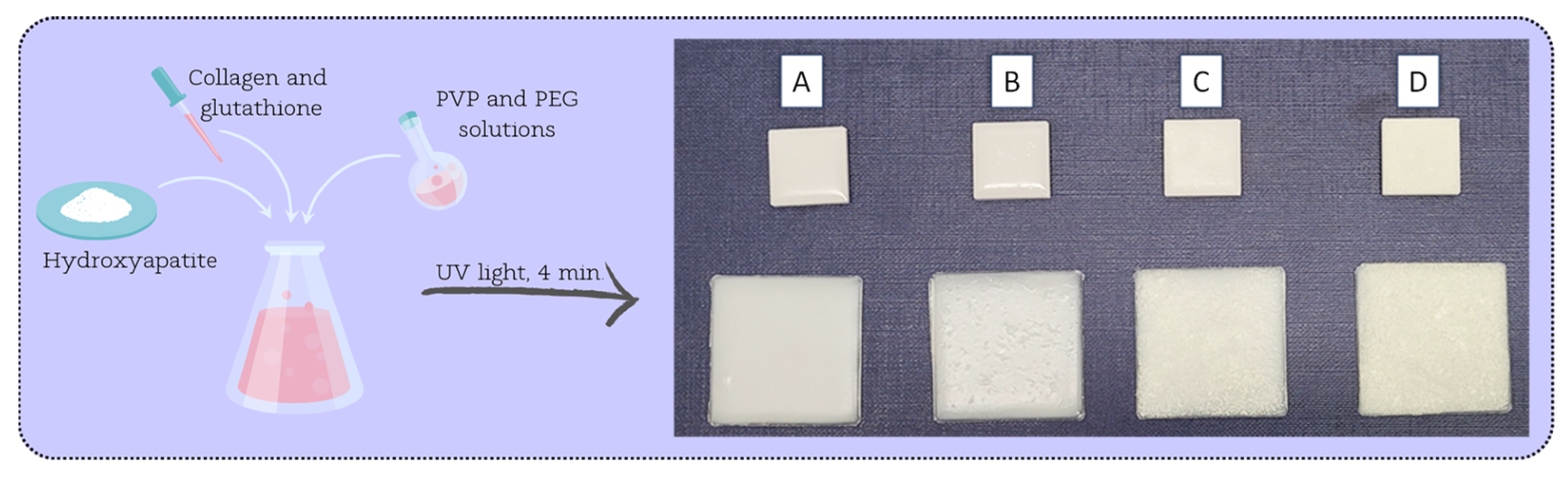
Figure 1 presents the research methodology of the study.
2. Materials and Methods
2.1. Reagents
The reagents used for the synthesis of HA, i.e., calcium acetate monohydrate (Ca(CH3CO2)2·H2O), sodium phosphate dibasic (Na2HPO4), and ammonia water (NH4OH, 25%), as well as polymers, i.e., polyethylene glycol (PEG), polyvinylpyrrolidone (PVP), poly(ethylene glycol) diacrylate Mn 575 (PEGDA), collagen from bovine Achilles tendon (COL), and other reagents, such as 2-hydroxy-2-methylpropiophenone 97% and peptide l-glutathione reduced 98% (GSH), were obtained from Sigma-Aldrich (Darmstadt, Germany). For the preparation of Ringer’s solution and simulated body fluid (SBF) fluid, NaCl, KCl, CaCl2, and Na2SO4 from Eurochem BGD (Tarów, Poland) were used. Additionally, NaHCO3 from DOR-CHEM (Krakow, Poland), K2HPO2·3H2O and MgCl2·6H2O from Chempur (Piekary Slaskie, Poland), 2-amino-2-(hydroxymethyl)- propane-1,3-diol (Tris) from POCH (Gliwice, Poland), and HCl 35–38% solution from Stanlab (Lublin, Poland) were purchased.
2.2. Preparation of Coatings
The coatings were prepared as previously described [
30], and their composition is presented in
Table 1. The crosslinking process was used, exposing the samples to UV light for 4 min with a lamp power of 0.8 J/cm
2, and a distance from the lamp of 5 cm.
The developed method of coating preparation and application is a completely waste-free technique and generates no by-products. Ultraviolet light crosslinking resulted in a fully crosslinked material, and no crumbling of the ceramics was observed even at higher HA concentrations. Also, good integrity was obtained as the coatings were fully continuous and uniformly crosslinked with no significant irregularities or holes.
Figure 2 demonstrates the resulting materials applied to hard polylactide (PLA) plates.
2.3. Methodology of Incubation Tests
2.3.1. Fluid Preparation
A pH-metric study of the coatings was conducted to determine their bioactivity. Three incubation fluids were selected: SBF (simulated body fluid), artificial saliva, and Ringer’s solution (60 mL). These were placed in sterile sealed containers, and 1 g coating discs were immersed in them.
The purpose of this study was to confirm the interactions occurring between the sample and the incubation fluids. The molecules and ions contained in the fluids, interacting with the biomaterial, cause a change in pH value. The materials were incubated in a POL-EKO incubator, model ST 5 B SMART (Wodzisław Śląski, Poland), at 36.6 °C for 40 days. The pH values were measured using an Elmetron CX-701 multifunctional device (Zabrze, Poland).
Ringer’s fluid and SBF were prepared according to the details in
Table 2,
Table 3 and
Table 4. For all fluids, each subsequent component was added after the previous component was completely dissolved. Ringer’s solution was obtained at room temperature. To prepare the SBF solution, 700 mL of distilled water was heated to 36.5 °C (±0.5 °C). One by one, all the ingredients were added, and finally, with a solution of HCl and (CH
2OH)
3CNH
2, the pH was brought to 7.4–7.45. After that distilled water was added to a volume of 1000 mL.
2.3.2. Electrochemical Analysis—Potentiometry
Potentiometric analysis was carried out in order to determine the change in pH values of the solutions in which the coatings were incubated. This assay enables the in vitro stability of the material to be determined in an incubation medium, which simulates conditions in a living organism by its composition. The purpose of this research was to confirm the interactions occurring between the sample and the incubation medium. The ions and molecules contained in the fluids, interfering with the biomaterial, cause a change in the pH value. The obtained dried coating samples with an initial mass of 1 g were incubated at a constant temperature of 36.6 °C in the POL-EKO incubator, model ST 5 B SMART, in prepared artificial biological fluids and distilled water (100 mL) for 40 days. Distilled water was chosen as the reference fluid. Three replicates were performed for each coating composition. The pH value of the fluids was systematically measured using the Elmetron CX-701 multifunctional device with an EPS-1 pH-metric electrode (Zabrze, Poland).
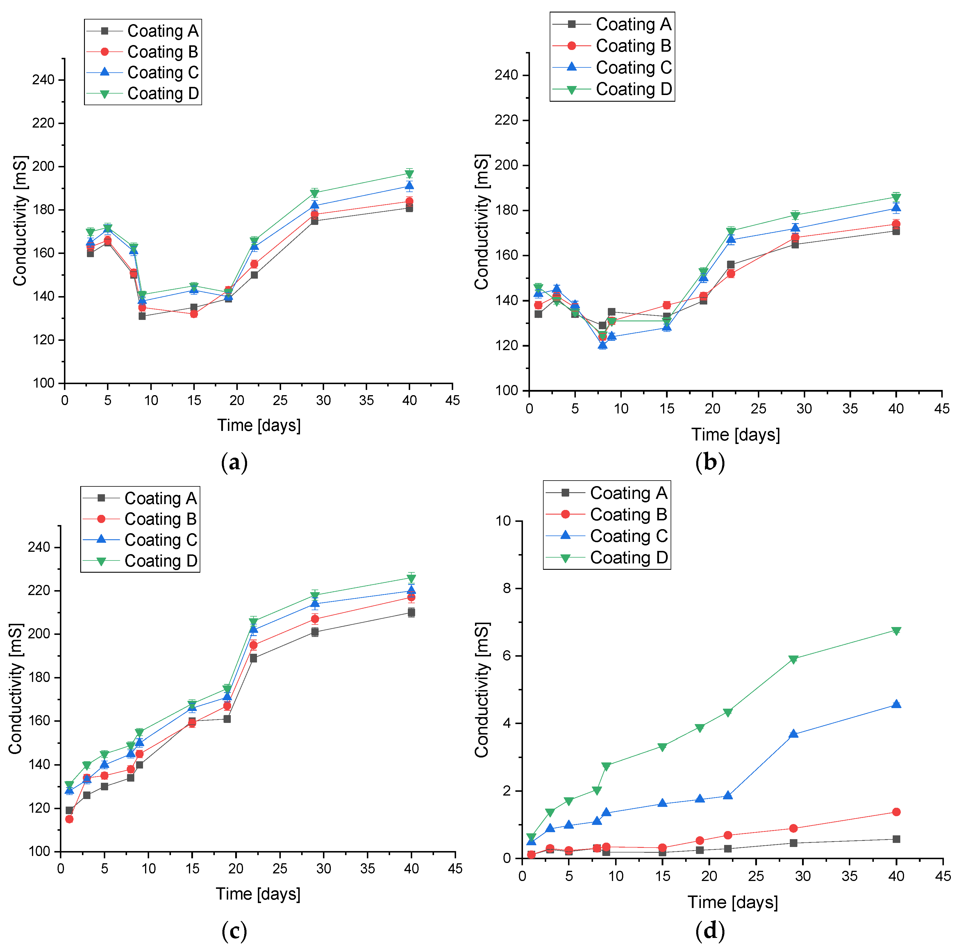
2.3.3. Electroanalytical Analysis—Conductivity
Conductometric analysis was carried out in order to evaluate the ionic conductivity in the incubation medium. The ion exchange occurring at the material/liquid interface causes changes in the conductometric value, as well as gives an indication that the material is not inert under the given conditions. The movement of ions and the appearance of their increasing number causes changes in the conductivity value. Analogous to potentiometric measurement, dried samples of 1 g were placed in artificial biological fluids and distilled water as the reference (100 mL) and incubated in a POL-EKO incubator, model ST 5 B SMART, for 40 days at a constant temperature of 36.6 °C. Three replicates were performed for each coating composition. The conductometric value of the fluids was systematically measured using the Elmetron CX-701 multifunctional device with an ECF-1 conductivity sensor (Zabrze, Poland).
2.3.4. Determination of Sorption Capacity
One of the strategies for delivering drugs and others active ingredients in targeted therapies is to determine the swelling capacity of materials. The composition of composite materials can affect not only their structure, but also the ability of these materials to swell (to bind water within their structure). In order to investigate the relationship between composition and swelling ability, a swelling kinetics study was carried out. For this purpose, 1 g discs were placed in sterile containers filled with the selected incubation fluids, i.e., SBF, artificial saliva, Ringer’s fluid, and distilled water as the reference (100 mL), at 36.6 °C. After 15 min, the samples were removed, excess liquid was collected using filter paper, and the samples were weighed. The measurement was repeated for all samples analogously after 15 min, 30 min, 45 min, 1 h, 2 h, 24 h, 7 days, and 14 days. Three replicates were performed for each coating composition.
The swelling ability of the coatings immersed in fluids was calculated in accordance with the following formula (Equation (1)):
where
m1 is the weight of the immersed sample (g) and
m0 is the initial weight of the sample (g).
The kinetics of coating swelling was investigated using the Voigt-based viscoelastic model (Equation (2)) [
32].
where
St is swelling at a given time
t (%),
Se is equilibrium swelling (power parameter) (%),
t is time of swelling
St (min), and
τ is the rate parameter (min).
2.3.5. Degradation Studies
Depending on the purpose of the biomaterial in the body as well as the functions they are expected to take over, it is expected that they will be stable or able to degrade over time. Degradation allows biomaterials to overgrow with new, natural tissue. To determine the stability of coatings in a biological environment, a degradation study was conducted. For this purpose, discs weighing approximately 1 g were placed in sterile containers filled with selected incubation fluids, i.e., SBF, artificial saliva, Ringer’s fluid (100 mL), and distilled water as the reference, at 36.6 °C. The containers remained unopened for a period of 60 days. Three replicates were performed for each coating composition. After this time, the samples were removed, dried, and weighed. Based on the equation below (Equation (3)), the degree of degradation in each liquid was determined.
where
mt is the weight of the dry sample after incubation time (g) and
mt0 is the initial dry sample mass at time t
0 (g).
2.4. Surface Morphology
The imaging and observation of the structure of the materials provides information on the possible mineralization processes that may occur on the surface under the influence of interaction with the incubation medium. After a 14-day incubation in SBF, the coatings were dried and referred for examination. To determine differences in surface morphology before and after incubation in artificial biological fluids, imaging was performed using a JEOL 5510LV (Tokyo, Japan) scanning electron microscope (SEM) with an EDS IXRF detector. Before SEM measurement, the samples were lyophilized and coated with a conductive gold nanolayer. EDS microanalysis was performed with points in order to detect specific elements on the surface of the samples.
2.5. Hardness Measurement
The composition of the materials as well as the addition of ceramics can affect the mechanical parameters of the coatings obtained. In order to determine the influence of the ceramic phase on the hardness of the coatings, Shore A hardness was measured using a ZwickRoell 3130 (Ulm, Germany). The measurement was performed at a load of 10 N. The instrument was pressed against the material. The indenter, extending from the base, was pressed into the material, as a result of which the balance between the force of the spring and the response of the material was established. Once the equilibrium is established, the pointer stops at the corresponding range of the scale represented in Shore degrees (0–100) [
33]. Three replicates were performed for each coating composition. The hardness of the materials was measured by the Shore method according to the PN-ISO 868 standard, with an indenter according to the PN-93/C-04206 standard [
34,
35].
2.6. Static Tensile Test
Biomaterials in the environment of a living organism are subjected to various loads; hence, it is crucial to study not only their physicochemical but also mechanical parameters. Static tensile tests were carried out on a Shimadzu AGS-X 10 kN testing machine (Kyoto, Japan). The tensile test was conducted in accordance with the PN-EN ISO 527-1 standard at a crosshead speed of 1 mm/min [
36]. A load value of 5 N was applied. Longitudinal paddle-shaped coating samples were prepared in order to perform the static tensile test. Three replicates were performed for each coating composition.
2.7. Statistical Analysis
The results of the experiments were subjected to statistical analysis. Statistical significance was calculated using one-way analysis of variance (ANOVA) (alpha value = 5%). The sorption capacity of the coatings and hardness measurement results were subjected to this analysis. For all other experiments, measurements were performed in triplicate and are presented as mean value and standard deviation (SD).
4. Discussion
Polymer-based composite materials with a suspended ceramic phase are well-known and extensively studied materials in skeletal regeneration applications. However, their potential for use as coating materials represents a niche. The key is the selection of components to obtain a fully crosslinked and homogeneous material. This study presents a method for obtaining coating materials. These were subjected to in vitro incubation tests in order to determine their behavior under conditions similar to those in a living organism. The mechanical parameters of the materials were also determined. The incubation studies were conducted in four incubation fluids—SBF, artificial saliva, Ringer’s fluid, and distilled water—in order to check the behavior of the coatings in environments with slightly different environmental conditions. Changes in pH value, ionic conductivity, and sorption capacity were examined. Some correlations between the first two parameters were shown. In artificial saliva where the greatest changes in pH were observed, there was the greatest increase in ionic conductivity values. Moreover, the greatest degree of degradation was also observed in this fluid. The materials demonstrated the greatest pH stability in Ringer’s fluid and SBF, and in these fluids, the ionic conductivity was also similar. In all cases, the conductivity values were higher for composite coatings than for polymeric ones. The effect of the proportion of the ceramic phase on the sorption capacity was also observed. The higher the proportion of the ceramic phase, the lower the swelling capacity of the biomaterials. The swelling results were compared to a similar work dealing with the development of injectable hydrogels for hard tissue. Although the purpose was different, the chemical composition of the material was similar, consisting of PEG, nanometer-sized ceramics, and PEGDA. In this case, a significantly higher increase in swelling capacity was observed [
43]. However, the cited work used PEG with a molecular weight of 35,000 g/mol, while for the coatings, PEG with a molecular weight of 10,000 g/mol was used. It can therefore be speculated that molecular weight may affect the sorption capacity. Unfortunately, the exact weight for PEGDA was not given. Significantly, the changes in conductivity values confirm the existence of interactions between the incubation fluids and the coatings, as confirmed by SEM and EDS analysis. The formation of new apatite crystals and probably chloride crystals was detected. Once again, a correlation due to the presence of HA in the material was demonstrated, as no new apatite precipitates were observed in coatings where it was absent. On the other hand, as little as 5% of HA was enough for the entire surface to be covered with new apatite layers within 14 days, which is a very desirable phenomenon, indicating the ability of the coatings to undergo biomineralization, which is needed for the target application of the developed materials: coating metallic and polymeric implants and ensuring their bioactive nature. Observations of surface changes and the biomineralization process are consistent with reports in the literature. For composites based on PVP and nano-HA, new apatite crystals were already observed after incubation in SBF at 3 and 7 days [
44]. However, the origin of the hydroxyapatite should also be considered in this type of application, as its bioactivity can vary depending on whether it is natural or synthetic [
45].
The analysis of mechanical properties demonstrated that the materials did not have a high modulus of elasticity despite the presence of HA, which has high strength parameters. This is probably because the coatings were relatively thin (the thickness of the coatings was previously reported in [
30]). However, in this case, its influence on the measured parameter can also be clearly seen, with the modulus of elasticity being higher with more ceramics in the polymer phase. A similar relationship was observed for the measurement of hardness; relating this to previous studies, it can be concluded that once the coating is applied to the target implant/biomaterial, the mechanical strength of the entire system will not be determined by the presence of the developed coating, but by the properties of the covered material. The effect of hydroxyapatite reinforcement on polymeric and hydrogel materials has been reported in the literature, where it has been confirmed that it increases the modulus of elasticity. In other composites based on PEG or PVP and polyvinyl alcohol, the value of modulus of elasticity was up to four times higher, and the final material was able to carry greater loads [
43,
46].
Nevertheless, with reference to the discussion presented above, it should be emphasized that the factors analyzed in the presented manuscript, mainly mechanical resistance and incubation fluids, are not the only ones that affect the implanted biomaterial in the body. Biological fluids, besides the considered ions, contain various types of proteins and other active components that can affect the implanted material via surface energy changes, hydrophilic/hydrophobic interactions, and charge changes through cationic/anionic binding [
47].


