
Biological and Mechanical Performance of Dual-Setting Brushite–Silica Gel Cements
 , , ,
, , ,
Abstract
:1. Introduction
2. Materials and Methods
2.1. Sample Fabrication
2.2. Characterisation
2.3. Biological Testing
3. Results
3.1. Phase Analysis
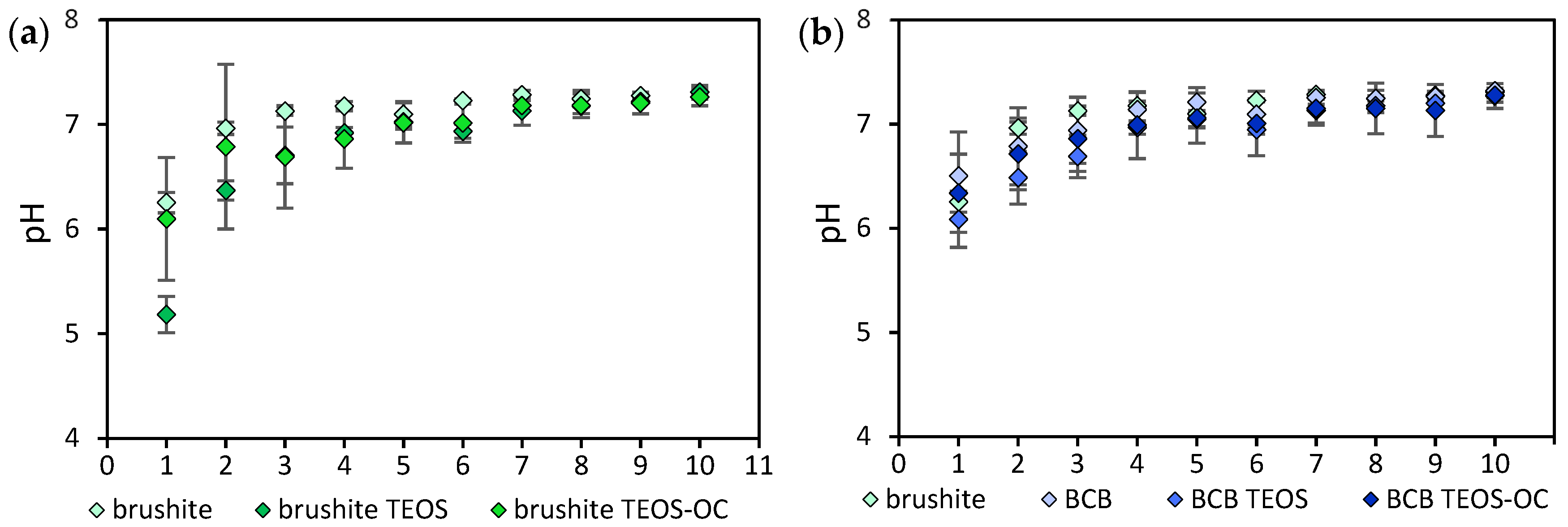
3.2. pH Value
3.3. Compressive Strength
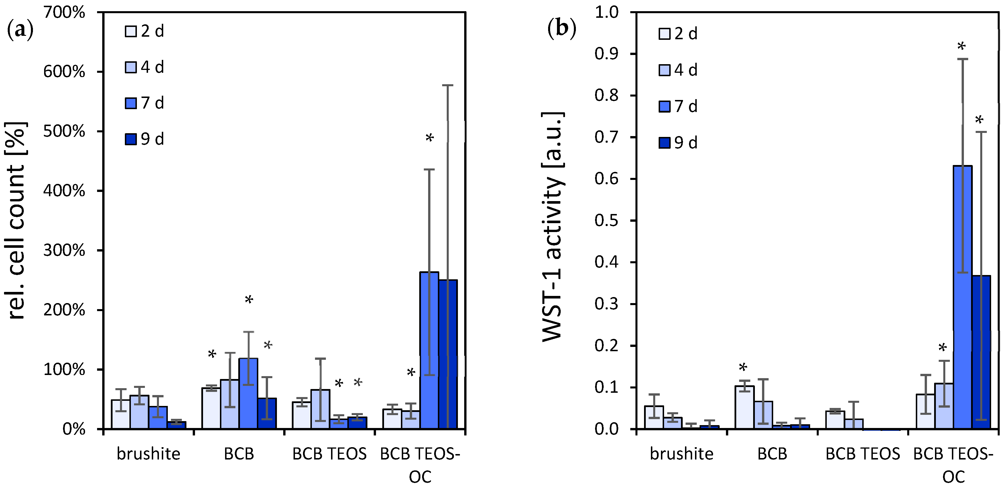
3.4. Osteoblastic-Like Cells
3.5. TRAP Activity Quantitative
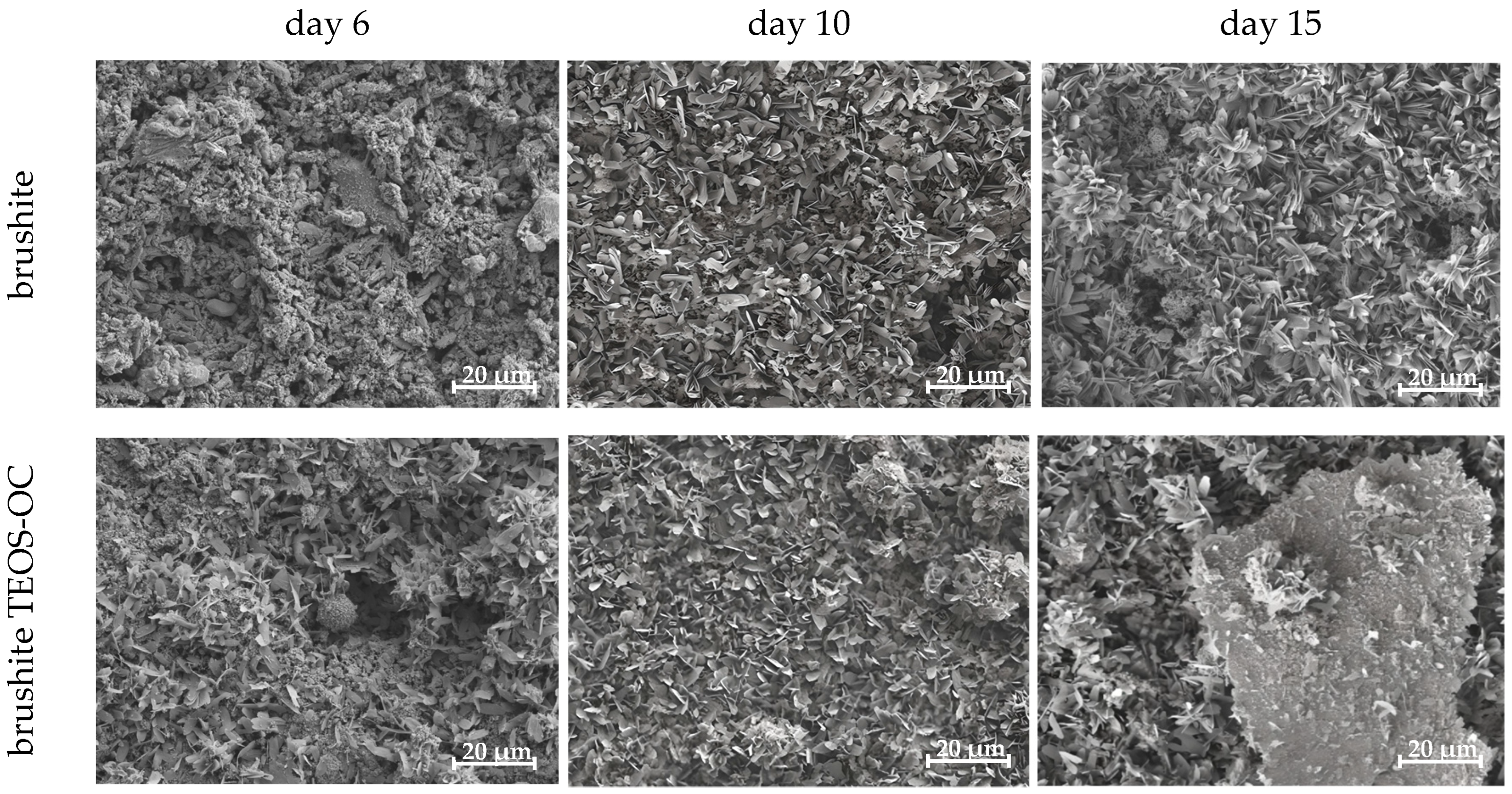
3.6. SEM
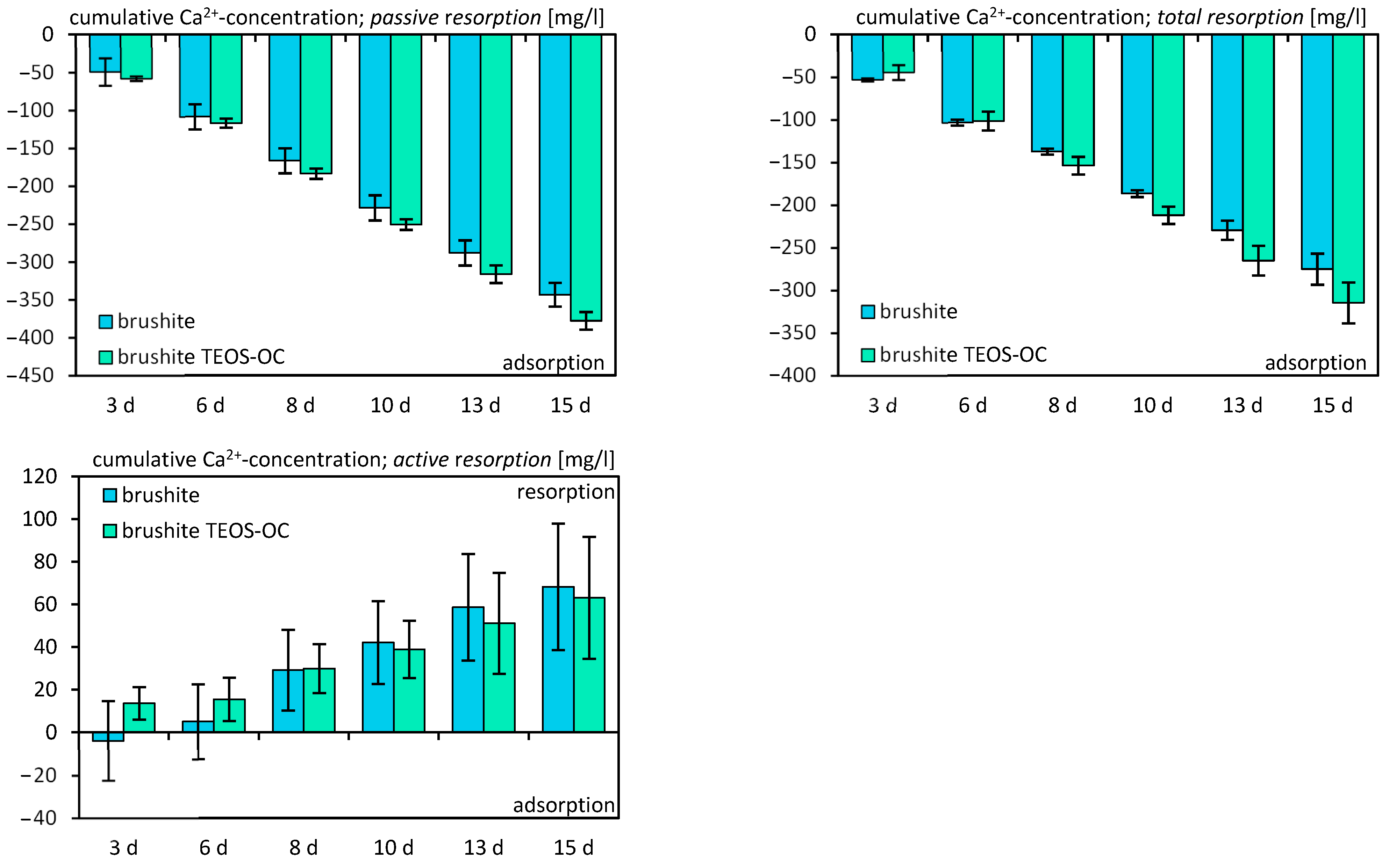
3.7. Solubility
4. Discussion
5. Conclusions
Author Contributions
Funding
Institutional Review Board Statement
Informed Consent Statement
Data Availability Statement
Conflicts of Interest
References
- Bohner, M. Calcium orthophosphates in medicine: From ceramics to calcium phosphate cements. Injury 2000, 31, D37–D47. [Google Scholar] [CrossRef]
- Zhang, J.; Liu, W.; Schnitzler, V.; Tancret, F.; Bouler, J.-M. Calcium phosphate cements for bone substitution: Chemistry, handling and mechanical properties. Acta Biomater. 2014, 10, 1035–1049. [Google Scholar] [CrossRef] [PubMed]
- Ishikawa, K.; Takagi, S.; Chow, L.C.; Ishikawa, Y.; Eanes, E.D.; Asaoka, K. Behavior of a calcium phosphate cement in simulated blood plasma in vitro. Dent. Mater. 1994, 10, 26–32. [Google Scholar] [CrossRef]
- Andrianjatovo, H.; Jose, F.; Lemaitre, J. Effect of β-TCP granularity on setting time and strength of calcium phosphate hydraulic cements. J. Mater. Sci. Mater. Med. 1996, 7, 34–39. [Google Scholar] [CrossRef]
- Bohner, M.; Merkle, H.P.; Lemai^tre, J. In vitro aging of a calcium phosphate cement. J. Mater. Sci. Mater. Med. 2000, 11, 155–162. [Google Scholar] [CrossRef]
- Meininger, S.; Blum, C.; Schamel, M.; Barralet, J.; Ignatius, A.; Gbureck, U. Phytic acid as alternative setting retarder enhanced biological performance of dicalcium phosphate cement in vitro OPEN. Sci. Rep. 2017, 7, 558. [Google Scholar] [CrossRef] [PubMed]
- Apelt, D.; Theiss, F.; El-Warrak, A.O.; Zlinszky, K.; Bettschart-Wolfisberger, R.; Bohner, M.; Matter, S.; Auer, J.A.; von Rechenberg, B. In vivo behavior of three different injectable hydraulic calcium phosphate cements. Biomaterials 2004, 25, 1439–1451. [Google Scholar] [CrossRef]
- Sheikh, Z.; Zhang, Y.L.; Grover, L.; Merle, G.E.; Tamimi, F.; Barralet, J. In vitro degradation and in vivo resorption of dicalcium phosphate cement based grafts. Acta Biomater. 2015, 26, 338–346. [Google Scholar] [CrossRef] [PubMed]
- Brown, W.E. A New Calcium Phosphate Water Setting Cement; Cements Research Progress: Westerville, OH, USA, 1986; pp. 352–379. [Google Scholar]
- Luo, J.; Engqvist, H.; Persson, C. A ready-to-use acidic, brushite-forming calcium phosphate cement. Acta Biomater. 2018, 81, 304–314. [Google Scholar] [CrossRef]
- Engstrand, J.; Persson, C.; Engqvist, H. The effect of composition on mechanical properties of brushite cements. J. Mech. Behav. Biomed. Mater. 2014, 29, 81–90. [Google Scholar] [CrossRef]
- Canal, C.; Ginebra, M.P. Fibre-reinforced calcium phosphate cements: A review. J. Mech. Behav. Biomed. Mater. 2011, 4, 1658–1671. [Google Scholar] [CrossRef] [PubMed]
- Krüger, R.; Groll, J. Fiber reinforced calcium phosphate cements—On the way to degradable load bearing bone substitutes? Biomaterials 2012, 33, 5887–5900. [Google Scholar] [CrossRef] [PubMed]
- dos Santos, L.A.; Carrodéguas, R.G.; Boschi, A.O.; Fonseca de Arruda, A.C. Fiber-enriched double-setting calcium phosphate bone cement. J. Biomed. Mater. Res. Part A 2003, 65A, 244–250. [Google Scholar] [CrossRef] [PubMed]
- Shi, S.-C.; Zeng, X.-X. Silica silanization graft-strengthening bone cement poly(methyl methacrylate): Process and dynamic mechanical properties. Mater. Res. Express 2024, 11, 025005. [Google Scholar] [CrossRef]
- Chiriac, Ş.; Popescu, R.-C.; Pele, M.-M.; Ghiţulică, C.-D.; Cucuruz, A.; Geanaliu-Nicolae, R.-E.; Stancu, I.-C.; Voicu, G.; Ciocan, L.-T. New 3D Printed Scaffolds Based on Walstromite Synthesized by Sol–Gel Method. J. Funct. Biomater. 2024, 15, 19. [Google Scholar] [CrossRef] [PubMed]
- dos Santos, L.A.; Carrodeguas, R.G.; Boschi, A.O.; de Arruda, A.C. Dual-setting calcium phosphate cement modified with ammonium polyacrylate. Artif. Organs 2003, 27, 412–418. [Google Scholar] [CrossRef] [PubMed]
- Wang, J.; Liu, C.; Liu, Y.; Zhang, S. Double-Network Interpenetrating Bone Cement via in situ Hybridization Protocol. Adv. Funct. Mater. 2010, 20, 3997–4011. [Google Scholar] [CrossRef]
- Zhou, P.; Xia, D.; Ni, Z.; Ou, T.; Wang, Y.; Zhang, H.; Mao, L.; Lin, K.; Xu, S.; Liu, J. Calcium silicate bioactive ceramics induce osteogenesis through oncostatin M. Bioact. Mater. 2021, 6, 810–822. [Google Scholar] [CrossRef]
- Uribe, P.; Johansson, A.; Jugdaohsingh, R.; Powell, J.J.; Magnusson, C.; Davila, M.; Westerlund, A.; Ransjö, M. Soluble silica stimulates osteogenic differentiation and gap junction communication in human dental follicle cells. Sci. Rep. 2020, 10, 9923. [Google Scholar] [CrossRef]
- Geffers, M.; Barralet, J.E.; Groll, J.; Gbureck, U. Dual-setting brushite–silica gel cements. Acta Biomater. 2015, 11, 467–476. [Google Scholar] [CrossRef]
- Lee, B.S.; Lin, H.P.; Chan, J.C.; Wang, W.C.; Hung, P.H.; Tsai, Y.H.; Lee, Y.L. A novel sol-gel-derived calcium silicate cement with short setting time for application in endodontic repair of perforations. Int. J. Nanomed. 2018, 13, 261–271. [Google Scholar] [CrossRef]
- Jmal, N.; Bouaziz, J. Synthesis, characterization and bioactivity of a calcium-phosphate glass-ceramics obtained by the sol-gel processing method. Mater. Sci. Eng. C 2017, 71, 279–288. [Google Scholar] [CrossRef]
- Castricum, H.L.; Sah, A.; Geenevasen, J.A.J.; Kreiter, R.; Blank, D.H.A.; Vente, J.F.; ten Elshof, J.E. Structure of hybrid organic–inorganic sols for the preparation of hydrothermally stable membranes. J. Sol-Gel Sci. Technol. 2008, 48, 11–17. [Google Scholar] [CrossRef]
- No, Y.J.; Holzmeister, I.; Lu, Z.; Prajapati, S.; Shi, J.; Gbureck, U.; Zreiqat, H. Effect of Baghdadite Substitution on the Physicochemical Properties of Brushite Cements. Materials 2019, 12, 1719. [Google Scholar] [CrossRef]
- Holzmeister, I. Branched Silica Precursors as Additives for Mineral Bone Cements—Verzweigte Silica-Vorläufer als Additive für Mineralische Knochenzemente. Ph.D. Thesis, Universität Würzburg, Würzburg, Germany, 2023. [Google Scholar]
- Alkhraisat, M.H.; Rueda, C.; Jerez, L.B.; Tamimi Mariño, F.; Torres, J.; Gbureck, U.; Lopez Cabarcos, E. Effect of silica gel on the cohesion, properties and biological performance of brushite cement. Acta Biomater. 2010, 6, 257–265. [Google Scholar] [CrossRef]
- Tamimi-Mariño, F.; Mastio, J.; Rueda, C.; Blanco, L.; López-Cabarcos, E. Increase of the final setting time of brushite cements by using chondroitin 4-sulfate and silica gel. J. Mater. Sci. Mater. Med. 2007, 18, 1195–1201. [Google Scholar] [CrossRef] [PubMed]
- Gerber, T.; Holzhüter, G.; Götz, W.; Bienengräber, V.; Henkel, K.-O.; Rumpel, E. Nanostructuring of Biomaterials—A Pathway to Bone Grafting Substitute. Eur. J. Trauma 2006, 32, 132–140. [Google Scholar] [CrossRef]
- Heinemann, S.; Heinemann, C.; Bernhardt, R.; Reinstorf, A.; Nies, B.; Meyer, M.; Worch, H.; Hanke, T. Bioactive silica–collagen composite xerogels modified by calcium phosphate phases with adjustable mechanical properties for bone replacement. Acta Biomater. 2009, 5, 1979–1990. [Google Scholar] [CrossRef]
- Milea, C.; Bogatu, C.; Duta, A. The influence of parameters in silica sol-gel process. Bull. Transilv. Univ. Bras. 2011, 4, 53. [Google Scholar]
- Brinker, C.J.; Scherer, G.W. Sol-Gel Science: The Physics and Chemistry of Sol-Gel Processing; Academic Press: Cambridge, MA, USA, 1990. [Google Scholar]
- Jamshidi, P.; Bridson, R.H.; Wright, A.J.; Grover, L.M. Brushite cement additives inhibit attachment to cell culture beads. Biotechnol. Bioeng. 2013, 110, 1487–1494. [Google Scholar] [CrossRef]
- Elma, M.; Setyawan, H. Synthesis of Silica Xerogels Obtained in Organic Catalyst via Sol Gel Route. IOP Conf. Ser. Earth Environ. Sci. 2018, 175, 012008. [Google Scholar] [CrossRef]
- Pizzoferrato, A.; Ciapetti, G.; Stea, S.; Cenni, E.; Arciola, C.R.; Granchi, D.; Savarino, L. Cell culture methods for testing biocompatibility. Clin. Mater. 1994, 15, 173–190. [Google Scholar] [CrossRef] [PubMed]
- Hsu, H.; Lacey, D.L.; Dunstan, C.R.; Solovyev, I.; Colombero, A.; Timms, E.; Tan, H.L.; Elliott, G.; Kelley, M.J.; Sarosi, I.; et al. Tumor necrosis factor receptor family member RANK mediates osteoclast differentiation and activation induced by osteoprotegerin ligand. Proc. Natl. Acad. Sci. USA 1999, 96, 3540–3545. [Google Scholar] [CrossRef] [PubMed]
- Collin-Osdoby, P.; Osdoby, P. RANKL-Mediated Osteoclast Formation from Murine RAW 264.7 Cells. In Bone Research Protocols; Helfrich, M.H., Ralston, S.H., Eds.; Humana Press: Totowa, NJ, USA, 2012; pp. 187–202. [Google Scholar] [CrossRef]
- Piper, K.; Boyde, A.; Jones, S.J. The relationship between the number of nuclei of an osteoclast and its resorptive capability in vitro. Anat. Embryol. 1992, 186, 291–299. [Google Scholar] [CrossRef] [PubMed]
- Lees, R.L.; Heersche, J.N.M. Differences in regulation of pHi in large (≥10 nuclei) and small (≤5 nuclei) osteoclasts. Am. J. Physiol.-Cell Physiol. 2000, 279, C751–C761. [Google Scholar] [CrossRef] [PubMed]
- Vahabzadeh, S.; Roy, M.; Bose, S. Effects of silicon on osteoclast cell mediated degradation, in vivo osteogenesis and vasculogenesis of brushite cement. J. Mater. Chem. B 2015, 3, 8973–8982. [Google Scholar] [CrossRef] [PubMed]
- Johnsson, M.S.-A.; Nancollas, G.H. The role of brushite and octacalcium phosphate in apatite formation. Crit. Rev. Oral Biol. Med. 1992, 3, 61–82. [Google Scholar] [CrossRef] [PubMed]
- Possenti, E.; Colombo, C.; Bersani, D.; Bertasa, M.; Botteon, A.; Conti, C.; Lottici, P.P.; Realini, M. New insight on the interaction of diammonium hydrogenphosphate conservation treatment with carbonatic substrates: A multi-analytical approach. Microchem. J. 2016, 127, 79–86. [Google Scholar] [CrossRef]
- Constantz, B.R.; Barr, B.M.; Ison, I.C.; Fulmer, M.T.; Baker, J.; McKinney, L.; Goodman, S.B.; Gunasekaren, S.; Delaney, D.C.; Ross, J.; et al. Histological, chemical, and crystallographic analysis of four calcium phosphate cements in different rabbit osseous sites. J. Biomed. Mater. Res. 1998, 43, 451–461. [Google Scholar] [CrossRef]
- Schamel, M.; Barralet, J.E.; Groll, J.; Gbureck, U. In vitro ion adsorption and cytocompatibility of dicalcium phosphate ceramics. Biomater. Res. 2017, 21, 10. [Google Scholar] [CrossRef]


| Sample Labelling | Baghdadite [g] | β-TCP [g] | MCPA [g] |
|---|---|---|---|
| brushite | 0 | 11.04 | 8.31 |
| brushite TEOS | 0 | 11.04 | 8.31 |
| brushite TEOS-OC | 0 | 11.04 | 8.31 |
| BCB | 2.24 | 8.82 | 8.31 |
| BCB TEOS | 2.24 | 8.82 | 8.31 |
| BCB TEOS-OC | 2.24 | 8.82 | 8.31 |
| Sample Labelling | H2O [mL] | TEOS [mL] | 1,8-Bis(triethoxysilyl)octane [mL] |
|---|---|---|---|
| brushite | 5.11 | 0 | 0 |
| brushite TEOS | 5.11 | 7 | 0 |
| brushite TEOS-OC | 8.31 | 7 | 6.39 |
| BCB | 5.11 | 0 | 0 |
| BCB TEOS | 5.11 | 7 | 0 |
| BCB TEOS-OC | 8.31 | 7 | 6.39 |
| Sample | Brushite [%] | β-TCP [%] | Monetite [%] | Baghdadite [%] |
|---|---|---|---|---|
| brushite | 26.7 | 29.5 | 43.8 | 0 |
| brushite TEOS | 11.1 | 2.9 | 85.0 | 0 |
| brushite TEOS-OC | 32.5 | 23.0 | 44.4 | 0 |
| BCB | 46.3 | 9.5 | 35.6 | 8.6 |
| BCB TEOS | 6.2 | 8.9 | 71.2 | 6.2 |
| BCB TEOS-OC | 15.6 | 5.5 | 69.8 | 9.4 |
Disclaimer/Publisher’s Note: The statements, opinions and data contained in all publications are solely those of the individual author(s) and contributor(s) and not of MDPI and/or the editor(s). MDPI and/or the editor(s) disclaim responsibility for any injury to people or property resulting from any ideas, methods, instructions or products referred to in the content. |
© 2024 by the authors. Licensee MDPI, Basel, Switzerland. This article is an open access article distributed under the terms and conditions of the Creative Commons Attribution (CC BY) license (https://creativecommons.org/licenses/by/4.0/).
Share and Cite
Steinacker, V.C.; Renner, T.; Holzmeister, I.; Gubik, S.; Müller-Richter, U.; Breitenbücher, N.; Fuchs, A.; Straub, A.; Scheurer, M.; Kübler, A.C.; et al. Biological and Mechanical Performance of Dual-Setting Brushite–Silica Gel Cements. J. Funct. Biomater. 2024, 15, 108. https://doi.org/10.3390/jfb15040108
Steinacker VC, Renner T, Holzmeister I, Gubik S, Müller-Richter U, Breitenbücher N, Fuchs A, Straub A, Scheurer M, Kübler AC, et al. Biological and Mechanical Performance of Dual-Setting Brushite–Silica Gel Cements. Journal of Functional Biomaterials. 2024; 15(4):108. https://doi.org/10.3390/jfb15040108
Chicago/Turabian StyleSteinacker, Valentin C., Tobias Renner, Ib Holzmeister, Sebastian Gubik, Urs Müller-Richter, Niko Breitenbücher, Andreas Fuchs, Anton Straub, Mario Scheurer, Alexander C. Kübler, and et al. 2024. "Biological and Mechanical Performance of Dual-Setting Brushite–Silica Gel Cements" Journal of Functional Biomaterials 15, no. 4: 108. https://doi.org/10.3390/jfb15040108