Next Issue
Volume 15, May
Previous Issue
Volume 15, March


J. Funct. Biomater., Volume 15, Issue 4 (April 2024) – 32 articles

Cover Story (view full-size image): 
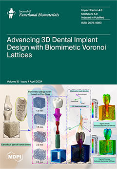
This work uses finite element analysis (FEA) to combine biomimetic Voronoi lattice structures that simulate the cancellous bone of the human jaw to advance dental implant designs. It utilizes nTOP 4.17.3 and Creo Parametric 8.0 to accurately mimic the trabecular bone's complicated porosity and biomechanical properties. The research presented improves the biomechanical performance of dental implants during dynamic mastication by varying pore diameters from 1.0 to 2.5 mm. These findings indicate the ideal pore size, which is consistent with natural bone mechanics, thereby significantly improving osseointegration and stability. Indeed, this is an important step in the development of long-lasting, patient-specific implants, thus laying the foundations for the next generation of dental treatments. View this paper
  • Issues are regarded as officially published after their release is announced to the table of contents alert mailing list.
  • You may sign up for e-mail alerts to receive table of contents of newly released issues.
  • PDF is the official format for papers published in both, html and pdf forms. To view the papers in pdf format, click on the "PDF Full-text" link, and use the free Adobe Reader to open them.
Order results
Result details
Section
Select all
Export citation of selected articles as:
11 pages, 3878 KiB  
Article
Deferoxamine-Loaded Chitosan-Based Hydrogel on Bone Implants Showing Enhanced Bond Strength and Pro-Angiogenic Effects
by Huan Liu, Kai Li, Deliang Yi, Yi Ding, Yanfeng Gao and Xuebin Zheng
J. Funct. Biomater. 2024, 15(4), 112; https://doi.org/10.3390/jfb15040112 - 22 Apr 2024
Viewed by 344
Abstract
Angiogenesis is vital for bone fracture healing and plays a significant role in the fate of orthopedic implants. The growth and maintenance of new blood vessels at the fracture site of patients is essential, which promotes the clinical outcome of plasma sprayed Ti [...] Read more.
Angiogenesis is vital for bone fracture healing and plays a significant role in the fate of orthopedic implants. The growth and maintenance of new blood vessels at the fracture site of patients is essential, which promotes the clinical outcome of plasma sprayed Ti (PST) coated orthopedic implants. In order to endow the PST coating with pro-angiogenic effects, deferoxamine-loaded chitosan-based hydrogel was fabricated on the coating surface. Polydopamine-modified chitosan (CS/PDA) hydrogel exhibited enhanced bonding strength to PST coatings as evidenced by scratch test. The deferoxamine-loaded CS/PDA (CS/PDA-DFO) exhibited a sustained drug-release property, and the cumulative concentration of released DFO reached 20.21 μg/mL on day 7. PST-CS/PDA with higher wettability and active group quantity enhanced the viability and adhesion characteristics of human umbilical vein endothelial cells (HUVECs) and upregulated the secretion level of nitric oxide and vascular endothelial growth factor. Moreover, the introduction of DFO in PST-CS/PDA further enhanced the pro-angiogenic effects. Above all, this study offers a novel approach for developing hydrogel coating on orthopedic implants showing enhanced bonding strength and pro-angiogenic effects. Full article
(This article belongs to the Section Bone Biomaterials)
Show Figures

Figure 1

17 pages, 3600 KiB  
Article
Natural Biomolecule Ovomucin–Chitosan Oligosaccharide Self-Assembly Nanogel for Lutein Application Enhancement: Characterization, Environmental Stability and Bioavailability
by Qi Xu, Haoye Teng, Xuanchen Li, Zhenqing Zhang, Yumeng Han and Haixin Sun
J. Funct. Biomater. 2024, 15(4), 111; https://doi.org/10.3390/jfb15040111 - 21 Apr 2024
Viewed by 294
Abstract
As an essential nutrient, lutein (LUT) has the ability to aid in the prevention of eye diseases, cardiovascular diseases, and cancer. However, the application of LUT is largely restricted by its poor solubility and susceptibility to oxidative degradation. Thus, in this study, LUT-loaded [...] Read more.
As an essential nutrient, lutein (LUT) has the ability to aid in the prevention of eye diseases, cardiovascular diseases, and cancer. However, the application of LUT is largely restricted by its poor solubility and susceptibility to oxidative degradation. Thus, in this study, LUT-loaded nanogel (OVM-COS-LUT) was prepared by a self-assembly of ovomucin (OVM) and chitosan oligosaccharide (COS) to enhance the effective protection and bioavailability of LUT. The nanogel had excellent dispersion (PDI = 0.25) and an 89.96% LUT encapsulation rate. XRD crystal structure analysis confirmed that the encapsulated LUT maintained an amorphous morphology. In addition, the nanogel showed satisfactory stability with pH levels ranging from 2 to 9 and high ionic strengths (>100 mM). Even under long-term storage, the nanogel maintained an optimistic stabilization and protection capacity; its effective retention rates could reach 96.54%. In vitro, digestion simulation showed that the bioaccessibility and sustained release of OVM-COS-LUT nanogel was superior to that of free LUT. The nanogel provided significant antioxidant activity, and no significant harmful effects were detected in cytotoxicity analyses at higher concentrations. In summary, OVM-COS-LUT can be utilized as a potential safe oral and functional carrier for encapsulating LUT. Full article
(This article belongs to the Special Issue State of the Art in Biomaterials for Drug Delivery)
Show Figures

Figure 1

34 pages, 23488 KiB  
Article
Performance of Austenitic High-Nitrogen Steels under Gross Slip Fretting Corrosion in Bovine Serum
by Alfons Fischer, Philipe Telouk, Christian Beckmann, Saskia Heermant, Adrian Wittrock, Jörg Debus and Markus A. Wimmer
J. Funct. Biomater. 2024, 15(4), 110; https://doi.org/10.3390/jfb15040110 - 18 Apr 2024
Viewed by 319
Abstract
Modular artificial hip joints are a clinical standard today. However, the release of wear products from the head–taper interface, which includes wear particles in the nm size range, as well as metal ions, have raised concerns. Depending on the loading of such taper [...] Read more.
Modular artificial hip joints are a clinical standard today. However, the release of wear products from the head–taper interface, which includes wear particles in the nm size range, as well as metal ions, have raised concerns. Depending on the loading of such taper joints, a wide variety of different mechanisms have been found by retrieval analyses. From these, this paper concentrates on analyzing the contribution of gross slip fretting corrosion at ultra-mild wear rates using a bovine calf serum solution (BCS) as the lubricant. The parameters were chosen based on biomechanical considerations, producing wear rates of some ng/m wear path. In parallel, the evolution of tribomaterial (third bodies) was analyzed as to its constituents and generation rates. It has already been shown earlier that, by an advantageous combination of wear mechanisms and submechanisms, certain constituents of the tribomaterial remain inside the contact area and act like extreme-pressure lubricant additives. For the known wear and corrosion resistance of austenitic high-nitrogen steels (AHNSs), which outperform CoCrMo alloys even under inflammatory conditions, we hypothesized that such steels will generate ultra-mild wear rates under gross slip fretting. While testing AHNSs against commercially available biomedical-grade materials of CoCrMo and TiAlV alloys, as well as zirconia-toughened alumina (ZTA) and against itself, it was found that AHNSs in combination with a Ti6Al4V alloy generated the smallest wear rate under gross slip fretting corrosion. This paper then discusses the wear behavior on the basis of ex situ analyses of the worn surfaces as to the acting wear mechanisms and submechanisms, as well as to the tribological reaction products. Full article
(This article belongs to the Special Issue Metallic Biomaterials for Medical Applications)
Show Figures

Figure 1

16 pages, 5700 KiB  
Article
3D Printing of a Porous Zn-1Mg-0.1Sr Alloy Scaffold: A Study on Mechanical Properties, Degradability, and Biosafety
by Xiangyu Cao, Xinguang Wang, Jiazheng Chen, Xiao Geng and Hua Tian
J. Funct. Biomater. 2024, 15(4), 109; https://doi.org/10.3390/jfb15040109 - 18 Apr 2024
Viewed by 302
Abstract
In recent years, the use of zinc (Zn) alloys as degradable metal materials has attracted considerable attention in the field of biomedical bone implant materials. This study investigates the fabrication of porous scaffolds using a Zn-1Mg-0.1Sr alloy through a three-dimensional (3D) printing technique, [...] Read more.
In recent years, the use of zinc (Zn) alloys as degradable metal materials has attracted considerable attention in the field of biomedical bone implant materials. This study investigates the fabrication of porous scaffolds using a Zn-1Mg-0.1Sr alloy through a three-dimensional (3D) printing technique, selective laser melting (SLM). The results showed that the porous Zn-1Mg-0.1Sr alloy scaffold featured a microporous structure and exhibited a compressive strength (CS) of 33.71 ± 2.51 MPa, a yield strength (YS) of 27.88 ± 1.58 MPa, and an elastic modulus (E) of 2.3 ± 0.8 GPa. During the immersion experiments, the immersion solution showed a concentration of 2.14 ± 0.82 mg/L for Zn2+ and 0.34 ± 0.14 mg/L for Sr2+, with an average pH of 7.61 ± 0.09. The porous Zn-1Mg-0.1Sr alloy demonstrated a weight loss of 12.82 ± 0.55% and a corrosion degradation rate of 0.36 ± 0.01 mm/year in 14 days. The Cell Counting Kit-8 (CCK-8) assay was used to check the viability of the cells. The results showed that the 10% and 20% extracts significantly increased the activity of osteoblast precursor cells (MC3T3-E1), with a cytotoxicity grade of 0, which indicates safety and non-toxicity. In summary, the porous Zn-1Mg-0.1Sr alloy scaffold exhibits outstanding mechanical properties, an appropriate degradation rate, and favorable biosafety, making it an ideal candidate for degradable metal bone implants. Full article
(This article belongs to the Special Issue Advanced 3D Printing Biomaterials)
Show Figures

Figure 1

16 pages, 4806 KiB  
Article
Biological and Mechanical Performance of Dual-Setting Brushite–Silica Gel Cements
by Valentin C. Steinacker, Tobias Renner, Ib Holzmeister, Sebastian Gubik, Urs Müller-Richter, Niko Breitenbücher, Andreas Fuchs, Anton Straub, Mario Scheurer, Alexander C. Kübler and Uwe Gbureck
J. Funct. Biomater. 2024, 15(4), 108; https://doi.org/10.3390/jfb15040108 - 18 Apr 2024
Viewed by 367
Abstract
Bone defects resulting from trauma, diseases, or surgical procedures pose significant challenges in the field of oral and maxillofacial surgery. The development of effective bone substitute materials that promote bone healing and regeneration is crucial for successful clinical outcomes. Calcium phosphate cements (CPCs) [...] Read more.
Bone defects resulting from trauma, diseases, or surgical procedures pose significant challenges in the field of oral and maxillofacial surgery. The development of effective bone substitute materials that promote bone healing and regeneration is crucial for successful clinical outcomes. Calcium phosphate cements (CPCs) have emerged as promising candidates for bone replacement due to their biocompatibility, bioactivity, and ability to integrate with host tissues. However, there is a continuous demand for further improvements in the mechanical properties, biodegradability, and bioactivity of these materials. Dual setting of cements is one way to improve the performance of CPCs. Therefore, silicate matrices can be incorporated in these cements. Silicate-based materials have shown great potential in various biomedical applications, including tissue engineering and drug delivery systems. In the context of bone regeneration, silicate matrices offer unique advantages such as improved mechanical stability, controlled release of bioactive ions, and enhanced cellular responses. Comprehensive assessments of both the material properties and biological responses of our samples were conducted. Cytocompatibility was assessed through in vitro testing using osteoblastic (MG-63) and osteoclastic (RAW 264.7) cell lines. Cell activity on the surfaces was quantified, and scanning electron microscopy (SEM) was employed to capture images of the RAW cells. In our study, incorporation of tetraethyl orthosilicate (TEOS) in dual-curing cements significantly enhanced physical properties, attributed to increased crosslinking density and reduced pore size. Higher alkoxysilyl group concentration improved biocompatibility by facilitating greater crosslinking. Additionally, our findings suggest citrate’s potential as an alternative retarder due to its positive interaction with the silicate matrix, offering insights for future dental material research. This paper aims to provide an overview of the importance of silicate matrices as modifiers for calcium phosphate cements, focusing on their impact on the mechanical properties, setting behaviour, and biocompatibility of the resulting composites. Full article
Show Figures

Figure 1

4 pages, 178 KiB  
Editorial
Functional Biomaterials and Digital Technologies in Dentistry: From Bench to Bedside
by Ping Li, Guojiang Wan, Shulan Xu and An Li
J. Funct. Biomater. 2024, 15(4), 107; https://doi.org/10.3390/jfb15040107 - 17 Apr 2024
Viewed by 303
Abstract
This Special Issue, “Functional Biomaterials and Digital Technologies in Dentistry: From Bench to Bedside”, highlights the integration of advanced materials science and digital technologies in dental and maxillofacial applications [...] Full article
12 pages, 978 KiB  
Article
Effects of Erythropoietin-Promoted Fracture Healing on Bone Turnover Markers in Cats
by Radina Vasileva, Tsvetan Chaprazov and Aneliya Milanova
J. Funct. Biomater. 2024, 15(4), 106; https://doi.org/10.3390/jfb15040106 - 17 Apr 2024
Viewed by 319
Abstract
In orthopaedics, erythropoietin (EPO) is applied in the preoperative management of anaemic patients, but also as a stimulating factor to assist bone regeneration due to its angiogenic and osteoinductive potential. Since orthopaedists mainly rely on their clinical experience to assess bone healing, additional [...] Read more.
In orthopaedics, erythropoietin (EPO) is applied in the preoperative management of anaemic patients, but also as a stimulating factor to assist bone regeneration due to its angiogenic and osteoinductive potential. Since orthopaedists mainly rely on their clinical experience to assess bone healing, additional and more objective methods such as studying the dynamics of bone markers are needed. Therefore, the aim of this study was to investigate the plasma activity of bone-specific alkaline phosphatase (BALP), the N-terminal propeptide of type I collagen (PINP), the C-terminal telopeptide of type I collagen (CTX), and deoxypyridinoline (DPD) during the first 2 months of healing of comminuted fractures in cats, either non-stimulated or locally stimulated with recombinant human erythropoietin (rhEPO). The study included twelve cats of mixed breeds, aged 7.2 ± 4 months, weighing 2.11 ± 1.1 kg, with comminuted diaphyseal fractures of the femur. Surgical treatment with plate osteosynthesis was performed in all animals. The cats were randomly divided into two groups—a control (n = 6) and an EPO group (n = 6). The locally applied EPO leads to the increased activity of bone formation markers (BALP and PINP) during the second week after the osteosynthesis, preceding the peaks in the control group by two weeks. The studied bone resorption markers (DPD, CTX) varied insignificantly during the studied period. In conclusion, erythropoietin could serve as a promoter of bone healing in comminuted fractures in cats. Full article
(This article belongs to the Special Issue Advances in Bone Substitute Biomaterials)
Show Figures

Figure 1

13 pages, 14329 KiB  
Article
Strontium-Doped Bioglass-Laden Gelatin Methacryloyl Hydrogels for Vital Pulp Therapy
by Sepideh Aminmansour, Ana Beatriz Gomes de Carvalho, Lais Medeiros Cardoso, Caroline Anselmi, Maedeh Rahimnejad, Renan Dal-Fabbro, Erika Benavides, Tiago Moreira Bastos Campos, Alexandre Luiz Souto Borges and Marco C. Bottino
J. Funct. Biomater. 2024, 15(4), 105; https://doi.org/10.3390/jfb15040105 - 17 Apr 2024
Viewed by 459
Abstract
This study aimed to develop gelatin methacryloyl (GelMA)-injectable hydrogels incorporated with 58S bioactive glass/BG-doped with strontium for vital pulp therapy applications. GelMA hydrogels containing 0% (control), 5%, 10%, and 20% BG (w/v) were prepared. Their morphological and chemical properties [...] Read more.
This study aimed to develop gelatin methacryloyl (GelMA)-injectable hydrogels incorporated with 58S bioactive glass/BG-doped with strontium for vital pulp therapy applications. GelMA hydrogels containing 0% (control), 5%, 10%, and 20% BG (w/v) were prepared. Their morphological and chemical properties were evaluated by scanning electron microscopy/SEM, energy dispersive spectroscopy/EDS, and Fourier transform infrared spectroscopy/FTIR (n = 3). Their swelling capacity and degradation ratio were also measured (n = 4). Cell viability (n = 8), mineralized matrix formation, cell adhesion, and spreading (n = 6) on DPSCs were evaluated. Data were analyzed using ANOVA/post hoc tests (α = 5%). SEM and EDS characterization confirmed the incorporation of BG particles into the hydrogel matrix, showing GelMA’s (C, O) and BG’s (Si, Cl, Na, Sr) chemical elements. FTIR revealed the main chemical groups of GelMA and BG, as ~1000 cm−1 corresponds to Si-O and ~1440 cm−1 to C-H. All the formulations were degraded by day 12, with a lower degradation ratio observed for GelMA+BG20%. Increasing the concentration of BG resulted in a lower mass swelling ratio. Biologically, all the groups were compatible with cells (p > 0.6196), and cell adhesion increased over time, irrespective of BG concentration, indicating great biocompatibility. GelMA+BG5% demonstrated a higher deposition of mineral nodules over 21 days (p < 0.0001), evidencing the osteogenic potential of hydrogels. GelMA hydrogels incorporated with BG present great cytocompatibility, support cell adhesion, and have a clinically relevant degradation profile and suitable mineralization potential, supporting their therapeutic potential as promising biomaterials for pulp capping. Full article
(This article belongs to the Section Dental Biomaterials)
Show Figures

Figure 1

12 pages, 4067 KiB  
Article
Binary Mixtures of Meloxicam and L-Tartaric Acid for Oral Bioavailability Modulation of Pharmaceutical Dosage Forms
by Cristina Macasoi, Viorica Meltzer, Ioana Stanculescu, Cosmin Romanitan and Elena Pincu
J. Funct. Biomater. 2024, 15(4), 104; https://doi.org/10.3390/jfb15040104 - 16 Apr 2024
Viewed by 268
Abstract
Binary mixtures of active pharmaceutical ingredients (API) are researched to improve the oral bioavailability of pharmaceutical dosage forms. The purpose of this study was to obtain mixtures of meloxicam and L-tartaric acid because tartaric acid improves intestinal absorption and meloxicam is more soluble [...] Read more.
Binary mixtures of active pharmaceutical ingredients (API) are researched to improve the oral bioavailability of pharmaceutical dosage forms. The purpose of this study was to obtain mixtures of meloxicam and L-tartaric acid because tartaric acid improves intestinal absorption and meloxicam is more soluble in a weakly basic environment. The mixtures in the 0–1 molar fraction range, obtained from solvent-assisted mechanosynthesis, were investigated by differential scanning calorimetry (DSC), Fourier Transform Infrared (FTIR) spectroscopy, Fourier Transform Raman spectroscopy (FT-Raman), X-ray powder diffraction (XRD) and solubility tests. The physicochemical characteristics of the compounds obtained from DSC data reveal, for the first time, the formation of a co-crystal at meloxicam molar fraction of 0.5. FTIR spectroscopy data show the existence of hydrogen bonds between the co-crystal components meloxicam and L-tartaric acid. FT-Raman spectroscopy was used complementary with FT-IR spectroscopy to analyze the pure APIs and their mixtures, to emphasize the appearance/disappearance and the shifts of the position/intensity of vibrational bands, following the formation of hydrogen-bonded structures or van der Waals interactions, and to especially monitor the crystal lattice vibrations below 400 cm−1. The experimental results obtained by X-ray powder diffraction confirmed the formation of the co-crystal by the loss and, respectively, the apparition of peaks from the single components in the co-crystal diffractogram. The solubility tests showed that the co-crystal product has a lower aqueous solubility due to the acidic character of the other component, tartaric acid. However, when the solubility tests were performed in buffer solution of pH 7.4, the solubility of meloxicam from the co-crystal mixture was increased by 57% compared to that of pure meloxicam. In conclusion, the studied API mixtures may be considered potential biomaterials for improved drug release molecular solids. Full article
(This article belongs to the Special Issue Biomaterials for Translational Medicine)
Show Figures

Figure 1

29 pages, 4486 KiB  
Review
Zinc Oxide-Based Nanomaterials for Microbiostatic Activities: A Review
by Alemtsehay Tesfay Reda, Jae Yeon Park and Yong Tae Park
J. Funct. Biomater. 2024, 15(4), 103; https://doi.org/10.3390/jfb15040103 - 15 Apr 2024
Viewed by 651
Abstract
The world is fighting infectious diseases. Therefore, effective antimicrobials are required to prevent the spread of microbes and protect human health. Zinc oxide (ZnO) nano-materials are known for their antimicrobial activities. Because of their distinctive physical and chemical characteristics, they can be used [...] Read more.
The world is fighting infectious diseases. Therefore, effective antimicrobials are required to prevent the spread of microbes and protect human health. Zinc oxide (ZnO) nano-materials are known for their antimicrobial activities. Because of their distinctive physical and chemical characteristics, they can be used in medical and environmental applications. ZnO-based composites are among the leading sources of antimicrobial research. They are effective at killing (microbicidal) and inhibiting the growth (microbiostatic) of numerous microorganisms, such as bacteria, viruses, and fungi. Although most studies have focused on the microbicidal features, there is a lack of reviews on their microbiostatic effects. This review provides a detailed overview of available reports on the microbiostatic activities of ZnO-based nano-materials against different microorganisms. Additionally, the factors that affect the efficacy of these materials, their time course, and a comparison of the available antimicrobials are highlighted in this review. The basic properties of ZnO, challenges of working with microorganisms, and working mechanisms of microbiostatic activities are also examined. This review underscores the importance of further research to better understand ZnO-based nano-materials for controlling microbial growth. Full article
Show Figures

Graphical abstract

18 pages, 19905 KiB  
Article
A Novel Method for Fabricating the Undulating Structures at Dermal—Epidermal Junction by Composite Molding Process
by Hao Qiao, Chuang Gao, Chunxiang Lu, Huazhen Liu, Yi Zhang, Aoxiang Jin, Qiqi Dai, Shihmo Yang, Bing Zhang and Yuanyuan Liu
J. Funct. Biomater. 2024, 15(4), 102; https://doi.org/10.3390/jfb15040102 - 15 Apr 2024
Viewed by 435
Abstract
The dermal–epidermal junction (DEJ), located between the dermal–epidermal layers in human skin tissue, plays a significant role in its function. However, the limitations of biomaterial properties and microstructure fabrication methods mean that most current tissue engineered skin models do not consider the existence [...] Read more.
The dermal–epidermal junction (DEJ), located between the dermal–epidermal layers in human skin tissue, plays a significant role in its function. However, the limitations of biomaterial properties and microstructure fabrication methods mean that most current tissue engineered skin models do not consider the existence of DEJ. In this study, a nanofiber membrane that simulates the fluctuating structure of skin DEJ was prepared by the composite molding process. Electrospinning is a technique for the production of nanofibers, which can customize the physical and biological properties of biomaterials. At present, electrospinning technology is widely used in the simulation of customized natural skin DEJ. In this study, four different concentration ratios of poly (lactic-co-glycolic acid) (PLGA) and polycaprolactone (PCL) nanofiber membranes were prepared based on electrospinning technology. We selected a 15%PLGA + 5%PCL nanofiber membrane with mechanical properties, dimensional stability, hydrophilicity, and biocompatibility after physical properties and biological characterization. Then, the array-based microstructure model was prepared by three-dimensional (3D) printing. Subsequently, the microstructure was created on a 15%PLGA + 5%PCL membrane by the micro-imprinting process. Finally, the cell proliferation and live/dead tests of keratinocytes (HaCaTs) and fibroblasts (HSFs) were measured on the microstructural membrane and flat membrane. The results showed that 15%PLGA + 5%PCL microstructure membrane was more beneficial to promote the adhesion and proliferation of HaCaTs and HSFs than a flat membrane. Full article
Show Figures

Figure 1

13 pages, 2376 KiB  
Article
One-Piece Mini Dental Implant-Retained Mandibular Overdentures: 10-Year Clinical and Radiological Outcomes of a Non-Comparative Longitudinal Observational Study
by Nicole Schenk, Hristina Bukvic, Martin Schimmel, Samir Abou-Ayash and Norbert Enkling
J. Funct. Biomater. 2024, 15(4), 99; https://doi.org/10.3390/jfb15040099 - 11 Apr 2024
Viewed by 470
Abstract
This study presents the first 10-year follow-up investigation of the implant survival and peri-implant outcomes of one-piece mini dental implants (MDIs) retaining mandibular implant overdentures (IODs), including marginal bone level alterations (ΔMBLs), clinical peri-implant parameters, and complications. Twenty participants with horizontally atrophied mandibles [...] Read more.
This study presents the first 10-year follow-up investigation of the implant survival and peri-implant outcomes of one-piece mini dental implants (MDIs) retaining mandibular implant overdentures (IODs), including marginal bone level alterations (ΔMBLs), clinical peri-implant parameters, and complications. Twenty participants with horizontally atrophied mandibles received complete dentures and four MDIs (diameter 1.8 mm) at baseline. The dentures were converted into IODs with O-ring attachments. The 10-year follow-up comprised a radiological assessment of ΔMBLs, peri-implant parameters, as well as biological and technical complications. Results from a 10-year follow-up of 14 participants showed a 100% implant survival rate for all 56 implants. The mean ΔMBL after 10 years was −1.12 ± 0.80 mm, with 49 implants classified as successful (ΔMBL < 2 mm) and 7 implants with satisfactory survival (ΔMBL 2–4 mm). Time after implant placement significantly influenced ΔMBL, with stable MBLs after 5 years. The prosthetic survival rate after 10 years was 93%. ΔMBLs were not influenced by implant position or gender but were significantly smaller in subjects older than 65 years. Conclusively, one-piece MDIs with O-ring attachments offer a reliable treatment option for horizontally atrophied mandibles after 10 years, with high implant and prosthetic survival rates, potentially benefiting from advanced age regarding peri-implant bone stability. Full article
(This article belongs to the Special Issue Advanced Biomaterials and Oral Implantology—Volume II)
Show Figures

Figure 1

14 pages, 3275 KiB  
Article
Biomechanical Effects of Ti-Base Abutment Height on the Dental Implant System: A Finite Element Analysis
by Miguel Beltrán-Guijarro, Esteban Pérez-Pevida, David Chávarri-Prado, Alejandro Estrada-Martínez, Markel Diéguez-Pereira, Fernando Sánchez-Lasheras and Aritza Brizuela-Velasco
J. Funct. Biomater. 2024, 15(4), 101; https://doi.org/10.3390/jfb15040101 - 11 Apr 2024
Viewed by 469
Abstract
This study aims to analyse, using a finite element analysis, the effects of Ti-base abutment height on the distribution and magnitude of transferred load and the resulting bone microstrain in the bone-implant system. A three-dimensional bone model of the mandibular premolar section was [...] Read more.
This study aims to analyse, using a finite element analysis, the effects of Ti-base abutment height on the distribution and magnitude of transferred load and the resulting bone microstrain in the bone-implant system. A three-dimensional bone model of the mandibular premolar section was created with an implant placed in a juxta-osseous position. Three prosthetic models were designed: a 1 mm-high titanium-base (Ti-base) abutment with an 8 mm-high cemented monolithic zirconia crown was designed for model A, a 2 mm-high Ti-base abutment with a 7 mm-high crown for model B, and a 3 mm-high abutment with a 6 mm-high crown for model C. A static load of 150 N was applied to the central fossa at a six-degree angle with respect to the axial axis of the implant to evaluate the magnitude and distribution of load transfer and microstrain. The results showed a trend towards a direct linear association between the increase in the height of the Ti-base abutments and the increase in the transferred stress and the resulting microstrain to both the prosthetic elements and the bone/implant system. An increase in transferred stress and deformation of all elements of the system, within physiological ranges, was observed as the size of the Ti-base abutment increased. Full article
(This article belongs to the Special Issue Biomaterials and Biomechanics Modelling in Dental Implantology)
Show Figures

Figure 1

13 pages, 2750 KiB  
Article
Antibacterial Properties of an Experimental Dental Resin Loaded with Gold Nanoshells for Photothermal Therapy Applications
by Shayan Darvish, Dana-Gabriela Budala and Ancuta Goriuc
J. Funct. Biomater. 2024, 15(4), 100; https://doi.org/10.3390/jfb15040100 - 11 Apr 2024
Viewed by 429
Abstract
This study explored the chemical and antibacterial properties of a dental resin loaded with gold nanoshells (AuNPs) in conjunction with photothermal therapy (PTT) as a novel method against Streptococcus mutans (S. mutans) to prevent secondary caries. First, a 20-h minimum inhibitory [...] Read more.
This study explored the chemical and antibacterial properties of a dental resin loaded with gold nanoshells (AuNPs) in conjunction with photothermal therapy (PTT) as a novel method against Streptococcus mutans (S. mutans) to prevent secondary caries. First, a 20-h minimum inhibitory concentration (MIC) assay was performed on solutions of AuNPs with planktonic S. mutans under an LED device and laser at 660 nm. Next, resin blends containing 0, 1 × 10¹⁰, or 2 × 10¹⁰ AuNPs/mL were fabricated, and the degree of conversion (DC) was measured using an FTIR spectroscopy. Lastly, a colony forming unit (CFU) count was performed following 24 h growth of S. mutans on 6 mm diameter resin disks with different light treatments of an LED device and a laser at 660 nm. The MIC results only showed a reduction in S. mutans at AuNP concentrations less than 3.12 µg/mL under a laser illumination level of 95.5 J/cm2 compared to the dark treatment (p < 0.010 for each). CFU and DC results showed no significant dependence on any light treatment studied. The AuNPs expressed antibacterial effects following PPT against planktonic S. mutans but not in a polymerized dental adhesive resin. Future studies should focus on different shapes, structure, and concentrations of AuNPs loaded in a resin blend. Full article
(This article belongs to the Special Issue Recent Advances in Dental Resin Composites)
Show Figures

Figure 1

14 pages, 4119 KiB  
Article
Plasma Gel Matrix as a Promising Carrier of Epigallocatechin Gallate for Regenerative Medicine
by Takashi Ushiki, Tomoharu Mochizuki, Mami Osawa, Katsuya Suzuki, Tetsuhiro Tsujino, Taisuke Watanabe, Carlos Fernando Mourão and Tomoyuki Kawase
J. Funct. Biomater. 2024, 15(4), 98; https://doi.org/10.3390/jfb15040098 - 10 Apr 2024
Viewed by 410
Abstract
Plasma gel (PG) is a protein matrix prepared from platelet-poor plasma and can be utilized as a drug carrier for controlled release. We previously demonstrated its applicability as a carrier of polyphosphate. Epigallocatechin-3-gallate (EGCG) is the main flavonoid found in green tea and [...] Read more.
Plasma gel (PG) is a protein matrix prepared from platelet-poor plasma and can be utilized as a drug carrier for controlled release. We previously demonstrated its applicability as a carrier of polyphosphate. Epigallocatechin-3-gallate (EGCG) is the main flavonoid found in green tea and functions as a strong antioxidant. To explore the applicability of PG as an EGCG carrier, we examined the release of EGCG from the PG matrix using an in vitro system. Pooled platelet-poor plasma (PPP) was prepared from four healthy adult male donors, mixed with EGCG, and heated at 75 °C for 10 or 20 min to prepare the PG matrix. The PG–EGCG matrix was incubated in PBS at 37 °C, and the EGCG released into PBS was determined using spectrophotometry. The antioxidant capacity was determined based on the principle of the iodine decolorization reaction. EGCG precipitated and incorporated into the PG matrix during thermal preparation. Trypsin, used to simulate the in vivo degradation of PG, released EGCG from the PG matrix over time. The released EGCG maintained its antioxidant capacity during incubation. These results indicate that thermally prepared PG matrices can be utilized as a promising EGCG carrier in the fields of tissue engineering and regenerative medicine. Full article
Show Figures

Figure 1

15 pages, 4307 KiB  
Article
Functionalization of PCL-Based Fiber Scaffolds with Different Sources of Calcium and Phosphate and Odontogenic Potential on Human Dental Pulp Cells
by Caroline Anselmi, Igor Paulino Mendes Soares, Rafaella Lara Maia Mota, Maria Luísa Leite, Rafael Antonio de Oliveira Ribeiro, Lídia de Oliveira Fernandes, Marco C. Bottino, Carlos Alberto de Souza Costa and Josimeri Hebling
J. Funct. Biomater. 2024, 15(4), 97; https://doi.org/10.3390/jfb15040097 - 10 Apr 2024
Viewed by 420
Abstract
This study investigated the incorporation of sources of calcium, phosphate, or both into electrospun scaffolds and evaluated their bioactivity on human dental pulp cells (HDPCs). Additionally, scaffolds incorporated with calcium hydroxide (CH) were characterized for degradation, calcium release, and odontogenic differentiation by HDPCs. [...] Read more.
This study investigated the incorporation of sources of calcium, phosphate, or both into electrospun scaffolds and evaluated their bioactivity on human dental pulp cells (HDPCs). Additionally, scaffolds incorporated with calcium hydroxide (CH) were characterized for degradation, calcium release, and odontogenic differentiation by HDPCs. Polycaprolactone (PCL) was electrospun with or without 0.5% w/v of calcium hydroxide (PCL + CH), nano-hydroxyapatite (PCL + nHA), or β-glycerophosphate (PCL + βGP). SEM/EDS analysis confirmed fibrillar morphology and particle incorporation. HDPCs were cultured on the scaffolds to assess cell viability, adhesion, spreading, and mineralized matrix formation. PCL + CH was also evaluated for gene expression of odontogenic markers (RT-qPCR). Data were submitted to ANOVA and Student’s t-test (α = 5%). Added CH increased fiber diameter and interfibrillar spacing, whereas βGP decreased both. PCL + CH and PCL + nHA improved HDPC viability, adhesion, and proliferation. Mineralization was increased eightfold with PCL + CH. Scaffolds containing CH gradually degraded over six months, with calcium release within the first 140 days. CH incorporation upregulated DSPP and DMP1 expression after 7 and 14 days. In conclusion, CH- and nHA-laden PCL fiber scaffolds were cytocompatible and promoted HDPC adhesion, proliferation, and mineralized matrix deposition. PCL + CH scaffolds exhibit a slow degradation profile, providing sustained calcium release and stimulating HDPCs to upregulate odontogenesis marker genes. Full article
(This article belongs to the Special Issue Biomaterials for Dental Pulp Tissue)
Show Figures

Figure 1

15 pages, 8413 KiB  
Article
Dental Implant Abutment Screw Loss: Presentation of 10 Cases
by Maryam Soleimani, Jarosław Żmudzki, Wojciech Pakieła, Anna Jaśkowska and Kornel Krasny
J. Funct. Biomater. 2024, 15(4), 96; https://doi.org/10.3390/jfb15040096 - 09 Apr 2024
Viewed by 446
Abstract
Re-tightening the loosened dental implant abutment screw is an accepted procedure, however the evidence that such screw will hold sufficiently is weak. The purpose of this study was material analysis of lost dental implant abutment screws made of the TiAlV alloy from various [...] Read more.
Re-tightening the loosened dental implant abutment screw is an accepted procedure, however the evidence that such screw will hold sufficiently is weak. The purpose of this study was material analysis of lost dental implant abutment screws made of the TiAlV alloy from various manufacturers, which became lost due to unscrewing or damaged when checking if unscrewed; undamaged screws could be safely re-tightened. Among 13 failed screws retrieved from 10 cases, 10 screws were removed due to untightening and 3 were broken but without mechanical damage at the threads. Advanced corrosion was found on nine screws after 2 years of working time on all surfaces, also not mechanically loaded. Sediments observed especially in the thread area did not affect the corrosion process because of no pit densification around sediments. Pitting corrosion visible in all long-used screws raises the question of whether the screws should be replaced after a certain period during service, even if they are well-tightened. This requires further research on the influence of the degree of corrosion on the loss of the load-bearing ability of the screw. Full article
Show Figures

Figure 1

24 pages, 3189 KiB  
Article
Green Synthesis of Gold Nanoparticles Using Liquiritin and Other Phenolics from Glycyrrhiza glabra and Their Anti-Inflammatory Activity
by Ali O. E. Eltahir, Kim L. Lategan, Oladipupo M. David, Edmund J. Pool, Robert C. Luckay and Ahmed A. Hussein
J. Funct. Biomater. 2024, 15(4), 95; https://doi.org/10.3390/jfb15040095 - 06 Apr 2024
Viewed by 723
Abstract
Phenolic compounds are the main phytochemical constituents of many higher plants. They play an important role in synthesizing metal nanoparticles using green technology due to their ability to reduce metal salts and stabilize them through physical interaction/conjugation to the metal surface. Six pure [...] Read more.
Phenolic compounds are the main phytochemical constituents of many higher plants. They play an important role in synthesizing metal nanoparticles using green technology due to their ability to reduce metal salts and stabilize them through physical interaction/conjugation to the metal surface. Six pure phenolic compounds were isolated from licorice (Glycyrrhiza glabra) and employed in synthesizing gold nanoparticles (AuNPs). The isolated compounds were identified as liquiritin (1), isoliquiritin (2), neoisoliquiritin (3), isoliquiritin apioside (4), liquiritin apioside (5), and glabridin (6). The synthesized AuNPs were characterized using UV, zeta sizer, HRTEM, and IR and tested for their stability in different biological media. The phenolic isolates and their corresponding synthesized NP conjugates were tested for their potential in vitro cytotoxicity. The anti-inflammatory effects were investigated in both normal and inflammation-induced settings, where inflammatory biomarkers were stimulated using lipopolysaccharides (LPSs) in the RAW 264.7 macrophage cell line. LPS, functioning as a mitogen, promotes cell growth by reducing apoptosis, potentially contributing to observed outcomes. Results indicated that all six pure phenolic isolates inhibited cell proliferation. The AuNP conjugates of all the phenolic isolates, except liquiritin apioside (5), inhibited cell viability. LPS initiates inflammatory markers by binding to cell receptors and setting off a cascade of events leading to inflammation. All the pure phenolic isolates, except isoliquiritin, neoisoliquiritin, and isoliquiritin apioside inhibited the inflammatory activity of RAW cells in vitro. Full article
(This article belongs to the Special Issue Nanoparticles: Fabrication, Properties and Biomedical Application)
Show Figures

Figure 1

22 pages, 14658 KiB  
Article
Advancing 3D Dental Implant Finite Element Analysis: Incorporating Biomimetic Trabecular Bone with Varied Pore Sizes in Voronoi Lattices
by Dawit Bogale Alemayehu, Masahiro Todoh and Song-Jeng Huang
J. Funct. Biomater. 2024, 15(4), 94; https://doi.org/10.3390/jfb15040094 - 04 Apr 2024
Viewed by 768
Abstract
The human mandible’s cancellous bone, which is characterized by its unique porosity and directional sensitivity to external forces, is crucial for sustaining biting stress. Traditional computer- aided design (CAD) models fail to fully represent the bone’s anisotropic structure and thus depend on simple [...] Read more.
The human mandible’s cancellous bone, which is characterized by its unique porosity and directional sensitivity to external forces, is crucial for sustaining biting stress. Traditional computer- aided design (CAD) models fail to fully represent the bone’s anisotropic structure and thus depend on simple isotropic assumptions. For our research, we use the latest versions of nTOP 4.17.3 and Creo Parametric 8.0 software to make biomimetic Voronoi lattice models that accurately reflect the complex geometry and mechanical properties of trabecular bone. The porosity of human cancellous bone is accurately modeled in this work using biomimetic Voronoi lattice models. The porosities range from 70% to 95%, which can be achieved by changing the pore sizes to 1.0 mm, 1.5 mm, 2.0 mm, and 2.5 mm. Finite element analysis (FEA) was used to examine the displacements, stresses, and strains acting on dental implants with a buttress thread, abutment, retaining screw, and biting load surface. The results show that the Voronoi model accurately depicts the complex anatomy of the trabecular bone in the human jaw, compared to standard solid block models. The ideal pore size for biomimetic Voronoi lattice trabecular bone models is 2 mm, taking in to account both the von Mises stress distribution over the dental implant, screw retention, cortical bone, cancellous bone, and micromotions. This pore size displayed balanced performance by successfully matching natural bone’s mechanical characteristics. Advanced FEA improves the biomechanical understanding of how bones and implants interact by creating more accurate models of biological problems and dynamic loading situations. This makes biomechanical engineering better. Full article
Show Figures

Figure 1

33 pages, 4014 KiB  
Review
Advancements in Hybrid Cellulose-Based Films: Innovations and Applications in 2D Nano-Delivery Systems
by Ghazaleh Ramezani, Ion Stiharu, Theo G. M. van de Ven and Vahe Nerguizian
J. Funct. Biomater. 2024, 15(4), 93; https://doi.org/10.3390/jfb15040093 - 04 Apr 2024
Viewed by 518
Abstract
This review paper delves into the realm of hybrid cellulose-based materials and their applications in 2D nano-delivery systems. Cellulose, recognized for its biocompatibility, versatility, and renewability, serves as the core matrix for these nanomaterials. The paper offers a comprehensive overview of the latest [...] Read more.
This review paper delves into the realm of hybrid cellulose-based materials and their applications in 2D nano-delivery systems. Cellulose, recognized for its biocompatibility, versatility, and renewability, serves as the core matrix for these nanomaterials. The paper offers a comprehensive overview of the latest advancements in the creation, analysis, and application of these materials, emphasizing their significance in nanotechnology and biomedical domains. It further illuminates the integration of nanomaterials and advanced synthesis techniques that have significantly improved the mechanical, chemical, and biological properties of hybrid cellulose-based materials. Full article
(This article belongs to the Special Issue Feature Review Papers on Functional Biomaterials)
Show Figures

Figure 1

11 pages, 2449 KiB  
Article
Nitric Oxide Photorelease from Silicone Films Doped with N-Nitroso BODIPY
by Natalia A. Virts, Tatyana Yu. Karogodina, Mikhail A. Panfilov, Alexey Yu. Vorob’ev and Alexander E. Moskalensky
J. Funct. Biomater. 2024, 15(4), 92; https://doi.org/10.3390/jfb15040092 - 02 Apr 2024
Viewed by 619
Abstract
Nitric oxide (NO) is a unique biochemical mediator involved in the regulation of vital processes. Light-controllable NO releasers show promise in the development of smart therapies. Here, we present a novel biocompatible material based on polydimethylsiloxane (PDMS) doped with BODIPY derivatives containing an [...] Read more.
Nitric oxide (NO) is a unique biochemical mediator involved in the regulation of vital processes. Light-controllable NO releasers show promise in the development of smart therapies. Here, we present a novel biocompatible material based on polydimethylsiloxane (PDMS) doped with BODIPY derivatives containing an N-nitroso moiety that is capable of the photoinduced generation of NO. We study the green-light-induced NO-release properties with the following three methods: electrochemical gas-phase sensor, liquid-phase sensor, and the Griess assay. Prolonged release of NO from the polymer films after short irradiation by narrow-band LED light sources and a laser beam is demonstrated. Importantly, this was accompanied by no or little release of the parent compound (BODIPY-based photodonor). Silicone films with the capability of controllable and clean NO release can potentially be used as a highly portable NO delivery system for different therapeutic applications. Full article
(This article belongs to the Special Issue Advanced Biomaterials for Drug Delivery)
Show Figures

Figure 1

11 pages, 533 KiB  
Review
Comparison of Zirconia Implant Surface Modifications for Optimal Osseointegration
by Hyun Woo Jin, Sammy Noumbissi and Thomas G. Wiedemann
J. Funct. Biomater. 2024, 15(4), 91; https://doi.org/10.3390/jfb15040091 - 02 Apr 2024
Viewed by 579
Abstract
Zirconia ceramic implants are commercially available from a rapidly growing number of manufacturers. Macroscopic and microscopic surface design and characteristics are considered to be key determining factors in the success of the osseointegration process. It is, therefore, crucial to assess which surface modification [...] Read more.
Zirconia ceramic implants are commercially available from a rapidly growing number of manufacturers. Macroscopic and microscopic surface design and characteristics are considered to be key determining factors in the success of the osseointegration process. It is, therefore, crucial to assess which surface modification promotes the most favorable biological response. The purpose of this study was to conduct a comparison of modern surface modifications that are featured in the most common commercially available zirconia ceramic implant systems. A review of the currently available literature on zirconia implant surface topography and the associated bio-physical factors was conducted, with a focus on the osseointegration of zirconia surfaces. After a review of the selected articles for this study, commercially available zirconia implant surfaces were all modified using subtractive protocols. Commercially available ceramic implant surfaces were modified or enhanced using sandblasting, acid etching, laser etching, or combinations of the aforementioned. From our literature review, laser-modified surfaces emerged as the ones with the highest surface roughness and bone–implant contact (BIC). It was also found that surface roughness could be controlled to achieve optimal roughness by modifying the laser output power during manufacturing. Furthermore, laser surface modification induced a very low amount of preload microcracks in the zirconia. Osteopontin (OPN), an early–late osteogenic differentiation marker, was significantly upregulated in laser-treated surfaces. Moreover, surface wettability was highest in laser-treated surfaces, indicating favorable hydrophilicity and thus promoting early bone forming, cell adhesion, and subsequent maturation. Sandblasting followed by laser modification and sandblasting followed by acid etching and post-milling heat treatment (SE-H) surfaces featured comparable results, with favorable biological responses around zirconia implants. Full article
(This article belongs to the Special Issue Surface Properties and Modifications of Zirconia)
Show Figures

Figure 1

14 pages, 6661 KiB  
Article
Synthesis of Alginate/Collagen Bioink for Bioprinting Respiratory Tissue Models
by Amanda Zimmerling, Yan Zhou and Xiongbiao Chen
J. Funct. Biomater. 2024, 15(4), 90; https://doi.org/10.3390/jfb15040090 - 01 Apr 2024
Viewed by 872
Abstract
Synthesis of bioinks for bioprinting of respiratory tissue requires considerations related to immunogenicity, mechanical properties, printability, and cellular compatibility. Biomaterials can be tailored to provide the appropriate combination of these properties through the synergy of materials with individual pros and cons. Sodium alginate, [...] Read more.
Synthesis of bioinks for bioprinting of respiratory tissue requires considerations related to immunogenicity, mechanical properties, printability, and cellular compatibility. Biomaterials can be tailored to provide the appropriate combination of these properties through the synergy of materials with individual pros and cons. Sodium alginate, a water-soluble polymer derived from seaweed, is a cheap yet printable biomaterial with good structural properties; however, it lacks physiological relevance and cell binding sites. Collagen, a common component in the extra cellular matrix of many tissues, is expensive and lacks printability; however, it is highly biocompatible and exhibits sites for cellular binding. This paper presents our study on the synthesis of bioinks from alginate and collagen for use in bioprinting respiratory tissue models. Bioinks were synthesized from 40 mg/mL (4%) alginate and 3 mg/mL (0.3%) collagen in varying ratios (1:0, 4:1, 3:1, 2:1, and 1:1); then examined in terms of rheological properties, printability, compressive, and tensile properties and cellular compatibility. The results illustrate that the ratio of alginate to collagen has a profound impact on bioink performance and that, among the examined ratios, the 3:1 ratio is the most appropriate for use in bioprinting respiratory tissue scaffolds. Full article
Show Figures

Figure 1

17 pages, 3630 KiB  
Article
A New Hyaluronic Emulgel of Hesperetin for Topical Application—An In Vitro Evaluation
by Raquel Taléns-Visconti, Yousra Belarbi, Octavio Díez-Sales, Jesus Vicente de Julián-Ortiz, Ofelia Vila-Busó and Amparo Nácher
J. Funct. Biomater. 2024, 15(4), 89; https://doi.org/10.3390/jfb15040089 - 01 Apr 2024
Viewed by 582
Abstract
The present study aimed to formulate and characterize a hesperetin formulation to achieve adequate deposition and retention of hesperetin in the epidermis as a target for some cosmetic/dermatological actions. To derive the final emulgel, various formulations incorporating different proportions of Polysorbate 80 and [...] Read more.
The present study aimed to formulate and characterize a hesperetin formulation to achieve adequate deposition and retention of hesperetin in the epidermis as a target for some cosmetic/dermatological actions. To derive the final emulgel, various formulations incorporating different proportions of Polysorbate 80 and hyaluronic acid underwent testing through a Box–Behnken experimental design. Nine formulations were created until the targeted emulgel properties were achieved. This systematic approach, following the principles of a design of experiment (DoE) methodology, adheres to a quality-by-design (QbD) paradigm, ensuring a robust and purposeful formulation and highlighting the commitment to a quality-driven design approach. The emulsions were developed using the phase inversion method, optimizing the emulgel with the incorporation of hyaluronic acid. Physically stable optimized emulgels were evaluated for their globule size, surface charge, viscosity, pH, electrical conductivity, and hesperetin content. These assays, along with the temperature swing test, were used to select the optimal formulation. It was characterized by a droplet size, d[4,3], of 4.02 μm, a Z-potential of −27.8 mV, an O/W sign, a pH of 5.2, and a creamy texture and proved to be stable for at least 2 months at room temperature. Additionally, in vitro release kinetics from the selected emulgel exhibited a sustained release profile of hesperetin. Skin assays revealed adequate retention of hesperetin in the human epidermis with minimum permeation. Altogether, these results corroborate the promising future of the proposed emulgel in cosmetic or dermatological use on healthy or diseased skin. Full article
(This article belongs to the Topic Advanced Manufacturing and Surface Technology)
Show Figures

Graphical abstract

17 pages, 1389 KiB  
Article
Influence of Magnesium Degradation on Schwannoma Cell Responses to Nerve Injury Using an In Vitro Injury Model
by Krathika Bhat, Lisa Hanke, Heike Helmholz, Eckhard Quandt, Sarah Pixley and Regine Willumeit-Römer
J. Funct. Biomater. 2024, 15(4), 88; https://doi.org/10.3390/jfb15040088 - 31 Mar 2024
Viewed by 542
Abstract
Nerve guidance conduits for peripheral nerve injuries can be improved using bioactive materials such as magnesium (Mg) and its alloys, which could provide both structural and trophic support. Therefore, we investigated whether exposure to Mg and Mg-1.6wt%Li thin films (Mg/Mg-1.6Li) would alter acute [...] Read more.
Nerve guidance conduits for peripheral nerve injuries can be improved using bioactive materials such as magnesium (Mg) and its alloys, which could provide both structural and trophic support. Therefore, we investigated whether exposure to Mg and Mg-1.6wt%Li thin films (Mg/Mg-1.6Li) would alter acute Schwann cell responses to injury. Using the RT4-D6P2T Schwannoma cell line (SCs), we tested extracts from freeze-killed cells (FKC) and nerves (FKN) as in vitro injury stimulants. Both FKC and FKN induced SC release of the macrophage chemoattractant protein 1 (MCP-1), a marker of the repair SC phenotype after injury. Next, FKC-stimulated cells exposed to Mg/Mg-1.6Li reduced MCP-1 release by 30%, suggesting that these materials could have anti-inflammatory effects. Exposing FKC-treated cells to Mg/Mg-1.6Li reduced the gene expression of the nerve growth factor (NGF), glial cell line-derived neurotrophic factor (GDNF), and myelin protein zero (MPZ), but not the p75 neurotrophin receptor. In the absence of FKC, Mg/Mg-1.6Li treatment increased the expression of NGF, p75, and MPZ, which can be beneficial to nerve regeneration. Thus, the presence of Mg can differentially alter SCs, depending on the microenvironment. These results demonstrate the applicability of this in vitro nerve injury model, and that Mg has wide-ranging effects on the repair SC phenotype. Full article
Show Figures

Figure 1

18 pages, 5731 KiB  
Article
A Novel Viscoelastic Deformation Mechanism Uncovered during Vickers Hardness Study of Bone
by Ahmed Ibrahim, Zhenting Jiang, Khosro Shirvani, Alireza Dalili and Z. Abdel Hamid
J. Funct. Biomater. 2024, 15(4), 87; https://doi.org/10.3390/jfb15040087 - 31 Mar 2024
Viewed by 519
Abstract
This study investigates the viscoelastic deformation mechanisms of bone as a response to Vickers hardness indentation. We utilized advanced high-resolution scanning electron microscopy (SEM) to investigate a distinct deformation pattern that originates from the indentation site within the bone matrix. The focus of [...] Read more.
This study investigates the viscoelastic deformation mechanisms of bone as a response to Vickers hardness indentation. We utilized advanced high-resolution scanning electron microscopy (SEM) to investigate a distinct deformation pattern that originates from the indentation site within the bone matrix. The focus of our research was to analyze a unique deformation mechanism observed in bone tissue, which has been colloquially termed as “screw-like” due to its resemblance to a screw thread when viewed under an optical microscope. The primary goals of this research are to investigate the distinctive characteristics of the “screw-like” deformation pattern and to determine how the microstructure of bone influences the initiation and control of this mechanism. These patterns, emerging during the dwell period of indentation, underscore the viscoelastic nature of bone, indicating its propensity for energy dissipation and microstructural reconfiguration under load. This study uncovered a direct correlation between the length of the “screw-like” deformation and the duration of the indentation dwell time, providing quantifiable evidence of the bone’s viscoelastic behavior. This finding is pivotal in understanding the mechanical properties of bone, including its fracture toughness, as it relates to the complex interplay of factors such as energy dissipation, microstructural reinforcement, and stress distribution. Furthermore, this study discusses the implications of viscoelastic properties on the bone’s ability to resist mechanical challenges, underscoring the significance of viscoelasticity in bone research. Full article
(This article belongs to the Section Bone Biomaterials)
Show Figures

Figure 1

22 pages, 11621 KiB  
Article
A Finite Element Method Study on a Simulation of the Thermal Behaviour of Four Methods for the Restoration of Class II Cavities
by Adela Nicoleta Staicu, Mihaela Jana Țuculină, Cristian Niky Cumpătă, Ana Maria Rîcă, Maria Cristina Beznă, Dragoș Laurențiu Popa, Alexandru Dan Popescu and Oana Andreea Diaconu
J. Funct. Biomater. 2024, 15(4), 86; https://doi.org/10.3390/jfb15040086 - 30 Mar 2024
Viewed by 535
Abstract
The possibility of dental pulp damage during dental procedures is well known. According to studies, during finishing and polishing without cooling, temperatures of up to 140 °C or more can be generated. There are many studies that have analysed the influence of the [...] Read more.
The possibility of dental pulp damage during dental procedures is well known. According to studies, during finishing and polishing without cooling, temperatures of up to 140 °C or more can be generated. There are many studies that have analysed the influence of the finishing and polishing of fillings on the mechanical parameters, but the analysis of thermal parameters has led to uncertain results due to the difficulty of performing this in vivo. Background: We set out to conduct a study, using the finite element method, to determine the extent to which the type of class II cavity and the volume of the composite filling influence the duration of heat transfer to the pulp during finishing and polishing without cooling. Materials and Methods: A virtual model of an upper primary molar was used, with a caries process located on the distal aspect, in which four types of cavities were digitally prepared: direct access, horizontal slot, vertical slot and occlusal–proximal. All four cavity types were filled using a Filtek Supreme XT nanocomposite. Results: The study showed that the filling volume almost inversely proportionally influences the time at which the dental pulp reaches the critical temperature of irreversible damage. The lowest duration occurred in occlusal–distal restorations and the highest in direct access restorations. Conclusions: based on the results of the study, a working protocol can be issued so that finishing and polishing restorations without cooling are safe for pulpal health. Full article
(This article belongs to the Special Issue Biomaterials in Conservative Dentistry and Prosthodontics)
Show Figures

Figure 1

13 pages, 8355 KiB  
Article
Assessment of the Cyclic Fatigue Performance of the Novel Protaper Ultimate File System Used in Different Kinematics: An In Vitro Study
by Cezar Tiberiu Diaconu, Anca Elena Diaconu, Mihaela Jana Tuculina, Laurența Lelia Mihai, Mircea Gheorghiță, Lelia Mihaela Gheorghiță, Petre Mărășescu, Alexandru Gliga and Oana Andreea Diaconu
J. Funct. Biomater. 2024, 15(4), 85; https://doi.org/10.3390/jfb15040085 - 29 Mar 2024
Viewed by 583
Abstract
This in vitro study aims to assess the cyclic fatigue resistance of the Protaper Ultimate (PTU) files compared to the Protaper Gold (PTG) and the M3 UDG (M3) files using various motion kinematics in simulated canals. As far as the authors are aware, [...] Read more.
This in vitro study aims to assess the cyclic fatigue resistance of the Protaper Ultimate (PTU) files compared to the Protaper Gold (PTG) and the M3 UDG (M3) files using various motion kinematics in simulated canals. As far as the authors are aware, no study has previously compared the three file systems before this current investigation. Therefore, closing this information gap is the goal of the current research. Methods: A total of (60 new endodontic files were randomly divided into 6 groups (10 files per group); groups 1, 3, and 5 used continuous rotation (CR), while groups 2, 4, and 6 used forward reciprocating motion (FRM). A manufactured stainless-steel artificial canal was used to perform the cyclic fatigue testing. The results were analyzed using Student’s t-test and two-way ANOVA. All pairwise comparisons revealed statistically significant differences in the time to failure (TTF) for every study group (p < 0.001), with the exception of the PTG and M3 files, which performed similarly using both CR and FRM. Conclusions: The PTU files performed better than the PTG and M3 files in terms of the TTF and number of cycles to failure (NCF) using both CR and FRM. Full article
Show Figures

Figure 1

35 pages, 8310 KiB  
Review
Biomaterials for Regenerative Cranioplasty: Current State of Clinical Application and Future Challenges
by Lizhe He
J. Funct. Biomater. 2024, 15(4), 84; https://doi.org/10.3390/jfb15040084 - 28 Mar 2024
Viewed by 795
Abstract
Acquired cranial defects are a prevalent condition in neurosurgery and call for cranioplasty, where the missing or defective cranium is replaced by an implant. Nevertheless, the biomaterials in current clinical applications are hardly exempt from long-term safety and comfort concerns. An appealing solution [...] Read more.
Acquired cranial defects are a prevalent condition in neurosurgery and call for cranioplasty, where the missing or defective cranium is replaced by an implant. Nevertheless, the biomaterials in current clinical applications are hardly exempt from long-term safety and comfort concerns. An appealing solution is regenerative cranioplasty, where biomaterials with/without cells and bioactive molecules are applied to induce the regeneration of the cranium and ultimately repair the cranial defects. This review examines the current state of research, development, and translational application of regenerative cranioplasty biomaterials and discusses the efforts required in future research. The first section briefly introduced the regenerative capacity of the cranium, including the spontaneous bone regeneration bioactivities and the presence of pluripotent skeletal stem cells in the cranial suture. Then, three major types of biomaterials for regenerative cranioplasty, namely the calcium phosphate/titanium (CaP/Ti) composites, mineralised collagen, and 3D-printed polycaprolactone (PCL) composites, are reviewed for their composition, material properties, and findings from clinical trials. The third part discusses perspectives on future research and development of regenerative cranioplasty biomaterials, with a considerable portion based on issues identified in clinical trials. This review aims to facilitate the development of biomaterials that ultimately contribute to a safer and more effective healing of cranial defects. Full article
Show Figures

Graphical abstract

17 pages, 4509 KiB  
Review
Animal Models for Investigating Osseointegration: An Overview of Implant Research over the Last Three Decades
by Antonio Scarano, Ahmad G. A. Khater, Sergio Alexandre Gehrke, Francesco Inchingolo and Sergio Rexhep Tari
J. Funct. Biomater. 2024, 15(4), 83; https://doi.org/10.3390/jfb15040083 - 27 Mar 2024
Viewed by 1017
Abstract
Dental implants and bone augmentation are among dentistry’s most prevalent surgical treatments; hence, many dental implant surfaces and bone grafts have been researched to improve bone response. Such new materials were radiologically, histologically, and histomorphometrically evaluated on animals before being used on humans. [...] Read more.
Dental implants and bone augmentation are among dentistry’s most prevalent surgical treatments; hence, many dental implant surfaces and bone grafts have been researched to improve bone response. Such new materials were radiologically, histologically, and histomorphometrically evaluated on animals before being used on humans. As a result, several studies used animals to evaluate novel implant technologies, biocompatibility, surgical techniques, and osseointegration strategies, as preclinical research on animal models is essential to evaluate bioactive principles (on cells, compounds, and implants) that can act through multiple mechanisms and to predict animal behavior, which is difficult to predict from in vitro studies alone. In this study, we critically reviewed all research on different animal models investigating the osseointegration degree of new implant surfaces, reporting different species used in the osseointegration research over the last 30 years. Moreover, this is the first study to summarize reviews on the main animal models used in the translational research of osseointegration, including the advantages and limitations of each model and determining the ideal location for investigating osseointegration in small and large animal models. Overall, each model has advantages and disadvantages; hence, animal selection should be based on the cost of acquisition, animal care, acceptability to society, availability, tolerance to captivity, and housing convenience. Among small animal models, rabbits are an ideal model for biological observations around implants, and it is worth noting that osseointegration was discovered in the rabbit model and successfully applied to humans. Full article
(This article belongs to the Section Dental Biomaterials)
Show Figures

Figure 1

Previous Issue
Next Issue
Back to TopTop