Next Issue
Volume 9, December
Previous Issue
Volume 9, June


J. Funct. Biomater., Volume 9, Issue 3 (September 2018) – 14 articles

Cover Story (view full-size image): 
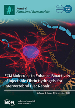
Fibrin is a promising delivery vehicle to introduce cells into the intervertebral disc (IVD) to regenerate damaged tissue for low back pain. However, it lacks key extracellular matrix (ECM) components normally found in native nucleus pulposus (NP) tissue. The overall aim of this work was to create a fibrin-based hydrogel by incorporating collagen (Col) and Hyaluronan (HA) into the matrix to enhance an NP-like matrix accumulation of cells. Results showed that the incorporation of HA promoted sGAG accumulation and tended to suppress collagen formation at higher concentrations. This work suggests that the incorporation of key ECM components can enhance the bioactivity of fibrin-based hydrogels. View this paper.
  • Issues are regarded as officially published after their release is announced to the table of contents alert mailing list.
  • You may sign up for e-mail alerts to receive table of contents of newly released issues.
  • PDF is the official format for papers published in both, html and pdf forms. To view the papers in pdf format, click on the "PDF Full-text" link, and use the free Adobe Reader to open them.
Order results
Result details
Section
Select all
Export citation of selected articles as:
13 pages, 2861 KiB  
Article
Specialized Living Wound Dressing Based on the Self-Assembly Approach of Tissue Engineering
by Laurence Cantin-Warren, Rina Guignard, Sergio Cortez Ghio, Danielle Larouche, François A. Auger and Lucie Germain
J. Funct. Biomater. 2018, 9(3), 53; https://doi.org/10.3390/jfb9030053 - 15 Sep 2018
Cited by 20 | Viewed by 7317
Abstract
There is a high incidence of failure and recurrence for chronic skin wounds following conventional therapies. To promote healing, the use of skin substitutes containing living cells as wound dressings has been proposed. The aim of this study was to produce a scaffold-free [...] Read more.
There is a high incidence of failure and recurrence for chronic skin wounds following conventional therapies. To promote healing, the use of skin substitutes containing living cells as wound dressings has been proposed. The aim of this study was to produce a scaffold-free cell-based bilayered tissue-engineered skin substitute (TES) containing living fibroblasts and keratinocytes suitable for use as wound dressing, while considering production time, handling effort during the manufacturing process, and stability of the final product. The self-assembly method, which relies on the ability of mesenchymal cells to secrete and organize connective tissue sheet sustaining keratinocyte growth, was used to produce TESs. Three fibroblast-seeding densities were tested to produce tissue sheets. At day 17, keratinocytes were added onto 1 or 3 (reference method) stacked tissue sheets. Four days later, TESs were subjected either to 4, 10, or 17 days of culture at the air–liquid interface (A/L). All resulting TESs were comparable in terms of their histological aspect, protein expression profile and contractile behavior in vitro. However, signs of extracellular matrix (ECM) digestion that progressed over culture time were noted in TESs produced with only one fibroblast-derived tissue sheet. With lower fibroblast density, the ECM of TESs was almost completely digested after 10 days A/L and lost histological integrity after grafting in athymic mice. Increasing the fibroblast seeding density 5 to 10 times solved this problem. We conclude that the proposed method allows for a 25-day production of a living TES, which retains its histological characteristics in vitro for at least two weeks. Full article
(This article belongs to the Special Issue Biomaterial Enhanced Regeneration)
Show Figures

Graphical abstract

13 pages, 3100 KiB  
Article
Formulation and Evaluation of Silymarin-Loaded Chitosan-Montmorilloite Microbeads for the Potential Treatment of Gastric Ulcers
by Ameya Sharma, Vivek Puri, Vandita Kakkar and Inderbir Singh
J. Funct. Biomater. 2018, 9(3), 52; https://doi.org/10.3390/jfb9030052 - 10 Sep 2018
Cited by 16 | Viewed by 5997
Abstract
Silymarin-loaded mucoadhesive microbeads of Chitosan-MMT were developed using the ionotropic gelation technique. Characterization of the microbeads was performed by DSC, XRD, SEM, and FTIR techniques. In vitro mucoadhesion and drug release studies; gastroprotective studies including the measurement of ulcerative index; the determination of [...] Read more.
Silymarin-loaded mucoadhesive microbeads of Chitosan-MMT were developed using the ionotropic gelation technique. Characterization of the microbeads was performed by DSC, XRD, SEM, and FTIR techniques. In vitro mucoadhesion and drug release studies; gastroprotective studies including the measurement of ulcerative index; the determination of gastric wall mucus; and the determination of percentage protection, biochemical, and histopathological studies were also performed. Microbeads batches were evaluated for particle size (120–140 µm), actual drug content, (49.36–58.18%) and entrapment efficiency (72.52–92.39%).Biochemical estimation of myeloperoxidase was found to be 0.10–0.75 µmoles/g/tissue. Significant reduction in the ulcerative index showed the gastroprotective effect of the formulation. Silymarin-loaded beads of Chitosan-MMT were found to exhibit good mucoadhesion and efficient release of the drug, and were found to be a promising drug carrier system for the treatment of gastric ulcers. Full article
(This article belongs to the Special Issue Clay-Based Biomaterials: From Synthesis to Applications)
Show Figures

Figure 1

12 pages, 7923 KiB  
Article
Fabrication and Multiscale Structural Properties of Interconnected Porous Biomaterial for Tissue Engineering by Freeze Isostatic Pressure (FIP)
by Mythili Prakasam, Ali Chirazi, Grzegorz Pyka, Anna Prokhodtseva, Daniel Lichau and Alain Largeteau
J. Funct. Biomater. 2018, 9(3), 51; https://doi.org/10.3390/jfb9030051 - 24 Aug 2018
Cited by 7 | Viewed by 5550
Abstract
Biomaterial for tissue engineering is a topic of huge progress with a recent surge in fabrication and characterization advances. Biomaterials for tissue engineering applications or as scaffolds depend on various parameters such as fabrication technology, porosity, pore size, mechanical strength, and surface available [...] Read more.
Biomaterial for tissue engineering is a topic of huge progress with a recent surge in fabrication and characterization advances. Biomaterials for tissue engineering applications or as scaffolds depend on various parameters such as fabrication technology, porosity, pore size, mechanical strength, and surface available for cell attachment. To serve the function of the scaffold, the porous biomaterial should have enough mechanical strength to aid in tissue engineering. With a new manufacturing technology, we have obtained high strength materials by optimizing a few processing parameters such as pressure, temperature, and dwell time, yielding the monolith with porosity in the range of 80%–93%. The three-dimensional interconnectivity of the porous media through scales for the newly manufactured biomaterial has been investigated using newly developed 3D correlative and multi-modal imaging techniques. Multiscale X-ray tomography, FIB-SEM Slice & View stacking, and high-resolution STEM-EDS electronic tomography observations have been combined allowing quantification of morphological and geometrical spatial distributions of the multiscale porous network through length scales spanning from tens of microns to less than a nanometer. The spatial distribution of the wall thickness has also been investigated and its possible relationship with pore connectivity and size distribution has been studied. Full article
(This article belongs to the Special Issue Advanced Functional Nanobiomaterials)
Show Figures

Figure 1

31 pages, 1898 KiB  
Review
Tailoring the Interface of Biomaterials to Design Effective Scaffolds
by Ludovica Parisi, Andrea Toffoli, Giulia Ghiacci and Guido M. Macaluso
J. Funct. Biomater. 2018, 9(3), 50; https://doi.org/10.3390/jfb9030050 - 21 Aug 2018
Cited by 42 | Viewed by 7661
Abstract
Tissue engineering (TE) is a multidisciplinary science, which including principles from material science, biology and medicine aims to develop biological substitutes to restore damaged tissues and organs. A major challenge in TE is the choice of suitable biomaterial to fabricate a scaffold that [...] Read more.
Tissue engineering (TE) is a multidisciplinary science, which including principles from material science, biology and medicine aims to develop biological substitutes to restore damaged tissues and organs. A major challenge in TE is the choice of suitable biomaterial to fabricate a scaffold that mimics native extracellular matrix guiding resident stem cells to regenerate the functional tissue. Ideally, the biomaterial should be tailored in order that the final scaffold would be (i) biodegradable to be gradually replaced by regenerating new tissue, (ii) mechanically similar to the tissue to regenerate, (iii) porous to allow cell growth as nutrient, oxygen and waste transport and (iv) bioactive to promote cell adhesion and differentiation. With this perspective, this review discusses the options and challenges facing biomaterial selection when a scaffold has to be designed. We highlight the possibilities in the final mold the materials should assume and the most effective techniques for its fabrication depending on the target tissue, including the alternatives to ameliorate its bioactivity. Furthermore, particular attention has been given to the influence that all these aspects have on resident cells considering the frontiers of materiobiology. In addition, a focus on chitosan as a versatile biomaterial for TE scaffold fabrication has been done, highlighting its latest advances in the literature on bone, skin, cartilage and cornea TE. Full article
Show Figures

Figure 1

11 pages, 1589 KiB  
Article
Experimental and Theoretical Studies on the Adsorption Mechanisms of Uranium (VI) Ions on Chitosan
by Kenji Mishima, Xiaoyu Du, Naoto Miyamoto, Naoki Kano and Hiroshi Imaizumi
J. Funct. Biomater. 2018, 9(3), 49; https://doi.org/10.3390/jfb9030049 - 09 Aug 2018
Cited by 27 | Viewed by 5896
Abstract
An experiment on the adsorption of uranium (VI) by chitosan was conducted to investigate the efficiency of chitosan as an adsorbent for U(VI). The adsorption potential of U(VI) by chitosan was investigated with ICP-MS by varying the experimental conditions such as the pH [...] Read more.
An experiment on the adsorption of uranium (VI) by chitosan was conducted to investigate the efficiency of chitosan as an adsorbent for U(VI). The adsorption potential of U(VI) by chitosan was investigated with ICP-MS by varying the experimental conditions such as the pH in order to obtain the optimum conditions. Adsorption dependence on the pH was confirmed, and the highest uptake of U(VI) was observed at pH 5. In addition, to scrutinize the experimental results, quantum chemistry calculations were performed. The results, taking into account the experimental conditions, show that the adsorption efficiency increases as the total charge of the adsorbent and adsorbate species decreases if both of them are positively charged. It was also found that a slight change in the adsorption geometric configuration controls the adsorption efficiency. Full article
Show Figures

Figure 1

11 pages, 2088 KiB  
Article
Histologic and Histomorphometric Analysis of Bone Regeneration with Bovine Grafting Material after 24 Months of Healing. A Case Report
by Renzo Guarnieri, Fabrizio Belleggia, Patricia DeVillier and Luca Testarelli
J. Funct. Biomater. 2018, 9(3), 48; https://doi.org/10.3390/jfb9030048 - 08 Aug 2018
Cited by 15 | Viewed by 6098
Abstract
Anorganic bovine bone mineral matrix (ABBMM) has been reported to have osteoconductive properties and no inflammatory or adverse responses when used as grafting material in sinus augmentation procedures. However, controversy remains in regard to degradation rate of ABBMM. The aim of this study [...] Read more.
Anorganic bovine bone mineral matrix (ABBMM) has been reported to have osteoconductive properties and no inflammatory or adverse responses when used as grafting material in sinus augmentation procedures. However, controversy remains in regard to degradation rate of ABBMM. The aim of this study was to histologically and histomorphometrically evaluate the degradation of ABBMM in human bone samples obtained in one patient 24 months after sinus augmentation. Materials and Methods: The histologic and histomorphometric analysis was performed by means of light microscopy in three specimens harvested from the same patient, Results: After 24 months the tissue pattern appeared to be composed of residual particles, some in close contact with the newly formed bone, others separated by translucent areas and osteoid tissues. Newly-formed bone presented different levels of maturation and numerous osteocytes, with greater numbers in bone closer to the grafted particles (27.3% vs. 11.2%, p < 0.05). The histomorphometric analysis showed mean values of 40.84% newly-formed bone, 33.58% residual graft material, 23.84% marrow spaces, and 1.69% osteoid tissue, Conclusions: Even though ABBMM underwent considerable resorption, a great amount of residual grafting material was still present after two years of healing following sinus augmentation. This study confirms that the bovine grafts can be classified as long-term degradation materials. Full article
(This article belongs to the Special Issue Dental Implant Materials and Biomaterials)
Show Figures

Figure 1

21 pages, 9724 KiB  
Article
Incorporating Germanium Oxide into the Glass Phase of Novel Zinc/Magnesium-Based GPCs Designed for Bone Void Filling: Evaluating Their Physical and Mechanical Properties
by Basel A. Khader, Omar Rodriguez and Mark R. Towler
J. Funct. Biomater. 2018, 9(3), 47; https://doi.org/10.3390/jfb9030047 - 31 Jul 2018
Cited by 4 | Viewed by 5996
Abstract
The structural role of Germanium (Ge), when substituting for Zinc (Zn) up to 8 mol % in the 0.48SiO2–0.12CaO–0.36ZnO–0.04MgO glass series, was investigated with respect to both the glass chemistry and also the properties of glass polyalkenoate cements (GPCs) manufactured from [...] Read more.
The structural role of Germanium (Ge), when substituting for Zinc (Zn) up to 8 mol % in the 0.48SiO2–0.12CaO–0.36ZnO–0.04MgO glass series, was investigated with respect to both the glass chemistry and also the properties of glass polyalkenoate cements (GPCs) manufactured from them. The Network connectivity (NC) of the glass was calculated to increase from 1.83 to 2.42 with the addition of GeO2 (0–8 mol %). Differential thermal analysis (DTA) results confirmed an increase in the glass transition temperature (Tg) of the glass series with GeO2 content. X-ray photoelectron spectroscopy (XPS) showed an increase in the ratio of bridging oxygens (BO) to non-bridging oxygens (NBO) with the addition of GeO2, supporting the NC and DTA results. 29Si magic angle spinning nuclear magnetic resonance spectroscopy (29Si MAS-NMR) determined a chemical shift from −80.3 to −83.7 ppm as the GeO2 concentration increased. These ionomeric glasses were subsequently used as the basic components in a series of GPCs by mixing them with aqueous polyacrylic acid (PAA). The handling properties of the GPCs resulting were evaluated with respect to the increasing concentration of GeO2 in the glass phase. It was found that the working times of these GPCs increased from 3 to 15 min, while their setting times increased from 4 to 18 min, facilitating the injectability of the Zn/Mg-GPCs through a 16-gauge needle. These Ge-Zn/Mg-GPCs were found to be injectable up to 96% within 12 min. Zn/Mg-GPCs containing GeO2 show promise as injectable cements for use in bone void filling. Full article
Show Figures

Figure 1

18 pages, 9231 KiB  
Article
Tissue Engineering Scaffolds Fabricated in Dissolvable 3D-Printed Molds for Patient-Specific Craniofacial Bone Regeneration
by Angela Alarcon De la Lastra, Katherine R. Hixon, Lavanya Aryan, Amanda N. Banks, Alexander Y. Lin, Andrew F. Hall and Scott A. Sell
J. Funct. Biomater. 2018, 9(3), 46; https://doi.org/10.3390/jfb9030046 - 24 Jul 2018
Cited by 17 | Viewed by 6940
Abstract
The current gold standard treatment for oral clefts is autologous bone grafting. This treatment, however, presents another wound site for the patient, greater discomfort, and pediatric patients have less bone mass for bone grafting. A potential alternative treatment is the use of tissue [...] Read more.
The current gold standard treatment for oral clefts is autologous bone grafting. This treatment, however, presents another wound site for the patient, greater discomfort, and pediatric patients have less bone mass for bone grafting. A potential alternative treatment is the use of tissue engineered scaffolds. Hydrogels are well characterized nanoporous scaffolds and cryogels are mechanically durable, macroporous, sponge-like scaffolds. However, there has been limited research on these scaffolds for cleft craniofacial defects. 3D-printed molds can be combined with cryogel/hydrogel fabrication to create patient-specific tissue engineered scaffolds. By combining 3D-printing technology and scaffold fabrication, we were able to create scaffolds with the geometry of three cleft craniofacial defects. The scaffolds were then characterized to assess the effect of the mold on their physical properties. While the scaffolds were able to completely fill the mold, creating the desired geometry, the overall volumes were smaller than expected. The cryogels possessed porosities ranging from 79.7% to 87.2% and high interconnectivity. Additionally, the cryogels swelled from 400% to almost 1500% of their original dry weight while the hydrogel swelling did not reach 500%, demonstrating the ability to fill a defect site. Overall, despite the complex geometry, the cryogel scaffolds displayed ideal properties for bone reconstruction. Full article
(This article belongs to the Special Issue 3D Printing of Biomaterials)
Show Figures

Figure 1

17 pages, 3015 KiB  
Article
A Decellularized Porcine Xenograft-Derived Bone Scaffold for Clinical Use as a Bone Graft Substitute: A Critical Evaluation of Processing and Structure
by Daniel N. Bracey, Thorsten M. Seyler, Alexander H. Jinnah, Mark O. Lively, Jeffrey S. Willey, Thomas L. Smith, Mark E. Van Dyke and Patrick W. Whitlock
J. Funct. Biomater. 2018, 9(3), 45; https://doi.org/10.3390/jfb9030045 - 12 Jul 2018
Cited by 47 | Viewed by 9231
Abstract
Background: Bone grafts are used in approximately one half of all musculoskeletal surgeries. Autograft bone is the historic gold standard but is limited in supply and its harvest imparts significant morbidity to the patient. Alternative sources of bone graft include allografts, synthetics and, [...] Read more.
Background: Bone grafts are used in approximately one half of all musculoskeletal surgeries. Autograft bone is the historic gold standard but is limited in supply and its harvest imparts significant morbidity to the patient. Alternative sources of bone graft include allografts, synthetics and, less commonly, xenografts which are taken from animal species. Xenografts are available in unlimited supply from healthy animal donors with controlled biology, avoiding the risk of human disease transmission, and may satisfy current demand for bone graft products. Methods: In the current study, cancellous bone was harvested from porcine femurs and subjected to a novel decellularization protocol to derive a bone scaffold. Results: The scaffold was devoid of donor cellular material on histology and DNA sampling (p < 0.01). Microarchitectural properties important for osteoconductive potential were preserved after decellularization as shown by high resolution imaging modalities. Proteomics data demonstrated similar profiles when comparing the porcine bone scaffold against commercially available human demineralized bone matrix approved for clinical use. Conclusion: We are unaware of any porcine-derived bone graft products currently used in orthopaedic surgery practice. Results from the current study suggest that porcine-derived bone scaffolds warrant further consideration to serve as a potential bone graft substitute. Full article
Show Figures

Graphical abstract

13 pages, 935 KiB  
Article
Interconnected PolymerS TeChnology (IPSTiC): An Effective Approach for the Modulation of 5α-Reductase Activity in Hair Loss Conditions
by Ortensia Ilaria Parisi, Luca Scrivano, Fabio Amone, Rocco Malivindi, Mariarosa Ruffo, Anna Francesca Vattimo, Vincenzo Pezzi and Francesco Puoci
J. Funct. Biomater. 2018, 9(3), 44; https://doi.org/10.3390/jfb9030044 - 12 Jul 2018
Cited by 8 | Viewed by 7696
Abstract
Hair loss represents a condition that adversely affects the social life of patients. The most common cause is androgenetic alopecia (AGA), which is a genetically determined progressive hair-loss condition involving 5α-reductase. In this study, a novel anti-baldness agent based on Interconnected PolymerS TeChnology [...] Read more.
Hair loss represents a condition that adversely affects the social life of patients. The most common cause is androgenetic alopecia (AGA), which is a genetically determined progressive hair-loss condition involving 5α-reductase. In this study, a novel anti-baldness agent based on Interconnected PolymerS TeChnology (IPSTiC), which is an effective strategy for the delivery of bioactive molecules, was developed. This product (IPSTiC patch hair) is based on a polymeric blend consisting of high molecular weight hyaluronic acid and soybean proteins and is able to improve efficacy and stability of bioactive ingredients such as Origanum vulgare leaf extract, Camellia Sinensis leaf extract, and Capsicum Annuum fruit extract. The efficacy of the developed anti-baldness agent was investigated by performing several tests including NO radical and 5α-reductase inhibition assays, stability studies under different conditions, and in vitro diffusion studies using Franz cells. The biocompatibility of IPSTiC patch hair was also evaluated by in vitro analysis of the pro-sensitising potential and EPISKIN model. The obtained results confirmed both the efficacy and safety of IPSTiC patch hair supporting the potential use of this product in the topical treatment of AGA. Full article
(This article belongs to the Special Issue Functional Materials for Healthcare)
Show Figures

Graphical abstract

19 pages, 5529 KiB  
Article
Incorporation of Collagen and Hyaluronic Acid to Enhance the Bioactivity of Fibrin-Based Hydrogels for Nucleus Pulposus Regeneration
by Jennifer Gansau and Conor Timothy Buckley
J. Funct. Biomater. 2018, 9(3), 43; https://doi.org/10.3390/jfb9030043 - 10 Jul 2018
Cited by 11 | Viewed by 8711
Abstract
Hydrogels, such as fibrin, offer a promising delivery vehicle to introduce cells into the intervertebral disc (IVD) to regenerate damaged disc tissue as a potential treatment for low back pain. However, fibrin lacks key extracellular matrix (ECM) components, such as collagen (Col) and [...] Read more.
Hydrogels, such as fibrin, offer a promising delivery vehicle to introduce cells into the intervertebral disc (IVD) to regenerate damaged disc tissue as a potential treatment for low back pain. However, fibrin lacks key extracellular matrix (ECM) components, such as collagen (Col) and hyaluronan (HA), normally found in native nucleus pulposus (NP) tissue. The overall aim of this work was to create a fibrin-based hydrogel, by incorporating Col and HA into the matrix to enhance NP-like matrix accumulation using articular chondrocytes (CC). Firstly, we assessed the effect of fibrin concentrations on hydrogel stability, and the viability and proliferation kinetics of articular chondrocytes. Secondly, we investigated the effect of incorporating Col and HA to enhance NP-like matrix accumulation, and finally, examined the influence of various HA concentrations. Results showed that increasing fibrin concentration enhanced cell viability and proliferation. Interestingly, incorporation of HA promoted sGAG accumulation and tended to suppress collagen formation at higher concentrations. Taken together, these results suggest that incorporation of ECM components can enhance the bioactivity of fibrin-based hydrogels, which may help advance the clinical potential of commercial cell and biomaterial ventures in the treatment of IVD regeneration. Full article
(This article belongs to the Special Issue Biomaterials for Spinal Applications)
Show Figures

Graphical abstract

9 pages, 1012 KiB  
Review
Glass Ionomer Cements for the Restoration of Non-Carious Cervical Lesions in the Geriatric Patient
by Nikolaos-Stefanos Kampanas and Maria Antoniadou
J. Funct. Biomater. 2018, 9(3), 42; https://doi.org/10.3390/jfb9030042 - 08 Jul 2018
Cited by 10 | Viewed by 9068
Abstract
Background: The restoration of non-carious cervical lesions in geriatric patients is a demanding process. Glass ionomer cements can be promising materials for the management of these lesions in older adults. The aim of this literature review is to present the benefits of glass [...] Read more.
Background: The restoration of non-carious cervical lesions in geriatric patients is a demanding process. Glass ionomer cements can be promising materials for the management of these lesions in older adults. The aim of this literature review is to present the benefits of glass ionomers and how they can be used for the restoration of non-carious cervical lesions of older adults depending on the geriatric patient’s profile. Data sources: All available in vitro and in vivo studies from Google Scholar, PubMed and Scopus search engines corresponding to glass ionomer cements, geriatric dentistry, elderly patients, and non-carious lesions as key words were reviewed. Data synthesis: The advantages of glass ionomer cements, such as good retention and fluoride release, make them suitable for the restoration of non-carious cervical lesions. However, several factors related to the geriatric patient’s profile determine the most suitable material type. Conclusion: In general, the resin modified glass ionomer cements (RMGICs) appear to be preferred, but under certain circumstances the use of the conventional product is more appropriate, despite its poorer mechanical features. Further studies are required for more reliable data analysis and clinical interpretation of the relevant results. Full article
Show Figures

Figure 1

14 pages, 10312 KiB  
Article
Implementation of Industrial Additive Manufacturing: Intelligent Implants and Drug Delivery Systems
by Jan Sher Akmal, Mika Salmi, Antti Mäkitie, Roy Björkstrand and Jouni Partanen
J. Funct. Biomater. 2018, 9(3), 41; https://doi.org/10.3390/jfb9030041 - 29 Jun 2018
Cited by 43 | Viewed by 9833
Abstract
The purpose of this study is to demonstrate the ability of additive manufacturing, also known as 3D printing, to produce effective drug delivery devices and implants that are both identifiable, as well as traceable. Drug delivery devices can potentially be used for drug [...] Read more.
The purpose of this study is to demonstrate the ability of additive manufacturing, also known as 3D printing, to produce effective drug delivery devices and implants that are both identifiable, as well as traceable. Drug delivery devices can potentially be used for drug release in the direct vicinity of target tissues or the selected medication route in a patient-specific manner as required. The identification and traceability of additively manufactured implants can be administered through radiofrequency identification systems. The focus of this study is to explore how embedded medication and sensors can be added in different additive manufacturing processes. The concept is extended to biomaterials with the help of the literature. As a result of this study, a patient-specific drug delivery device can be custom-designed and additively manufactured in the form of an implant that can identify, trace, and dispense a drug to the vicinity of a selected target tissue as a patient-specific function of time for bodily treatment and restoration. Full article
(This article belongs to the Special Issue 3D Printing of Biomaterials)
Show Figures

Graphical abstract

17 pages, 3910 KiB  
Article
Genipin-Enhanced Fibrin Hydrogel and Novel Silk for Intervertebral Disc Repair in a Loaded Bovine Organ Culture Model
by Daniela A. Frauchiger, Rahel D. May, Ezgi Bakirci, Adel Tekari, Samantha C. W. Chan, Michael Wöltje, Lorin M. Benneker and Benjamin Gantenbein
J. Funct. Biomater. 2018, 9(3), 40; https://doi.org/10.3390/jfb9030040 - 24 Jun 2018
Cited by 44 | Viewed by 7161
Abstract
(1) Background: Intervertebral disc (IVD) repair represents a major challenge. Using functionalised biomaterials such as silk combined with enforced hydrogels might be a promising approach for disc repair. We aimed to test an IVD repair approach by combining a genipin-enhanced fibrin hydrogel with [...] Read more.
(1) Background: Intervertebral disc (IVD) repair represents a major challenge. Using functionalised biomaterials such as silk combined with enforced hydrogels might be a promising approach for disc repair. We aimed to test an IVD repair approach by combining a genipin-enhanced fibrin hydrogel with an engineered silk scaffold under complex load, after inducing an injury in a bovine whole organ IVD culture; (2) Methods: Bovine coccygeal IVDs were isolated from ~1-year-old animals within four hours post-mortem. Then, an injury in the annulus fibrosus was induced by a 2 mm biopsy punch. The repair approach consisted of genipin-enhanced fibrin hydrogel that was used to fill up the cavity. To seal the injury, a Good Manufacturing Practise (GMP)-compliant engineered silk fleece-membrane composite was applied and secured by the cross-linked hydrogel. Then, IVDs were exposed to one of three loading conditions: no load, static load and complex load in a two-degree-of-freedom bioreactor for 14 days. Followed by assessing DNA and matrix content, qPCR and histology, the injured discs were compared to an uninjured control IVD that underwent the same loading profiles. In addition, the genipin-enhanced fibrin hydrogel was further investigated with respect to cytotoxicity on human stem cells, annulus fibrosus, and nucleus pulposus cells; (3) Results: The repair was successful as no herniation could be detected for any of the three loading conditions. Disc height was not recovered by the repair DNA and matrix contents were comparable to a healthy, untreated control disc. Genipin resulted being cytotoxic in the in vitro test but did not show adverse effects when used for the organ culture model; (4) Conclusions: The current study indicated that the combination of the two biomaterials, i.e., genipin-enhanced fibrin hydrogel and an engineered silk scaffold, was a promising approach for IVD repair. Furthermore, genipin-enhanced fibrin hydrogel was not suitable for cell cultures; however, it was highly applicable as a filler material. Full article
(This article belongs to the Special Issue Biomaterials for Spinal Applications)
Show Figures

Graphical abstract

Previous Issue
Next Issue
Back to TopTop