
Bioactive Glasses Containing Strontium or Magnesium Ions to Enhance the Biological Response in Bone Regeneration

 , , ,
, , ,  ,
,  , and
, and
Abstract
:1. Introduction
2. Materials and Methods
2.1. Materials Synthesis
2.2. Thermal Analysis
2.3. Structural Characterization
2.3.1. XRD
2.3.2. FTIR
2.4. Cytotoxicity Assay
2.5. Bioactivity
2.6. Antimicrobial Effect
3. Results
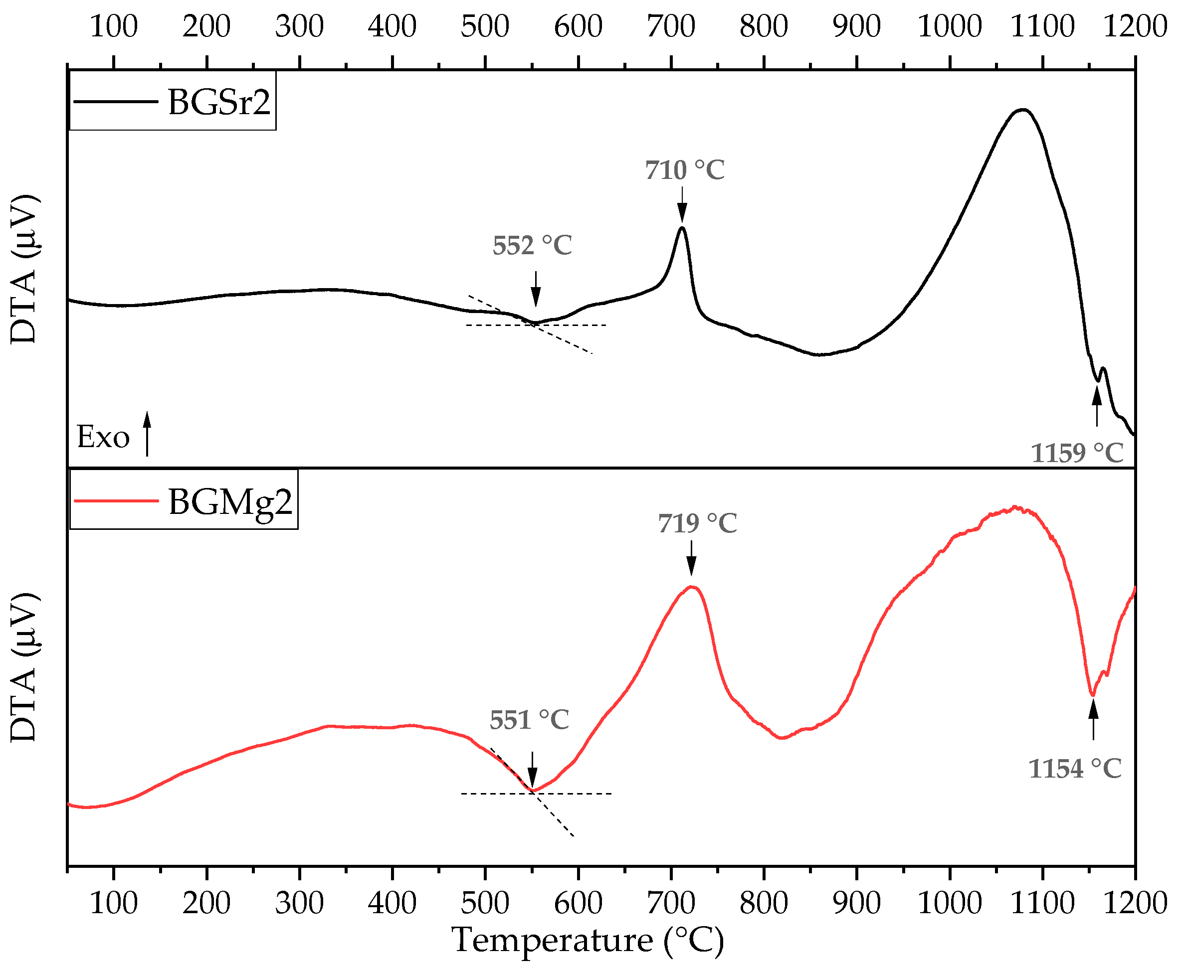
3.1. Differential Thermal Analysis (DTA)
3.2. Physical Characterization
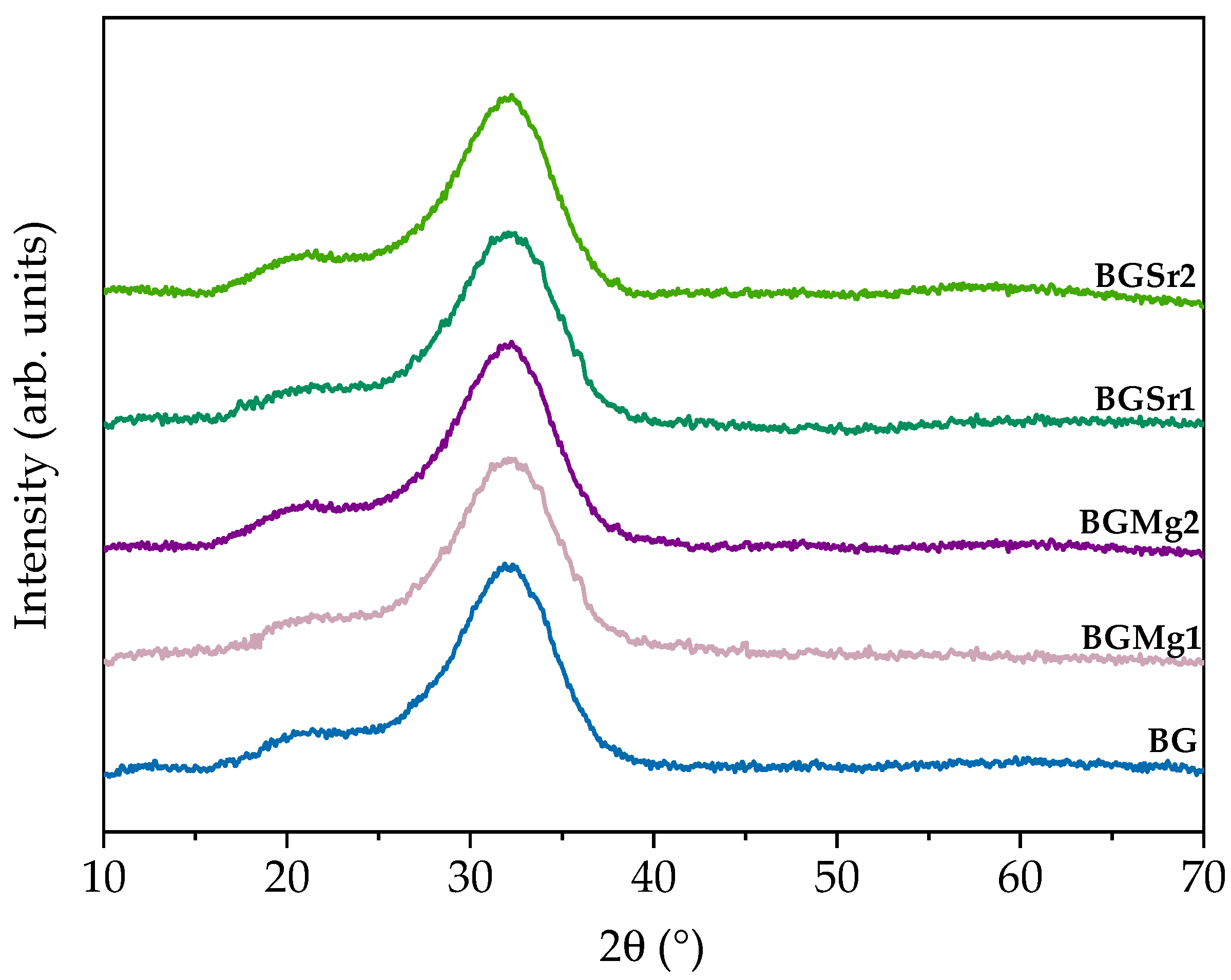
3.2.1. X-ray Diffraction (XRD)
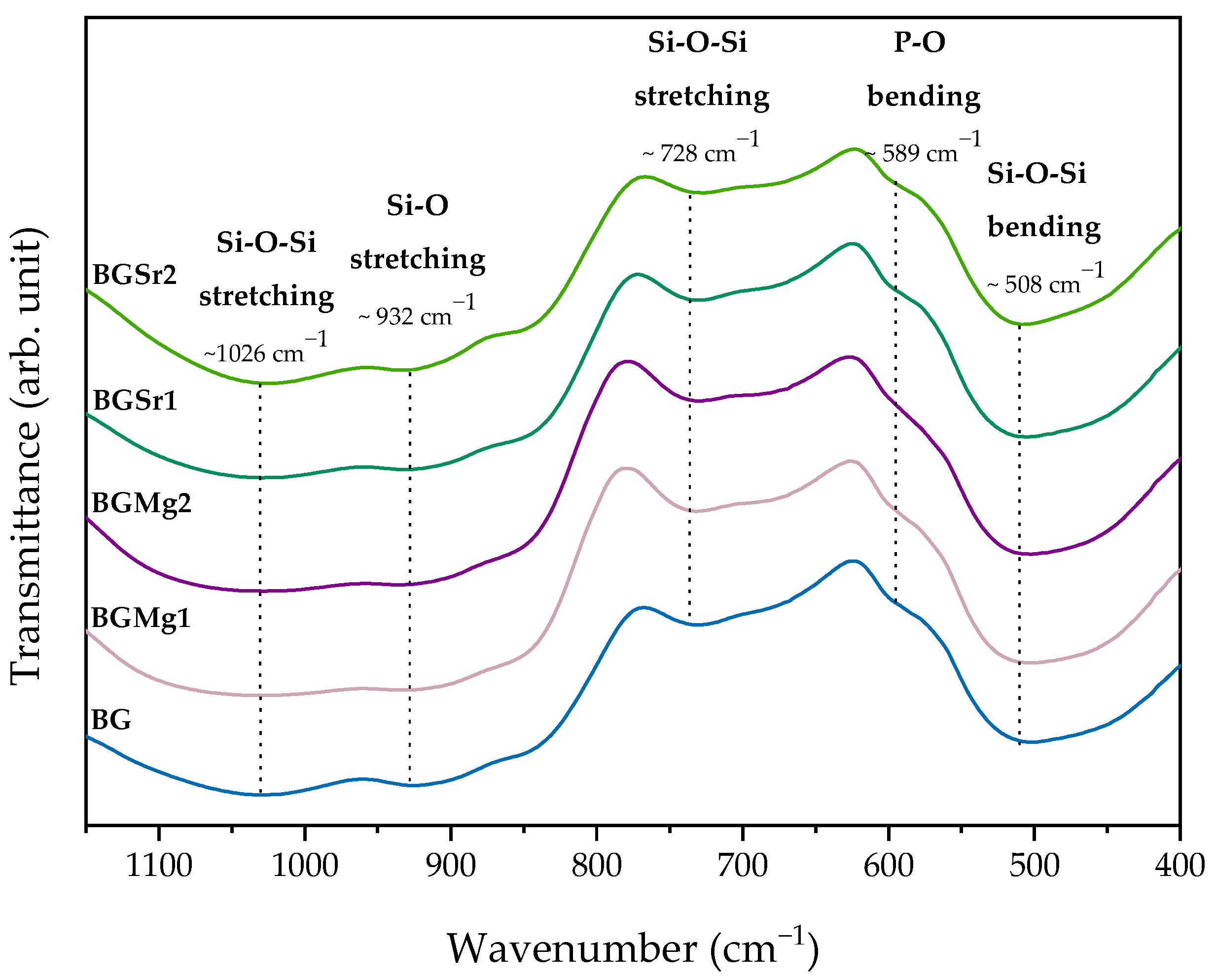
3.2.2. Fourier Transform Infrared Spectroscopy (FTIR)
3.3. Biological Behaviour
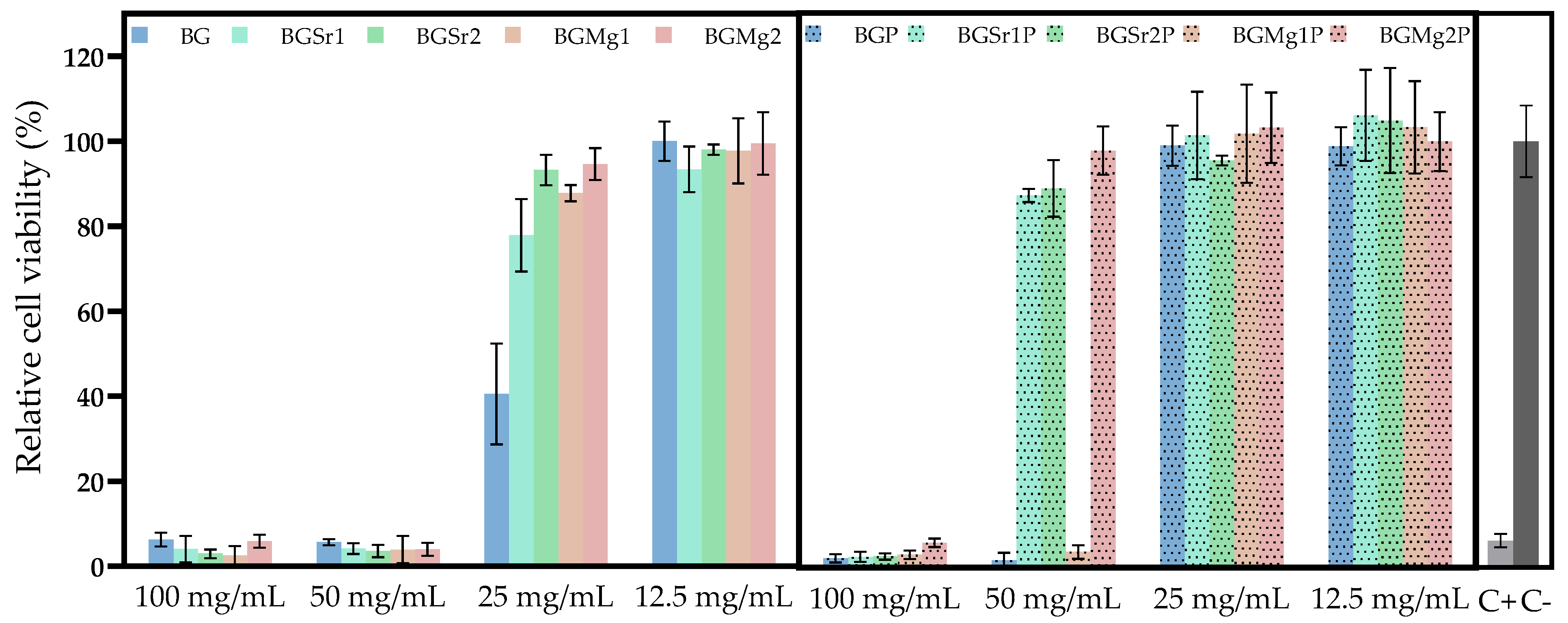
3.3.1. Cytotoxicity
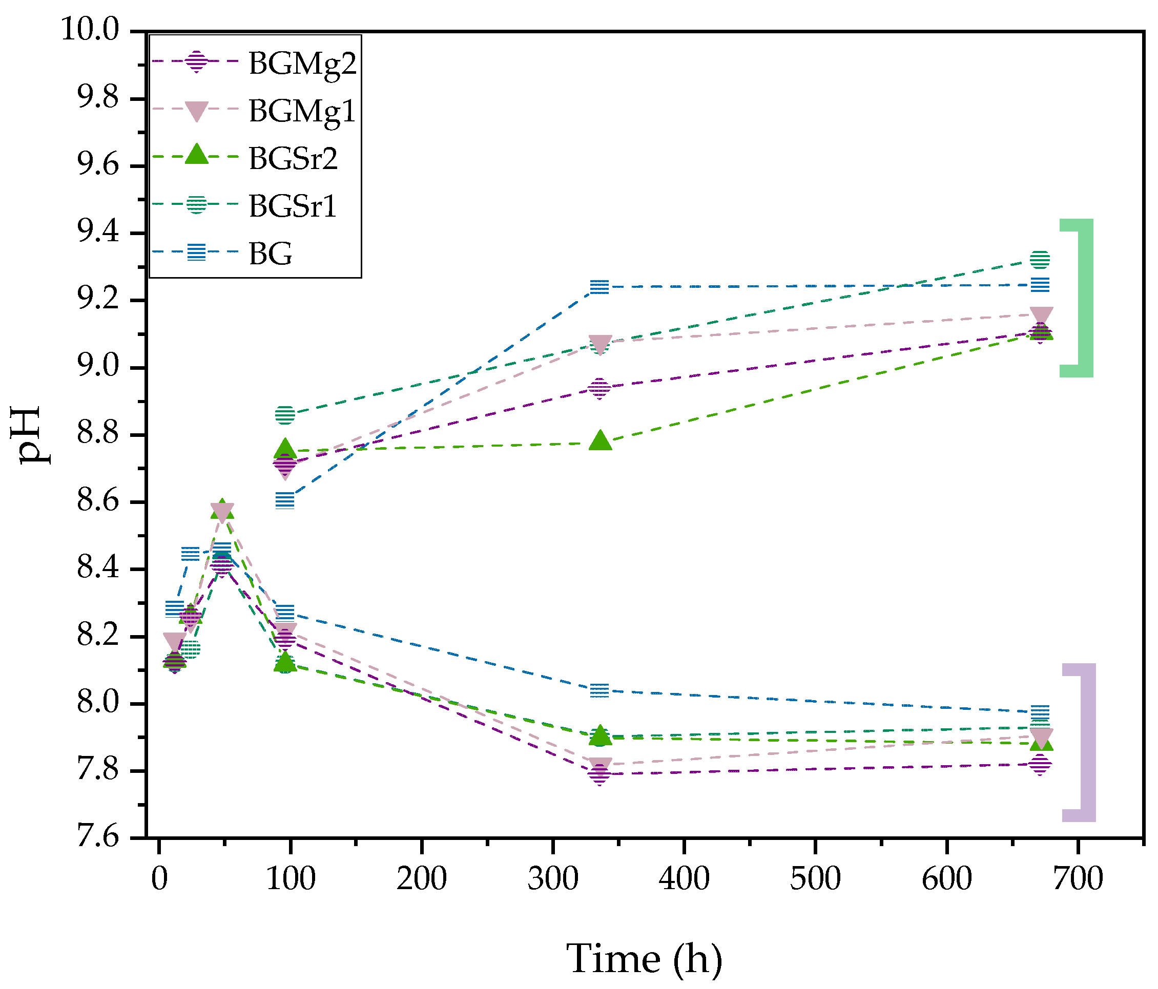
3.3.2. Bioactivity
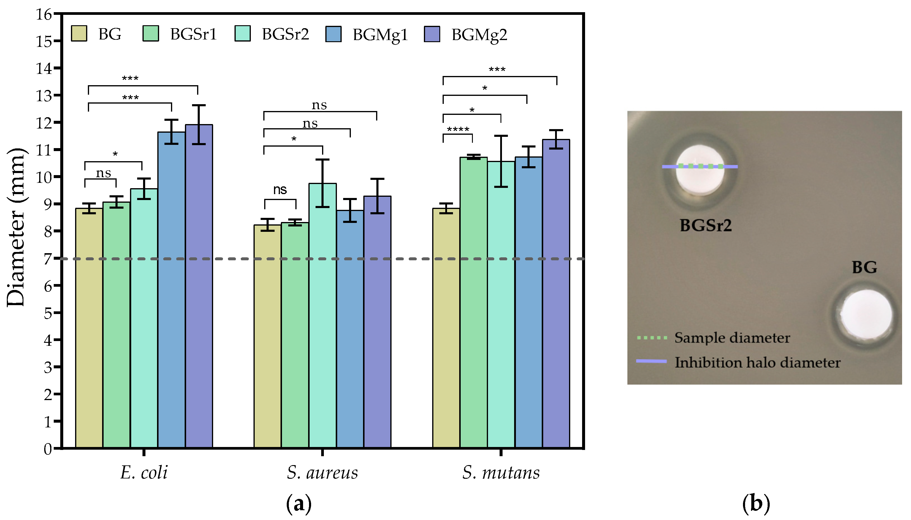
3.3.3. Antimicrobial Activity
4. Discussion
5. Conclusions
Supplementary Materials
Author Contributions
Funding
Acknowledgments
Conflicts of Interest
References
- Esfahanizadeh, N.; Montazeri, M.; Nourani, M.; Harandi, M. Use of Bioactive Glass Doped with Magnesium or Strontium for Bone Regeneration: A Rabbit Critical-Size Calvarial Defects Study. Dent. Res. J. 2022, 19, 18. [Google Scholar] [CrossRef]
- Zhou, J.; Zhang, Z.; Joseph, J.; Zhang, X.; Ferdows, B.E.; Patel, D.N.; Chen, W.; Banfi, G.; Molinaro, R.; Cosco, D.; et al. Biomaterials and Nanomedicine for Bone Regeneration: Progress and Future Prospects. Exploration 2021, 1, 20210011. [Google Scholar] [CrossRef]
- Singh, A.K.; Pramanik, K.; Biswas, A. MgO Enables Enhanced Bioactivity and Antimicrobial Activity of Nano Bioglass for Bone Tissue Engineering Application. Mater. Technol. 2019, 34, 818–826. [Google Scholar] [CrossRef]
- Sharma, K.; Mujawar, M.A.; Kaushik, A. State-of-Art Functional Biomaterials for Tissue Engineering. Front. Mater. 2019, 6, 172. [Google Scholar] [CrossRef]
- Tang, G.; Liu, Z.; Liu, Y.; Yu, J.; Wang, X.; Tan, Z.; Ye, X. Recent Trends in the Development of Bone Regenerative Biomaterials. Front. Cell Dev. Biol. 2021, 9, 665813. [Google Scholar] [CrossRef]
- Tiskaya, M.; Shahid, S.; Gillam, D.; Hill, R. The Use of Bioactive Glass (BAG) in Dental Composites: A Critical Review. Dent. Mater. 2020, 37, 296–310. [Google Scholar] [CrossRef]
- Raszewski, Z.; Kulbacka, J. Mechanical Properties, Cytotoxicity, and Fluoride Ion Release Capacity of Bioactive Glass-Modified Methacrylate Resin Used in Three-Dimensional Printing Technology. Materials 2022, 15, 1133. [Google Scholar] [CrossRef]
- Majumdar, S.; Gupta, S.; Krishnamurthy, S. Multifarious Applications of Bioactive Glasses in Soft Tissue Engineering. Biomater. Sci. 2021, 9, 8111–8147. [Google Scholar] [CrossRef]
- Schatkoski, V.M.; Larissa do Amaral Montanheiro, T.; Canuto de Menezes, B.R.; Pereira, R.M.; Rodrigues, K.F.; Ribas, R.G.; Morais da Silva, D.; Thim, G.P. Current Advances Concerning the Most Cited Metal Ions Doped Bioceramics and Silicate-Based Bioactive Glasses for Bone Tissue Engineering. Ceram. Int. 2021, 47, 2999–3012. [Google Scholar] [CrossRef]
- Gavinho, S.R.; Prezas, P.R.; Graça, M.P.F. Synthesis, Structural and Electrical Properties of the 45S5 Bioglass®. In Electrical Measurements: Introduction, Concepts and Applications; Nova Science Publishers: New York, NY, USA, 2017; ISBN 9781536129748. [Google Scholar]
- Larry, R.; Hench, L. The Story of Bioglass. J. Mater. Sci. Mater. Med. 2006, 17, 967–978. [Google Scholar] [CrossRef]
- Jones, J.R. Reprint of: Review of Bioactive Glass: From Hench to Hybrids. Acta Biomater. 2015, 23, 53–82. [Google Scholar] [CrossRef] [PubMed]
- Hench, L.L. The Story of Bioglass®. J. Mater. Sci. Mater. Med. 2006, 17, 967–978. [Google Scholar] [CrossRef] [PubMed]
- Battafarano, G.; Rossi, M.; De Martino, V.; Marampon, F.; Borro, L.; Secinaro, A.; Fattore, A. Del Strategies for Bone Regeneration: From Graft to Tissue Engineering. Int. J. Mol. Sci. 2021, 22, 1128. [Google Scholar] [CrossRef] [PubMed]
- Dash, P.; Thirumurugan, S.; Hu, C.C.; Wu, C.J.; Shih, S.J.; Chung, R.J. Preparation and Characterization of Polyelectrolyte Multilayer Coatings on 316L Stainless Steel for Antibacterial and Bone Regeneration Applications. Surf. Coat. Technol. 2022, 435, 128254. [Google Scholar] [CrossRef]
- Chaichana, W.; Insee, K.; Chanachai, S.; Benjakul, S.; Aupaphong, V.; Naruphontjirakul, P.; Panpisut, P. Physical/Mechanical and Antibacterial Properties of Orthodontic Adhesives Containing Sr-Bioactive Glass Nanoparticles, Calcium Phosphate, and Andrographolide. Sci. Rep. 2022, 12, 6635. [Google Scholar] [CrossRef]
- Ningsih, H.S.; Liu, Y.C.; Chen, J.W.; Chou, Y.J. Effects of Strontium Dopants on the in Vitro Bioactivity and Cytotoxicity of Strontium-Doped Spray-Dried Bioactive Glass Microspheres. J. Non. Cryst. Solids 2022, 576, 121284. [Google Scholar] [CrossRef]
- Kumar, A.; Banrjee, S.; Roy, P.; Xu, H.; Mariappan, C.R. Osteogenic Commitment of Strontium Nanoparticles Doped Mesoporous Bioactive Glass-Ceramics. Mater. Sci. Eng. B Solid-State Mater. Adv. Technol. 2022, 286, 116068. [Google Scholar] [CrossRef]
- Naruphontjirakul, P.; Li, S.; Pinna, A.; Barrak, F.; Chen, S.; Redpath, A.N.; Rankin, S.M.; Porter, A.E.; Jones, J.R. Interaction of Monodispersed Strontium Containing Bioactive Glass Nanoparticles with Macrophages. Biomater. Adv. 2022, 133, 112610. [Google Scholar] [CrossRef]
- Mutreja, I.; Kumar, D.; Hogan, K.; Campbell, E.; Mansky, K.; Aparicio, C. Strontium- and Peptide-Modified Silicate Nanostructures for Dual Osteogenic and Antimicrobial Activity. Biomater. Adv. 2022, 135, 212735. [Google Scholar] [CrossRef]
- Manoochehri, H.; Ghorbani, M.; Moosazadeh Moghaddam, M.; Nourani, M.R.; Makvandi, P.; Sharifi, E. Strontium Doped Bioglass Incorporated Hydrogel-Based Scaffold for Amplified Bone Tissue Regeneration. Sci. Rep. 2022, 12, 10160. [Google Scholar] [CrossRef]
- Moghanian, A.; Sedghi, A.; Ghorbanoghli, A.; Salari, E. The effect of Magnesium Content on in Vitro Bioactivity, Biological Behavior and Antibacterial Activity of Sol–Gel Derived 58S Bioactive Glass. Ceram. Int. 2018, 44, 9422–9432. [Google Scholar] [CrossRef]
- Damian-buda, A.I.; Voicu, G.; Stefan, B.; Banciu, A.; Iordache, F.; Toma, L. Development of Mesoporous Borosilicate Bioactive Glass Nanoparticles Containing Mg2+ and Zn2+: Biocompatibility, Bioactivity and Antibacterial Activity. J. Non. Cryst. Solids 2022, 594, 121819. [Google Scholar] [CrossRef]
- Coelho, C.C.; Araújo, R.; Quadros, P.A.; Sousa, S.R.; Monteiro, F.J. Antibacterial Bone Substitute of Hydroxyapatite and Magnesium Oxide to Prevent Dental and Orthopaedic Infections. Mater. Sci. Eng. C 2019, 97, 529–538. [Google Scholar] [CrossRef] [PubMed]
- Coelho, C.C.; Padrão, T.; Costa, L.; Pinto, M.T.; Costa, P.C.; Domingues, V.F.; Quadros, P.A.; Monteiro, F.J.; Sousa, S.R. The Antibacterial and Angiogenic Effect of Magnesium Oxide in a Hydroxyapatite Bone Substitute. Sci. Rep. 2020, 10, 19098. [Google Scholar] [CrossRef] [PubMed]
- Sergi, R.; Bellucci, D.; Salvatori, R.; Anesi, A.; Cannillo, V. A Novel Bioactive Glass Containing Therapeutic Ions with Enhanced Biocompatibility. Materials 2020, 13, 4600. [Google Scholar] [CrossRef] [PubMed]
- Liu, J.; Rawlinson, S.C.F.; Hill, R.G.; Fortune, F. Strontium-Substituted Bioactive Glasses in Vitro Osteogenic and Antibacterial Effects. Dent. Mater. 2016, 32, 412–422. [Google Scholar] [CrossRef]
- Dai, L.L.; Mei, M.L.; Chu, C.H.; Lo, E.C.M. Effect of Strontium-Doped Bioactive Glass-Ceramic Containing Toothpaste on Prevention of Artificial Dentine Caries Formation: An in Vitro Study. BMC Oral Health 2022, 22, 288. [Google Scholar] [CrossRef]
- Dai, L.L.; Mei, M.L.; Chu, C.H.; Zhao, I.S.; Lo, E.C.M. Effect of Strontium-Doped Bioactive Glass on Preventing Formation of Demineralized Lesion. Materials 2021, 14, 4645. [Google Scholar] [CrossRef]
- Aboutalebianaraki, N.; Neal, C.J.; Seal, S.; Razavi, M. Biodegradable Mg-Sc-Sr Alloy Improves Osteogenesis and Angiogenesis to Accelerate Bone Defect Restoration. J. Funct. Biomater. 2022, 13, 261. [Google Scholar] [CrossRef]
- Salem, R.M.; Zhang, C.; Chou, L. Effect of Magnesium on Dentinogenesis of Human Dental Pulp Cells. Int. J. Biomater. 2021, 2021, 6567455. [Google Scholar] [CrossRef]
- Kong, Y.; Hu, X.; Zhong, Y.; Xu, K.; Wu, B.; Zheng, J. Magnesium-Enriched Microenvironment Promotes Odontogenic Differentiation in Human Dental Pulp Stem Cells by Activating ERK/BMP2/Smads Signaling. Stem Cell Res. Ther. 2019, 10, 378. [Google Scholar] [CrossRef]
- Almehmadi, A.H. Effect of Magnesium-Based Coatings on Titanium or Zirconia Substrates on Bone Regeneration and Implant Osseointegration—A Systematic Review. Front. Mater. 2021, 8, 754697. [Google Scholar] [CrossRef]
- Kis, V.K.; Sulyok, A.; Hegedűs, M.; Kovács, I.; Rózsa, N.; Kovács, Z. Magnesium Incorporation into Primary Dental Enamel and Its Effect on Mechanical Properties. Acta Biomater. 2021, 120, 104–115. [Google Scholar] [CrossRef] [PubMed]
- Vujović, S.; Desnica, J.; Stanišić, D.; Ognjanović, I.; Stevanovic, M.; Rosic, G. Applications of Biodegradable Magnesium-Based Materials in Reconstructive Oral and Maxillofacial Surgery: A Review. Molecules 2022, 27, 5529. [Google Scholar] [CrossRef] [PubMed]
- Jones, J.R.; Brauer, D.S.; Hupa, L.; Greenspan, D.C. Bioglass and Bioactive Glasses and Their Impact on Healthcare. Int. J. Appl. Glas. Sci. 2016, 7, 423–434. [Google Scholar] [CrossRef]
- Hohenbild, F.; Arango-Ospina, M.; Moghaddam, A.; Boccaccini, A.R.; Westhauser, F. Preconditioning of Bioactive Glasses before Introduction to Static Cell Culture: What Is Really Necessary? Methods Protoc. 2020, 3, 38. [Google Scholar] [CrossRef]
- Vieira, T.; Carvalho, J.; Botelho, A.M.; Borges, J.P. Materials Science & Engineering C Electrospun Biodegradable Chitosan Based-Poly (Urethane Urea) Sca Ff Olds for Soft Tissue Engineering. Mater. Sci. Eng. C 2019, 103, 109819. [Google Scholar] [CrossRef]
- Kokubo, T.; Takadama, H. How Useful Is SBF in Predicting in Vivo Bone Bioactivity? Biomaterials 2006, 27, 2907–2915. [Google Scholar] [CrossRef]
- Gavinho, S.R.; Pádua, A.S.; Sá-Nogueira, I.; Silva, J.C.; Borges, J.P.; Costa, L.C.; Graça, M.P.F. Biocompatibility, Bioactivity, and Antibacterial Behaviour of Cerium-Containing Bioglass®. Nanomaterials 2022, 12, 4479. [Google Scholar] [CrossRef]
- Gavinho, S.R.; Pádua, A.S.; Sá-Nogueira, I.; Silva, J.C.; Borges, J.P.; Costa, L.C.; Graça, M.P.F. Fabrication, Structural and Biological Characterization of Zinc-Containing Bioactive Glasses and Their Use in Membranes for Guided Bone Regeneration. Materials 2023, 16, 956. [Google Scholar] [CrossRef]
- Schneider, C.A.; Rasband, W.S.; Eliceiri, K.W. HISTORICAL Commentary NIH Image to ImageJ: 25 Years of Image Analysis. Nat. Methods 2012, 9, 671–675. [Google Scholar] [CrossRef] [PubMed]
- Gavinho, S.R.; Graça, M.P.F.; Prezas, P.R.; Kumar, J.S.; Melo, B.M.G.; Sales, A.J.M.; Almeida, A.F.; Valente, M.A. Structural, Thermal, Morphological and Dielectric Investigations on 45S5 Glass and Glass-Ceramics. J. Non. Cryst. Solids 2021, 562, 120780. [Google Scholar] [CrossRef]
- Wetzel, R.; Bartzok, O.; Brauer, D.S. Influence of Low Amounts of Zinc or Magnesium Substitution on Ion Release and Apatite Formation of Bioglass 45S5. J. Mater. Sci. Mater. Med. 2020, 31, 86. [Google Scholar] [CrossRef] [PubMed]
- Cacciotti, I. Bivalent Cationic Ions Doped Bioactive Glasses: The Influence of Magnesium, Zinc, Strontium and Copper on the Physical and Biological Properties. J. Mater. Sci. 2017, 52, 8812–8831. [Google Scholar] [CrossRef]
- Watts, S.J.; Hill, R.G.; O’Donnell, M.D.; Law, R.V. Influence of Magnesia on the Structure and Properties of Bioactive Glasses. J. Non. Cryst. Solids 2010, 356, 517–524. [Google Scholar] [CrossRef]
- Fredholm, Y.C.; Karpukhina, N.; Law, R.V.; Hill, R.G. Strontium Containing Bioactive Glasses: Glass Structure and Physical Properties. J. Non. Cryst. Solids 2010, 356, 2546–2551. [Google Scholar] [CrossRef]
- Gavinho, S.R.; Prezas, P.R.; Ramos, D.J.; Sá-Nogueira, I.; Borges, J.P.; Lança, M.C.; Silva, J.C.; Henriques, C.M.R.; Pires, E.; Kumar, J.S.; et al. Nontoxic Glasses: Preparation, Structural, Electrical and Biological Properties. Int. J. Appl. Ceram. Technol. 2019, 16, 1885–1894. [Google Scholar] [CrossRef]
- Gavinho, R.; Miguel, B.; Melo, G.; Silva, J.C.; Pedro, M.; Graça, F. Thermal, Structural, Morphological and Electrical Characterization of Cerium-Containing 45S5 for Metal Implant Coatings. Coatings 2023, 13, 294. [Google Scholar] [CrossRef]
- Guo, R.; Hou, X.; Zhao, D.; Wang, H.; Shi, C.; Zhou, Y. Mechanical Stability and Biological Activity of Mg–Sr Co-Doped Bioactive Glass/Chitosan Composite Scaffolds. J. Non. Cryst. Solids 2022, 583, 121481. [Google Scholar] [CrossRef]
- Gavinho, S.R.; Soares, M.C.; Borges, J.P.; Silva, J.C.; Nogueira, I.S.; Graça, M.P.F. Preparation and Characterization of Zinc and Magnesium Doped Bioglasses. In NATO Science for Peace and Security Series B: Physics and Biophysics; Petkov, P., Achour, M., Popov, C., Eds.; Springer: Berlin/Heidelberg, Germany, 2020; pp. 465–475. [Google Scholar]
- Boccaccini, A.R.; Chen, Q.; Lefebvre, L.; Gremillard, L.; Chevalier, J. Sintering, Crystallisation and Biodegradation Behaviour of Bioglass®-Derived Glass-Ceramics. Faraday Discuss. 2007, 136, 27–44. [Google Scholar] [CrossRef]
- Ismail, N.; Mohamad, H.; Ahmad, N. Fabrication and Characterization of 45S5 Bioactive Glass Microspheres. AIP Conf. Proc. 2020, 2267, 020041–020048. [Google Scholar] [CrossRef]
- Baino, F.; Yamaguchi, S. The Use of Simulated Body Fluid (SBF) for Assessing Materials Bioactivity in the Context of Tissue Engineering: Review and Challenges. Biomimetics 2020, 5, 57. [Google Scholar] [CrossRef] [PubMed]
- Fiorilli, S.; Molino, G.; Pontremoli, C.; Iviglia, G.; Torre, E.; Cassinelli, C.; Morra, M.; Vitale-Brovarone, C. The Incorporation of Strontium to Improve Bone-Regeneration Ability of Mesoporous Bioactive Glasses. Materials 2018, 11, 678. [Google Scholar] [CrossRef] [PubMed]
- Hohenbild, F.; Ospina, M.A.; Schmitz, S.I.; Moghaddam, A.; Boccaccini, A.R.; Westhauser, F. An In Vitro Evaluation of the Biological and Osteogenic Properties of Magnesium-Doped Bioactive Glasses for Application in Bone Tissue Engineering. Int. J. Mol. Sci. 2021, 22, 12701. [Google Scholar] [CrossRef]
- Dorozhkin, S.V. Bioceramics of Calcium Orthophosphates. Biomaterials 2010, 31, 1465–1485. [Google Scholar] [CrossRef]
- Bano, S.; Ahmed, I.; Grant, D.M.; Nommeots-Nomm, A.; Hussain, T. Effect of Processing on Microstructure, Mechanical Properties and Dissolution Behaviour in SBF of Bioglass (45S5) Coatings Deposited by Suspension High Velocity Oxy Fuel (SHVOF) Thermal Spray. Surf. Coat. Technol. 2019, 372, 229–238. [Google Scholar] [CrossRef]
- Özarslan, A.C.; Yücel, S. Comprehensive Assessment of SrO and CuO Co-Incorporated 50S6P Amorphous Silicate Bioactive Glasses in Vitro: Revealing Bioactivity Properties of Bone Graft Biomaterial for Bone Tissue Engineering Applications. Ceram. Int. 2023, 49, 13940–13952. [Google Scholar] [CrossRef]
- Tabia, Z.; El Mabrouk, K.; Bricha, M.; Nouneh, K. Mesoporous Bioactive Glass Nanoparticles Doped with Magnesium: Drug Delivery and Acellular: In Vitro Bioactivity. RSC Adv. 2019, 9, 12232–12246. [Google Scholar] [CrossRef]
- Tahir, M.A.; Saif ur Rahman, M.; Nisha, F.; Shahzad, F.; Jawad, M.T.; Bahadur, A.; Qamar, M.A.; Shoaib, M. Hydroxycarbonate Apatite Formation and 5-Fluorouracil Delivery by Strontium Containing Mesoporous Bioactive Glass Nanoparticles. Ceram. Int. 2022, 48, 15862–15867. [Google Scholar] [CrossRef]
- Fiume, E.; Barberi, J.; Verné, E.; Baino, F. Bioactive Glasses: From Parent 45S5 Composition to Scaffold-Assisted Tissue-Healing Therapies. J. Funct. Biomater. 2018, 9, 24. [Google Scholar] [CrossRef]
- Miola, M.; Verné, E.; Ciraldo, F.E.; Cordero-Arias, L.; Boccaccini, A.R. Electrophoretic Deposition of Chitosan/45S5 Bioactive Glass Composite Coatings Doped with Zn and Sr. Front. Bioeng. Biotechnol. 2015, 3, 159. [Google Scholar] [CrossRef] [PubMed]
- Ciraldo, F.E.; Boccardi, E.; Melli, V.; Westhauser, F.; Boccaccini, A.R. Tackling Bioactive Glass Excessive in Vitro Bioreactivity: Preconditioning Approaches for Cell Culture Tests. Acta Biomater. 2018, 75, 3–10. [Google Scholar] [CrossRef]
- Kargozar, S.; Milan, P.B.; Amoupour, M.; Kermani, F.; Gorgani, S.; Nazarnezhad, S.; Hooshmand, S.; Baino, F. Osteogenic Potential of Magnesium (Mg)-Doped Multicomponent Bioactive Glass: In Vitro and In Vivo Animal Studies. Materials 2022, 15, 318. [Google Scholar] [CrossRef] [PubMed]
- Thadavirul, N.; Pavasant, P.; Supaphol, P. Improvement of Dual-Leached Polycaprolactone Porous Scaffolds by Incorporating with Hydroxyapatite for Bone Tissue Regeneration. J. Biomater. Sci. Polym. Ed. 2014, 25, 1986–2008. [Google Scholar] [CrossRef] [PubMed]


| Samples | SiO2 | P2O5 | Na2O | CaO | SrO | MgO |
|---|---|---|---|---|---|---|
| BG | 46.1 | 2.6 | 24.35 | 26.91 | - | - |
| BGSr1 | 45.64 | 2.57 | 24.11 | 26.64 | 1.00 | - |
| BGSr2 | 45.18 | 2.55 | 23.86 | 26.37 | 2.00 | - |
| BGMg1 | 45.64 | 2.57 | 24.11 | 26.64 | - | 1.00 |
| BGMg2 | 45.18 | 2.55 | 23.86 | 26.37 | - | 2.00 |
| Grade | Reaction | Culture Conditions |
|---|---|---|
| 0 | None | No cell lysis, no reduction of cell growth |
| 1 | Slight | Not more than 20 % of the cells loosely attached or show changes in morphology; only slight growth inhibition visible |
| 2 | Mild | No extensive cell lysis; not more than 50 % growth inhibition visible |
| 3 | Moderate | Cell layers not completely destroyed, but more than 50 % growth inhibition visible |
| 4 | Severe | Nearly complete or complete destruction of cell layers |
| Ions | Concentration (10−3 mol) | |
|---|---|---|
| SBF (ISO 23317:2014) | Human Blood Plasma | |
| 142.0 | 142.0 | |
| 147.8 | 103.0 | |
| 4.2 | 27.0 | |
| 5.0 | 5.0 | |
| 1.5 | 1.5 | |
| 2.5 | 2.5 | |
| 1.0 | 1.0 | |
| 0.5 | 0.5 | |
| Tg (°C) | Tc (°C) | Tm (°C) | |
|---|---|---|---|
| BG [43] | 559 | 728 | 1175 |
| BGSr2 | 552 | 710 | 1159 |
| BGMg2 | 551 | 719 | 1154 |
| Samples | 24 h | 96 h | 336 h |
|---|---|---|---|
| BG | 0.27 ± 0.05 | 2.14 ± 0.25 | 5.99 ± 1.67 |
| BGSr2 | 0.20 ± 0.03 | 2.8 ± 0.33 | 6.57 ± 0.91 |
| BGMg2 | 0.37 ± 0.10 | 2.73 ± 0.64 | 5.78 ± 0.61 |
Disclaimer/Publisher’s Note: The statements, opinions and data contained in all publications are solely those of the individual author(s) and contributor(s) and not of MDPI and/or the editor(s). MDPI and/or the editor(s) disclaim responsibility for any injury to people or property resulting from any ideas, methods, instructions or products referred to in the content. |
© 2023 by the authors. Licensee MDPI, Basel, Switzerland. This article is an open access article distributed under the terms and conditions of the Creative Commons Attribution (CC BY) license (https://creativecommons.org/licenses/by/4.0/).
Share and Cite
Gavinho, S.R.; Pádua, A.S.; Holz, L.I.V.; Sá-Nogueira, I.; Silva, J.C.; Borges, J.P.; Valente, M.A.; Graça, M.P.F. Bioactive Glasses Containing Strontium or Magnesium Ions to Enhance the Biological Response in Bone Regeneration. Nanomaterials 2023, 13, 2717. https://doi.org/10.3390/nano13192717
Gavinho SR, Pádua AS, Holz LIV, Sá-Nogueira I, Silva JC, Borges JP, Valente MA, Graça MPF. Bioactive Glasses Containing Strontium or Magnesium Ions to Enhance the Biological Response in Bone Regeneration. Nanomaterials. 2023; 13(19):2717. https://doi.org/10.3390/nano13192717
Chicago/Turabian StyleGavinho, Sílvia Rodrigues, Ana Sofia Pádua, Laura Isabel Vilas Holz, Isabel Sá-Nogueira, Jorge Carvalho Silva, João Paulo Borges, Manuel Almeida Valente, and Manuel Pedro Fernandes Graça. 2023. "Bioactive Glasses Containing Strontium or Magnesium Ions to Enhance the Biological Response in Bone Regeneration" Nanomaterials 13, no. 19: 2717. https://doi.org/10.3390/nano13192717