Article

Enhanced Photocatalytic Performance under Visible and Near-Infrared Irradiation of Cu1.8Se/Cu3Se2 Composite via a Phase Junction

State Key Laboratory of New Ceramics and Fine Processing, School of Materials Science and Engineering, Tsinghua University, Beijing 100084, China
*
Author to whom correspondence should be addressed.
Nanomaterials 2017, 7(1), 19; https://doi.org/10.3390/nano7010019
Submission received: 14 November 2016 / Revised: 2 January 2017 / Accepted: 10 January 2017 / Published: 18 January 2017
(This article belongs to the Special Issue Nanoscale in Photocatalysis)

Abstract

:
A novel Cu1.8Se/Cu3Se2 composite photocatalyst was prepared by the simple precipitation method. This composite possesses a wide photoabsorption until the range of near-infrared light, and exhibits significantly enhanced photocatalytic activity for methyl orange degradation under visible and near-infrared light irradiation compared with bare Cu1.8Se and Cu3Se2. The mechanism of this outstanding photocatalytic behavior can be explained by the calculated energy band positions. The efficient charge separation via a phase junction of Cu1.8Se/Cu3Se2 composite would make a great contribution to its much-enhanced photocatalytic efficiency.

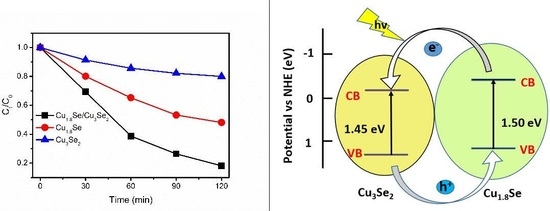
Graphical Abstract

1. Introduction

Semiconductor photocatalysis for the degradation of organic pollutants using solar light energy is a promising approach to solving environmental issue. Since TiO2 was first reported on photocatalytic water splitting under ultraviolet (UV) light [1], photocatalysts have been widely studied in recent years [2,3,4,5]. Recently, photocatalysts with outstanding photocatalytic properties have been found, such as TiO2 and ZnO in the UV spectral range [6,7], and CdS and Bi2WO6 in the visible range [8,9], and only few semiconductors present near-infrared (NIR) photoactivity, such as Cu2(OH)PO4 and WS2 [10,11]. Nowadays, the development of effective ways to exploit near-infrared light (which accounts for more than 50% of solar energy) and develop UV-visible-near-infrared (UV-Vis-NIR) broad light spectrum photocatalysts is an important emerging topic [12]. Forming heterogeneous structures with matched band potentials is one approach [13,14,15]. For example, Au-tipped PbSe/CdSe/CdS core/shell/shell heterostructure nanocrystals are active NIR photocatalysts for the degradation of methylene blue (MB) [16], and Bi2WO6/TiO2 heterojunction can harness UV, visible, and near-infrared light, and exhibits enhanced broad-spectrum photocatalytic properties to decompose methyl orange (MO) [17]. Thus, the design of a broad light spectrum photocatalyst with heterogeneous structure is of great significance.
Selenides of copper are metal chalcogenide semiconductors with different compositions (Cu2Se, Cu3Se2, CuSe, and Cu1.8Se) and various crystallographic forms, which are usually dependent on the preparation process [18,19,20,21,22,23,24,25]. The thermal stability and band gaps of copper selenides vary with their compositions or phases [19,20,21]. These semiconductors with p-type conductivity and direct band gaps have wide applications in solar cells, superconductors, thermoelectric, and photoelectric transformers [26,27,28,29,30]. Therefore, considerable progress on the study of copper selenides has been made, and a number of methods for the synthesis of copper selenides have been explored, such as microwave-assisted heating, sonochemical method, hydrothermal method, and vacuum evaporation [31,32,33,34].
Herein, we report on the aqueous precipitation synthesis of Cu1.8Se/Cu3Se2 heterogeneous structure, bare Cu1.8Se, and Cu3Se2 based on the redox reaction between alkaline selenium solution and cupric aqueous solution under atmospheric pressure. Compared with other reported methods, this method established in aqueous solution is rather simple and safe. The photocatalytic degradation of MO under visible and near-infrared (NIR) light demonstrated that Cu1.8Se/Cu3Se2 composite exhibits much enhanced photocatalytic activity compared with bare Cu1.8Se or Cu3Se2. The mechanism of enhanced photocatalytic activity for Cu1.8Se/Cu3Se2 composite based on the calculated energy band positions was also proposed.

2. Results and Discussion

The composition of products was controlled by varying the addition of NaOH. As shown in Figure 1, when 0.4 g NaOH was added, the X-ray diffraction (XRD) pattern of Figure 1a matched the JCPDS reference file for tetragonal Cu3Se2 [PDF#65-1656]. When 4 g NaOH was included, the diffraction profile of Figure 1b shows that the composite was composed of tetragonal Cu3Se2 [PDF#65-1656] and cubic Cu1.8Se [JCPDF 06-0680]. By increasing the addition of NaOH to 8 g, the XRD pattern of Figure 1c indicates that all the peaks can be well indexed to cubic Cu1.8Se [JCPDF 06-0680]. No characteristic peaks of any impurities were detected in the patterns, and the high diffraction intensity reveals that all the samples had good crystallinity. In addition, the particle sizes calculated from Scherrer’s equation and the BET (Brunauer-Emmett-Teller) specific surface area of different samples are shown in Table 1. There are notbig difference in their particle sizes, and Cu1.8Se displays a larger specific surface area than Cu1.8Se/Cu3Se2 composite and Cu3Se2, which could lead to the exposure of more active sites for the photocatalytic experiment.
The morphology and microstructure of Cu1.8Se/Cu3Se2 composite are shown in the TEM (transmission electron microscopy) image of Figure 2c, with homogeneous size distribution of about 50–80 nm (in agreement with the calculated particle size in Table 1), and the bare Cu1.8Se (Figure 2a) and Cu3Se2 (Figure 2b) nanocrystals (NCs) have similar morphologies. The lattice resolved HRTEM (high resolution transmission electron microscopy) image of Cu1.8Se/Cu3Se2 composite indicates the longitudinal fringe spacing of 0.331 nm and the horizontal fringe spacing of 0.354 nm (as seen in Figure 2d), which is consistent with the spacing of (111) and (101) planes of cubic Cu1.8Se and tetragonal Cu3Se2, respectively. The high-quality image of the interface between Cu1.8Se and Cu3Se2 can be observed clearly, which could be considered as a phase junction structure [35].
The UV-Vis diffuse reflection spectrum (DRS) of the as-prepared samples is shown in Figure 3a. It can be seen that these samples present effective optical absorption from the UV light region to the whole range of NIR light, which can be attributed to their small bandgaps. As shown in Figure 3b (converted from the DRS spectrum according to the Kubelka–Munk function [36,37,38]), the bandgap energies (Eg) of Cu1.8Se/Cu3Se2 composite, Cu3Se2, and Cu1.8Se were estimated to be around 1.42, 1.45, and 1.5 eV, respectively, which are similar to the reported bandgap [31,32]. It is worth noting that these bandgap values are very close, and Cu1.8Se/Cu3Se2 composite has a relatively narrower band gap and stronger light absorption than that of Cu3Se2 and Cu1.8Se.
The photocatalytic activities of these samples were evaluated by the photodegradation of MO under visible and NIR light irradiation. As shown in Figure 4a,b, under identical experimental conditions, the Cu1.8Se/Cu3Se2 composite exhibited strongly enhanced photocatalytic activity compared to both bare Cu1.8Se and Cu3Se2. The photodegradation rate of MO reached 80% and 75% in the presence of Cu1.8Se/Cu3Se2 composite after 3 h of visible and NIR light irradiation, respectively. Thus, the Cu1.8Se/Cu3Se2 composite shows relatively good visible-NIR broad light spectrum photocatalytic property, which is comparative with that of WS2 [11] or Bi2WO6/TiO2 [17]. By contrast, 50% and 46% of MO was degraded by bare Cu1.8Se, whereas only 20% and 17% of MO was degraded by bare Cu3Se2 within the same time. Moreover, photodegradation of MO under full solar light without optical filter was performed. As shown in Figure 4c, after 2 h of full solar light irradiation, 82%, 52%, and 20% of the MO was degraded with Cu1.8Se/Cu3Se2 composite, pure Cu1.8Se, and Cu3Se2 as catalysts, respectively. In addition, the stabilities of these photocatalysts are shown in Figure 5. After four successive cycles, the photodegradation rate of MO under full solar light slightly decreased to 70%, 42%, and 18% within 120 min for Cu1.8Se/Cu3Se2 composite, Cu1.8Se, and Cu3Se2, respectively, indicating that these materials could be re-used without appreciable loss of photocatalytic ability.
To further understand the reaction kinetics of MO degradation under full solar light, we applied the Langmuir−Hinshelwood model, which is well-designed for photocatalytic experiments when the concentration of the organic pollutant is in the millimolar range [39], as expressed by
ln(C0/Ct) = kt
where C0 and Ct are the concentrations of pollutant in solution at time t0 and t, respectively, and k is the kinetic constant, which is calculated to be 0.0145, 0.0062, and 0.0018 min−1 for Cu1.8Se/Cu3Se2 composite, bare Cu1.8Se, and Cu3Se2, respectively (Figure 4d). Then, the apparent reaction rate constant (K) was calculated to get deeper insight into the photodegradation rate per unit surface area according to the following formula:
K = k/(mS)
where k is the kinetic constant from Formula (1), m is the mass of photocatalyst (0.1 g), and S is the specific surface area from Table 1. The apparent reaction rate constant (K) was calculated to be 0.0248, 0.0072, and 0.0027 min−1 for Cu1.8Se/Cu3Se2 composite, bare Cu1.8Se, and Cu3Se2, respectively. In other words, the photocatalytic activity per unit surface area of Cu1.8Se/Cu3Se2 is around 3.4 and 9.1 times higher than that of bare Cu1.8Se and Cu3Se2.
On the other hand, X-ray photoelectron spectroscopy (XPS) analysis was carried out to investigate the chemical binding states of Cu1.8Se/Cu3Se2 composite, as shown in Figure 6a,b. The Cu 2p peaks of Cu3Se2 and Cu1.8Se/Cu3Se2 composite broaden and undergo splitting, while pronounced satellite peaks (marked as Sat.) form due to the existence of Cu2+ vacancy [40,41]. Two strong peaks are consistent with the literature data of Cu+ 2p3/2 and 2p1/2 [42,43,44]. On the sides of Cu+ 2p3/2 and 2p1/2 peaks, two low-intensity components appeared, which can be assigned to the Cu2+ oxidation state [45,46]. As illustrated in Figure 6c, the Cu 2p3/2 and Cu 2p1/2 peaks are symmetric, narrow, and devoid of satellite peaks, which is indicative of monovalent copper of Cu+ for bare Cu1.8Se. Therefore, the existence of Cu3Se2 leads to the presence of Cu2+ in the Cu1.8Se/Cu3Se2 composite.
In view of the mentioned XRD result (Figure 1b), Cu1.8Se is the main phase of Cu1.8Se/Cu3Se2 composite, resulting in the smaller relative concentration ratio of Cu2+ to Cu+ (1.04) than that (2.32) of bare Cu3Se2. Combined with the photodegradation results (Figure 3), it could be inferred that decreasing the concentration ratio of Cu2+ to Cu+ is conducive to enhancing photoactivity. It is worth noting that Cu1.8Se/Cu3Se2 composite has a larger concentration ratio of Cu2+ to Cu+ (1.04) than that (0) of bare Cu1.8Se, but the best photocatalytic activity—mainly due to the existence of phase junction between Cu1.8Se and Cu3Se2 mentioned in the HRTEM image (Figure 2d).
The relative band positions of the two semiconductors were determined to account for the enhanced photocatalytic activity of the Cu1.8Se/Cu3Se2 composite. The energy of conduction band (CB) bottoms (ECB) were calculated empirically according to formula [47]
ECB = X − 0.5Eg + E0
where Eg is the band gap of the semiconductor, E0 is −4.5 eV for a normal hydrogen electrode, and X is the electronegativity of the semiconductor, expressed as the geometric mean of the absolute electronegativity of the constituent atoms [48]. The X values for Cu1.8Se and Cu3Se2 were calculated to be 4.94 and 5.0 eV, respectively, and the band gaps of Cu1.8Se and Cu3Se2 are 1.5 and 1.45 eV, respectively. On the basis of the equation above, the conduction band bottoms (ECB) of Cu1.8Se and Cu3Se2 were calculated to be −0.31 and −0.22 eV, respectively. Correspondingly, the valence band tops (EVB) of Cu1.8Se and Cu3Se2 are 1.19 and 1.23 eV, respectively. Thus, both the conduction band bottom (ECB) and the valence band top (EVB) of Cu1.8Se are higher than that of Cu3Se2. The calculated band positions of Cu1.8Se/Cu3Se2 composite was conducive to the separation and transportation of photogenerated carriers.
As shown in Figure 7, Cu1.8Se and Cu3Se2 are easily excited by visible or NIR light, and photoinduced electrons and holes are generated. The CB edge potentials of the two phases enable photogenerated electrons to easily transfer from Cu1.8Se to Cu3Se2. Simultaneously, holes on the valence band of Cu3Se2 can be transferred to that of Cu1.8Se under the band energy potential difference. In such a way, long-lived reactive photogenerated carriers can be yielded, and thus enhanced charge separation efficiency through the phase junction can be achieved.
To further confirm the effect of phase junction, the photoelectrochemical (PEC) behavior of Cu1.8Se/Cu3Se2 composite has been explored. As shown in Figure 8, the photocurrent responses were recorded under visible light irradiation. The electrodes (1 × 1 cm2) demonstrated photocurrent responses around 1 μA/cm2 and 2.5 μA/cm2 for Cu1.8Se and Cu1.8Se/Cu3Se2 composite, respectively, while the photocurrent responses of Cu3Se2 were not apparent. This result provides strong evidence that the formation of a phase junction between Cu1.8Se and Cu3Se2 would efficiently accelerate the separation efficiency of charge carriers. Therefore, the Cu1.8Se/Cu3Se2 composite presents highly enhanced performance as compared to bare Cu1.8Se and Cu3Se2.

3. Materials and Methods

3.1. Preparation

All reagents were of 99.9% purity and were used without further purification. A typical synthesis process of Cu1.8Se/Cu3Se2 composite was described as follows: 4 g NaOH, 0.76 g NaBH4, and 0.3 g elemental Se were added to 100 mL distilled water under constant stirring. The mixture reached about 80 °C in a few minutes to form alkaline selenium aqueous solution. Meanwhile, 10 mL Cu(NO3)2 aqueous solution (0.5 M) was prepared, and the mixture was combined with the alkaline selenium solution through rapid stirring. Finally, 0.1 g SDS was included as surfactant to control the morphology. After stirring for 8 h, the resulting products were separated by filtration, washed several times with distilled water and absolute alcohol, and then dried at 60 °C for 6 h. For comparison, bare Cu3Se2 and Cu1.8Se were also prepared by the hydrothermal method under the same conditions mentioned above by adding 0.4 g and 8 g NaOH, respectively.

3.2. Characterization

The phase and composition of the as-prepared samples was examined with an X-ray diffractometer (XRD, Bruker, D8 Advance, Beijing, China) using Cu Kα radiation (λ = 1.5418 Å). The morphology and microstructures were characterized by transmission electron microscope (TEM, JEOL JEM-2100, Beijing, China). X-ray photoelectron spectroscopy (XPS) measurements were conducted on a Thermo XPS ESCALAB 250Xi instrument (Thermo Scientific, Shanghai, China). UV-Vis-NIR diffuser reflectance (DRS) measurements were carried out on a UV/Vis/NIR spectrometer (PerkinElmer, Lambda 950, Shanghai, China). Specific surface area measurements were conducted by Autosorb-iQ2-Mp (Quantachrome, Shanghai, China).

3.3. Photocatalytic Test

The photocatalytic activities of as-prepared samples were investigated by photodegradation of MO under visible and NIR light. A 300 W xenon lamp (CEL-HXF300, Beijing, China) with cutoff filters (420 nm, 800 nm) was used as the light source. The specific process was as follows: 0.1 g photocatalyst was added into 100 mL MO (50 mg/L). Prior to irradiation, the slurry was continuously stirred in the dark for 1 h to ensure an adsorption–desorption equilibrium between photocatalyst and MO. Ice-water bath and magnetic stirring always existed to prevent thermal effect in the photocatalytic process. At a given time interval, 3 mL suspensions were centrifuged to remove the photocatalyst. The concentration of MO was analyzed by the UV-Vis spectrophotometer by recording the variations of the absorption band maximum (465 nm).

3.4. Photoelectrochemical Testing

Photocurrent under visible light was measured with a standard three electrode system on an electrochemical workstation (CHI 660, ChenHua, Shanghai, China). Ag/AgCl and Pt plate were used as reference electrode and counter electrode, respectively, in Na2SO4 solution (0.5 mol/L) as electrolyte. The working electrode was made by depositing photoctalyst on the FTO (fluorine-doped tin oxide) substrate (Beijing, China).

4. Conclusions

The Cu1.8Se/Cu3Se2 heterogeneous structure prepared by the simple precipitation method exhibits excellent photocatalytic activity in the degradation of MO under visible and NIR light irradiation, which is 3.4 and 9.1 times higher than that of bare Cu1.8Se and Cu3Se2. We also present that decreasing the concentration ratio of Cu2+ to Cu+ is advantageous for the enhancement of photoactivity. The mechanism of enhanced photocatalytic activity for the Cu1.8Se/Cu3Se2 composite was investigated based on the calculated energy band positions. The formation of a phase junction between the two semiconductors leads to the effective separation of photogenerated electron-hole pairs, which mainly account for the strongly enhanced photoactivity.

Acknowledgments

This work was supported by the NSF of China (51221291, 51328203 and 51532003).

Author Contributions

Li-Na Qiao wrote the manuscript under the guidance of Huan-Chun Wang, Yang Shen, Yuan-Hua Lin and Ce-Wen Nan. Additionally, manuscript was corrected and edited by Huan-Chun Wang.

Conflicts of Interest

The authors declare no conflict of interest.

References

  1. Fujishima, A. Electrochemical photolysis of water at a semiconductor electrode. Nature 1972, 238, 37–38. [Google Scholar] [CrossRef] [PubMed]
  2. Asahi, R.; Morikawa, T.; Ohwaki, T.; Aoki, K.; Taga, Y. Visible-light photocatalysis in nitrogen-doped titanium oxides. Science 2001, 293, 269–271. [Google Scholar] [CrossRef] [PubMed]
  3. Zou, Z.; Ye, J.; Sayama, K.; Arakawa, H. Direct splitting of water under visible light irradiation with an oxide semiconductor photocatalyst. Nature 2001, 414, 625–627. [Google Scholar] [CrossRef] [PubMed]
  4. Gao, F.; Chen, X.; Yin, K.; Dong, S.; Ren, Z.; Yuan, F.; Yu, T.; Zou, Z.; Liu, J.M. Visible-Light Photocatalytic Properties of Weak Magnetic BiFeO3 Nanoparticles. Adv. Mater. 2007, 19, 2889–2892. [Google Scholar] [CrossRef]
  5. Abdullah, H.; Khan, M.R.; Pudukudy, M.; Yaakob, Z.; Ismail, N.A. CeO2-TiO2 as a visible light active catalyst for the photoreduction of CO2 to methanol. J. Rare Earths 2015, 33, 1155–1161. [Google Scholar] [CrossRef]
  6. Nakata, K.; Ochiai, T.; Murakami, T.; Fujishima, A. Photoenergy conversion with TiO2 photocatalysis: New materials and recent applications. Electrochim. Acta 2012, 84, 103–111. [Google Scholar] [CrossRef]
  7. He, Y.; Wang, Y.; Zhang, L.; Teng, B.; Fan, M. High-efficiency conversion of CO2 to fuel over ZnO/gC3N4 photocatalyst. Appl. Catal. B 2015, 168, 1–8. [Google Scholar]
  8. Hou, Y.-F.; Liu, S.-J.; Zhang, J.; Cheng, X.; Wang, Y. Facile hydrothermal synthesis of TiO2–Bi2WO6 hollow superstructures with excellent photocatalysis and recycle properties. Dalton Trans. 2014, 43, 1025–1031. [Google Scholar] [CrossRef] [PubMed]
  9. Wang, L.; Wei, H.; Fan, Y.; Gu, X.; Zhan, J. One-dimensional CdS/α-Fe2O3 and CdS/Fe3O4 heterostructures: Epitaxial and nonepitaxial growth and photocatalytic activity. J. Phys. Chem. C 2009, 113, 14119–14125. [Google Scholar] [CrossRef]
  10. Wang, G.; Huang, B.; Ma, X.; Wang, Z.; Qin, X.; Zhang, X.; Dai, Y.; Whangbo, M.H. Cu2(OH)PO4, a Near-Infrared-Activated Photocatalyst. Angew. Chem. Int. Ed. 2013, 52, 4810–4813. [Google Scholar] [CrossRef] [PubMed]
  11. Sang, Y.; Zhao, Z.; Zhao, M.; Hao, P.; Leng, Y.; Liu, H. From UV to Near-Infrared, WS2 Nanosheet: A Novel Photocatalyst for Full Solar Light Spectrum Photodegradation. Adv. Mater. 2015, 27, 363–369. [Google Scholar] [CrossRef] [PubMed]
  12. Tong, H.; Ouyang, S.; Bi, Y.; Umezawa, N.; Oshikiri, M.; Ye, J. Nano-photocatalytic materials: Possibilities and challenges. Adv. Mater. 2012, 24, 229–251. [Google Scholar] [CrossRef] [PubMed]
  13. Hou, Y.; Li, X.; Zhao, Q.; Quan, X.; Chen, G. Fabrication of Cu2O/TiO2 nanotube heterojunction arrays and investigation of its photoelectrochemical behavior. Appl. Phys. Lett. 2009, 95, 093108. [Google Scholar] [CrossRef]
  14. Georgieva, J.; Armyanov, S.; Valova, E.; Poulios, I.; Sotiropoulos, S. Enhanced photocatalytic activity of electrosynthesised tungsten trioxide–titanium dioxide bi-layer coatings under ultraviolet and visible light illumination. Electrochem. Commun. 2007, 9, 365–370. [Google Scholar] [CrossRef]
  15. Zhang, Z.; Wang, W.; Wang, L.; Sun, S. Enhancement of Visible-Light Photocatalysis by Coupling with Narrow-Band-Gap Semiconductor: A Case Study on Bi2S3/Bi2WO6. ACS Appl. Mater. Interfaces 2012, 4, 593–597. [Google Scholar] [CrossRef] [PubMed]
  16. Pak, C.; Woo, J.Y.; Lee, K.; Kim, W.D.; Yoo, Y.; Lee, D.C. Extending the Limit of Low-Energy Photocatalysis: Dye Reduction with PbSe/CdSe/CdS Core/Shell/Shell Nanocrystals of Varying Morphologies under Infrared Irradiation. J. Phys. Chem. C 2012, 116, 25407–25414. [Google Scholar] [CrossRef]
  17. Tian, J.; Sang, Y.; Yu, G.; Jiang, H.; Mu, X.; Liu, H. A Bi2WO6-based hybrid photocatalyst with broad spectrum photocatalytic properties under UV, visible, and near-infrared irradiation. Adv. Mater. 2013, 25, 5075–5080. [Google Scholar] [CrossRef] [PubMed]
  18. Stevels, A.; Jellinek, F. Phase transitions in copper chalcogenides: I. The copper-selenium system. Recl. Trav. Chim. Pays-Bas. 1971, 90, 273–283. [Google Scholar] [CrossRef]
  19. Haram, S.K.; Santhanam, K.; Neumann-Spallart, M.; Levy-Clement, C. Electroless deposition on copper substrates and characterization of thin films of copper (I) selenide. Mater. Res. Bull. 1992, 27, 1185–1191. [Google Scholar] [CrossRef]
  20. Shafizade, R.; Ivanova, I.; Kazinets, M. Electron diffraction study of phase transformations of the compound CuSe. Thin Solid Films 1978, 55, 211–220. [Google Scholar] [CrossRef]
  21. Garcıa, V.; Nair, P.; Nair, M. Copper selenide thin films by chemical bath deposition. J. Cryst. Growth 1999, 203, 113–124. [Google Scholar] [CrossRef]
  22. Lakshmi, M.; Bindu, K.; Bini, S.; Vijayakumar, K.; Kartha, C.S.; Abe, T.; Kashiwaba, Y. Chemical bath deposition of different phases of copper selenide thin films by controlling bath parameters. Thin Solid Films 2000, 370, 89–95. [Google Scholar] [CrossRef]
  23. Lakshmi, M.; Bindu, K.; Bini, S.; Vijayakumar, K.; Kartha, C.S.; Abe, T.; Kashiwaba, Y. Reversible Cu2−xSe↔Cu3Se2 phase transformation in copper selenide thin films prepared by chemical bath deposition. Thin Solid Films 2001, 386, 127–132. [Google Scholar] [CrossRef]
  24. Heyding, R.D.; Murray, R.M. The crystal structures of Cu1.8Se, Cu3Se2, α-and γCuSe, CuSe2, and CuSe2II. Can. J. Chem. 1976, 54, 841–848. [Google Scholar] [CrossRef]
  25. Heyding, R. The copper/selenium system. Can. J. Chem. 1966, 44, 1233–1236. [Google Scholar] [CrossRef]
  26. Bhuse, V.; Hankare, P.; Garadkar, K.; Khomane, A. A simple, convenient, low temperature route to grow polycrystalline copper selenide thin films. Mater. Chem. Phys. 2003, 80, 82–88. [Google Scholar] [CrossRef]
  27. Lévy-Clément, C.; Neumann-Spallart, M.; Haram, S.; Santhanam, K. Chemical bath deposition of cubic copper (I) selenide and its room temperature transformation to the orthorhombic phase. Thin Solid Films 1997, 302, 12–16. [Google Scholar] [CrossRef]
  28. Chen, W.S.; Stewart, J.; Mickelsen, R. Polycrystalline thin-film Cu2−xSe/CdS solar cell. Appl. Phys. Lett. 1985, 46, 1095–1097. [Google Scholar] [CrossRef]
  29. Dorfs, D.; Härtling, T.; Miszta, K.; Bigall, N.C.; Kim, M.R.; Genovese, A.; Falqui, A.; Povia, M.; Manna, L. Reversible Tunability of the Near-Infrared Valence Band Plasmon Resonance in Cu2–xSe Nanocrystals. J. Am. Chem. Soc. 2011, 133, 11175–11180. [Google Scholar] [CrossRef] [PubMed]
  30. Liu, X.; Wang, X.; Zhou, B.; Law, W.C.; Cartwright, A.N.; Swihart, M.T. Size-Controlled Synthesis of Cu2−xE (E = S, Se) Nanocrystals with Strong Tunable Near-Infrared Localized Surface Plasmon Resonance and High Conductivity in Thin Films. Adv. Funct. Mater. 2013, 23, 1256–1264. [Google Scholar] [CrossRef]
  31. Zhu, J.; Palchik, O.; Chen, S.; Gedanken, A. Microwave assisted preparation of CdSe, PbSe, and Cu2−xSe nanoparticles. J. Phys. Chem. B 2000, 104, 7344–7347. [Google Scholar] [CrossRef]
  32. Xie, Y.; Zheng, X.; Jiang, X.; Lu, J.; Zhu, L. Sonochemical Synthesis and Mechanistic Study of Copper Selenides Cu2−xSe, β-CuSe, and Cu3Se2. Inorg. Chem. 2002, 41, 387–392. [Google Scholar] [CrossRef] [PubMed]
  33. Xu, S.; Wang, H.; Zhu, J.-J.; Chen, H.-Y. Sonochemical synthesis of copper selenides nanocrystals with different phases. J. Cryst. Growth 2002, 234, 263–266. [Google Scholar] [CrossRef]
  34. Kemmler, M.; Lazell, M.; O'brien, P.; Otway, D.; Park, J.-H.; Walsh, J. The growth of thin films of copper chalcogenide films by MOCVD and AACVD using novel single-molecule precursors. J. Mater. Sci. Mater. Electron. 2002, 13, 531–535. [Google Scholar] [CrossRef]
  35. Liu, D.; Wang, J.; Zhang, M.; Liu, Y.; Zhu, Y. A superior photocatalytic performance of a novel Bi2SiO5 flower-like microsphere via a phase junction. Nanoscale 2014, 6, 15222–15227. [Google Scholar] [CrossRef] [PubMed]
  36. Butler, M. Photoelectrolysis and physical properties of the semiconducting electrode WO2. J. Appl. Phys. 1977, 48, 1914–1920. [Google Scholar] [CrossRef]
  37. Zhao, W.; Ma, W.; Chen, C.; Zhao, J.; Shuai, Z. Efficient degradation of toxic organic pollutants with Ni2O3/TiO2−xBx under visible irradiation. J. Am. Chem. Soc. 2004, 126, 4782–4783. [Google Scholar] [CrossRef] [PubMed]
  38. Xu, C.; Liu, Y.; Huang, B.; Li, H.; Qin, X.; Zhang, X.; Dai, Y. Preparation, characterization, and photocatalytic properties of silver carbonate. Appl. Surf. Sci. 2011, 257, 8732–8736. [Google Scholar] [CrossRef]
  39. Sakkas, V.; Arabatzis, I.; Konstantinou, I.; Dimou, A.; Albanis, T.; Falaras, P. Metolachlor photocatalytic degradation using TiO2 photocatalysts. Appl. Catal. B 2004, 49, 195–205. [Google Scholar] [CrossRef]
  40. Zhang, J.; Yu, J.; Zhang, Y.; Li, Q.; Gong, J.R. Visible light photocatalytic H2-production activity of CuS/ZnS porous nanosheets based on photoinduced interfacial charge transfer. Nano Lett. 2011, 11, 4774–4779. [Google Scholar] [CrossRef] [PubMed]
  41. Wong, L.; Chiam, S.; Huang, J.; Wang, S.; Pan, J.; Chim, W. Growth of Cu2O on Ga-doped ZnO and their interface energy alignment for thin film solar cells. J. Appl. Phys. 2010, 108, 033702. [Google Scholar] [CrossRef]
  42. Xing, C.; Zhang, Y.; Wu, Z.; Jiang, D.; Chen, M. Ion-exchange synthesis of Ag/Ag2S/Ag3CuS2 ternary hollow microspheres with efficient visible-light photocatalytic activity. Dalton Trans. 2014, 43, 2772–2780. [Google Scholar] [CrossRef] [PubMed]
  43. Yu, S.; Liu, J.; Zhu, W.; Hu, Z.-T.; Lim, T.-T.; Yan, X. Facile room-temperature synthesis of carboxylated graphene oxide-copper sulfide nanocomposite with high photodegradation and disinfection activities under solar light irradiation. Sci. Rep. 2015, 5, 16369. [Google Scholar] [CrossRef] [PubMed]
  44. Riha, S.C.; Johnson, D.C.; Prieto, A.L. Cu2Se nanoparticles with tunable electronic properties due to a controlled solid-state phase transition driven by copper oxidation and cationic conduction. J. Am. Chem. Soc. 2010, 133, 1383–1390. [Google Scholar] [CrossRef] [PubMed]
  45. Ludwig, J.; An, L.; Pattengale, B.; Kong, Q.; Zhang, X.; Xi, P.; Huang, J. Ultrafast hole trapping and relaxation dynamics in p-type CuS nanodisks. J. Phys. Chem. Lett. 2015, 6, 2671–2675. [Google Scholar] [CrossRef] [PubMed]
  46. Savariraj, A.D.; Viswanathan, K.K.; Prabakar, K. Influence of Cu vacancy on knit coir mat structured CuS as counter electrode for quantum dot sensitized solar cells. ACS Appl. Mater. Interfaces 2014, 6, 19702–19709. [Google Scholar] [CrossRef] [PubMed]
  47. Jun, L.; Kako, T.; Jinhua, Y.; Zhigang, Z. Band structure design and photocatalytic activity of In2O3/N-InNbO4 composite. Appl. Phys. Lett. 2009, 95. [Google Scholar] [CrossRef]
  48. Xu, Y.; Schoonen, M.A. The absolute energy positions of conduction and valence bands of selected semiconducting minerals. Am. Mineral. 2000, 85, 543–556. [Google Scholar] [CrossRef]
Figure 1. X-ray diffraction profiles of as-prepared copper selenides, (a) Cu3Se2, (b) Cu1.8Se/Cu3Se2 composite, (c) Cu1.8Se.
Nanomaterials 07 00019 g001
Figure 2. TEM (transmission electron microscopy) images of (a) Cu1.8Se; (b) Cu3Se2; (c) Cu1.8Se/Cu3Se2 composite; (d) HRTEM (high resolution transmission electron microscopy) of Cu1.8Se/Cu3Se2 composite.
Nanomaterials 07 00019 g002
Figure 3. (a) UV-visible-near-infrared (UV-Vis-NIR) diffuse reflectance absorption (DRS) spectrum of copper selenides; (b) A plot transformed according to the Kubelka–Munk function versus energy of light.
Nanomaterials 07 00019 g003
Figure 4. Photocatalytic degradation of methyl orange (MO) solution with copper selenides as catalysts under (a) visible light; (b) NIR light; (c) full solar light; (d) the degradation kinetics by means of plotting ln(C0/Ct) versus time.
Nanomaterials 07 00019 g004
Figure 5. Cycling runs for the photocatalytic degradation of MO under full solar light in the presence of (a) Cu1.8Se/Cu3Se2 composite; (b) Cu1.8Se; (c) Cu3Se.
Nanomaterials 07 00019 g005
Figure 6. High-resolution X-ray photoelectron spectroscopy (XPS) scan of Cu 2p (a) Cu3Se2; (b) Cu1.8Se/Cu3Se2 composite; (c) Cu1.8Se.
Nanomaterials 07 00019 g006
Figure 7. Diagram of energy band levels of Cu1.8Se/Cu3Se2 composites vs. NHE (normal hydrogen electrode) and the possible charge separation process.
Nanomaterials 07 00019 g007
Figure 8. The photocurrent responses of copper selenides in 0.5 M Na2SO4 electrolyte under visible light.
Nanomaterials 07 00019 g008
Table 1. The BET (Brunauer-Emmett-Teller) specific surface area of different samples.
SamplesCu1.8Se/Cu3Se2 CompositeCu1.8SeCu3Se2
Size (nm)66.348.657.8
Specific surface area (m2/g)5.8428.6556.556