Facet-Dependent Cuprous Oxide Nanocrystals Decorated with Graphene as Durable Photocatalysts under Visible Light

Department of Environmental Engineering, National Cheng Kung University, Tainan 70101, Taiwan
*
Author to whom correspondence should be addressed.
Nanomaterials 2018, 8(6), 423; https://doi.org/10.3390/nano8060423
Submission received: 25 May 2018
/
Revised: 8 June 2018
/
Accepted: 10 June 2018
/
Published: 11 June 2018
Abstract
:Three morphologies (octahedral, hierarchical and rhombic dodecahedral) of crystal Cu2O with different facets ({111}, {111}/{110}, and {110}) incorporating graphene sheets (denoted as o-Cu2O-G, h-Cu2O-G and r-Cu2O-G, respectively) have been fabricated by using simple solution-phase techniques. Among these photocatalysts, the r-Cu2O-G possesses the best photocatalytic performance of 98% removal efficiency of methyl orange (MO) with outstanding kinetics for 120 min of visible light irradiation. This enhancement is mainly due to the dangling “Cu” atoms in the highly active {110} facets, resulting in the increased adsorption of negatively charged MO. More importantly, the unique interfacial structures of Cu2O rhombic dodecahedra connected to graphene nanosheets can not only decrease the recombination of electron-hole pairs but also stabilize the crystal structure of Cu2O, as verified by a series of spectroscopic analyses (e.g., X-ray diffraction (XRD), X-ray photoelectron spectroscopy (XPS), scanning electron microscopy (SEM) and transmission electron microscopy (TEM)). The effective photocatalysts developed in this work could be applied to the efficient decolorization of negatively charged organic dyes by employing solar energy.
1. Introduction
Metal oxide semiconductors have been intensively investigated for photocatalytic degradation of organic pollutants for many years [1,2,3]. However, these photocatalysts, e.g., TiO2 and ZnO (band gap ≅ 3.2 eV), can exhibit superior photocatalysis properties in the ultraviolet (UV) light region, leading to their limited practical application in wastewater treatments due to the restricted use of solar energy [4,5,6,7,8]. Cuprous oxide (Cu2O), which is a p-type semiconductor with a direct band gap of ca. 2.17 eV, has been widely studied as an efficient photocatalyst [9,10,11,12,13] because of its abundance, low cost, environmental-friendliness and good visible-light response. Nonetheless, the photocatalytic activity of Cu2O is constrained by fast recombination of the electron/hole (e/h) pairs [14] and has low durability [15]. Therefore, many studies have been dedicated to enhancing visible light-active photocatalysis by enhancing the segregation of electron-hole pairs. For instance, incorporating ions into the semiconductor [16,17,18,19,20,21], sensitization with dyer and surface complex [22,23,24], and coupling two or more semiconductors [25,26]. Moreover, well-defined facets of Cu2O which exhibit unique crystallographic properties related to different atomic terminated arrangements have been demonstrated to make a great improvement to photocatalysis [27,28,29]. The {110} facets of Cu2O were found to have a superior photocatalytic activity toward the degradation of methyl orange [27]. In addition, the Cu2O octahedra crystals consisting of {111} facets showed higher photocatalytic performance as compared to truncated cubic crystals abundant in {100} facets which has been attributed to the lower surface energy density of {100} facets than that of {111} facets [30]. In comparison to the pure Cu2O octahedra with {111} surfaces and pure cubes with {100} surfaces, the combination of 26-facet and 18-facet polyhedra with main {110} was observed to have better adsorption and photocatalytic activities [31]. However, the aforementioned Cu2O crystals may suffer from the deterioration of their crystal structure during long-term operation [32].
To further increase the stability of Cu2O nanocrystals, one of the possible methods was carried out by the incorporation of carbonaceous materials onto Cu2O [33,34,35]. Graphene is a well-known two-dimensional (2D) carbon material [36], which has distinguishing physiochemical properties such as a theoretical surface area (~2965 m2 g−1) [37], high intrinsic electron mobility (2 × 105 cm2 V−1 s−1) [38], and exceptional chemical durability [39,40,41]. Owing to these unique properties, visible light-driven photocatalysts based on the synthesis of Cu2O-graphene composites [35,42,43] for fuel production and pollutant degradation have been proposed. However, limited studies have been reported in terms of different facets of Cu2O-graphene nanoheterostructures on their visible light-responsive activity and corresponding durability.
In this research, three different morphologies of Cu2O crystals with low-index facets ({111}, {111}/{110}, and {110}) decorated with graphene sheets were prepared via simple wet-chemical methods. These as-synthesized Cu2O crystals were thoroughly characterized by a variety of analytical spectroscopies and used as visible light-driven photocatalysts in the degradation of methyl orange (MO).
2. Experiment
2.1. Photocatalyts Preparation
Typically, the synthesis of graphene oxide (GO) was carried out by referring to a modified route described earlier [44]. For preparation of octahedral Cu2O [27] and octahedral Cu2O-graphene, ca. 88 mL of deionized water or 1% GO solution was mixed with ca. 1 mL of copper(II) chloride solution and 0.87 g of sodium dodecyl sulfate (SDS) solids under vigorous stirring until the dissolution of the SDS powder. Then, 8.5 mL of 0.2 M NH2OH·HCl and 2.5 mL of 1.0 M NaOH solution were consecutively added to the resulting mixture. Lastly, the precipitate was treated by centrifugation and dried under a vacuum. The aforementioned photocatalysts prepared by using deionized water and 1% GO solution in the synthesis process were labelled as o-Cu2O and o-Cu2O-G, respectively.
In terms of the synthesis of hierarchical facets of Cu2O [30] and hierarchical facets of Cu2O-graphene, 48 mL of 1% GO solution was mixed well with 1 mL of copper(II) chloride solution. Afterward, 40 mL of ethylene glycol (EG) was introduced to the aforementioned mixture. Then, 24 mL of 0.1 M NH2OH·HCl was added to the above solution for 10 min. The resultant solution was moved to a Teflon-lined stainless steel autoclave, followed by ramping from room temperature to 180 °C for 1 h. The solid products were filtrated, washed and dried at ambient temperature. The aforementioned samples prepared without and with 1% GO solution in the synthesis process were denoted as h-Cu2O and h-Cu2O-G, respectively.
For the preparation of rhombic dodecahedral Cu2O [27] and rhombic dodecahedral Cu2O-graphene, ca. 34.6 mL of deionized water or 1% GO solution was heated to 34 °C in a water bath. Then, ca. 2.5 mL of copper(II) chloride solution (0.1 M) and 0.44 g of SDS solids were added into the above mixture with continuous stirring. While the SDS was dissolved, 2.5 mL of NaOH solution was introduced, followed by adding 12 mL of 0.1 M NH2OH·HCl to the resulting mixture. Finally, the precipitated products were separated by centrifuge, washed by using a water–ethanol mixture and dried. The aforementioned samples prepared by using deionized water and 1% GO solution in the synthesis process were denoted as r-Cu2O and r-Cu2O-G, respectively.
2.2. Characterizations of Photocatalyts
Powder X-ray diffraction (XRD) patterns of the samples were examined by using a PANalytical X’Pert PRO diffractometer with Cu Kα radiation (λ = 1.541 Å). The elemental compositions of samples were analysed by X-ray photoelectron spectroscopy (XPS) using a Kratos AXIS Ultra DLD spectrometer (Kratos Analytical Ltd., Stretford, Manchester, UK) and monochromated Al Kα X-ray source. The morphologies of the photocatlysts were observed through a scanning electron microscope (SEM, JEOL-7000F, JEOL Ltd., Akishima, Tokyo, Japan) with an accelerating voltage of 20 kV. In addition, to further study the detailed surface characteristics of the Cu2O nanocrystals, a high-resolution transmission electron microscopy (TEM, JEOL 2100F, JEOL Ltd., Akishima, Tokyo, Japan) was carried out. The UV-visible (UV-Vis) diffuse reflection spectra of the samples were collected on a UV-Vis spectrophotometer (Varian, Cary 100, Palo Alto, CA, USA).
2.3. Photocatalytic Degradation of Organic Pollutants
Photocatalytic degradation tests of MO were performed at 25 °C in a photoreactor (100 mL) described previously [35]. The test solution was prepared via mixing 5.0 mg of photocatalysts into 80 mL of MO solution (15 mg L−1). Prior to irradiation, the aqueous solution was kept stirring in the dark for 2 h to establish the adsorption equilibrium. The pH value of the solution was 5.6. Then, the photocatalytic reaction was performed by using a 300 W Xe lamp combined with a 420 nm cutoff filter as the light source. About 2 mL of aliquots was periodically withdrawn for every 30 min, centrifuged to separate solid samples, and the variations of concentration were measured by using a Hitachi UV-Visible (UV-Vis) spectroscope (Model U-2910).
3. Results and Discussion
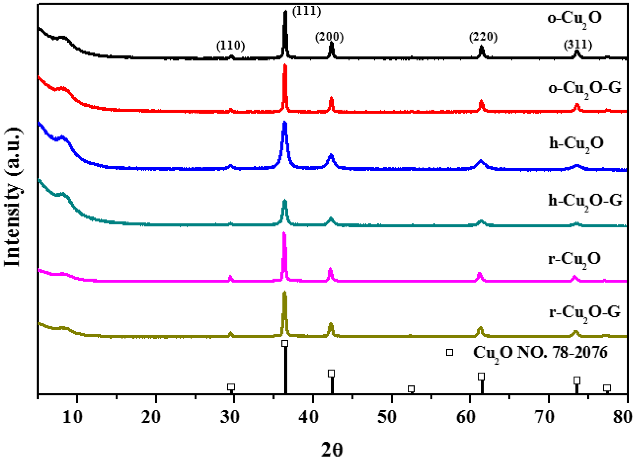
As displayed in Figure 1, the XRD patterns of various shapes of Cu2O- and Cu2O-incorporated graphene samples possess five characteristic reflections at 2θ = 29.6°, 36.4°, 42.3°, 61.4° and 73.6° which are attributed to the (110), (111), (200), (220), (311) planes of cuprous oxide. This result shows the synthesized samples are classified to the cubic phase Cu2O (JCPDS No. 78-2076). The sharp diffraction peaks indicate that the high crystallinity of Cu2O with different morphologies in all samples can be prepared by using wet-chemical methods. In the previous study [27], the intensity of the (220) peak to that of the (200) peak (I(220)/I(200)) is able to be used to evaluate the degree of crystal structure. For instance, the index of I(220)/I(200) is nearly 0.79 in terms of rhombic dodecahedra Cu2O. As can be seen in Figure 1, while incorporating graphene onto various crystals of Cu2O (o-Cu2O-G, h-Cu2O-G and r-Cu2O-G), the value of I(220)/I(200) is practically unchanged, indicating that no apparent perturbation of crystal structure is observed. This result implies that the introduction of graphene may cause little impact on the growth of nanocrystal Cu2O in the samples.
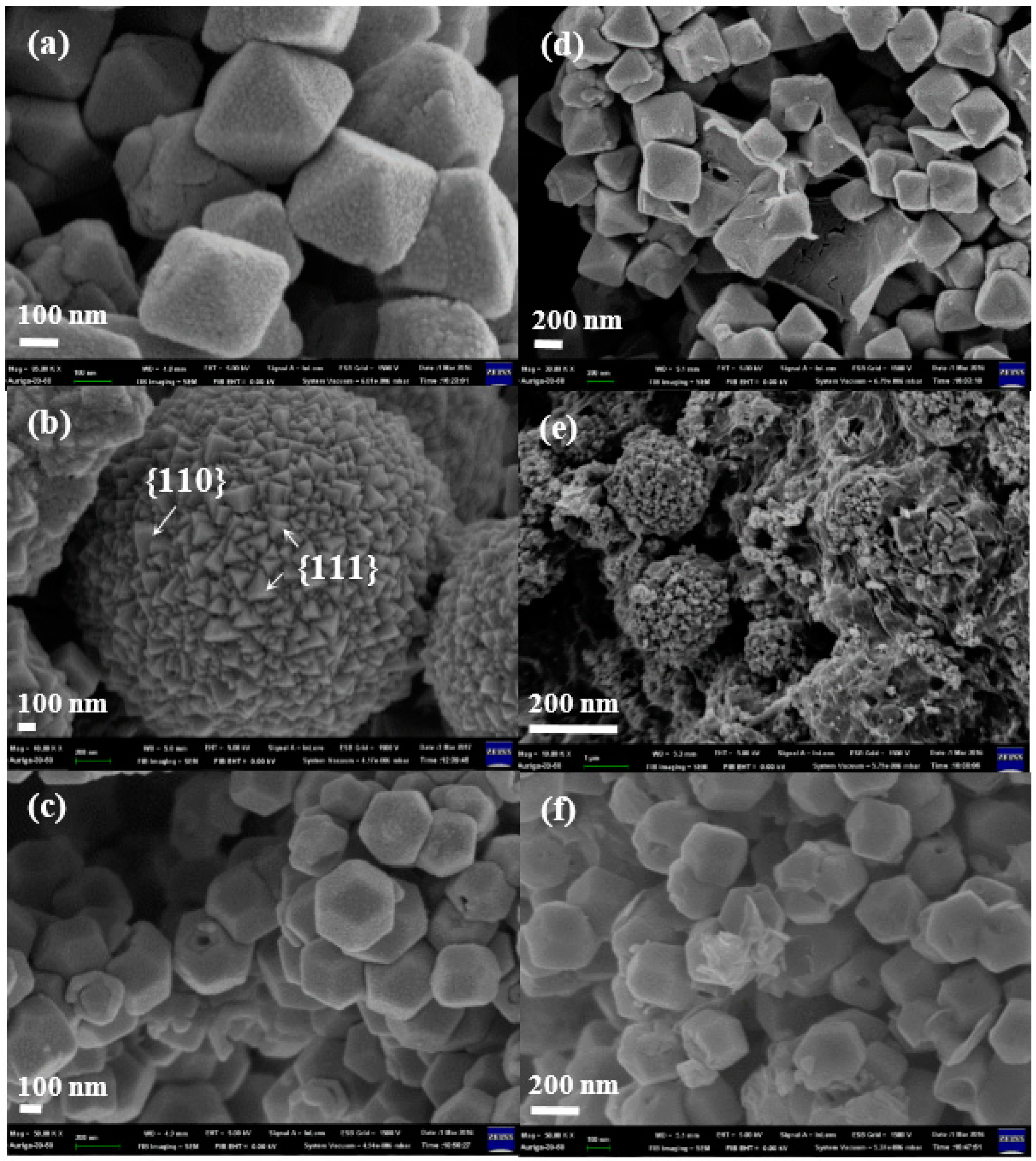
The morphologies of the crystal Cu2O, o-Cu2O-G, h-Cu2O-G and r-Cu2O-G were investigated by using field emission scanning electron microscopy (FESEM). As can be seen in Figure 2a, the pristine o-Cu2O possesses octahedron morphology of eight {111} facets. Figure 2b presents the typical image of the microspherical h-Cu2O with randomly crosslinked polyhedrons which are composed of {110} and {111} facets. In terms of r-Cu2O, a 12-facet polyhedral with mostly {110} facets can be observed, as displayed in Figure 2c. Based on the center of the Gaussian distribution, the particle sizes of o-Cu2O-G, h-Cu2O-G and r-Cu2O-G are calculated to be 315 ± 35, 218 ± 19 and 289 ± 38 nm, respectively. Upon incorporating graphene onto various crystals of Cu2O (o-Cu2O-G, h-Cu2O-G and r-Cu2O-G), the morphologies and crystal structure Cu2O are almost the same as those without graphene, as shown in Figure 2c–e. The microstructures of the Cu2O nanocrystals were additionally identified by TEM. It can be observed that the TEM images (Figure 3) show that crystal Cu2O is incorporating with the wrinkled, thin and transparent graphene nanosheets. The high-resolution TEM (HRTEM) images of o-Cu2O-G, h-Cu2O-G and r-Cu2O-G in Figure 3d–f show that the interplanar lattice with d-spacings of 0.24 and 0.30 nm are, respectively, assigned to the (111) and (110) planes of Cu2O. Moreover, selected area electron diffraction (SAED) and HRTEM suggest that these crystal Cu2O samples possess a single-crystal structure, which matches well with the high crystallinity explored by XRD. Therefore, different shapes of highly crystallized Cu2O decorated with graphene nanosheets have been fabricated by using simple wet-chemical methods.
The surface atomic compositions and interfacial electronic states of the samples can be investigated by XPS analysis, as displayed in Figure 4. The peaks at 284.6, 286.5 and 288.4 eV in the C1s spectra (Figure 4a) are assigned to non-oxygenated, epoxy/hydroxyl and carboxyl carbons, respectively [45,46]. The intensities observed for the peaks at 286.5 and 288.4 eV in the o-Cu2O-G, h-Cu2O-G and r-Cu2O-G samples are decreased slightly in comparison to the pure graphene oxide sheets, suggesting that the graphene sheets may maintain the reduced states even with the existence of Cu2O nanocrystals in the samples. The XPS of spectra of Cu2p (Figure 4b) indicate the photocatalysts have the principal and satellite features at ca. 934 eV and 944 eV which are attributed to Cu(II), while the features at ca. 932 and 952 eV are assigned to Cu(I) 2p3/2 and Cu(I) 2p1/2 peaks [9], respectively. In addition, the O1s (see Figure 4c) peaks at ca. 532.5 and 530.3 eV are assigned to C-O and Cu-O bindings, respectively. As a result, the contents of copper(I) for o-Cu2O-G and r-Cu2O-G composites are much higher, indicating that Cu2O nanoparticles may exist stably when dispersed on graphene nanosheets, which can maintain the photocatalytic performance during photodegradation process [47]. Moreover, it is hard to attain bulk information of the atoms by using XPS because a small number of atomic layers on the surface are identified. In addition, it is noteworthy that the diffraction features of copper oxide could hardly be found for all photocatalysts by using the XRD, which is possibly because of the identification of the crystal structure in the bulk phase (see Figure 1).
To understand the optical properties of o-Cu2O-G, h-Cu2O-G and r-Cu2O-G nanocomposites, diffused reflectance UV-Vis (DR UV-Vis) absorption spectroscopies were carried out. Figure 5a shows that o-Cu2O-G possesses a band edge of ca. 580 nm in the visible region, suggesting that o-Cu2O-G photocatalysts having the bandgap of ca. 2.2 eV (see Figure 5b) should be a visible light-sensitive semiconductor. However, the r-Cu2O-G photocatalysts show slightly increased absorbance intensities in the visible-light region (400–800 nm) and absorption edge positions as compared to o-Cu2O-G and h-Cu2O-G. Moreover, the band gap of these photocatalysts can be obtained by plotting transformed Kubelka–Munk functions, as illustrated in Figure 5b. The band gaps of o-Cu2O-G, h-Cu2O-G and r-Cu2O-G photocatalysts are ca. 2.20, 2.12 and 2.06 eV, respectively. This finding may be due to their different crystal sizes and distinct exposed facets in the photocatalysts. Combining the outcomes of the XRD, TEM, SEM, XPS and DR UV-Vis spectra, various morphologies of crystal Cu2O incorporated with graphene nanosheets may be able to take advantage of natural light for decontamination and also be recycled for reuse after photocatalytic reactions.
The photocatalytic performance of pure cuprous oxides (o-Cu2O, h-Cu2O and r-Cu2O) and various graphene-incorporated cuprous oxides (o-Cu2O-G, h-Cu2O-G and r-Cu2O-G) were studied by the photocatalytic oxidation of the MO solution under visible light illumination at room temperature. About 8.3, 4.5, 17.7, 21.2, 37.4 and 20.2% of MO were adsorbed by o-Cu2O, h-Cu2O, r-Cu2O, o-Cu2O-G, h-Cu2O-G and r-Cu2O-G after 120 min in the dark, respectively, indicating that the presence of a high surface area of graphene can increase MO adsorption. Figure 6a shows the concentration variations of MO as a function of irradiation time. It can be seen that little photocatalytic degradation of MO on the graphene is apparent. The order of MO photodegradation ratios of r-Cu2O (79.4%) > h-Cu2O (35.4%) > o-Cu2O (26.9%) can be observed after 120 min of visible light irradiation. Based on this result, it is concluded that the rhombic dodecahedra Cu2O nanocrystals which expose mainly {110} facets exhibit superior photocatalytic activity. However, the octahedral Cu2O composed of {111} facets have the lowest photocatalytic activity. The hierarchical facets of Cu2O exposing both {110} and {111} facets possess moderate photodegradation activity. Upon the incorporation of graphene sheets onto the aforementioned Cu2O crystals, all the photoactivities of prepared catalysts are highly enhanced, suggesting that the existence of graphene owing to the unique interface contact between Cu2O facets and graphene [48] can enhance the photocatalytic efficiency. Among them, ca. 98% photodegradation of MO can be observed for the r-Cu2O-G photocatalysts. It has been reported that the level of conduction band (CB) potential for Cu2O (−3.0 eV vs. vacuum) [48] was greater than the level of lowest unoccupied molecular orbital (LUMO) for MO (−3.3 eV vs. vacuum) [49]. As a result, the electron transfer from the excited MO to the Cu2O can barely happen, i.e., the MO sensitization during photocatalysis should hardly occur. Moreover, kinetic studies can be used to confirm the photocatalytic activities of various catalysts. As can be seen in Figure 6b, the photocatalytic reactions follow pseudo-first order kinetics and the corresponding data for rate constants (k) can be attained. Consequently, the k values of o-Cu2O, h-Cu2O, r-Cu2O, o-Cu2O-G, h-Cu2O-G and r-Cu2O-G are found to be ca. 0.0026, 0.0036, 0.0131, 0.0043, 0.0089 and 0.0292 min−1, respectively. It should be noted that the photodegradation rate of r-Cu2O-G is 6.8 and 3.3 times higher than those of o-Cu2O-G and h-Cu2O-G, respectively.
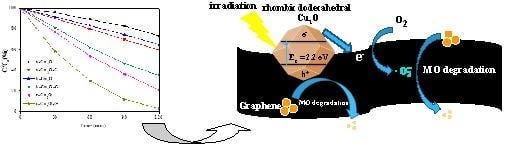
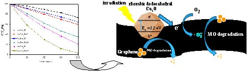
It is crucial to explore the durability and reusability in terms of practical applications of these photocatalysts. We take r-Cu2O and r-Cu2O-G as the example photocatalysts to test the stability of MO degradation. As can be seen in Figure 7a, an obvious decrease of photocatalytic performance (ca. 54.8%, i.e., from 80.0 to 25.2%) can be found for r-Cu2O after three consecutive tests. However, only ca. 10% of efficiency decline has been observed for r-Cu2O-G, which is probably due to the assistance of graphene. It is noteworthy that no obvious dissimilarity can be found in the fresh and used r-Cu2O-G photocatalysts (see XRD patterns in Figure 7b) in which Cu2O is still the dominant species. The slight decline of catalytic performance in the r-Cu2O-G photocatalysts may be due to the occurrence of intermediates [46] during photoreaction. It should be noted that the synthesized r-Cu2O-G photocatalysts can perform the photodegradation of MO with the highest kinetic rate (degradation efficiency = ca. 98% within 120 min) upon the presence of ultra-low content of samples (0.06 g L−1) using visible light illumination. Moreover, these r-Cu2O-G photocatalysts can be obtained via a facile and low-cost liquid-phase method. Above all, our r-Cu2O-G nanocomposites also have a remarkable enhancement of their long-term durability by the assistance of 1 wt % of graphene. In comparison to previously reported Cu2O-based photocatalysts, the synthesized r-Cu2O-G photocatalysts with dominant {110} facets of crystal Cu2O exhibit an excellent degradation kinetic in a low concentration of photocatalysts under visible-light irradiation that could be practically used to make the best use of daylight for the remediation of organic wastewater.
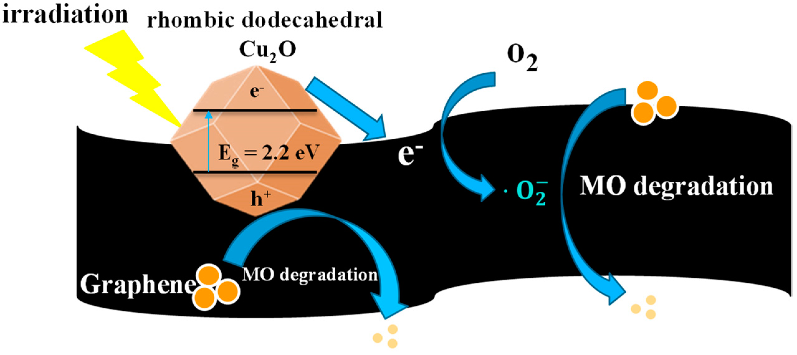
As shown in Figure 8, according to the aforementioned results a probable mechanism for MO degradation by crystal Cu2O-graphene photocatalysts can be proposed. Upon visible-light illumination, the crystal Cu2O (e.g., rhombic dodecahedra Cu2O) is excited to produce electrons (in the conduction band (CB)) and holes (in the valence band (VB)). Because of the stronger contact between crystal facets and the graphene surface, the photogenerated electrons can be rapidly transported to the graphene nanosheets. As a consequence of the small difference between the Fermi potential of graphene and the reduction potential of O2/O2− [50], O2− and H2O2 can be observed after the reaction of high-energy electrons with the dissolved oxygen. In this way, the recombination of electron/hole pairs can be greatly prohibited. In addition, compared to octahedral Cu2O (mainly {111} facets), the holes (h+) with positive charge generated on the valence band (ca. 1.92 eV vs. normal hydrogen electrode (NHE)) of rhombic dodecahedra Cu2O (mainly {110} facets) also can degrade more MO molecules (ca. 1.48 eV vs. NHE) [51,52] since the number of dangling Cu atoms on the {110} plane per unit surface area was nearly 1.5 times higher than that on the {111} plane, resulting in a more positively charged surface on {110} facets [28]. Therefore, the superoxide radical anions, hydrogen peroxide and h+ may be the dominant species governing the photodegradation of MO under visible-light irradiation. It should be noted that the graphene sheets not only serve as acceptors of the photogenerated electrons from Cu2O but also as stabilizers to prevent the crystal Cu2O from structural destruction under irradiation over a long period.
4. Conclusions
In this study, three different crystal facets of Cu2O photocatalysts decorated with graphene sheets were synthesized by using simple solution-phase methods. The order in terms of photocatalytic MO degradation was observed to be as follows: rhombic dodecahedral Cu2O-graphene (r-Cu2O-G) > hierarchical Cu2O-graphene (h-Cu2O-G) > octahedral Cu2O-graphene (o-Cu2O-G), which was demonstrated to be related to the morphologies and crystal structure of facets (i.e., the {110} facets are most active toward MO degradation). In earlier reports regarding Cu2O-based photocatalysts, the r-Cu2O-G photocatalysts possessed high kinetics of photocatalytic degradation, i.e., by using an ultra-low content of samples (0.06 g L−1) to reach 98% of MO photodegradation under 120 min irradiation of visible light. More importantly, the cycling tests indicate that the resulting r-Cu2O-G composites show a surpassing durability compared to pure r-Cu2O nanocrystals. These significant enhancements are possibly because of the unique interfacial interaction of rhombic dodecahedra Cu2O (more positively charged {110} facets) with the graphene nanosheets, which could lead to the effective isolation of electron/hole pairs, stabilization of the crystal Cu2O, and an increase of MO adsorption. Consequently, the development of a cost-effective and facile method to prepare r-Cu2O-G composites with mostly {110} facets and graphene sheets, which exhibit superior photocatalytic performance (kinetics and stability), offers the potential for a promising application in the treatment of organic wastewater by utilizing natural sunlight.
Author Contributions
The design, writing, review and editing of the paper were undertaken by S.-H.L.; the experiments and analysis of the data were undertaken by J.-S.L. Both authors reviewed and approved the manuscript.
Funding
The work was financially supported by Ministry of Science and Technology of Taiwan (MOST 105-2221-E-006-015-MY5).
Conflicts of Interest
The authors declare no conflict of interest.
References
- Gilja, V.; Novakovic, K.; Travas-Sejdic, J.; Hrnjak-Murgic, Z.; Rokovic, M.K.; Zic, M. Stability and synergistic effect of polyaniline/TiO2 photocatalysts in degradation of Azo Dye in wastewater. Nanomaterials 2018, 8, 30. [Google Scholar] [CrossRef] [PubMed]
- Liao, T.W.; Verbruggen, S.W.; Claes, N.; Yadav, A.; Grandjean, D.; Bals, S.; Lievens, P. TiO2 films modified with Au nanoclusters as self-cleaning surfaces under visible light. Nanomaterials 2018, 8, 30. [Google Scholar] [CrossRef] [PubMed]
- Ye, L.Q.; Su, Y.R.; Jin, X.L.; Xie, H.Q.; Zhang, C. Recent advances in BiOX (X = Cl, Br and I) photocatalysts: Synthesis, modification, facet effects and mechanisms. Environ. Sci. Nano 2014, 1, 90–112. [Google Scholar] [CrossRef]
- Liu, S.-H.; Syu, H.-R. One-step fabrication of N-doped mesoporous TiO2 nanoparticles by self-assembly for photocatalytic water splitting under visible light. Appl. Energy 2012, 100, 48–154. [Google Scholar] [CrossRef]
- Pang, D.D.; Wang, Y.T.; Ma, X.D.; Ouyang, F. Fluorine promoted and silica supported TiO2 for photocatalytic decomposition of acrylonitrile under simulant solar light irradiation. Chem. Eng. J. 2014, 258, 43–50. [Google Scholar] [CrossRef]
- Liu, S.-H.; Syu, H.-R. High visible-light photocatalytic hydrogen evolution of C,N-codoped mesoporous TiO2 nanoparticles prepared via an ionic-liquid template approach. Int. J. Hydrogen Energy 2013, 38, 13856–13865. [Google Scholar] [CrossRef]
- Song, X.L.; Li, Y.Y.; Wei, Z.D.; Ye, S.Y.; Dionysiou, D.D. Synthesis of BiVO4/P25 composites for the photocatalytic degradation of ethylene under visible light. Chem. Eng. J. 2017, 314, 443–452. [Google Scholar] [CrossRef]
- Liu, Y.Z.; Xu, J.A.; Wang, L.Q.; Zhang, H.Y.; Xu, P.; Duan, X.G.; Sun, H.Q.; Wang, S.B. Three-dDimensional BiOI/BiOX (X = Cl or Br) nanohybrids for enhanced visible-light photocatalytic activity. Nanomaterials 2017, 7, 64. [Google Scholar] [CrossRef] [PubMed]
- Tian, L.Y.; Rui, Y.L.; Sun, K.L.; Cui, W.Q.; An, W.J. Surface decoration of ZnWO4 nanorods with Cu2O nanoparticles to bBuild heterostructure with enhanced photocatalysis. Nanomaterials 2018, 8, 33. [Google Scholar] [CrossRef] [PubMed]
- Kumar, A.; Kumar, A.; Sharma, G.; Al-Muhtaseb, A.H.; Naushad, M.; Ghfar, A.A.; Stadler, F.J. Quaternary magnetic BiOCl/g-C3N4/Cu2O/Fe3O4 nano-junction for visible light and solar powered degradation of sulfamethoxazole from aqueous environment. Chem. Eng. J. 2018, 334, 462–478. [Google Scholar] [CrossRef]
- Singh, M.; Jampaiah, D.; Kandjani, A.E.; Sabri, Y.M.; Della Gaspera, E.; Reineck, P.; Judd, M.; Langley, J.; Cox, N.; van Embden, J.; et al. Oxygen-deficient photostable Cu2O for enhanced visible light photocatalytic activity. Nanoscale 2018, 10, 6039–6050. [Google Scholar] [CrossRef] [PubMed]
- Sakar, M.; Balakumar, S. Reverse Ostwald ripening process induced dispersion of Cu2O nanoparticles in silver-matrix and their interfacial mechanism mediated sunlight driven photocatalytic properties. J. Photochem. Photobiol. A 2018, 356, 150–158. [Google Scholar] [CrossRef]
- Wei, Q.; Wang, Y.; Qin, H.Y.; Wu, J.M.; Lu, Y.F.; Chi, H.Z.; Yang, F.; Zhou, B.; Yu, H.L.; Liu, J.B. Construction of rGO wrapping octahedral Ag-Cu2O heterostructure for enhanced visible light photocatalytic activity. Appl. Catal. B Environ. 2018, 227, 132–144. [Google Scholar] [CrossRef]
- Su, Y.; Li, H.F.; Ma, H.B.; Wang, H.; Robertson, J.; Nathan, A. Dye-assisted transformation of Cu2O nanocrystals to amorphous CuxO nanoflakes for enhanced photocatalytic performance. ACS Omega 2018, 3, 1939–1945. [Google Scholar] [CrossRef]
- Sun, S.D. Recent advances in hybrid Cu2O-based heterogeneous nanostructures. Nanoscale 2015, 7, 10850–10882. [Google Scholar] [CrossRef] [PubMed]
- Chen, X.Q.; Wu, Z.S.; Gao, Z.Z.; Ye, B.C. Effect of different activated carbon as carrier on the photocatalytic activity of Ag-N-ZnO photocatalyst for methyl orange degradation under visible light irradiation. Nanomaterials 2018, 7, 258. [Google Scholar] [CrossRef] [PubMed]
- Choi, H.; Shin, D.; Yeo, B.C.; Song, T.; Han, S.S.; Park, N.; Kim, S. Simultaneously controllable doping sites and the activity of a W-N codoped TiO2 photocatalyst. ACS Catal. 2016, 6, 2745–2753. [Google Scholar] [CrossRef]
- Luster, E.; Avisar, D.; Horovitz, I.; Lozzi, L.; Baker, M.A.; Grilli, R.; Mamane, H. N-doped TiO2-coated ceramic membrane for carbamazepine degradation in different water qualities. Nanomaterials 2017, 7, 206. [Google Scholar] [CrossRef] [PubMed]
- Bailón-García, E.; Elmouwahidi, A.; Álvarez, M.A.; Carrasco-Marín, F.; Pérez-Cadenas, A.F.; Maldonado-Hóda, F.J. New carbon xerogel-TiO2 composites with high performance as visible-light photocatalysts for dye mineralization. Appl. Catal. B Environ. 2017, 201, 29–40. [Google Scholar] [CrossRef]
- Klaysri, R.; Ratova, M.; Praserthdam, P.; Kelly, P.J. Deposition of visible light-active C-doped titania films via magnetron sputtering using CO2 as a source of carbon. Nanomaterials 2017, 7, 113. [Google Scholar] [CrossRef] [PubMed]
- Nica, I.C.; Stan, M.S.; Dinischiotu, A.; Popa, M.; Chifiriuc, M.C.; Lazar, V.; Pircalabioru, G.G.; Bezirtzoglou, E.; Iordache, O.G.; Varzaru, E.; et al. Innovative self-cleaning and biocompatible polyester textiles nano-decorated with Fe-N-doped titanium dioxide. Nanomaterials 2016, 6, 214. [Google Scholar] [CrossRef] [PubMed]
- Reddy, P.A.K.; Reddy, P.V.L.; Kwon, E.; Kim, K.H.; Akter, T.; Kalagara, S. Recent advances in photocatalytic treatment of pollutants in aqueous media. Environ. Int. 2016, 91, 94–103. [Google Scholar] [CrossRef] [PubMed]
- Chang, M.L.; Hu, H.W.; Zhang, Y.Y.; Chen, D.C.; Wu, L.P.; Li, X.J. Improving visible light-absorptivity and photoelectric conversion efficiency of a TiO2 nanotube anode film by sensitization with Bi2O3 Nanoparticles. Nanomaterials 2017, 7, 104. [Google Scholar] [CrossRef] [PubMed]
- Singh, R.; Dutta, S. A review on H2 production through photocatalytic reactions using TiO2/TiO2-assisted catalysts. Fuel 2018, 220, 607–620. [Google Scholar] [CrossRef]
- Hu, J.L.; Tu, J.H.; Li, X.Y.; Wang, Z.Y.; Li, Y.; Li, Q.S.; Wang, F.P. Enhanced UV-Visible light photocatalytic activity by constructing appropriate heterostructures between mesopore TiO2 nanospheres and Sn3O4 nanoparticles. Nanomaterials 2017, 7, 336. [Google Scholar] [CrossRef] [PubMed]
- Petronella, F.; Truppi, A.; Ingrosso, C.; Placido, T.; Striccoli, M.; Curri, M.L.; Agostiano, A.; Comparelli, R. Nanocomposite materials for photocatalytic degradation of pollutants. Catal. Today 2017, 281, 85–100. [Google Scholar] [CrossRef]
- Huang, W.-C.; Lyu, L.-M.; Yang, Y.-C.; Huang, M.H. Synthesis of Cu2O nanocrystals from cubic to rhombic dodecahedral structures and their comparative photocatalytic activity. J. Am. Chem. Soc. 2012, 134, 1261–1267. [Google Scholar] [CrossRef] [PubMed]
- Shang, Y.; Guo, L. Facet-controlled synthetic strategy of Cu2O-based crystals for catalysis and sensing. Adv. Sci. 2015, 2, 1500140. [Google Scholar] [CrossRef] [PubMed]
- Yuan, G.-Z.; Hsia, C.-F.; Lin, Z.-W.; Chiang, C.; Chiang, Y.-W.; Huang, M.H. Highly facet-dependent photocatalytic properties of Cu2O crystals established through the formation of Au-decorated Cu2O heterostructures. Chem. Eur. J. 2016, 22, 12548–12556. [Google Scholar] [CrossRef] [PubMed]
- Chen, D.S.; Yu, W.B.; Deng, Z.; Liu, J.; Jin, J.; Li, Y.; Wu, M.; Chen, L.H.; Su, B.L. Hollow Cu2O microspheres with two active {111} and {110} facets for highly selective adsorption and photodegradation of anionic dye. RSC Adv. 2015, 5, 55520–55526. [Google Scholar] [CrossRef]
- Zhang, Y.; Deng, B.; Zhang, T.R.; Gao, D.M.; Xu, A.W. Shape effects of Cu2O polyhedral microcrystals on photocatalytic activity. J. Phys. Chem. C 2010, 114, 5073–5079. [Google Scholar] [CrossRef]
- Xu, L.; Zhang, F.Y.; Song, X.Y.; Yin, Z.L.; Bu, Y.X. Construction of reduced graphene oxide-supported Ag-Cu2O composites with hierarchical structures for enhanced photocatalytic activities and recyclability. J. Mater. Chem. A 2015, 3, 5923–5933. [Google Scholar] [CrossRef]
- Babu, S.G.; Vinoth, R.; Narayana, P.S.; Bahnemann, D.; Neppolian, B.S. Reduced graphene oxide wrapped Cu2O supported on C3N4: An efficient visible light responsive semiconductor photocatalyst. APL Mater. 2015, 3, 104415. [Google Scholar] [CrossRef] [Green Version]
- Yu, L.; Li, G.J.; Zhang, X.S.; Ba, X.; Shi, G.D.; Li, Y.; Wong, P.K.; Yu, J.C.; Yu, Y. Enhanced activity and stability of carbon-decorated cuprous oxide mesoporous nanorods for CO2 reduction in artificial photosynthesis. ACS Catal. 2016, 6, 6444–6454. [Google Scholar] [CrossRef]
- Liu, S.-H.; Wei, Y.-S.; Lu, J.-S. Visible-light-driven photodegradation of sulfamethoxazole and methylene blue by Cu2O/rGO photocatalysts. Chemosphere 2016, 154, 118–123. [Google Scholar] [CrossRef] [PubMed]
- Upadhyay, R.K.; Soin, N.; Roy, S.S. Role of graphene/metal oxide composites as photocatalysts, adsorbents and disinfectants in water treatment: A review. RSC Adv. 2014, 4, 3823–3851. [Google Scholar] [CrossRef]
- Stoller, M.D.; Park, S.; Zhu, Y.; An, J.; Ruoff, R.S. Graphene-based ultracapacitors. Nano Lett. 2008, 8, 3498–3502. [Google Scholar] [CrossRef] [PubMed]
- Mayorov, A.S.; Gorbachev, R.V.; Morozov, S.V.; Britnell, L.; Jalil, R.; Ponomarenko, L.A.; Blake, P.; Novoselov, K.S.; Watanabe, K.; Taniguchi, T.; et al. Micrometer-scale ballistic transport in encapsulated graphene at room temperature. Nano Lett. 2011, 11, 2396–2399. [Google Scholar] [CrossRef] [PubMed]
- Chen, Y.; Sun, H.Q.; Peng, W.C. 2D transition metal dichalcogenides and graphene-based ternary composites for photocatalytic hydrogen evolution and pollutants degradation. Nanomaterials 2017, 7, 62. [Google Scholar] [CrossRef] [PubMed]
- Tian, Y.; Sun, Z.H.; Zhang, Y.G.; Wang, X.; Bakenov, Z.; Yin, F.X. Micro-spherical sulfur/graphene oxide composite via spray drying for high performance lithium sulfur batteries. Nanomaterials 2018, 8, 50. [Google Scholar] [CrossRef] [PubMed]
- Gong, Y.X.; Wang, Y.; Sun, G.; Jia, T.K.; Jia, L.; Zhang, F.M.; Lin, L.; Zhang, B.Q.; Cao, J.L.; Zhang, Z.Y. Carbon nitride decorated ball-flower like Co3O4 hybrid composite: Hydrothermal synthesis and ethanol gas sensing application. Nanomaterials 2018, 8, 132. [Google Scholar] [CrossRef] [PubMed]
- Zhou, Y.X.; Jia, L.P.; Wang, T.X.; Du, Y.L.; Wang, C.M. Preparation of carbon nanotube and graphene doped polyphenylene sulfide flexible film electrodes and the electrodeposition of Cu2O nanocrystals for hydrogen-generation. Int. J. Hydrogen Energy 2018, 43, 7356–7365. [Google Scholar] [CrossRef]
- Sharma, K.; Maiti, K.; Kim, N.H.; Hui, D.; Lee, J.H. Green synthesis of glucose-reduced graphene oxide supported Ag-Cu2O nanocomposites for the enhanced visible-light photocatalytic activity. Compos. Part B 2018, 138, 35–44. [Google Scholar] [CrossRef]
- González, J.A.; Villanueva, M.E.; Piehl, L.L.; Copello, G.J. Development of a chitin/graphene oxide hybrid composite for the removal of pollutant dyes: Adsorption and desorption study. Chem. Eng. J. 2015, 280, 42–48. [Google Scholar] [CrossRef]
- Stankovich, S.; Dikin, D.A.; Piner, R.D.; Kohlhaas, K.A.; Kleinhammes, A.; Jia, Y.; Wu, Y.; Nguyen, S.T.; Ruoff, R.S. Synthesis of graphene-based nanosheets via chemical reduction of exfoliated graphite oxide. Carbon 2007, 45, 1558–1565. [Google Scholar] [CrossRef]
- Liu, S.-H.; Yang, S.-W. Highly efficient cuprous oxide nanocrystals assisted with graphene for decolorization using visible light. Water Air Soil Pollut. 2018, 229, 67. [Google Scholar] [CrossRef]
- Pu, Y.-C.; Chou, H.-Y.; Kuo, W.-S.; Wei, K.-H.; Hsu, Y.-J. Interfacial charge carrier dynamics of cuprous oxide-reduced graphene oxide (Cu2O-rGO) nanoheterostructures and their related visible-light-driven photocatalysis. Appl. Catal. B Environ. 2017, 204, 21–32. [Google Scholar] [CrossRef]
- Gao, Z.; Liu, J.; Xu, F.; Wu, D.; Wu, Z.; Jiang, K. One-pot synthesis of graphene-cuprous oxide composite with enhanced photocatalytic activity. Solid State Sci. 2012, 14, 276–280. [Google Scholar] [CrossRef]
- Chang, X.F.; Gondal, M.A.; Al-Saadi, A.A.; Ali, M.A.; Shen, H.; Zhou, Q.; Zhang, J.; Du, M.; Liu, Y.; Ji, G. Photodegradation of Rhodamine B over unexcited semiconductor compounds of BiOCl and BiOBr. J. Colloid Interface Sci. 2012, 377, 291–298. [Google Scholar] [CrossRef] [PubMed]
- Wang, J.T.W.; Ball, J.M.; Barea, E.M.; Abate, A.; Alexander-Webber, J.A.; Huang, J.; Saliba, M.; Mora-Sero, I.; Bisquert, J.; Snaith, H.J.; et al. Low-temperature processed electron collection layers of graphene/TiO2 nanocomposites in thin film perovskite solar cells. Nano Lett. 2014, 14, 724–730. [Google Scholar] [CrossRef] [PubMed]
- Yan, S.C.; Li, Z.S.; Zou, Z.G. Photodegradation of Rhodamine B and methyl orange over boron-doped g-C3N4 under visible light irradiation. Langmuir 2010, 26, 3894–3901. [Google Scholar] [CrossRef] [PubMed]
- Hu, X.; Zhou, X.; Wang, R.; Hu, C.; Qu, J. Characterization and photostability of Cu2O-Ag-AgBr/Al2O3 for the degradation of toxic pollutants with visible-light irradiation. Appl. Catal. B 2014, 154–155, 44–50. [Google Scholar] [CrossRef]
Figure 1.
X-ray diffraction (XRD) patterns of different photocatalysts.

Figure 2.
Scanning electron microscope (SEM) images of (a) o-Cu2O; (b) h-Cu2O; (c) r-Cu2O; (d) o-Cu2O-G; (e) h-Cu2O-G and (f) r-Cu2O-G.
Figure 2.
Scanning electron microscope (SEM) images of (a) o-Cu2O; (b) h-Cu2O; (c) r-Cu2O; (d) o-Cu2O-G; (e) h-Cu2O-G and (f) r-Cu2O-G.

Figure 3.
Transmission electron microscope (TEM) images of (a) o-Cu2O-G; (b) h-Cu2O-G; (c) r-Cu2O-G and high-resolution TEM (HRTEM) of (d) o-Cu2O-G; (e) h-Cu2O-G; (f) r-Cu2O-G. Inset: selected area electron diffraction (SAED) from the circle area.
Figure 3.
Transmission electron microscope (TEM) images of (a) o-Cu2O-G; (b) h-Cu2O-G; (c) r-Cu2O-G and high-resolution TEM (HRTEM) of (d) o-Cu2O-G; (e) h-Cu2O-G; (f) r-Cu2O-G. Inset: selected area electron diffraction (SAED) from the circle area.

Figure 4.
X-ray photoelectron spectroscopy (XPS) spectra of different photocatalysts at the (a) high-resolution C 1s core-level; (b) Cu 2p core-level; and (c) O 1s core-level.
Figure 4.
X-ray photoelectron spectroscopy (XPS) spectra of different photocatalysts at the (a) high-resolution C 1s core-level; (b) Cu 2p core-level; and (c) O 1s core-level.
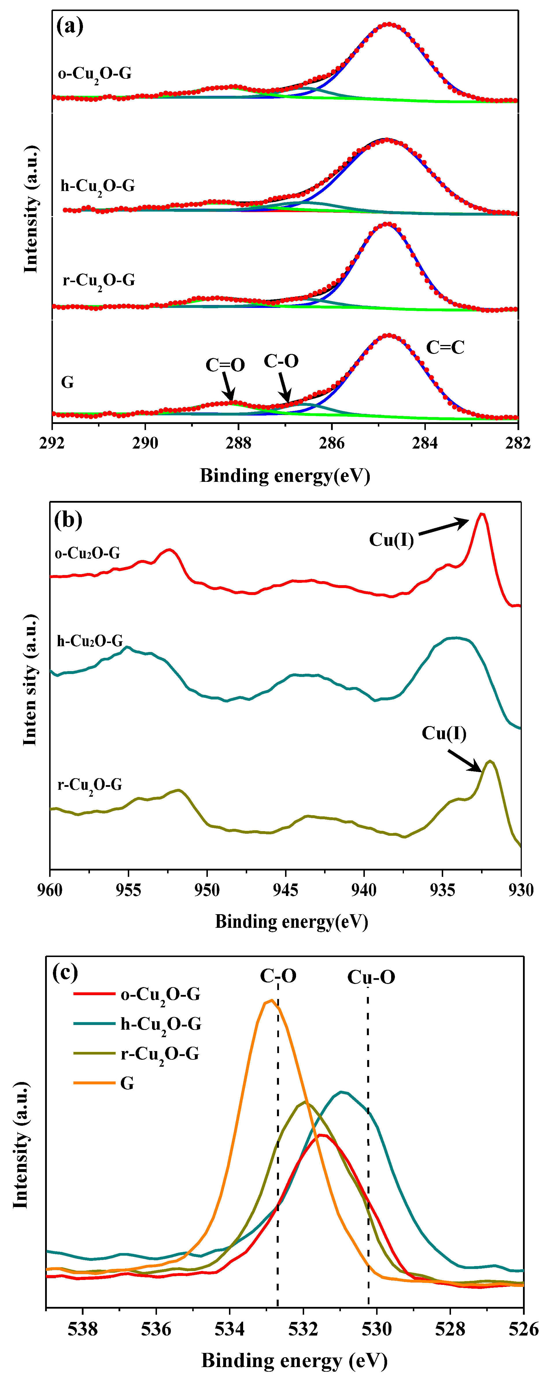
Figure 5.
(a) Ultraviolet-visible (UV-Vis) absorption spectra for different samples and (b) the corresponding Kubelka–Munk plots of photocatalysts.
Figure 5.
(a) Ultraviolet-visible (UV-Vis) absorption spectra for different samples and (b) the corresponding Kubelka–Munk plots of photocatalysts.

Figure 6.
(a) Relative concentration of methyl orange (MO) versus time by various photocatalysts under visible light and (b) kinetic plots from the data in (a).
Figure 6.
(a) Relative concentration of methyl orange (MO) versus time by various photocatalysts under visible light and (b) kinetic plots from the data in (a).

Figure 7.
(a) Cyclic tests of r-Cu2O and r-Cu2O-G photocatalysts for MO photodegradation under visible light; (b) XRD patterns of the fresh and used r-Cu2O-G.
Figure 7.
(a) Cyclic tests of r-Cu2O and r-Cu2O-G photocatalysts for MO photodegradation under visible light; (b) XRD patterns of the fresh and used r-Cu2O-G.

Figure 8.
Possible mechanism of MO photodegradation over r-Cu2O-G under visible-light illumination.

© 2018 by the authors. Licensee MDPI, Basel, Switzerland. This article is an open access article distributed under the terms and conditions of the Creative Commons Attribution (CC BY) license (http://creativecommons.org/licenses/by/4.0/).
Share and Cite
MDPI and ACS Style
Liu, S.-H.; Lu, J.-S. Facet-Dependent Cuprous Oxide Nanocrystals Decorated with Graphene as Durable Photocatalysts under Visible Light. Nanomaterials 2018, 8, 423. https://doi.org/10.3390/nano8060423
AMA Style
Liu S-H, Lu J-S. Facet-Dependent Cuprous Oxide Nanocrystals Decorated with Graphene as Durable Photocatalysts under Visible Light. Nanomaterials. 2018; 8(6):423. https://doi.org/10.3390/nano8060423
Chicago/Turabian StyleLiu, Shou-Heng, and Jun-Sheng Lu. 2018. "Facet-Dependent Cuprous Oxide Nanocrystals Decorated with Graphene as Durable Photocatalysts under Visible Light" Nanomaterials 8, no. 6: 423. https://doi.org/10.3390/nano8060423
Note that from the first issue of 2016, this journal uses article numbers instead of page numbers. See further details here.