Article

Facile Strategy for Synthesizing Non-Stoichiometric Monoclinic Structured Tungsten Trioxide (WO3−x) with Plasma Resonance Absorption and Enhanced Photocatalytic Activity

1
School of physics & optoelectronic engineering, Guangdong University of Technology, Guangdong 510006, China
2
Synergy Innovation Institute for Modern Industries, Guangdong University of Technology, Dongyuan 517500, China
3
School of Physics Science and Technology, Lingnan Normal University, Zhanjiang 524048, China
*
Author to whom correspondence should be addressed.
Nanomaterials 2018, 8(7), 553; https://doi.org/10.3390/nano8070553
Submission received: 6 June 2018 / Revised: 5 July 2018 / Accepted: 17 July 2018 / Published: 21 July 2018

Abstract

:
Oxygen vacancy defects play an important role in improving the light-capturing and photocatalytic activity of tungsten trioxide (WO3). However, the hydrogen treatment method that is commonly used to introduce oxygen vacancies is expensive and dangerous. Therefore, the introduction and control of oxygen vacancy defects in WO3 remains a challenge. Here, we demonstrated that oxygen vacancies could be successfully introduced into WO3−x while using a facile method through low temperature annealing in alcohol. The obtained WO3−x samples with optimal oxygen vacancies showed strong absorption of light, extending from the ultraviolet to the visible and near-infrared regions, and exhibits strong plasmon resonance from 400–1200 nm peaking at approximately 800 nm. When compared to pristine WO3, the photocatalytic activity of WO3−x was greatly improved in the ultraviolet and visible regions. This study provides a simple and efficient method to generate oxygen vacancies in WO3 for photocatalysis, which may be applied in the photoelectrochemical, electrochromic, and photochromic fields. Because oxygen vacancy is a common characteristic of metal oxides, the findings that are presented herein may be extended to other metal oxides.

Graphical Abstract

1. Introduction

Semiconductor photocatalysis has attracted significant attention due to its promising applications in solar energy conversion, since the discovery of water splitting on a titanium dioxide (TiO2) photoanode in the 1970s [1]. However, the photocatalytic efficiency of TiO2 is limited by its large band gap energy and fast electron-hole recombination due to its high density of trap states [2,3]. Great efforts have been dedicated to enhancing the visible light absorption of large band gap metal oxides. For instance, dye-sensitized and noble metal nanodot decorated TiO2 nanostructures were developed [4,5], forming heterojunctions with other semiconductors [6,7], and band gap narrowing was achieved via elemental doping in order to improve the conversion efficiency of metal oxide photoelectrodes. These methods modified the optical absorption coefficient and wavelength of the materials [8,9,10]. In addition to studies on reducing the band gap of TiO2 for enhanced visible light response, alternative semiconductor materials (WO3, ZnO, SnO2, ZnGaO4, and BiVO4) with intrinsic narrow band gaps are currently being explored [11,12,13,14]. Among them, tungsten trioxide is an attractive semiconductor material.
Tungsten trioxide (WO3) exhibits powerful oxidation properties, non-toxicity, and low cost and it has found widespread applications in electrochromic devices [15], gas sensors [16,17], photoelectrochemical water splitting, and photodegradation of organic compounds [18,19,20,21,22]. However, the photocatalytic activity of pristine WO3 is not high enough for practical use. Therefore, different strategies have been developed to increase the photocatalytic activity of WO3, including the preparation of WO3 with different nanostructures [23], supporting noble metal Pt/Au/Ag [24,25,26], composites with other materials [27,28,29], and oxygen vacancy defects [30,31,32,33,34]. The introduction of oxygen vacancies is a simple and efficient strategy to improve the photocatalytic performance of WO3 and it can greatly increase its conductivity and the absorption of visible light by local surface plasmon resonance (LSPR) in the near-infrared region, yielding an intensity that is comparable to bandgap absorption [35,36]. Over the past decades, persistent efforts have focused on improving the photocatalytic activity of WO3 by introducing oxygen vacancies, and various strategies have been proposed. At present, the main method for introducing oxygen vacancies is through the hydrogenation of WO3 [31,37]. Chen et al. produced black TiO2 using hydrogen treatment of white TiO2 in 2011, which triggered a surge in the production of hydrogenated semiconductors [38] and vacuum annealing procedures [37]. For instance, Yamashita et al. reported a facile H-spillover route to prepare heavily hydrogen-doped WO3 (HxWO3) [39]. These hydrogenated HxWO3 materials exhibited strong plasmonic absorption in the visible light region, which is tunable over a wide range by varying its stoichiometry. The electron concentration of the hydrogenated sample was high, up to 3.1 × 1021 cm−3. Gong et al. explored the exfoliation of layered tungstic acid to form WO3 nanosheets, and prepared sub-stoichiometric WO3 single crystal nanosheets, and subsequently introduced oxygen vacancies [35]. Simultaneously, the introduction of oxygen vacancies can promote the separation of photo-generated electrons and holes and inhibit their recombination, thus improving photocatalytic activity [40]. Therefore, the introduction of oxygen vacancies in WO3 plays an important role in photocatalytic performance. However, these methods require harsh experimental conditions and are expensive, which make them difficult to perform in most laboratories. Consequently, WO3-based semiconductors with adequate activity under visible or solar light are still under investigation.
Herein, we prepared monoclinic structured WO3 nanosheets via a one-step template-free hydrothermal route and then introduced oxygen vacancies through low temperature annealing in alcohol. These processes successfully synthesized non-stoichiometric monoclinic structured WO3 (WO3−x) nanosheets. Because of the introduction of oxygen vacancies, the WO3−x showed enhanced electron concentrations of up to 9.1 × 1021 cm−3, which are enough to induce LSPR. The WO3−x also exhibited very strong visible and infrared light absorption, which significantly increased its photocatalytic activity.

2. Experimental

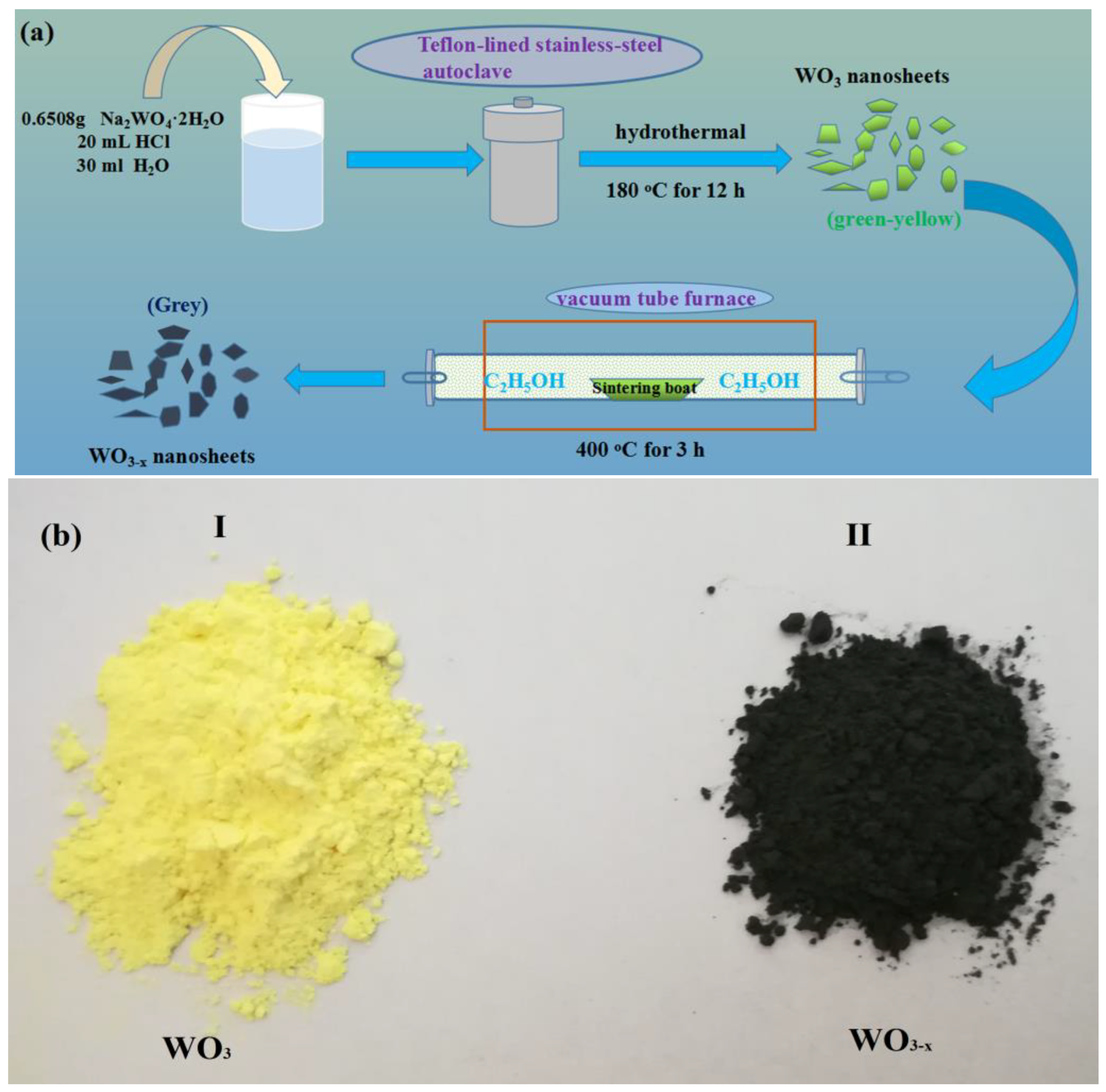
2.1. Preparation of the WO3 Nanosheets

The WO3 single crystal nanosheets were synthesized by a one-step template-free hydrothermal route [41]. Sodium tungstate dehydrate (Na2WO4·2H2O, 0.6398 g, 2 mmol) was added into distilled water (40 mL) and hydrochloric acid (40% HCl, 20 mL, adjusted pH = 1.5) was subsequently added into the Na2WO4 solution at room temperature (25 °C), stirring with vigorous magnetic for approximately 10 min. Then, the mixture was transferred to a 100 mL Teflon-lined stainless-steel autoclave. The autoclave was sealed and incubated at 180 °C for 12 h and then subsequently cooled down to room temperature naturally. The final products were collected by centrifuging the mixture, washed several times with distilled water and absolute ethanol, and drying at 60 °C in air.

2.2. Preparation of WO3−x Nanosheets

In a simple synthetic procedure, the prepared green-yellow WO3 nanosheets (0.5 g) were added into 10 mL absolute ethanol to form a suspension. After vigorous stirring for 10 min, the suspension was poured into a sintering boat (length 6 cm, width 3 cm, height 1.5 cm) and transferred to a vacuum tube furnace (SK-G06163, Φ60/50 × 1000 mm). The ends of the vacuum tube are sealed. The initial temperature is set to 50 °C, and the temperature is raised at a rate of 5 °C/min until the temperature reaches 400 °C, and the temperature is maintained at 400 °C for three hours. Then, the temperature is lowered at a rate of 5 °C/min until the temperature reaches 50 °C, and hence, cooled down to room temperature naturally. The obtained grey powders were collected for further characterization.

2.3. Characterization

The X-ray diffraction (XRD) patterns were recorded by X-ray diffractometer (D8 ADVANCE, BRUKER) with Cu-Kα radiation (λ = 1.541 Å) in a wide angle range from 10° to 80° on 2θ scale. Field emission scanning electron microscopy (FE-SEM) images, which were collected on a Hitachi SU8220 electron microscope with a working distance of 8 mm. Transmission electron microscopy (TEM) images and high-resolution TEM images were performed by using a FEI TecnaiG2 F20 with an acceleration voltage of 200 kV and TEM sample specimens were prepared by briefly ultrasonicating the sample powders in ethanol, followed by placing a drop of the suspension onto lacey support films that were dried before imaging. Fourier transformed infra-red (FT-IR) measurements were carried out using a 80/80 v Bruker TENSOR27 spectrometer. The FT-IR spectra were acquired with KBr discs under transmission mode. The scanned wavenumber range was from 4000 to 400 cm−1. The Raman spectra were recorded on a RENISHAW A-9570-2000 Raman spectrometer with an excitation wavelength of 785 nm. UV-Vis diffuse reflectance spectroscopy (DRS) measurement was conducted by a SHIMADZU UV-3600 spectrophotometer. X-ray photoelectron spectra (XPS) were recorded on a THERMO ESCALAB 250 Xi with a monochromatic Al Kα X-ray source. The electron paramagnetic resonance spectra were recorded while using a JES-FA200 spectrometer at room temperature.

2.4. Photocatalytic Test

2.4.1. UV Light Photocatalytic Degradation

Rhodamine B (RhB) is an organic dye with a bright red colour and it is widely used as a model pollutant in photocatalytic tests. Therefore, the photocatalytic activity of the WO3−x nanosheets samples was evaluated in terms of the decolorization of RhB dye under UV irradiation from a 500 W Hg lamp. The lamp was positioned in a cylindrical Pyrex vessel and cooled by circulating water to maintain the reaction temperature at approximately 27 °C. A quartz tube was used as the photo catalytic reactor. The catalysts (0.02 g) were dispersed into 40 mL of 2 × 10⁻⁵ M RhB solution and stirred in the dark for 20 min to reach a complete adsorption–desorption equilibrium between the photocatalyst and RhB solution. Then, the mixture was exposed to UV light. Vigorous magnetic stirring was maintained to keep WO3−x nanosheets suspended in the RhB solution. The concentration of aqueous RhB was determined with a UV-Vis spectrophotometer by measuring the absorption peak intensity at 553 nm.

2.4.2. Visible Light Photocatalytic Degradation

The photocatalytic activity of WO3−x nanosheets under visible light was also evaluated by monitoring the decomposition of RhB. The apparatus for studying the photocatalytic decomposition of RhB was identical under visible and ultraviolet light, except that an 800 W Xe lamp with a 420 nm cut-on filter was used instead of a Hg lamp as the light source.

3. Results and Discussion

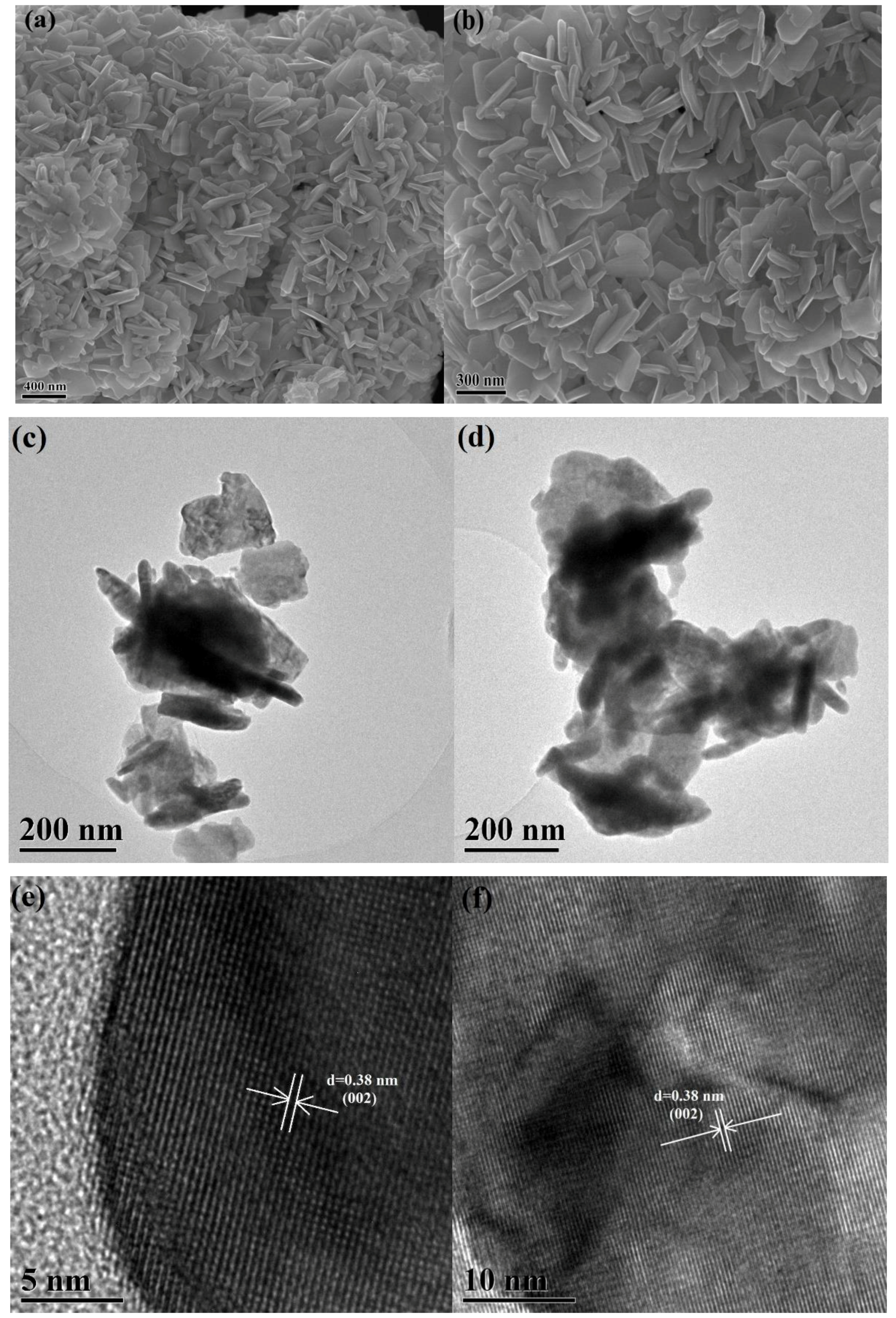
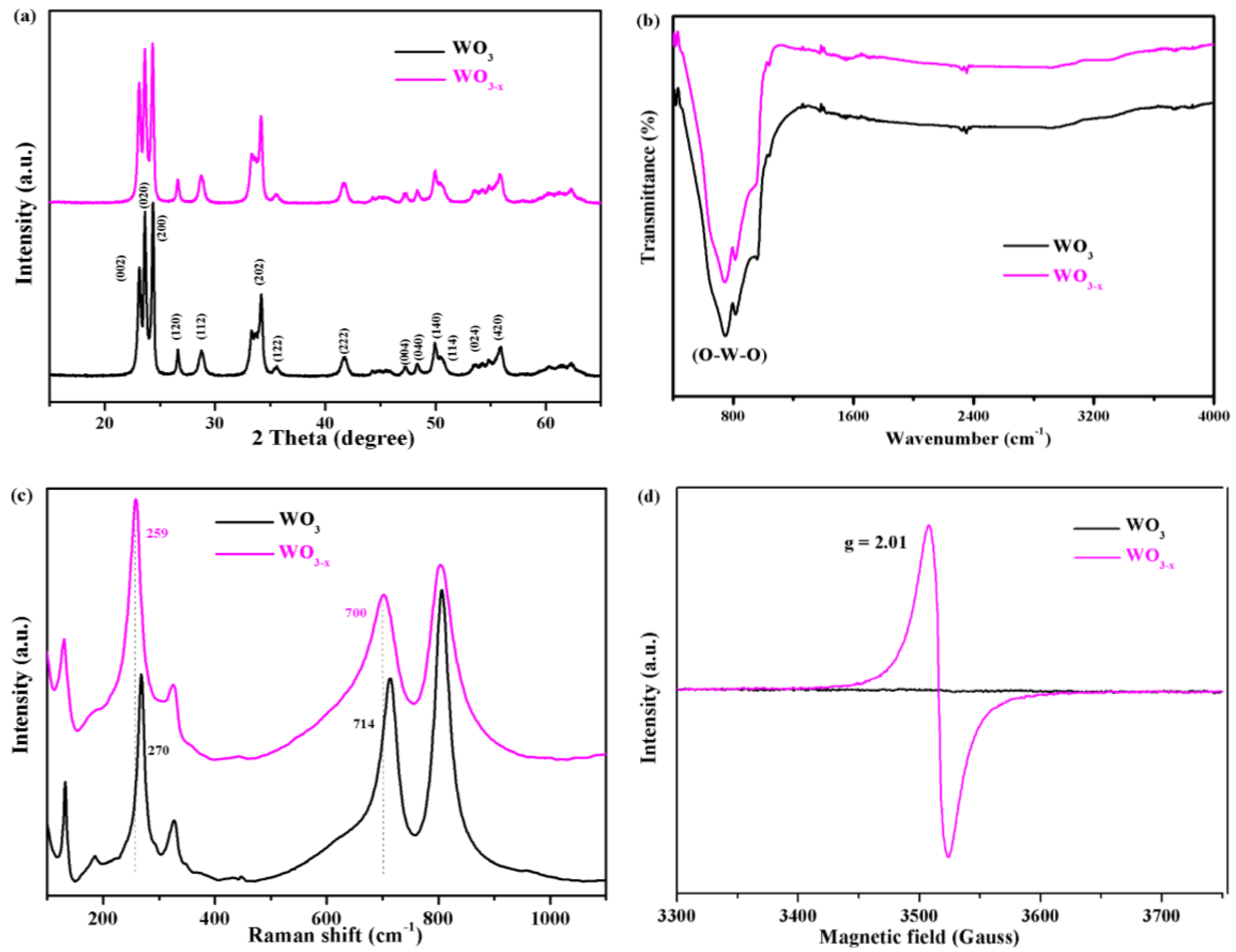
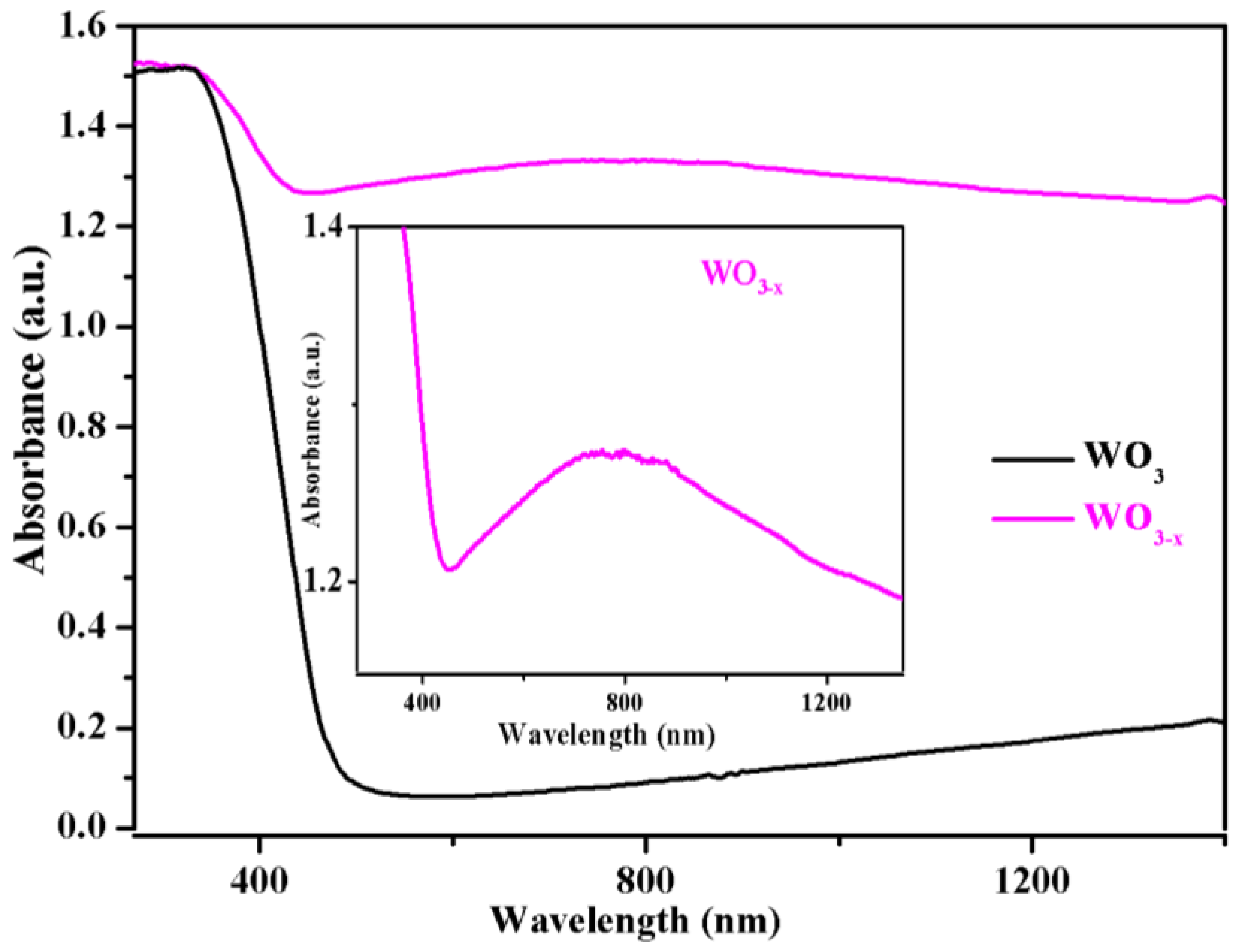
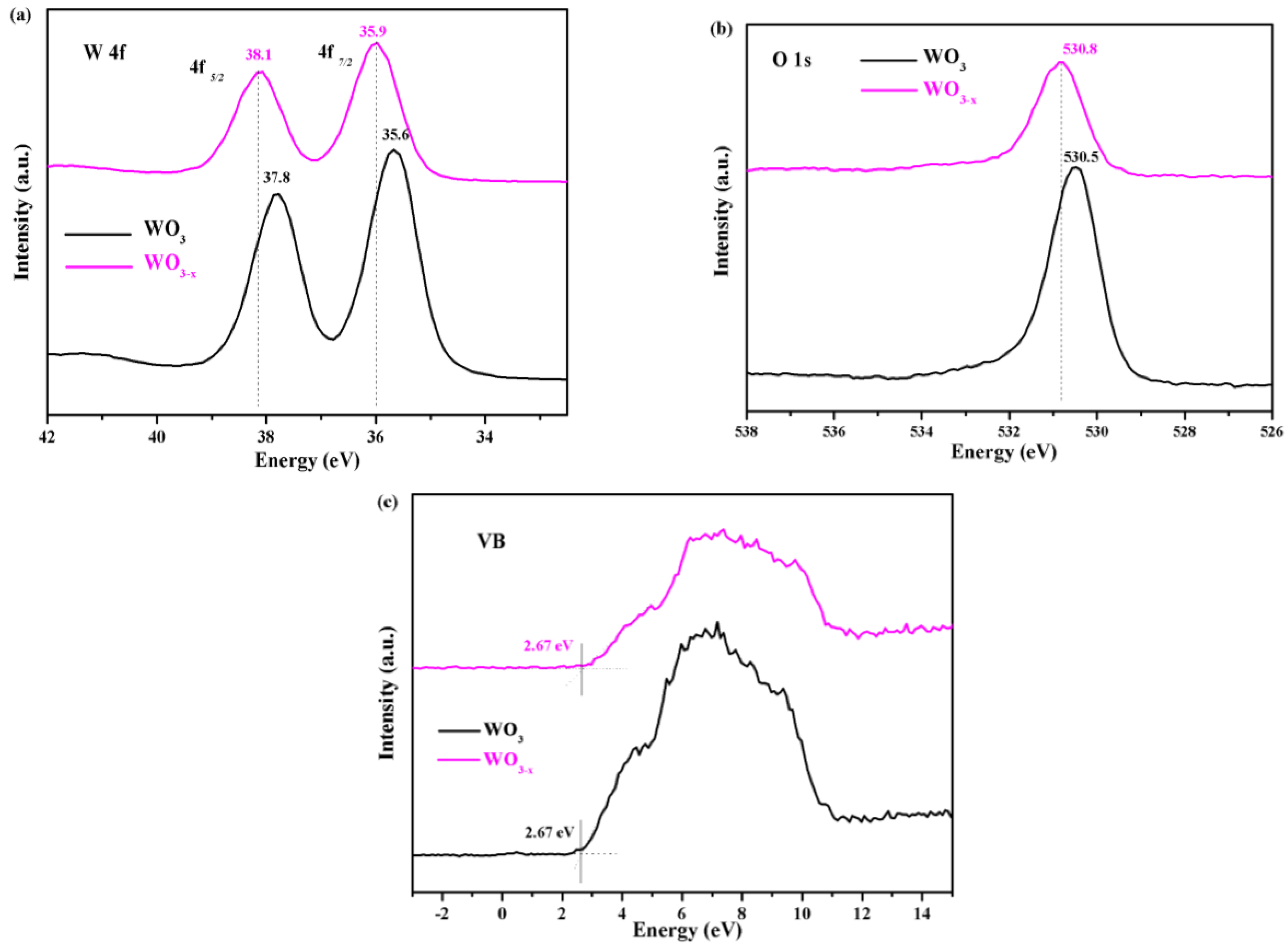
3.1. Characterization of the WO3−x Nanosheets

The WO3−x single crystal nanosheets were prepared by a one-step template-free hydrothermal route and oxygen vacancies were introduced through low-temperature annealing in ethanol. It is well-known that ethanol has reducing properties. When annealing WO3 in ethanol (anaerobic environment), ethanol can consume oxygen atoms in WO3, resulting in oxygen vacancies in the crystals. This strategy is simple, safe, and efficient when compared to the hydrogen treatment of WO3. The fabrication procedure of stable WO3−x nanosheets is schematically illustrated in Figure 1a. Meanwhile, the annealing process was accompanied by a strong colour change from the original light yellow WO3 to the final grey WO3−x (Figure 1b). This method can be used in most laboratories and mass production of WO3−x can also be achieved. The obtained WO3−x exhibits very good stability, as no change was observed after storage for six months. We performed detailed characterizations and photocatalytic performance tests of the prepared catalyst, as follows.
The morphologies of the WO3 and WO3−x samples were examined by FE-SEM and field emission transmission electron microscopy (FE-TEM), as shown in Figure 2. The SEM and TEM images clearly show the stacking morphology of the WO3 nanosheets and no obvious change in the size and morphology was observed after annealing. The thickness of the nanosheets was typically 20–30 nm, with a length of 100–150 nm. In addition, the similar morphologies of WO3 (Figure 2a,c) and WO3−x (Figure 2b,d) prove that the annealing treatment at 400 °C does not significantly change the nanosheet morphology of WO3−x. As shown in the representative HR-TEM image of an unannealed WO3 nanosheets (Figure 2e), clear lattice fringes can be identified, and the lattice fringe spacing between two adjacent crystal planes is 0.38 nm, corresponding to the (002) lattice plane of the monoclinic structured WO3 [42]. In contrast, the framework of the highly crystalline WO3−x can be clearly observed in Figure 2f, and the WO3−x nanosheets also crystallize well and have a lattice spacing of 0.38 nm between the (002) planes.
The monoclinic crystal structure of the as-prepared WO3−x and WO3 samples were confirmed by the X-ray diffraction patterns shown in Figure 3a. The WO3 and WO3−x samples exhibited similar diffraction peaks, with peak centers at 23.1, 23.6, 24.3, 26.6, 28.7, 33.4, 34.1, 41.6, 50.1, and 55.9 corresponding to the (002), (020), (200), (120), (112), (202), (122), (222), (140), and (420) crystal faces of the monoclinic structured WO3 that was indexed to the JCPDS card No. 43-1035 [43]. No characteristic peaks of other crystalline impurities were detected. In addition, no other crystalline forms of WO3 were observed after low temperature annealing.
To further study the chemical structure and surface composition of the as-fabricated WO3−x nanosheets, the samples were characterized, as shown in Figure 3b. Both samples only show a strong absorption peak at 800 cm−1, which can be attributed to (O–W–O) of the interconnected octahedron WO3 [44]. Meanwhile, no other characteristic peaks were observed after low temperature annealing. This indicates that no other chemicals or organic residues were present on the surface of the WO3−x nanosheets after the fabrication procedure. Raman spectroscopy was performed to investigate the chemical structure of the samples, as shown in Figure 3c. The oxygen deficiencies of WO3−x were unambiguously supported by the raman spectroscopy data. The raman spectra of the WO3 nanosheets display three major characteristic peaks at 270, 715, and 805 cm−1, arising from the bending vibration of δ(O–W–O) and stretching vibrations of ν(W–O–W) of the monoclinic phase, respectively [35]. However, the characteristic raman peaks for WO3−x broaden after annealing, thus suggesting slight changes in its structure. After annealing, the W6+–O stretching band at 270 and 715 cm−1 slightly shifted to lower wavelengths of 258 and 700 cm−1. Similar results were reported in previous studies [35]. These results indicate that oxygen vacancies were successfully introduced to the nanosheets. The formation of oxygen vacancies in the WO3 crystals caused an up-shift of the W6+-O bond in the raman spectrum, indicating that the structure of WO3−x was changed by the formation of oxygen vacancies. These results are consistent with the HR-TEM images of WO3−x (Figure 2d).
To further confirm the presence of oxygen vacancies in WO3−x, electron paramagnetic resonance (EPR) spectroscopy was performed. EPR is highly sensitive to paramagnetic species containing unpaired electrons and it has been widely used to characterize oxygen vacancies. The test was performed at room temperature and the analytical results are shown in Figure 3d. It is evident that the WO3 nanosheets did not contain any paramagnetic sites, as a flat line was observed in its EPR spectrum. In contrast, a strong EPR signal was observed at g = 2.01 and it could be ascribed to trapped electrons on oxygen vacancies in the WO3−x [34,45].
The optical properties of the pristine WO3 and WO3−x nanosheets were examined by measuring their UV-Vis diffuse reflectance spectra, as shown in Figure 4. For the two samples, the steep increase in absorption at wavelengths shorter than ≈ 476 nm (2.6 eV) can be attributed to the intrinsic bandgap absorption of crystalline pristine WO3. When examining the optical absorption beyond the band edge, WO3−x possess a significant absorption in the visible and near infrared region. Notably, there is a broad absorption band from 400 to 1200 nm peaking at approximately 800 nm (see the inset of Figure 4). The absorption spectrum is similar to the reported results for WO2.83 nanorods by Alivisatos et al. [46] and Meso-WO2.83 by Thomas et al [45]. The enhanced visible light absorption peak in WO3−x nanosheets samples can be undoubtedly assigned to the LSPR. The surface plasmon resonance in WO3−x is correlated with its abundant free electrons. It is well known that even a very small decrease in oxygen content yields a dramatic increase in the electrical conductivity of WO3. Consequently, oxygen vacancies provide abundant localized electrons in WO3−x to support its plasmon resonance, in contrast to the pristine WO3. Thus, we further estimated the free-carrier density from the LSPR peak, according to the Drude model [45,46].
At the resonance condition, the plasmonic frequency, ωsp can be expressed as:
ω s p = ω p 2 1 + 2 ε m γ 2 ,
where ωp is the bulk plasma, εm is the dielectric constant of the surrounding medium, and γ is the damping parameter that is equal to the line width of the plasmon resonance band [45]. In our case, the value of εm is 1. For WO3−x, the resonance energy is 1.55 eV at the plasmonic wavelength (800 nm) and the line width is 0.66 eV, as obtained by measuring the full width at half-maximum (FWHM) of the optical spectrum. Therefore, the bulk plasma frequency, ωp, was estimated to be approximately 2.92 eV. Moreover, ωp depends on the free electron density, N, by the formula:
ω P = Ne 2 ε 0 m * ,
where e is the elementary charge, ε0 is the permittivity of free space, and m* is the effective mass of the free carriers. According to a previous report [46], the effective mass of WO3−x is in the range of m0 to 1.4 m0. Therefore, the effective electron mass of WO3−x was set to m* = 1.2 m0, and m0 represents the electron rest mass. Therefore, the free electron concentration, N, was estimated to be 9.1 × 1021 cm−3 in the WO3−x sample. Table 1. summarizes the free electron concentration of a previously reported plasma-doped WO3−x.
X-ray photoelectron spectroscopy (XPS) was employed in order to investigate the transformation of surface chemical bonds and detect the electronic valence band position of the samples. The W 4f7/2 and 4f5/2 XPS peaks of WO3 are centred at binding energies of 35.6 and 37.8 eV, respectively, as shown in Figure 5a, which is typical for W6+–O bonds in WO3 [35]. For WO3−x, the W 4f XPS spectrum shows different features, where the W 4f7/2 and 4f5/2 peaks shift to higher energies at 35.9 and 38.1 eV, respectively (a shift of 0.3 eV). The XPS spectra of O 1s (Figure 5b) in the WO3 samples show a well-developed peak at 530.4 eV, which can be attributed to the lattice oxygen. However, the O 1s XPS peak of WO3−x (Figure 5b), which is located at ~530.7 eV, also showed a clear positive shift in binding energy of approximately 0.3 eV, which is consistent with shift that was observed in the W 4f peak. A reasonable explanation can be given for the shifting phenomena. Upon removal of an O atom, the nearest W atoms relax away from the vacancy, strengthening their bonding with the rest of the lattice. This outward relaxation decreases the overlap between the W dangling bonds and reduces the W–O bond length. To maintain the original crystal structure and stability, the W6+–O bond binding energy was increased [47,48,49]. Reasonably, the positive shifts in the binding energies of W 4f and O 1s can be ascribed to the strong interaction between W6+ and oxygen vacancies. Van de Walle et al. showed that when the oxygen atoms are removed, the outward motion of the W atoms cause their bonds with the next-nearest-neighbour O atoms along the W–Vo–W direction to be compressed by up to 20%, through hybrid density functional theory [47]. Therefore, the positive shift of the XPS peak can be attributed to a shortening of the correlation length because of the presence of oxygen vacancies. Similar results have been reported for TiO2−x by Dal Santo et al. [50]. They observed a lattice contraction of TiO2 induced by the presence of oxygen vacancies.
The valence band (VB) position was analysed through the VB XPS spectra of the samples (Figure 5c). Both VB spectra were similar and the VB maxima were estimated by linear extrapolation of the peaks to the baselines, which derives a band edge position of 2.76 eV below the Fermi energy for both samples. The annealing treatment exerted a negligible effect on the VB position of the WO3−x surface.

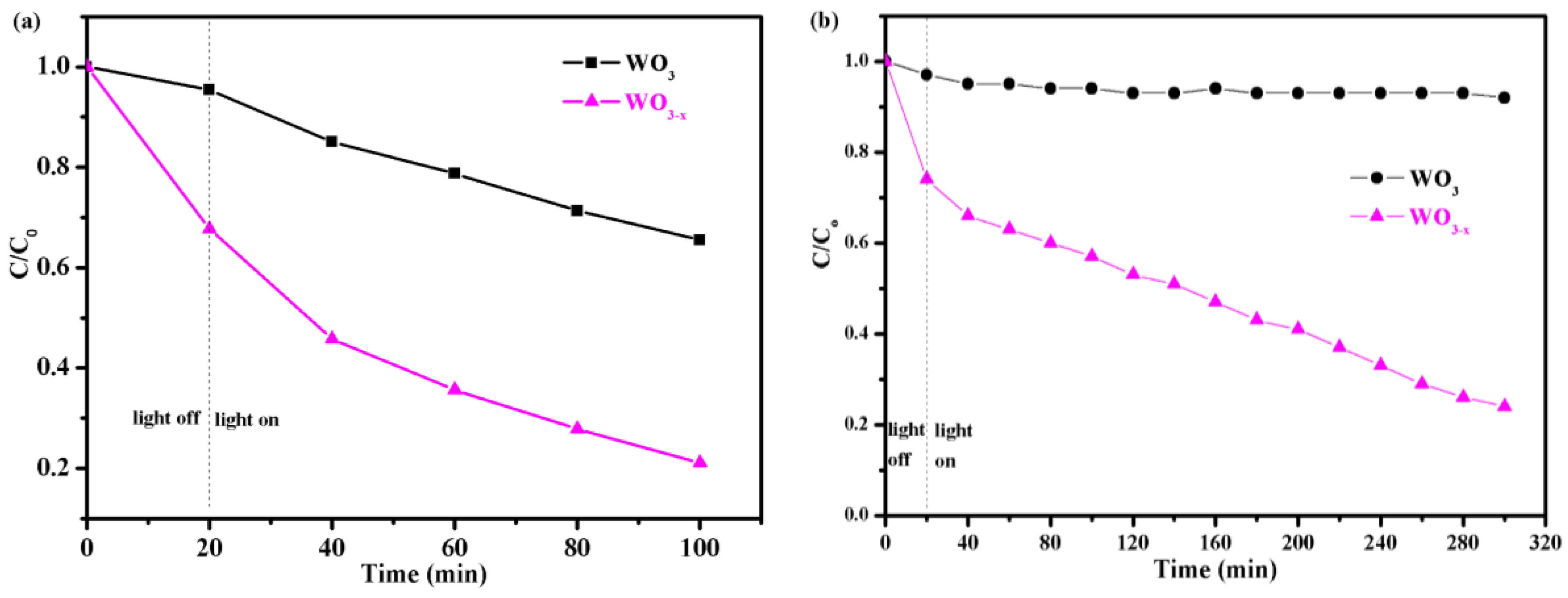
3.2. Photocatalytic Degradation Test

To evaluate the ability of the catalysts to degrade organic contaminants, RhB was used as a representative pollutant. The as-prepared WO3−x photocatalysts are expected to exhibit considerable UV-light photocatalysis as compared to pristine WO3. Before irradiation, an adsorption experiment was performed in the dark to ensure that adsorption equilibrium of RhB on the catalyst surface was adequately established. The results are shown in Figure 6a, where t, C, and C0 refer to the irradiation time, instantaneous RhB concentration, and RhB concentration before irradiation, respectively. The C/C0 ratio was used to describe the decomposition efficiency, which represents the concentration ratio before and after a certain period of reaction time. The WO3−x sample efficiently decomposed the RhB in 80 min under in the irradiation of UV light, while the pristine WO3 decomposed only approximately 30% of the RhB. The annealing-treated WO3−x samples exhibited better photocatalytic activity than the pristine WO3.
The photocatalytic activity of the WO3−x sample was also investigated under visible light (λ > 420 nm) illumination. As shown in Figure 6b, the WO3 samples showed little ability to decompose RhB after visible light irradiation for 320 min. In contrast, the WO3−x samples exhibited higher photocatalytic efficiency (77%) than that of WO3 (9%). The photocatalytic activity of visible light was greatly improved, because WO3−x has enhanced visible light absorption at 400–1400 nm and the plasmonic effect due to the introduction of oxygen vacancies (Figure 3a). The oxygen vacancies were demonstrated to be electron donors and contributed to the enhanced donor density [44]. The increased donor density improved charge transport in WO3−x. Therefore, the LSPR and charge transport are believed to be major mechanisms for the observed photocatalytic activity enhancement in the WO3−x samples.

4. Conclusions

We prepared monoclinic structured WO3 nanosheets via a one-step template-free hydrothermal route and then introduced oxygen vacancies through low temperature annealing in alcohol. x possesses a high concentration of oxygen vacancies, which were demonstrated to be electron donors and contributed to the enhanced donor density, which is believed to induce LSPR and to increase light harvesting efficiency in the visible and infrared regions. Simultaneously, the oxygen vacancies act as traps for reducing the recombination of electrons and holes and significantly improved the e-h separation efficiency, greatly enhancing the photocatalytic activity. This represents a novel approach for further improving the photocatalytic properties of WO3. In addition, this study illustrates a facile route to obtain plasmonic WO3−x nanosheets and may offer a generalized method for generating other semiconductor nanostructures with LSPR, such as deficient vanadium and molybdenum oxides.

Author Contributions

S.C. designed the study, analyzed the data, generated the figures and wrote the manuscript; Y.X. supervised rhodamine B products analysis in cooperation with S.C.; Y.W. conceived the concept, designed the experiments, analyzed the data and revised the manuscript; W.Z. revised the manuscript; Z.H. performed EPR analysis; H.Z. performed XPS analysis; W.X. performed FTIR and UV~analysis.

Funding

This work is supported by the National Nature Science Foundation of China [No.21271048], Natural Science Foundation of China [11747074], Guangdong province science and technology plan project public welfare fund and ability construction project [2016A010103041, 2017A010103025], Doctoral Program of Lingnan Normal University [ZL1503], China Spark Program (2015GA780058).

Conflicts of Interest

The authors declare no conflict of interest.

References

  1. Fujishima, A.; Honda, K. Electrochemical photolysis of water at a semiconductor electrode. Nature 1972, 238, 37–38. [Google Scholar] [CrossRef] [PubMed]
  2. Chen, X.B.; Mao, S.S. Titanium Dioxide Nanomaterials:  Synthesis, properties, modifications, and applications. Chem. Rev. 2007, 107, 289. [Google Scholar] [CrossRef] [PubMed]
  3. Ma, Y.; Wang, X.; Jia, Y.; Chen, X.; Han, H.; Li, C. Titanium Dioxide-based nanomaterials for photocatalytic fuel generations. Chem. Rev. 2014, 114, 9987. [Google Scholar] [CrossRef] [PubMed]
  4. Chen, X.; Shen, S.; Guo, L.; Mao, S.S. Semiconductor-based photocatalytic hydrogen generation. Chem. Rev. 2010, 110, 6503. [Google Scholar] [CrossRef] [PubMed]
  5. Tada, H.; Kiyonaga, T.; Naya, S. Rational design and applications of highly efficient reaction systems photocatalyzed by noble metal nanoparticle-loaded titanium(IV) dioxide. Chem. Soc. Rev. 2009, 38, 1849. [Google Scholar] [CrossRef] [PubMed]
  6. Dahl, M.; Liu, Y.; Yin, Y. Composite titanium dioxide nanomaterials. Chem. Rev. 2014, 114, 9853. [Google Scholar] [CrossRef] [PubMed]
  7. Qu, Y.; Duan, X. Challenge and perspective of heterogeneous photocatalysts. Chem. Soc. Rev. 2013, 42, 2568. [Google Scholar] [CrossRef] [PubMed]
  8. Asahi, R.; Morikawa, T.; Ohwaki, T.; Aoki, K.; Taga, Y. Visible-light photocatalysis in nitrogen-doped titanium oxides. Science 2001, 293, 269–271. [Google Scholar] [CrossRef] [PubMed]
  9. Liu, G.; Zhao, Y.; Sun, C.; Li, F.; Lu, G.Q.; Cheng, H.M. Synergistic effects of B/N doping on the visible-light photocatalytic activity of mesoporous TiO2. Angew. Chem. Int. Ed. 2008, 47, 4516–4520. [Google Scholar] [CrossRef] [PubMed]
  10. Yang, G.D.; Jiang, Z.; Shi, H.H.; Xiao, T.C.; Yan, Z.F. Preparation of highly visible-light active N-doped TiO2 photocatalyst. J. Mater. Chem. 2010, 20, 5301–5309. [Google Scholar] [CrossRef]
  11. Hernández-Alonso, M.D.; Fresno, F.; Suáreza, S.; Coronado, J.M. Development of alternative photocatalysts to TiO2: Challenges and opportunities. Energy Environ. Sci. 2009, 2, 1231. [Google Scholar] [CrossRef]
  12. Fernández-Domene, R.M.; Sánchez-Tovar, R.; Lucas-Granados, B.; García-Antón, J. Improvement in photocatalytic activity of stable WO3, nanoplatelet globular clusters arranged in a tree-like fashion: influence of rotation velocity during anodization. Appl. Catal. B 2016, 189, 266–282. [Google Scholar] [CrossRef]
  13. Bai, X.; Sun, C.; Liu, D.; Luo, X.; Li, D.; Wang, J. Photocatalytic degradation of deoxynivalenol using graphene/ZnO hybrids in aqueous suspension. Appl. Catal. B 2017, 204, 11–20. [Google Scholar] [CrossRef]
  14. Kudo, A.; Miseki, Y. Heterogeneous photocatalyst materials for water splitting. Chem. Soc. Rev. 2009, 38, 253. [Google Scholar] [CrossRef] [PubMed]
  15. Zheng, H.; Ou, J.Z.; Strano, M.S.; Kaner, R.B.; Mitchell, A.; Kalantar-zadeh, K. Nanostructured tungsten oxide-properties synthesis, and applications. Adv. Funct. Mater. 2011, 21, 2175–2196. [Google Scholar] [CrossRef]
  16. Chen, Q.; Li, J.; Zhou, B.; Long, M.; Chen, H.; Liu, Y.; Cai, W.; Shangguan, W.F. Preparation of well-aligned WO3 nanoflake arrays vertically grown on tungsten substrate as photoanode for photoelectrochemical water splitting. Electrochem. Commun. 2012, 20, 153–156. [Google Scholar] [CrossRef]
  17. An, X.; Yu, J.C.; Wang, Y.; Hu, Y.; Yu, X.; Zhang, G. WO3 nanorods/graphene nanocomposites for high-efficiency visible-light-driven photocatalysis and NO2 gas sensing. J. Mater. Chem. 2012, 22, 8525–8531. [Google Scholar] [CrossRef]
  18. Ng, C.; Ng, Y.H.; Iwase, A.; Amal, R. Influence of annealing temperature of WO3 in photoelectrochemical conversion and energy storage for water splitting. ACS Appl. Mater. Interfaces 2013, 5, 5269–5275. [Google Scholar] [CrossRef] [PubMed]
  19. Reyes-Gil, K.R.; Wiggenhorn, C.; Brunschwig, B.S.; Lewis, N.S. Comparison between the quantum yields of compact and porous WO3 photoanodes. J. Phys. Chem. C 2013, 117, 14947–14957. [Google Scholar] [CrossRef]
  20. Hu, X.X.; Xu, P.Q.; Gong, H.Y.; Yin, G.T. Synthesis and characterization of WO3/Graphene nanocomposites for enhanced photocatalytic activities by one-step in-situ hydrothermal reaction. Materials 2018, 11, 147. [Google Scholar]
  21. Székely, I.; Kovács, G.; Baia, L.; Danciu, V.; Pap, Z. Synthesis of shape-tailored WO3 micro-/nanocrystals and the photocatalytic activity of WO3/TiO2 composites. Materials 2016, 9, 258. [Google Scholar] [CrossRef] [PubMed]
  22. Li, W.; Da, P.; Zhang, Y.; Wang, Y.; Lin, X.; Gong, X.; Zheng, G. WO3 Nanoflakes for enhanced photoelectrochemical conversion. ACS Nano 2014, 8, 11770–11777. [Google Scholar] [CrossRef] [PubMed]
  23. Osterloh, F.E. Cheminform abstract: Inorganic nanostructures for photoelectrochemical and photocatalytic water splitting. Cheminform 2013, 42, 2294–2320. [Google Scholar] [CrossRef]
  24. Tanaka, A.; Hashimoto, K.; Kominami, H. Visible-light-induced hydrogen and oxygen formation over Pt/Au/WO3 photocatalyst utilizing two types of photoabsorption due to surface plasmon resonance and band-gap excitation. J. Am. Chem. Soc. 2014, 136, 586–589. [Google Scholar] [CrossRef] [PubMed]
  25. Xiang, Q.; Meng, G.F.; Zhao, H.B.; Zhang, Y.; Li, H.; Ma, W.J.; Xu, J.Q. Au nanoparticle modified WO3 nanorods with their enhanced properties for photocatalysis and gas sensing. J. Phys. Chem. C 2010, 114, 2049–2055. [Google Scholar] [CrossRef]
  26. Sun, S.M.; Wang, W.Z.; Zeng, S.Z.; Shang, M.; Zhang, L. Preparation of ordered mesoporous Ag/WO3 and its highly efficient degradation of acetaldehyde under visible-light irradiation. J. Hazard. Mater. 2010, 178, 427–433. [Google Scholar] [CrossRef] [PubMed]
  27. Cui, L.F.; Ding, X.; Wang, Y.G.; Shi, H.C.; Huang, L.H.; Zuo, Y.H.; Kang, S.F. Facile preparation of Z-scheme WO3/g-C3N4 composite photocatalyst with enhanced photocatalytic performance under visible light. Appl. Surf. Sci. 2017, 391, 202–210. [Google Scholar] [CrossRef]
  28. Jin, T.; Diao, P.; Wu, Q.Y.; Xu, D.; Hu, D.Y.; Xie, Y.H.; Zhang, M. WO3 nanoneedles/α-Fe2O3/cobalt phosphate composite photoanode for efficient photoelectrochemical water splitting. Appl. Catal. B 2014, 148–149, 304–310. [Google Scholar] [CrossRef]
  29. Momeni, M.M.; Ghayeb, Y.; Davarzadeh, M. Single-step electrochemical anodization for synthesis of hierarchical WO3-TiO2 nanotube arrays on titanium foil as a good photoanode for water splitting with visible light. J. Electroanal. Chem. 2015, 739, 149–155. [Google Scholar] [CrossRef]
  30. Bazarjani, M.S.; Hojamberdiev, M.; Morita, K.; Zhu, G.Q.; Cherkashinin, G.; Fasel, C.; Herrmann, T.; Breitzke, H.; Gurlo, A.; Riedel, R. Visible light photocatalysis with c-WO3−x/WO3·H2O nanoheterostructures in situ formed in mesoporous polycarbosilane-siloxane polymer. J. Am. Chem. Soc. 2013, 135, 4467–4475. [Google Scholar] [CrossRef] [PubMed]
  31. Song, J.J.; Huang, Z.F.; Pan, L.; Zou, J.J.; Zhang, X.W.; Wang, L. Oxygen-deficient tungsten oxide as versatile and efficient hydrogenation catalyst. ACS Catal. 2015, 5, 6594–6599. [Google Scholar] [CrossRef]
  32. Yan, M.; Li, G.L.; Guo, C.S.; Guo, W.; Ding, D.D.; Zhang, S.H.; Liu, S.Q. WO3−x sensitized TiO2 spheres with full-spectrum-driven photocatalytic activities from UV to near infrared. Nanoscale 2016, 8, 17828–17835. [Google Scholar] [CrossRef] [PubMed]
  33. Lou, Z.Z.; Xue, C. In situ growth of WO3−x nanowires on g-C3N4 nanosheets: 1D/2D heterostructures with enhanced photocatalytic activity. Cryst Eng Comm 2016, 18, 8406. [Google Scholar] [CrossRef]
  34. Li, Y.S.; Tang, Z.L.; Zhang, J.Y.; Zhang, Z.T. Defect engineering of air-treated WO3 and its enhanced visible light-driven photocatalytic and electrochemical performance. J. Phys. Chem. C 2016, 120, 9750–9763. [Google Scholar] [CrossRef]
  35. Yan, J.; Wang, T.; Wu, G.; Dai, W.; Guan, N.; Li, L.; Gong, J. Tungsten oxide single crystal nanosheets for enhanced multichannel solar light harvesting. Adv. Mater. 2015, 27, 1580. [Google Scholar] [CrossRef] [PubMed]
  36. Lu, Y.; Jiang, Y.; Gao, X.; Wang, X.; Chen, W. Strongly coupled Pd nanotetrahedron/tungsten oxide nanosheet hybrids with enhanced catalytic activity and stability as oxygen reduction electrocatalysts. J. Am. Chem. Soc. 2014, 136, 11687. [Google Scholar] [CrossRef] [PubMed]
  37. Wang, G.M.; Ling, Y.C.; Wang, H.Y.; Yang, X.Y.; Wang, C.C.; Zhang, J.Z.; Li, Y. Hydrogen-treated WO3 nanoflakes show enhanced photostability. Energy Environ. Sci. 2012, 5, 6180. [Google Scholar] [CrossRef]
  38. Chen, X.B.; Liu, L.; Yu, P.Y.; Mao, S.S. Increasing solar absorption for photocatalysis with black hydrogenated titanium dioxide nanocrystals. Science 2011, 331, 746–750. [Google Scholar] [CrossRef] [PubMed]
  39. Cheng, H.F.; Wen, M.C.; Ma, X.C.; Kuwahara, Y.; Mori, K.; Dai, Y.; Huang, B.B.; Yamashita, H. Hydrogen doped metal oxide semiconductors with exceptional and tunable localized surface plasmon resonances. J. Am. Chem. Soc. 2016, 138, 9316. [Google Scholar] [CrossRef] [PubMed]
  40. Hu, Y.H. A highly efficient photocatalyst-hydrogenated black TiO2 for the photocatalytic splitting of water. Angew. Chem. Int. Ed. 2012, 51, 12410–12412. [Google Scholar] [CrossRef] [PubMed]
  41. Zhang, H.L.; Yang, J.Q.; Li, D.; Guo, W.; Qin, Q.; Zhua, L.J.; Zheng, W.J. Template-free facile preparation of monoclinic WO3 nanoplates and their high photocatalytic activities. Appl. Surf. Sci. 2014, 305, 274–280. [Google Scholar] [CrossRef]
  42. Wu, H.Y.; Xu, M.; Da, P.M.; Li, W.J.; Jia, D.S.; Zheng, G.F. WO3-reduced graphene oxide composites with enhanced charge transfer for photoelectrochemical conversion. Phys. Chem. Chem. Phys. 2013, 15, 16138. [Google Scholar] [CrossRef] [PubMed]
  43. Li, Y.Y.; Wang, C.H.; Zheng, H.; Wan, F.X.; Yu, F.; Zhang, X.T.; Liu, Y.C. Surface oxygen vacancies on WO3 contributed to enhanced photothermo-synergistic effect. Appl. Surf. Sci. 2017, 391, 654–661. [Google Scholar] [CrossRef]
  44. Hilaire, S.; Süess, M.J.; Kränzlin, N.; Bieńkowski, K.; Solarska, R.; Augustyński, J.; Niederberger, M. Microwave-assisted nonaqueous synthesis of WO3 nanoparticles for crystallographically oriented photoanodes for water splitting. J. Mater. Chem. A 2014, 48, 20530–20537. [Google Scholar] [CrossRef]
  45. Cheng, H.F.; Klapproth, M.; Sagaltchik, A.; Li, S.; Thomas, A. Ordered mesoporous WO2.83: Selective reduction synthesis, exceptional localized surface plasmon resonance and enhanced hydrogen evolution reaction activity. J. Mater. Chem. A 2018, 6, 2249. [Google Scholar] [CrossRef]
  46. Manthiram, K.; Alivisatos, A.P. Tunable localized surface plasmon resonances in tungsten oxide nanocrystals. J. Am. Chem. Soc. 2012, 134, 3995–3998. [Google Scholar] [CrossRef] [PubMed]
  47. Janotti, A.; Varley, J.B.; Rinke, P.; Umezawa, N.; Kresse, G.; Van de Walle, C.G. Hybrid functional studies of the oxygen vacancy in TiO2. Phys. Rev. B 2010, 81, 085212. [Google Scholar] [CrossRef]
  48. Morgan, B.J.; Watson, G.W. Intrinsic n-type defect formation in TiO2: a comparison of rutile and anatase from GGA+U calculations. J. Phys. Chem. C 2010, 114, 2321. [Google Scholar] [CrossRef]
  49. Pan, X.Y.; Yang, M.Q.; Fu, X.Z.; Zhang, N.; Xu, Y.J. Defective TiO2 with oxygen vacancies: synthesis, properties and photocatalytic applications. Nanoscale 2013, 5, 3601–3614. [Google Scholar] [CrossRef] [PubMed]
  50. Naldoni, A.; Allieta, M.; Santangelo, S.; Marelli, M.; Fabbri, F.; Cappelli, S.; Bianchi, C.L.; Psaro, R.; Dal Santo, V. Effect of nature and location of defects on bandgap narrowing in black TiO2 nanoparticles. J. Am. Chem. Soc. 2012, 134, 7600. [Google Scholar] [CrossRef] [PubMed]
Figure 1. (a) Schematic illustration of the fabrication of tungsten trioxide (WO3) and non-stoichiometric monoclinic structured WO3 (WO3−x) nanosheets. (b) Digital photos of the WO3 (I) and WO3−x (II) nanosheets samples.
Nanomaterials 08 00553 g001
Figure 2. Field emission scanning electron microscopy (FE-SEM) images of the WO3 (a) and WO3−x (b) nanosheets. FE-TEM images of the WO3 (c) and WO3−x (d) nanosheets. Field emission transmission electron microscopy (HR-TEM) images of the WO3 (e) and WO3−x (f) nanosheets.
Nanomaterials 08 00553 g002
Figure 3. Comparison of the WO3 and WO3−x nanosheets. (a) X-ray diffraction (XRD) patterns; (b) Fourier-transform infrared spectroscopy (FTIR) spectra; (c) Raman spectra and (d) electron paramagnetic resonance (EPR) spectra at room temperature.
Nanomaterials 08 00553 g003
Figure 4. UV-Vis diffuse reflectance spectra of the WO3 and WO3−x nanosheets.
Nanomaterials 08 00553 g004
Figure 5. (a) The Ti 2p X-ray photoelectron spectra (XPS) spectra of the as-prepared WO3 and WO3−x; (b) O 1s XPS spectra of the as-prepared WO3 and WO3−x and (c) valence band (VB) XPS spectra of the as-prepared WO3 and WO3−x.
Nanomaterials 08 00553 g005
Figure 6. (a) UV light photocatalytic degradation of rhodamine B (RhB) and (b) visible-light photocatalytic degradation of RhB by the prepared photocatalysts.
Nanomaterials 08 00553 g006
Table 1. The free electron concentration of a previously reported plasma-doped WO3−x.
Plasmonic MaterialsLSPR WavelengthFree-Carrier Density (cm3)Reference
WO3−x800 nm9.1 × 1021This Work
WO2.83650 nm9.79 × 1021Ref. [45]
WO3−x1450 nm2.5 × 1021Ref. [35]
WO3−x900 nm6.3 × 1021Ref. [46]

