Article

Phenotypic and Genotypic Analysis of Bacterial Pathogens Recovered from Patients Diagnosed with Fever of Unknown Origin in Egypt

1
Microbiology Lab Department, Abbasia Fever Hospital, Cairo 11566, Egypt
2
Department of Microbiology and Immunology, Faculty of Pharmacy, Ain Shams University, Cairo 11566, Egypt
3
Department of Medical Physiology, College of Medicine, King Khalid University, Abha 61421, Saudi Arabia
4
Department of Microbiology and Immunology, Faculty of Pharmacy, October University for Modern Sciences and Arts (MSA), 6th of October, Giza 12451, Egypt
*
Author to whom correspondence should be addressed.
Antibiotics 2023, 12(8), 1294; https://doi.org/10.3390/antibiotics12081294
Submission received: 19 June 2023 / Revised: 27 July 2023 / Accepted: 3 August 2023 / Published: 7 August 2023
(This article belongs to the Special Issue Antibiotic Resistant Pathogens in Hospital)

Abstract

:
Fever of unknown origin (FUO) is a medical term describing fever that lasts for at least three weeks without a diagnosis being reached after extensive diagnostic evaluation. Therefore, this study aimed to identify the common pathogens causing FUO in patients admitted to Abbasia Fever Hospital in Egypt from January 2020 to December 2022, their antimicrobial susceptibility profiles, and associated resistance genes. The study also aimed to investigate the burden of multidrug-resistant (MDR) pathogens and the priority pathogens nominated by the World Health Organization (WHO) for posing the greatest threat to human health due to antibiotic resistance. During the study period, about 726 patients were diagnosed with FUO. After extensive investigations, the cause of the FUO was found to be infectious diseases in 479/726 patients (66.0%). Of them, 257 patients had positive bacterial cultures, including 202 Gram-negative isolates that comprised Klebsiella pneumoniae (85/202; 42.1%), Escherichia coli (71/202; 35.1%), Acinetobacter baumannii (26/202; 12.9%), and Pseudomonas aeruginosa (14/202; 6.9%) and 55 Gram-positive isolates, including Staphylococcus aureus (23/55; 41.8%), Streptococcus pneumoniae (7/55; 12.7%), and Enterococcus spp. (25/55; 45.5%). The MDR phenotype was shown by 68.3% and 65.5% of the Gram-negative and Gram-positive isolates, respectively. Carbapenem resistance (CR) was shown by 43.1% of the Gram-negative isolates. Of the 23 S. aureus isolates obtained from research participants, 15 (65.2%) were methicillin-resistant S. aureus (MRSA). A high-level aminoglycoside resistance (HLAR) phenotype was found in 52.0% of the Enterococcus sp. isolates. The PCR screening of resistance genes in the MDR isolates showed that blaOXA−48 was the most prevalent (84%) among the carbapenemase-coding genes, followed by blaVIM (9%) and then blaIMP (12%). The ESBL-coding genes blaTEM, blaCTX-M, aac(6′)-Ib, and blaSHV, were prevalent in 100%, 93.2%, 85,% and 53.4% of the MDR isolates, respectively. This study updates the range of bacteria that cause FUO and emphasizes the burden of multidrug resistance and priority infections in the region. The obtained data is of relevant medical importance for the implementation of evidence-based antimicrobial stewardship programs and tailoring existing empirical treatment guidelines.

1. Introduction

Fever of unknown origin (FUO) was defined as an illness of more than 3 weeks duration with a fever greater than 38.3 °C (101 °F) on several occasions, the cause of which is uncertain after 1 week of in-hospital investigations [1]. To keep with diagnostic capabilities, some modifications in the definition of FUO occurred throughout the years [1]. The results of the study conducted by Fusco et al. [2] carried out from 2005 to 2015 concluded that the most common causes of FUO are infectious diseases (37.8%), followed by non-infectious inflammatory diseases (20.9%), neoplasms (11.6%), and other diseases (6.5%), while the diagnosis remained unknown in 23.2% of cases [2]. Infectious diseases are the principal categories of diseases causing FUO, which may be caused by multidrug-resistant (MDR) bacterial infections [3].
MDR in bacteria is a great concern to humans’ and animals’ health and welfare [4]. Microbes may develop antimicrobial resistance after prolonged exposure to one or more antibiotics as a result of certain genetic mutations. In addition, the horizontal transfer of mobile genetic elements greatly contributes to the spread of antimicrobial resistance [5]. It has been reported that MDR bacterial infections kill around 50,000 individuals every year in the United States and Europe, and are estimated to kill more than 700,000 people worldwide [6]. If no action is taken to reduce MDR, 10 million people are predicted to die yearly from MDR infections by the year 2050 [6]. The World Health Organization (WHO) released a report titled “Microorganisms posing a substantial hazard to human health” that aimed to promote funding for the development of new antimicrobial drugs to counter the threat posed by a list of priority pathogens [7].
Gram-negative bacteria that are extensively drug-resistant (XDR) and carbapenem-resistant (CR) are thought to pose a worrying threat to human health worldwide [8,9,10,11]. The development of CR is currently gaining a lot of interest [8]. The excessive expression of class A, B, and D carbapenemase enzymes, such as Klebsiella pneumoniae carbapenemase (KPC), New Delhi Metallo-β-lactamase (NDM), imipenemase (IMP), and Verona Integron-encoded Metallo-β-lactamase (VIM) and oxacillinases (OXA−48) continues to be the most relevant route for the development of resistance among Gram-negative bacteria and has been responsible for the majority of nosocomial outbreaks in recent years [10,12]. In the current study, we sought to explore the contribution of bacterial infections in patients diagnosed with FUO in one of the major public fever hospitals in Egypt. In addition to defining the implicated bacterial species, we investigated the antimicrobial susceptibility of the bacterial isolates and explored the burden of MDR and WHO-priority pathogens. We extended our study to investigate some genetic determinants conferring resistance to clinically relevant classes of antimicrobial agents.

2. Results

2.1. Fever of Unknown Origin (FUO) Caused by Bacterial Infections

During the study period, from January 2020 to December 2022, about 726 patients were diagnosed with FUO. After extensive investigations, the cause of the FUO was found to be infectious diseases in 66.0% of cases, followed by returned travelers (9%), neoplastic diseases (4%), inflammatory diseases (2%), and other diseases (16%), and the diagnosis remained unknown in 2% of cases. The infectious disease cases of the FUO reached approximately 479/726 (66.0%). Of these, 257 positive bacterial cultures were obtained. These comprise 11.7% of the total number of specimens received by the hospital during the study period (n = 2200). The majority of positive bacterial cultures came from urine samples (n = 91, 35.4%), then blood (n = 82, 31.9%), sputum (n = 59, 22.9%), and cerebrospinal fluid (CSF) (n = 25, 9.72%).
A total of 55 Gram-positive isolates (20%) and 202 Gram-negative isolates (79%) were identified. The most-common Gram-positive species were Staphylococcus aureus and Enterococcus sp., whereas Escherichia coli and Klebsiella pneumoniae predominated among the recovered Gram-negative isolates. Figure 1 displays the full range of bacterial etiologies for FUO in the patient population included in the current study and the specimen distribution of various species.

2.2. Antimicrobial Susceptibility Profiles

The frequency of the antimicrobial resistance of various species to the studied antimicrobial drugs in Gram-negative and Gram-positive species are shown in Figure 2 and Figure 3, respectively. Only one Salmonella spp. isolate was included in the study. This was susceptible to cefaclor, gentamicin, amikacin, trimethoprim–sulfamethoxazole, ciprofloxacin, ofloxacin, ampicillin/sulbactam, piperacillin/tazobactam, cefepime, ceftriaxone, cefotaxime, ceftazidime, cefoxitin, imipenem, and meropenem. The isolate was non-susceptible only to amoxicillin-clavulanic acid and doxycycline. Interestingly, we found a significant association between non-susceptibility to some antimicrobial agents, as shown in Figures S1–S8.
The MDR phenotype was shown in 138/202 (68.31%) of the Gram-negative isolates. The MDR phenotype was most frequently shown by A. baumannii (23/26; 88.46%), E. coli (40/71; 56.33%), and Klebsiella pneumoniae (69/85; 81.17%). Some genera had a lower prevalence of MDR, such as P. aeruginosa (6/14; 42.85%). None of the isolates that belonged to the Enterobacter species and Proteus species showed the MDR phenotype. Of the Gram-positive isolates, only 65% (36/55) were MDR. These comprised Enterococcus sp. (19/25; 76.00%), S. pneumoniae (5/7; 71.42%), and finally S. aureus (12/23; 52.17%). Carbapenem resistance (CR) was shown by 43.06% of the Gram-negative isolates. Glucose non-fermenters had a higher prevalence of CR (27/40; 67.50%) compared to Enterobacteriaceae isolates (60/162; 37.03%). The three species with the highest rates of CR were A. baumannii (22/26; 84.61%), K. pneumoniae (46/85; 54.11%), and P. aeruginosa (6/14; 42.85%). Only 19.71% (14/71) of the E. coli isolates were CR.
Associations were found between the resistance phenotypes and more antimicrobial agents in Gram-negative species, as shown in Figure S1–S5. Moreover, the association between the resistance phenotypes to different antimicrobial agents showed a statistically significant association between resistance phenotypes to cefoxitin, levofloxacin, and gentamicin in S. aureus; chloramphenicol, ofloxacin, levofloxacin, and gentamicin in the Enterococci (Figure S6–S8).
Of the 23 S. aureus isolates obtained from research participants, 15 (65%) were methicillin-resistant S. aureus (MRSA), while all were susceptible to vancomycin (MIC range = 0.125–2 µg/mL). Thirteen isolates of Enterococcus sp. underwent high-level aminoglycoside resistance (HLAR) testing. The HLAR phenotype was found in 52% of all Enterococcus sp. isolates. In 86% of S. pneumoniae isolates, penicillin resistance was phenotypically evident. In the meantime, the E-test demonstrated that all S. pneumoniae isolates were sensitive to cefotaxime, ceftriaxone, or imipenem.
The resistance frequency for the tested antimicrobial drugs in all Gram-negative and Gram-positive isolates was examined to provide more helpful information for directing empirical treatment guidelines. A similar analysis was conducted on the MDR isolates. The analysis findings are displayed in Figure 4 and Figure 5.

2.3. Minimum Inhibitory Concentrations of the Tested Antibiotics

One hundred and three Gram-negative isolates showing the MDR phenotype were selected for the broth microdilution assays for the determination of the minimum inhibitory concentrations (MICs) of selected antimicrobial agents and correlating the MICs with some antimicrobial resistance genes. The MDR isolates selected for further analysis included 53 K. pneumoniae, 26 E. coli, 20 A. baumannii, and 4 P. aeruginosa isolates.
The MIC of imipenem ranged from 4 to 256 µg/mL with an MIC50 of 64 μg/mL, but the MIC of cefepime ranged from 24 to 512 µg/mL with an MIC50 of 256 µg/mL. On the other hand, the MIC of cefotaxime ranged from 24 to 512 µg/mL with an MIC50 of 512 µg/mL, and finally, the MIC of ciprofloxacin ranged from 6 to 512 µg/mL with an MIC50 of 128 µg/mL. All isolates were found to be susceptible to colistin by E-test. The data summary of the MICs is shown in Table S1.

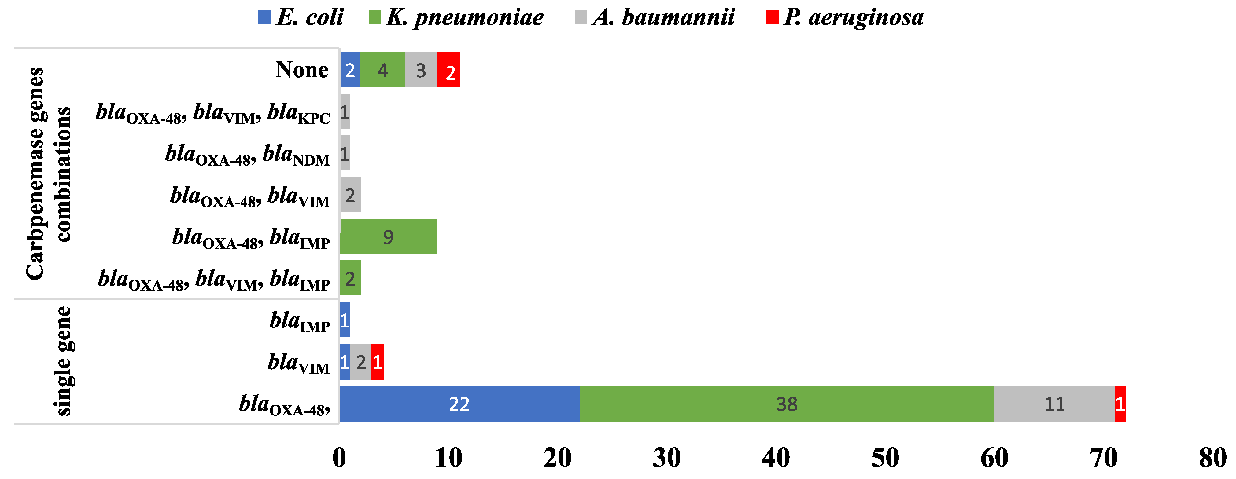
2.4. Molecular Detection of the Carbapenemase-, Extended-Spectrum β-Lactamase (ESBL)-, and aac(6′)-Ib-Coding Genes

Of all the carbapenemase-coding genes screened here, blaOXA−48 was the most prevalent and was carried by 87 (84%) isolates, followed by blaVIM, which was detected in 9 (9%) isolates, and blaIMP was observed in only 12 (12%) isolates. The carbapenemase-coding genes blaKPC and blaNDM−1 were the least prevalent; each was identified in one A. baumannii isolate. Screening our isolates for the ESBL-encoding genes showed that the predominant gene was blaTEM (100%), then blaCTX-M (93.2%), and the least prevalent was blaSHV (53.4%). Finally, the aac(6′)-Ib gene was found in a relatively high prevalence, reaching 85%. The total prevalence of all genes as well as their distribution in different species are shown in Figure 6.
Some isolates were found to carry CR genes while retaining phenotypic susceptibility to carbapenem antibiotics. Among E. coli, carbapenem susceptibility was retained by 12 isolates carrying blaOXA−48 and two isolates that carried at least one of the CR genes blaVIM or blaIMP. In K. pneumoniae, ten isolates carried blaOXA−48 as a single gene and two isolates had three combined genes (blaOXA−48, blaVIM, and blaIMP). Interestingly, all were susceptible to the tested carbapenems. In A. baumannii, only one carbapenem-susceptible isolate was found to carry the blaVIM gene.
A data summary of the antimicrobial resistance genes carried by different species correlated to the MICs of imipenem, cefepime, cefoxitin, and ciprofloxacin is shown in Table S1.
Remarkably, 15/103 (14.56%) MDR isolates co-harbored more than one CPase-coding gene. In comparison, 77/103 (74.75%) carried only a single gene (Figure 7). Out of the 103 isolates harboring CPases-encoding genes, the co-existence of blaOXA−48 and blaIMP was detected in nine isolates (8.7%) while both (blaOXA−48 and blaVIM) and (blaOXA−48, blaVIM and blaIMP) co-existed in two isolates for each. Finally, (blaOXA−48 and blaNDM) and (blaOXA−48, blaKPC and blaVIM) were recorded in one isolate (0. 97%). Testing the association between the resistance genes and the phenotypic resistance to different antimicrobial agents showed that only blaVIM had a statistically significant association with imipenem and meropenem resistance, with p-values of 0.011 and 0.026, respectively. This was only found among K. pneumoniae isolates. None of the ESBL-coding genes showed a statistically significant association with resistance to any of the tested β-lactam antibiotics. Finally, acc-(6′)-Ib showed a statistically significant association with gentamicin resistance (p-value 0.045) in A. baumannii but not with amikacin or any of the tested fluoroquinolones. The p-values calculated for all genes and antimicrobial agents are shown in Tables S2–S4.

2.5. Phenotypic and Genotypic Analysis Using Heatmap Analysis

Using the antimicrobial resistance profiles, carbapenemase genes, ESBL genes, and aac-(6′)-Ib gene results, a dendrogram depicting the heatmap signature of the isolates was produced. The 53, 26, 20, and 4 MDR K. pneumoniae, E. coli, A. baumannii, and P. aeruginosa isolates were clustered into 31, 21, 18, and 3 clusters, respectively (Figure 8).

3. Discussion

This is a two-year study conducted in the Abbasia Fever Hospital, a major infectious disease public hospital serving the area of Greater Cairo in Egypt. The study provides an update on the recent spectrum of bacterial pathogens causing FUO and their susceptibility profiles. Upon culturing 726 clinical specimens received by the hospital’s laboratory for the investigation of FUO, only 257 (35.4%) showed bacterial growth. Other infectious causes of FUO (30.6%) may include unculturable bacteria and those diagnosed principally through serological tests such as Salmonella sp. and Brucella sp. They may also include viruses, fungi, or parasites. These are typically underdiagnosed since they are primarily diagnosed through serological, molecular, or histological testing, which may not be practical in many hospitals, especially in nations with poor healthcare resources [13]. Reports on the prevalence and etiology of the FUO in Egypt are relatively scarce. FUO with an infectious etiology prevailed in most of the previous studies [14,15,16,17], with a prevalence that reached 72% [14]. In a study by Montasser et al. [17] conducted in 2015 on patients admitted to Abbassia Fever Hospital with FUO, cytomegalovirus infection was identified as the most common infectious cause, followed by urinary tract infections and respiratory tract infections. Less frequently, the infectious causes of the FUO included salmonellosis, brucellosis, tuberculosis, and infective endocarditis. In the same study, the Gram-negative isolates were predominated by E. coli, Klebsiella sp., and Enterobacter sp., while S. aureus was the most common Gram-positive species.
The bacterial isolates that were recovered from the patients enrolled in our study included 202 Gram-negative and 55 Gram-positive isolates. Compared to our previous study conducted in 2018 and 2019 [18], a lower percentage of positive bacterial cultures were found among the total number of specimens received by the microbiology laboratory of the Abbasia Fever Hosiptal in Cairo (24.7% versus 11.7%). This is likely due to the contribution of other viral infections such as COVID-19 that emerged early in 2020. A considerable share was found for the MDR isolates in the current study. Up to 67.7% of all isolates were MDR. This comprised 138/202 (68.3%) and 36/55 (65.5%) of the Gram-negative and Gram-positive isolates, respectively. The high prevalence of MDR strains in hospital settings has been widely reported in Egypt [19,20,21,22,23] and in other countries as well [24,25,26]. A further rise in the prevalence of the MDR strains was also evident during the pandemic of COVID-19 [23,26,27,28]. In FUO, MDR infections have been recently linked to prior colonization by MDR organisms that were in turn linked to a longer hospitalization [29].
Of 162 Enterobacteriaceae isolates investigated here, 109 (67.28%) showed MDR. This was most frequently found in K. pneumoniae (69/85; 81.17%) and E. coli (40/71; 56.33%) isolates. Yet, a higher prevalence of MDR (84.6%) was shown by Acinetobacter baumannii isolates. Carbapenems are among the last-resort treatment options for severe Gram-negative infections, particularly those that are MDR. As reported by El-Kholy et al. [19], A. baumannii, K. pneumoniae, and P. aeruginosa are among the top-ranked CR Gram-negative species in Egypt. At least 37% of Enterobacteriaceae isolates were non-susceptible to at least one carbapenem. Interestingly, 54% of the carbapenem-resistant Enterobacteriaceae belonged to K. pneumoniae. Other carbapenem-resistant isolates comprised E. coli (20%). Carbapenem-resistant K. pneumoniae (CRKP) is well-recognized as a leading cause of hospital-acquired infections worldwide [30] as well as in Egypt [31,32,33]. The susceptibility profiles of the ESBL producers were encountered in 79% of Enterobacteriaceae isolates. More than half of K. pneumoniae (70/85; 82%) isolates were predicted to be ESBL producers. Similar findings were reported by others [34]. Carbapenem-resistant A. baumannii (CRAB) is among the high-priority pathogens listed by the WHO. In our collection, CRAB was evident in 85% of the A. baumannii isolates. The wide dissemination of carbapenem-resistant Acinetobacter baumannii (CRAB) was previously reported by regional [35,36,37] and worldwide studies [38,39]. CRAB was also identified in a higher prevalence in some recent studies from Egypt [40]. Therapeutic options for the treatment of CRAB infections are limited to colistin, tigecycline, and some aminoglycosides [39].
MRSA is another high-priority pathogen presented here. A total of 23 S. aureus infections were found; 15/23 (65.2%) of the isolates were resistant to penicillinase-stable penicillins, and 52.17% were MDR. In comparison to other African [41] and Mediterranean [42] nations, previous investigations have indicated that Egypt has the highest MRSA scores. Vancomycin has long been acknowledged as the cornerstone of MRSA infection treatment [43]. Fortunately, none of the MRSA isolates were resistant to vancomycin.
Five out of seven (71.42%) S. pneumoniae isolates identified here showed the MDR phenotype. Nevertheless, medium priority was assigned by the WHO for penicillin-resistant S. pneumoniae. Using the oxacillin disk-diffusion test, penicillin resistance was inferred in 6/7 (85.7%) S. pneumoniae isolates. Of them, seven isolates were tested for susceptibility to cefotaxime, ceftriaxone, or imipenem using an E-test, as recommended by the Clinical and Laboratory Standards Institute (CLSI) guidelines [44]. All the isolates (100%) were susceptible to all of the tested agents. In contrast to our findings, a prevalence of 49.0% of penicillin resistance was reported by Wasfy et al. [45] in S. pneumoniae isolates collected between 1993 and 2003 in Egypt. Furthermore, susceptibility to penicillin was maintained in 84.2% of S. pneumoniae isolates tested by El-Kholy et al. in a more recent study [46]. The high prevalence of penicillin resistance identified here was balanced by acceptable levels of susceptibility to other antimicrobial classes, highlighting the critical need for empirical treatment regimen re-evaluation.
The human gut inhabitants, Enterococci, are well-recognized as challenging nosocomial pathogens frequently causing difficult-to-treat infections [47]. MDR Enterococci comprised 76% of the Enterococcal isolates investigated here. The combination therapy of cell-wall inhibitors with aminoglycosides has long been used for the treatment of serious enterococcal illnesses [48]. The effectiveness of this treatment regimen is eliminated by the reduced susceptibility to either of the combined antimicrobials. Hence, synergy is routinely predicted using the HLAR test. The test revealed high-level resistance to aminoglycosides in 13/25 (52.0%) Enterococcal isolates in our collection.
Investigating the association between the resistance phenotypes to different antimicrobial agents showed a statistically significant association between the resistance phenotypes to some antimicrobial agents. This was mostly found between antimicrobial agents belonging to the same class, which are likely affected by the same resistance determinants. Occasionally, a statistical significance was found between antimicrobial agents that belong to different classes. This is likely due to the co-existence of some resistance genes on the same plasmids or other mobile genetic elements [49,50]. It is worth mentioning that such associations were more frequently found among Gram-negative species (Figure S1–S8).
One of the goals of our study was to investigate the contribution of different classes of CPases to the CR phenotype shown by the MDR isolates identified here. For this purpose, we have molecularly characterized the carbapenemase-encoding genes in a group of MDR isolates.
The blaOXA−48 gene was the predominant carbapeneamse-coding gene, found in 84.5% of the isolates, followed by blaIMP (10.7%), blaVIM (8.7%), blaNDM (1%), and blaKPC (1%). According to Abdelaziz et al. [51], blaNDM (80.5%) was the predominant CR gene, followed by blaVIM (36.4%), blaKPC (28.6%), blaOXA−48 (26%) and blaIMP (6.5%]. Imipenem MIC50 values were 64, with MIC values ranging from 4 to 512 µg/mL. It is worth mentioning that blaNDM and blaKPC were exclusively found in A. baumannii. While blaNDM was frequently reported in A. baumannii [11,36,37,40], only a few reports about KPC-producer A. baumannii have been published [37,52,53]. Combinations of up to three CR genes were detected in our collection. The most frequently detected combination was blaOXA−48 and blaIMP, which was exclusively carried by K. pneumoniae isolates. No combinations of CR genes were found in E. coli or P. aeruginosa. Many studies have also noticed the co-existence of carbapenemase-coding genes, which resulted in decreased sensitivity to various antibiotics [49,54,55,56]. Interestingly, some of our isolates that carried one or more of the CR genes (blaKPC, blaIMP, blaVIM, or blaNDM) showed phenotypic susceptibility to imipenem. These results contrasted with other studies confirming that MBL genes confer phenotypic resistance to carbapenems [56,57]. In line with our findings, carbapenem-susceptible strains carrying CR genes were also reported by other authors [55,58,59,60,61]. Kayama et al. proposed that some carbapenemases may affect meropenem but not imipenem [62].
Extended-spectrum β-lactamases (ESBLs) are one of the most clinically significant subgroups of β-lactamases [63]. Numerous investigations have shown that patients who are infected with bacteria that encode ESBLs have higher mortality rates and, generally, worse clinical outcomes [63]. More than half of the isolates studied here carried at least one of the ESBL-coding genes blaSHV, blaCTX-M, and blaTEM. This was slightly higher than the percentage found in our previous study conducted in 2018 and 2019 [18]. ESBL-coding genes were found in combinations in the majority of the isolates, most frequently in K. pneumoniae (Table S1). Also, cefotaxime and cefepime MIC50 values were 512 and 256, respectively, with MIC values ranging from 24 to 512 µg/mL for both. No statistical significance was found between any of the tested ESBL genes and the resistance phenotypes of β-lactams. This was partly due to the high fraction of the isolates that were non-susceptible to most of the tested β-lactams. Moreover, all isolates were positive for blaTEM. Hence, the p-values could not be calculated in many cases, as shown in Tables S2–S4.
A major issue in the therapeutic management of infections is the concurrent quinolone resistance in ESBL-producing Enterobacteriaceae. The fluoroquinolone-acetylating aminoglycoside-(6′)-N-acetyltransferase (aac(6′)-Ib-cr) gene is one of the most frequently identified plasmid-mediated quinolone resistance (PMQR) determinants. The gene is commonly found to co-exist with other PMQR determinants, including qnr genes [64,65,66]. A remarkably high frequency of fluoroquinolone resistance was found in the current investigation. Such a high FQ resistance was accompanied by the existence of aac(6′)-Ib determinants in 85.4% of the clinical isolates. An MIC50 of 128 µg/mL was recorded for ciprofloxacin in our isolates, with MIC values ranging from 6 to 512 µg/mL. Nevertheless, the association between the aac(6′)-Ib gene and ciprofloxacin resistance was not significant in our isolates. This is likely because aac(6′)-Ib-cr is not sufficient to confer ciprofloxacin resistance. As reported by Hamed et al. [65], full resistance to ciprofloxacin occurs due to multiple mechanisms, among which target site alteration is the main player.
The clonal relatedness of the MDR isolates studied here was investigated by a hierarchical clustering based on both the genotypes and phenotypes. The analysis showed that most of the isolates were not clonal, reflecting that the screened genes were likely disseminated by horizontal transfer rather than vertically by clonal expansion (Figure 8).
Taken together, the results of the current study highlight the serious spread of MDR organisms in the community and their implications for FUO, whose definitive diagnosis is particularly challenging in the outpatient setting. In an attempt to contribute to developing treatment guidelines tailored to the currently circulating MDR strains, the antimicrobial susceptibility findings of the current study were used for ranking the tested antimicrobial agents based on their activity against all isolates as well as those showing MDR. The top-ranked antimicrobials that showed the highest activity against Gram-negative isolates were colistin, amikacin, and nitrofurantoin. However, one of the limitations of our study is that the colistin susceptibility of the isolates was tested using an E-test that was reported before to underestimate colistin MICs, resulting in a significant number of false-susceptible results [67,68,69]. A similar analysis was performed for Gram-positive isolates, and the top-ranked antimicrobials included vancomycin, linezolid, co-trimoxazole, chloramphenicol, and rifampicin. All showed acceptable activity against MDR Gram-positive isolates. Accordingly, our findings support other calls for reviving interest in older antimicrobials for the management of infections caused by MDR bacterial strains [70,71,72]. In addition, several alternative approaches have also been proposed by many researchers for managing MDR infections. Among others, these include antimicrobial combination therapy [73], bacteriophage therapy [74], and nanoparticles [73]. The proper implementation of antimicrobial stewardship and infection control programs is also crucial for reducing the emergence and spread of MDR strains [74].

4. Materials and Methods

4.1. Study Design

The current study is a prospective study conducted at Abbasia Fever Hospital in Cairo in the period from January 2020 to December 2022. The Abbassia Fever Hospital is one of the largest infectious disease hospitals in Egypt. It is affiliated with the Egyptian Ministry of Health, and all febrile patients from the Greater Cairo Area are referred there. The study protocol was in agreement with the ethical principles stated in the Declaration of Helsinki and was approved by the institutional ethics committee, Faculty of Pharmacy, Ain Shams University (ENREC-ASU-2019-268).

4.2. Microbiological Procedures

During the study period, about 2200 clinical specimens, including urine, blood, sputum, and cerebrospinal fluid (CSF) were received by the microbiology laboratory of the hospital. The specimens were collected from patients admitted to Abbassia Fever Hospital in 2020 and 2021 for investigation of the cause and etiology of FUO after a failure to establish a definitive diagnosis in the outpatient setting. Of them, positive bacterial cultures were obtained from 257 cases selected for enrollment in our study. The isolates were recovered from various clinical specimens obtained from febrile neutrophilic patients (>11,000 white blood cells/µL with oral temperature >38 °C, over at least 3 days for inpatient or at least 3 weeks for outpatient). Bacterial isolates were identified routinely by morphological and biochemical tests and supplemented as needed by specialized tests and special agar media, according to the guidelines of the Central Health Laboratories affiliated with the Egyptian Ministry of Health. Blood specimens were cultured in trypticase soya broth blood culture vials that were then incubated at 35–37 °C in a non-CO2 incubator for a maximum of 14–21 days. Subcultures were made on blood agar, chocolate agar, and McConkey agar for subsequent identification. Pathogens in CSF specimens were identified by Gram staining, and cultures were made on chocolate agar, blood agar, nutrient agar, and MacConkey agar after incubation at 37 °C for 24 h. Blood and chocolate agar plates were incubated in 5–10% CO2 conditions. Urine specimens were cultured on nutrient (or blood agar) and MacConkey agar or CLED (cystine-lactose-electrolyte-deficient) agar plates that were then incubated at 35 °C for 24 h. Sputum specimens were cultured on blood, chocolate, and MacConkey agar plates. Incubation conditions were aerobic for 24 h at 35 °C for MacConkey agar plates and 5–10% CO2 for blood and chocolate agar. The identification of the isolates that exhibited the MDR phenotype was confirmed by the VITEK2 automated system (bioMérieux, Marcy L’Etoile, France).

4.3. Antimicrobial Susceptibility Testing

The antimicrobial susceptibility testing of recovered isolates was carried out according to CLSI (2021) for each bacterial species [44]. The Kirby–Bauer disk-diffusion method was performed on Müller–Hinton agar (Hi media, Maharashtra, India) using the following antimicrobial disks (Bioanalyse, Ankara, Turkey): penicillin (P, 10 U), ampicillin (AMP, 10 μg), amoxicillin/clavulanic acid (AMC, 20/10 μg), ampicillin/sulbactam (SAM, 10/10 μg), piperacillin/tazobactam (TPZ, 10/100 μg), cefaclor (CEC, 30 μg), cefoxitin (FOX, 30 μg), cefepime (FEP, 30 μg), ceftriaxone (CRO, 30 μg), cefotaxime (CTX, 30 μg), ceftazidime (CAZ, 30 μg), imipenem (IMP, 10 μg), meropenem (MEM, 10 μg), gentamicin (CN, 10 μg and 120 μg) amikacin (AK, 30 μg), trimethoprim-sulfamethoxazole (cotrimoxazole) (TMP/SMX, 1.25/23.75 μg), ciprofloxacin (CIP, 5 μg), levofloxacin (LEV, 5 μg), norfloxacin (NOR, 10 μg), ofloxacin (OFX, 5 μg), doxycycline (DO, 30 μg), nitrofurantoin (F, 300 μg), erythromycin (E, 15 μg), azithromycin (AZM, 15 μg), chloramphenicol (C, 30 μg), clindamycin (DA, 2 μg), vancomycin (V, 30 μg), rifampin (R, 5 μg) and linezolid (LNZ, 30 μg). Susceptibility to colistin was examined using an E-test (Bioanalyse, Turkey) according to the manufacturer’s recommendations. Both E. coli ATCC 25922 and S. aureus ATCC® 25923 reference strains were used for the quality control.
The following issues were considered during antimicrobial susceptibility testing: (1) cefoxitin was used as a surrogate for oxacillin in the disk-diffusion test for the identification of methicillin (oxacillin)-resistant Staphylococci (MRS); (2) MRSA was considered resistant to all other β-lactams except cephalosporins with anti-MRSA activity (ceftaroline and ceftobiprole); (3) An E-test (Bioanalyse, Turkey) was used for testing the susceptibility of Staphylococci to vancomycin; (4) Oxacillin was used as a surrogate for β-lactams disk-diffusion tests in S. pneumoniae. (5) S. pneumoniae isolates with oxacillin zone diameters of ≤19 mm were tested for susceptibility to cefotaxime, ceftriaxone, or imipenem using an E-test (Bioanalyse, Turkey) when possible; and (6) HLAR in Enterococci was tested by disk-diffusion test using 120 µg gentamicin disks. The minimum inhibitory concentrations of some selected antimicrobial agents in the MDR Gram-negative isolates were determined using the broth microdilution assay/E-test according to the recommendations of the CLSI guidelines [44].

4.4. Identification of MDR and ESBL Phenotypes

The MDR phenotype was identified as previously reported by Magiorakos et al., who defined it as resistance to a minimum of one antimicrobial agent in three or more categories of antimicrobials [75].
For identifying the ESBL production phenotype, a modified version of the Jarlier double-disk synergy (DDS) approach was applied as recommended by the CLSI [44]. A disc containing amoxicillin (20 mg) and clavulanic acid (10 mg) was surrounded by ceftazidime (30 mg) and cefotaxime (5 mg) discs (Oxoid), with a distance of 25 to 30 mm from center to center. ESBL production was inferred by an extension of the margin of the inhibition zone of any disc towards the amoxicillin-clavulanic acid disc.

4.5. Identification of Carbapenemase-Coding and ESBL Genes

The DNA from the MDR isolates selected for PCR analysis was extracted using the Genomic DNA Purification Kit (Thermo Fisher Scientific, Waltham, MA, USA) as per the manufacturer’s recommendations. Using the appropriate primers created by Macrogen® (Macrogen®, Madrid, Spain), PCR was used for screening the MDR isolates for the antimicrobial resistance genes blaKPC, blaVIM, blaNDM, blaIMP, blaOxa−48, blaCTX-M, blaSHV, blaTEM, and aac(6‘)-Ib using the primers listed in Table S5. The following steps were used in the amplification reaction: initial 95 °C denaturation for 5 min; 30 cycles of denaturation at 95 °C for 40 s, annealing for 40 s, extension at 72 °C for 40 s; and a final extension step at 72 °C for 7 min. In order to electrophoretically separate the PCR products, a 1.5% agarose gel was stained with 3 µL of ethidium bromide. A 1000 bp DNA ladder (ThermoFisher Scientific, Waltham, MA, USA) was used to measure the size of DNA fragments, and agarose gel electrophoresis was used to analyze the amplified PCR data [8].

4.6. Phenotypic and Genotypic Analysis Using Heatmap Analysis:

The clonality of the MDR isolates was investigated based on the heatmap results. The Jaccard similarity measure was used for the hierarchical clustering of the isolates based on the phenotypic and genotypic data. This was computed using the online calculator (https://software.broadinstitute.org/morpheus/, accessed on 12 April 2023).

4.7. Statistical Analyses

Data analyses, including descriptive statistics, frequencies, and cross-tabulations, were carried out using IBM SPSS Statistics 20.0 software (SPSS, Inc., Chicago, IL, USA). Chi-Square and Fisher’s exact tests were used for categorical data analysis where appropriate. All tests of significance were two-tailed; a value of p < 0.05 was recognized as statistically significant.

5. Conclusions

This study updates the range of bacteria that cause FUO and emphasizes the high contribution of MDR and priority pathogens to this medical condition. The genetic basis of carbapenem resistance in MDR Gram-negative species was also explored. The findings of this study highlight the urgent need to effectively implement evidence-based antimicrobial stewardship and infection control programs. To help clinicians in tailoring existing empirical treatment guidelines for FUO complicated by resistant pathogens, we also provided a ranking of the tested antimicrobials with respect to their activity against Gram-positive and Gram-negative isolates recovered from patients suffering from this challenging medical condition.

Supplementary Materials

The following supporting information can be downloaded at: https://www.mdpi.com/article/10.3390/antibiotics12081294/s1, Figure S1: Matrix showing the association between antimicrobial resistance phenotypes to the tested antimicrobial in E. coli; Figure S2: Matrix showing the association between antimicrobial resistance phenotypes to the tested antimicrobial agents in K. pneumoniae; Figure S3: Matrix showing the association between antimicrobial resistance phenotypes to the tested antimicrobial agents in A. baumannii; Figure S4: Matrix showing the association between antimicrobial resistance phenotypes to the tested antimicrobial agents in P. aeruginosa; Figure S5: Matrix showing the association between antimicrobial resistance phenotypes to the tested antimicrobial agents in Proteus sp.; Figure S6: Matrix showing the association between antimicrobial resistance phenotypes to the tested antimicrobial agents in S. aureus; Figure S7: Matrix showing the association between antimicrobial resistance phenotypes to the tested antimicrobial agents in S. pneumoniae; Figure S8: Matrix showing the association between antimicrobial resistance phenotypes to the tested antimicrobial agents in Enterococci; Table S1: The MICs of the tested antimicrobial agents, phenotypic and molecular analysis of Carbapenemase-encoding genes, ESBLs and aac(6′)-Ib of the tested isolates (n = 103); Table S2: p-values of the association between CR genes and the resistance to carbapenems in various species; Table S3: p-values of the association between ESBL genes and the resistance to β-lactams in various species; Table S4: p-values of the association between the aac(6′)-Ib gene and the resistance to aminoglycosides and fluoroquinolones in various species; Table S5: Primers used in this study, expected PCR product sizes, and annealing temperatures (Ta). References [76,77,78,79,80,81] are cited in the supplementary materials.

Author Contributions

Conceptualization, S.H.M., S.E.S., E.F.K., R.M.B., S.M.H. and K.M.A.; methodology, S.H.M., S.E.S. and S.M.H.; writing—original draft preparation, S.H.M., E.F.K., R.M.B., S.E.S. and S.M.H.; writing—review and editing, S.H.M. and K.M.A.; supervision, S.E.S., S.H.M. and K.M.A.; statistical analysis, S.E.S., S.M.H. and K.M.A.; funding acquisition, E.F.K., R.M.B. and K.M.A. All authors have read and agreed to the published version of the manuscript.

Funding

This research was funded by the Deanship of Scientific Research at King Khalid University through the large group research Project Under grant number RGP2/321/44.

Institutional Review Board Statement

The study was conducted according to the guidelines of the Declaration of Helsinki, and approved by the Faculty of Pharmacy, Ain Shams University Research Ethics Committee (ENREC-ASU-2019-268).

Informed Consent Statement

Informed and oral consent was obtained from all subjects involved in the study.

Data Availability Statement

Data supporting the results is found in the main manuscript as well as in the Supplementary File.

Acknowledgments

The authors express their appreciation to the Deanship of Scientific Research at King Khalid University for funding this work through the large group research Project Under grant number RGP2/321/44 as well as to the Abbasia Fever Hospital, Clinical Microbiology Laboratory, Cairo, Egypt for administrative and laboratory support whenever necessary.

Conflicts of Interest

The authors declare no conflict of interest.

References

  1. Unger, M.; Karanikas, G.; Kerschbaumer, A.; Winkler, S.; Aletaha, D. Fever of unknown origin (FUO) revised. Wien. Klin. Wochenschr. 2016, 128, 796–801. [Google Scholar] [CrossRef] [Green Version]
  2. Fusco, F.M.; Pisapia, R.; Nardiello, S.; Cicala, S.D.; Gaeta, G.B.; Brancaccio, G. Fever of unknown origin (FUO): Which are the factors influencing the final diagnosis? A 2005–2015 systematic review. BMC Infect. Dis. 2019, 19, 653. [Google Scholar] [CrossRef] [PubMed]
  3. Cunha, B.A. Fever of unknown origin: Focused diagnostic approach based on clinical clues from the history, physical examination, and laboratory tests. Infect. Dis. Clin. N. Am. 2007, 21, 1137–1187. [Google Scholar] [CrossRef]
  4. Parry, C.M.; Threlfall, E.J. Antimicrobial resistance in typhoidal and nontyphoidal salmonellae. Curr. Opin. Infect. Dis. 2008, 21, 531–538. [Google Scholar] [CrossRef]
  5. Partridge, S.R.; Kwong, S.M.; Firth, N.; Jensen, S.O. Mobile Genetic Elements Associated with Antimicrobial Resistance. Clin. Microbiol. Rev. 2018, 31, e00088-17. [Google Scholar] [CrossRef] [PubMed] [Green Version]
  6. Santos, M.A.; Vianna, M.F.; Nishino, L.K.; Lazarini, P.R. Vestibular disorders in Bell’s palsy: A prospective study. Rev. Laryngol. Otol. Rhinol. 2015, 136, 29–31. [Google Scholar]
  7. Tacconelli, E.; Carrara, E.; Savoldi, A.; Harbarth, S.; Mendelson, M.; Monnet, D.L.; Pulcini, C.; Kahlmeter, G.; Kluytmans, J.; Carmeli, Y.; et al. Discovery, research, and development of new antibiotics: The WHO priority list of antibiotic-resistant bacteria and tuberculosis. Lancet Infect. Dis. 2018, 18, 318–327. [Google Scholar] [CrossRef] [PubMed]
  8. Mabrouk, S.S.; Abdellatif, G.R.; El-Ansary, M.R.; Aboshanab, K.M.; Ragab, Y.M. Carbapenemase Producers Among Extensive Drug-Resistant Gram-Negative Pathogens Recovered from Febrile Neutrophilic Patients in Egypt. Infect. Drug Resist. 2020, 13, 3113–3124. [Google Scholar] [CrossRef]
  9. Jean, S.S.; Harnod, D.; Hsueh, P.R. Global Threat of Carbapenem-Resistant Gram-Negative Bacteria. Front. Cell Infect. Microbiol. 2022, 12, 823684. [Google Scholar] [CrossRef]
  10. Elshamy, A.A.; Aboshanab, K.M. A review on bacterial resistance to carbapenems: Epidemiology, detection and treatment options. Future Sci. OA 2020, 6, FSO438. [Google Scholar] [CrossRef] [PubMed] [Green Version]
  11. Zafer, M.M.; Hussein, A.F.A.; Al-Agamy, M.H.; Radwan, H.H.; Hamed, S.M. Genomic Characterization of Extensively Drug-Resistant NDM-Producing Acinetobacter baumannii Clinical Isolates With the Emergence of Novel bla (ADC-257). Front. Microbiol. 2021, 12, 736982. [Google Scholar] [CrossRef] [PubMed]
  12. Nordmann, P.; Poirel, L.; Carrer, A.; Toleman, M.A.; Walsh, T.R. How to detect NDM-1 producers. J. Clin. Microbiol. 2011, 49, 718–721. [Google Scholar] [CrossRef] [PubMed] [Green Version]
  13. McGregor, A.C.; Moore, D.A. Infectious causes of fever of unknown origin. Clin. Med. 2015, 15, 285–287. [Google Scholar] [CrossRef] [Green Version]
  14. Albalakosy, A.; Hussein, M.; Khalil, M.; Hussein, H.; Abdallah, M.; Askar, S.R. Epidemiology of Fever of Unknown Origin (Fuo) at Imbaba Fever Hospital, Giza Governorate, Egypt. J. Egypt. Soc. Parasitol. 2022, 52, 169–176. [Google Scholar] [CrossRef]
  15. Abdelbaky, M.S.; Mansour, H.E.; Ibrahim, S.I.; Hassan, I.A. Prevalence of connective tissue diseases in egyptian patients presenting with Fever of unknown origin. Clin. Med. Insights Arthritis Musculoskelet. Disord. 2011, 4, 33–41. [Google Scholar] [CrossRef] [Green Version]
  16. Ali-Eldin, F.A.; Abdelhakam, S.M.; Ali-Eldin, Z.A. Clinical spectrum of fever of unknown origin among adult Egyptian patients admitted to Ain Shams University Hospitals: A hospital based study. J. Egypt. Soc. Parasitol. 2011, 41, 379–386. [Google Scholar]
  17. Montasser, M.F.; Abdelkader, N.A.; Montasser, I.F.; El Khouly, A.M. Changing the face of fever of unknown origin in Egypt: A single hospital study. Braz. J. Infect. Dis. 2015, 19, 334–335. [Google Scholar] [CrossRef] [PubMed]
  18. Mostafa, S.H.; Saleh, S.E.; Hamed, S.M.; Aboshanab, K.M. Febrile illness of bacterial etiology in a public fever hospital in Egypt: High burden of multidrug resistance and WHO priority Gram negative pathogens. Germs 2022, 12, 75–85. [Google Scholar] [CrossRef]
  19. El-Kholy, A.; El-Mahallawy, H.A.; Elsharnouby, N.; Abdel Aziz, M.; Helmy, A.M.; Kotb, R. Landscape of Multidrug-Resistant Gram-Negative Infections in Egypt: Survey and Literature Review. Infect. Drug Resist. 2021, 14, 1905–1920. [Google Scholar] [CrossRef]
  20. Fahim, N.A.E. Prevalence and antimicrobial susceptibility profile of multidrug-resistant bacteria among intensive care units patients at Ain Shams University Hospitals in Egypt-a retrospective study. J. Egypt. Public Health Assoc. 2021, 96, 7. [Google Scholar] [CrossRef]
  21. Taha, M.S.; Hagras, M.M.; Shalaby, M.M.; Zamzam, Y.A.; Elkolaly, R.M.; Abdelwahab, M.A.; Maxwell, S.Y. Genotypic Characterization of Carbapenem-Resistant Klebsiella pneumoniae Isolated from an Egyptian University Hospital. Pathogens 2023, 12, 121. [Google Scholar] [CrossRef]
  22. Alfeky, A.E.; Tawfick, M.M.; Ashour, M.S.; El-Moghazy, A.A. High Prevalence of Multi-drug Resistant Methicillin-Resistant Staphylococcus aureus in Tertiary Egyptian Hospitals. J. Infect. Dev. Ctries. 2022, 16, 795–806. [Google Scholar] [CrossRef] [PubMed]
  23. Elshimy, R.; Zedan, H.; Elmorsy, T.H.; Khattab, R.A. A Study on Multidrug-Resistant Escherichia coli Clinical Isolates from Different Hospitals in Greater Cairo. Microb. Drug Resist. 2021, 27, 1420–1432. [Google Scholar] [CrossRef] [PubMed]
  24. Catalano, A.; Iacopetta, D.; Ceramella, J.; Scumaci, D.; Giuzio, F.; Saturnino, C.; Aquaro, S.; Rosano, C.; Sinicropi, M.S. Multidrug Resistance (MDR): A Widespread Phenomenon in Pharmacological Therapies. Molecules 2022, 27, 616. [Google Scholar] [CrossRef]
  25. Salleh, M.Z.; Nik Zuraina, N.M.N.; Hajissa, K.; Ilias, M.I.; Banga Singh, K.K.; Deris, Z.Z. Prevalence of Multidrug-Resistant and Extended-Spectrum Beta-Lactamase-Producing Shigella Species in Asia: A Systematic Review and Meta-Analysis. Antibiotics 2022, 11, 11653. [Google Scholar] [CrossRef]
  26. Shbaklo, N.; Corcione, S.; Vicentini, C.; Giordano, S.; Fiorentino, D.; Bianco, G.; Cattel, F.; Cavallo, R.; Zotti, C.M.; De Rosa, F.G. An Observational Study of MDR Hospital-Acquired Infections and Antibiotic Use during COVID-19 Pandemic: A Call for Antimicrobial Stewardship Programs. Antibiotics 2022, 11, 695. [Google Scholar] [CrossRef] [PubMed]
  27. Bedenic, B.; Bratic, V.; Mihaljevic, S.; Lukic, A.; Vidovic, K.; Reiner, K.; Schoenthaler, S.; Barisic, I.; Zarfel, G.; Grisold, A. Multidrug-Resistant Bacteria in a COVID-19 Hospital in Zagreb. Pathogens 2023, 12, 117. [Google Scholar] [CrossRef]
  28. Rizk, N.A.; Moghnieh, R.; Haddad, N.; Rebeiz, M.C.; Zeenny, R.M.; Hindy, J.R.; Orlando, G.; Kanj, S.S. Challenges to Antimicrobial Stewardship in the Countries of the Arab League: Concerns of Worsening Resistance during the COVID-19 Pandemic and Proposed Solutions. Antibiotics 2021, 10, 1320. [Google Scholar] [CrossRef]
  29. Guarnera, L.; Trotta, G.E.; Boldrini, V.; Cardillo, L.; Cerroni, I.; Mezzanotte, V.; Pasqualone, G.; Savi, A.; Borsellino, B.; Buzzatti, E.; et al. Fever of Unknown Origin and Multidrug Resistant Organism Colonization in AML Patients. Mediterr. J. Hematol. Infect. Dis. 2023, 15, e2023013. [Google Scholar] [CrossRef]
  30. Grundmann, H.; Glasner, C.; Albiger, B.; Aanensen, D.M.; Tomlinson, C.T.; Andrasevic, A.T.; Canton, R.; Carmeli, Y.; Friedrich, A.W.; European Survey of Carbapenemase-Producing Enterobacteriaceae Working Group; et al. Occurrence of carbapenemase-producing Klebsiella pneumoniae and Escherichia coli in the European survey of carbapenemase-producing Enterobacteriaceae (EuSCAPE): A prospective, multinational study. Lancet Infect. Dis. 2017, 17, 153–163. [Google Scholar] [CrossRef] [Green Version]
  31. Ghaith, D.M.; Zafer, M.M.; Said, H.M.; Elanwary, S.; Elsaban, S.; Al-Agamy, M.H.; Bohol, M.F.F.; Bendary, M.M.; Al-Qahtani, A.; Al-Ahdal, M.N. Genetic diversity of carbapenem-resistant Klebsiella Pneumoniae causing neonatal sepsis in intensive care unit, Cairo, Egypt. Eur. J. Clin. Microbiol. Infect. Dis. 2020, 39, 583–591. [Google Scholar] [CrossRef] [PubMed]
  32. Zafer, M.M.; El Bastawisie, M.M.; Wassef, M.; Hussein, A.F.; Ramadan, M.A. Epidemiological features of nosocomial Klebsiella pneumoniae: Virulence and resistance determinants. Future Microbiol. 2022, 17, 27–40. [Google Scholar] [CrossRef] [PubMed]
  33. El-Badawy, M.F.; El-Far, S.W.; Althobaiti, S.S.; Abou-Elazm, F.I.; Shohayeb, M.M. The First Egyptian Report Showing the Co-Existence of bla (NDM-25), bla (OXA-23), bla (OXA-181), and bla (GES-1) Among Carbapenem-Resistant K. pneumoniae Clinical Isolates Genotyped by BOX-PCR. Infect. Drug Resist. 2020, 13, 1237–1250. [Google Scholar] [CrossRef]
  34. Osama, D.; El-Mahallawy, H.; Mansour, M.T.; Hashem, A.; Attia, A.S. Molecular Characterization of Carbapenemase-Producing Klebsiella pneumoniae Isolated from Egyptian Pediatric Cancer Patients Including a Strain with a Rare Gene-Combination of beta-Lactamases. Infect. Drug Resist. 2021, 14, 335–348. [Google Scholar] [CrossRef] [PubMed]
  35. El Far, M.Y.; El-Mahallawy, H.A.; Attia, A.S. Tracing the dissemination of the international clones of multidrug-resistant Acinetobacter baumannii among cancer patients in Egypt using the PCR-based open reading frame typing (POT) method. J. Glob. Antimicrob. Resist. 2019, 19, 210–215. [Google Scholar] [CrossRef]
  36. Hamed, S.M.; Hussein, A.F.A.; Al-Agamy, M.H.; Radwan, H.H.; Zafer, M.M. Genetic Configuration of Genomic Resistance Islands in Acinetobacter baumannii Clinical Isolates from Egypt. Front. Microbiol. 2022, 13, 878912. [Google Scholar] [CrossRef]
  37. Wasfi, R.; Rasslan, F.; Hassan, S.S.; Ashour, H.M.; Abd El-Rahman, O.A. Co-Existence of Carbapenemase-Encoding Genes in Acinetobacter baumannii from Cancer Patients. Infect. Dis. Ther. 2021, 10, 291–305. [Google Scholar] [CrossRef]
  38. Higgins, P.G.; Dammhayn, C.; Hackel, M.; Seifert, H. Global spread of carbapenem-resistant Acinetobacter baumannii. J. Antimicrob. Chemother. 2010, 65, 233–238. [Google Scholar] [CrossRef] [Green Version]
  39. Pogue, J.M.; Mann, T.; Barber, K.E.; Kaye, K.S. Carbapenem-resistant Acinetobacter baumannii: Epidemiology, surveillance and management. Expert. Rev. Anti Infect. Ther. 2013, 11, 383–393. [Google Scholar] [CrossRef]
  40. Jalal, D.; Elzayat, M.G.; Diab, A.A.; El-Shqanqery, H.E.; Samir, O.; Bakry, U.; Hassan, R.; Elanany, M.; Shalaby, L.; Sayed, A.A. Deciphering Multidrug-Resistant Acinetobacter baumannii from a Pediatric Cancer Hospital in Egypt. mSphere 2021, 6, e0072521. [Google Scholar] [CrossRef]
  41. Borg, M.A.; de Kraker, M.; Scicluna, E.; van de Sande-Bruinsma, N.; Tiemersma, E.; Monen, J.; Grundmann, H.; ARMed Project Members and Collaborators. Prevalence of methicillin-resistant Staphylococcus aureus (MRSA) in invasive isolates from southern and eastern Mediterranean countries. J. Antimicrob. Chemother. 2007, 60, 1310–1315. [Google Scholar] [CrossRef] [Green Version]
  42. Rodvold, K.A.; McConeghy, K.W. Methicillin-resistant Staphylococcus aureus therapy: Past, present, and future. Clin. Infect. Dis. 2014, 58 (Suppl. S1), S20–S27. [Google Scholar] [CrossRef]
  43. Cong, Y.; Yang, S.; Rao, X. Vancomycin resistant Staphylococcus aureus infections: A review of case updating and clinical features. J. Adv. Res. 2020, 21, 169–176. [Google Scholar] [CrossRef] [PubMed]
  44. CLSI. Performance Standards for Antimicrobial Susceptibility Testing; Clinical and Laboratory Standards Institute: Wayne, PA, USA, 2021. [Google Scholar]
  45. Wasfy, M.O.; Pimentel, G.; Abdel-Maksoud, M.; Russell, K.L.; Barrozo, C.P.; Klena, J.D.; Earhart, K.; Hajjeh, R. Antimicrobial susceptibility and serotype distribution of Streptococcus pneumoniae causing meningitis in Egypt, 1998–2003. J. Antimicrob. Chemother. 2005, 55, 958–964. [Google Scholar] [CrossRef] [Green Version]
  46. El-Kholy, A.; Badawy, M.; Gad, M.; Soliman, M. Serotypes and Antimicrobial Susceptibility of Nasopharyngeal Isolates of Streptococcus pneumoniae from Children Less Than 5 Years Old in Egypt. Infect. Drug Resist. 2020, 13, 3669–3677. [Google Scholar] [CrossRef]
  47. Willems, R.J.; van Schaik, W. Transition of Enterococcus faecium from commensal organism to nosocomial pathogen. Future Microbiol. 2009, 4, 1125–1135. [Google Scholar] [CrossRef] [PubMed]
  48. Herman, D.J.; Gerding, D.N. Screening and treatment of infections caused by resistant enterococci. Antimicrob. Agents Chemother. 1991, 35, 215–219. [Google Scholar] [CrossRef] [PubMed] [Green Version]
  49. Hamed, S.M.; Hussein, A.F.A.; Al-Agamy, M.H.; Radwan, H.H.; Zafer, M.M. Tn7382, a novel composite transposon harboring bla(NDM-1) and aphA6 in Acinetobacter baumannii. J. Glob. Antimicrob. Resist. 2022, 30, 414–417. [Google Scholar] [CrossRef]
  50. Pan, Y.; Zeng, J.; Li, L.; Yang, J.; Tang, Z.; Xiong, W.; Li, Y.; Chen, S.; Zeng, Z. Coexistence of Antibiotic Resistance Genes and Virulence Factors Deciphered by Large-Scale Complete Genome Analysis. mSystems 2020, 5, e00821-19. [Google Scholar] [CrossRef]
  51. Abdelaziz, N.A. Phenotype-genotype correlations among carbapenem-resistant Enterobacterales recovered from four Egyptian hospitals with the report of SPM carbapenemase. Antimicrob. Resist. Infect. Control 2022, 11, 13. [Google Scholar] [CrossRef] [PubMed]
  52. Hassan, R.M.; Salem, S.T.; Hassan, S.I.M.; Hegab, A.S.; Elkholy, Y.S. Molecular characterization of carbapenem-resistant Acinetobacter baumannii clinical isolates from Egyptian patients. PLoS ONE 2021, 16, e0251508. [Google Scholar] [CrossRef] [PubMed]
  53. El Bannah, A.M.S.; Nawar, N.N.; Hassan, R.M.M.; Salem, S.T.B. Molecular Epidemiology of Carbapenem-Resistant Acinetobacter baumannii in a Tertiary Care Hospital in Egypt: Clonal Spread of blaOXA-23. Microb. Drug Resist. 2018, 24, 269–277. [Google Scholar] [CrossRef]
  54. Chen, Z.; Qiu, S.; Wang, Y.; Wang, Y.; Liu, S.; Wang, Z.; Du, X.; Wang, L.; Guo, J.; Wang, Z.; et al. Coexistence of blaNDM-1 with the prevalent blaOXA23 and blaIMP in pan-drug resistant Acinetobacter baumannii isolates in China. Clin. Infect. Dis. 2011, 52, 692–693. [Google Scholar] [CrossRef] [Green Version]
  55. Urmi, U.L.; Nahar, S.; Rana, M.; Sultana, F.; Jahan, N.; Hossain, B.; Alam, M.S.; Mosaddek, A.S.M.; McKimm, J.; Rahman, N.A.A.; et al. Genotypic to Phenotypic Resistance Discrepancies Identified Involving beta-Lactamase Genes, blaKPC, blaIMP, blaNDM-1, and blaVIM in Uropathogenic Klebsiella pneumoniae. Infect. Drug Resist. 2020, 13, 2863–2875. [Google Scholar] [CrossRef]
  56. Cornaglia, G.; Giamarellou, H.; Rossolini, G.M. Metallo-beta-lactamases: A last frontier for beta-lactams? Lancet Infect. Dis. 2011, 11, 381–393. [Google Scholar] [CrossRef]
  57. Naas, T.; Dortet, L.; Iorga, B.I. Structural and Functional Aspects of Class A Carbapenemases. Curr. Drug Targets 2016, 17, 1006–1028. [Google Scholar] [CrossRef] [PubMed]
  58. Al-Agamy, M.H.; Jeannot, K.; El-Mahdy, T.S.; Shibl, A.M.; Kattan, W.; Plesiat, P.; Courvalin, P. First Detection of GES-5 Carbapenemase-Producing Acinetobacter baumannii Isolate. Microb. Drug Resist. 2017, 23, 556–562. [Google Scholar] [CrossRef]
  59. Gatya Al-Mayahie, S.M.; Al-Guranie, D.R.T.; Hussein, A.A.; Bachai, Z.A. Prevalence of common carbapenemase genes and multidrug resistance among uropathogenic Escherichia coli phylogroup B2 isolates from outpatients in Wasit Province/Iraq. PLoS ONE 2022, 17, e0262984. [Google Scholar] [CrossRef]
  60. Ortega, A.; Saez, D.; Bautista, V.; Fernandez-Romero, S.; Lara, N.; Aracil, B.; Perez-Vazquez, M.; Campos, J.; Oteo, J.; Spanish Collaborating Group for the Antibiotic Resistance Surveillance Programme. Carbapenemase-producing Escherichia coli is becoming more prevalent in Spain mainly because of the polyclonal dissemination of OXA-48. J. Antimicrob. Chemother. 2016, 71, 2131–2138. [Google Scholar] [CrossRef] [PubMed] [Green Version]
  61. Gauthier, L.; Dortet, L.; Cotellon, G.; Creton, E.; Cuzon, G.; Ponties, V.; Bonnin, R.A.; Naas, T. Diversity of Carbapenemase-Producing Escherichia coli Isolates in France in 2012–2013. Antimicrob. Agents Chemother. 2018, 62, e00266-18. [Google Scholar] [CrossRef] [Green Version]
  62. Kayama, S.; Koba, Y.; Shigemoto, N.; Kuwahara, R.; Kakuhama, T.; Kimura, K.; Hisatsune, J.; Onodera, M.; Yokozaki, M.; Ohge, H.; et al. Imipenem-susceptible, meropenem-resistant Klebsiella pneumoniae producing OXA-181 in Japan. Antimicrob. Agents Chemother. 2015, 59, 1379–1380. [Google Scholar] [CrossRef] [PubMed] [Green Version]
  63. Jones, C.H.; Tuckman, M.; Keeney, D.; Ruzin, A.; Bradford, P.A. Characterization and sequence analysis of extended-spectrum-beta-lactamase-encoding genes from Escherichia coli, Klebsiella pneumoniae, and Proteus mirabilis isolates collected during tigecycline phase 3 clinical trials. Antimicrob. Agents Chemother. 2009, 53, 465–475. [Google Scholar] [CrossRef] [Green Version]
  64. Shin, S.Y.; Kwon, K.C.; Park, J.W.; Song, J.H.; Ko, Y.H.; Sung, J.Y.; Shin, H.W.; Koo, S.H. Characteristics of aac(6′)-Ib-cr gene in extended-spectrum beta-lactamase-producing Escherichia coli and Klebsiella pneumoniae isolated from Chungnam area. Korean J. Lab. Med. 2009, 29, 541–550. [Google Scholar] [CrossRef] [Green Version]
  65. Hamed, S.M.; Elkhatib, W.F.; El-Mahallawy, H.A.; Helmy, M.M.; Ashour, M.S.; Aboshanab, K.M.A. Multiple mechanisms contributing to ciprofloxacin resistance among Gram negative bacteria causing infections to cancer patients. Sci. Rep. 2018, 8, 12268. [Google Scholar] [CrossRef] [PubMed] [Green Version]
  66. Hamed, S.M.; Aboshanab, K.M.A.; El-Mahallawy, H.A.; Helmy, M.M.; Ashour, M.S.; Elkhatib, W.F. Plasmid-Mediated Quinolone Resistance in Gram-Negative Pathogens Isolated from Cancer Patients in Egypt. Microb. Drug Resist. 2018, 24, 1316–1325. [Google Scholar] [CrossRef] [PubMed]
  67. Matuschek, E.; Ahman, J.; Webster, C.; Kahlmeter, G. Antimicrobial susceptibility testing of colistin—Evaluation of seven commercial MIC products against standard broth microdilution for Escherichia coli, Klebsiella pneumoniae, Pseudomonas aeruginosa, and Acinetobacter spp. Clin. Microbiol. Infect. 2018, 24, 865–870. [Google Scholar] [CrossRef] [Green Version]
  68. Chew, K.L.; La, M.V.; Lin, R.T.P.; Teo, J.W.P. Colistin and Polymyxin B Susceptibility Testing for Carbapenem-Resistant and mcr-Positive Enterobacteriaceae: Comparison of Sensititre, MicroScan, Vitek 2, and Etest with Broth Microdilution. J. Clin. Microbiol. 2017, 55, 2609–2616. [Google Scholar] [CrossRef] [Green Version]
  69. Sharafi, T.; Ardebili, A. Plastic binding feature of polymyxins: The effect on MIC susceptibility measurements. Infect. Drug Resist. 2019, 12, 2649–2653. [Google Scholar] [CrossRef] [Green Version]
  70. Sood, S. Chloramphenicol—A Potent Armament Against Multi-Drug Resistant (MDR) Gram Negative Bacilli? J. Clin. Diagn. Res. 2016, 10, DC01–DC03. [Google Scholar] [CrossRef]
  71. Bergen, P.J.; Landersdorfer, C.B.; Lee, H.J.; Li, J.; Nation, R.L. ‘Old’ antibiotics for emerging multidrug-resistant bacteria. Curr. Opin. Infect. Dis. 2012, 25, 626–633. [Google Scholar] [CrossRef] [Green Version]
  72. Theuretzbacher, U.; Van Bambeke, F.; Canton, R.; Giske, C.G.; Mouton, J.W.; Nation, R.L.; Paul, M.; Turnidge, J.D.; Kahlmeter, G. Reviving old antibiotics. J. Antimicrob. Chemother. 2015, 70, 2177–2181. [Google Scholar] [CrossRef] [PubMed] [Green Version]
  73. Adeniji, O.O.; Nontongana, N.; Okoh, J.C.; Okoh, A.I. The Potential of Antibiotics and Nanomaterial Combinations as Therapeutic Strategies in the Management of Multidrug-Resistant Infections: A Review. Int. J. Mol. Sci. 2022, 23, 15038. [Google Scholar] [CrossRef] [PubMed]
  74. Morris, S.; Cerceo, E. Trends, Epidemiology, and Management of Multi-Drug Resistant Gram-Negative Bacterial Infections in the Hospitalized Setting. Antibiotics 2020, 9, 196. [Google Scholar] [CrossRef] [PubMed] [Green Version]
  75. Magiorakos, A.P.; Srinivasan, A.; Carey, R.B.; Carmeli, Y.; Falagas, M.E.; Giske, C.G.; Harbarth, S.; Hindler, J.F.; Kahlmeter, G.; Olsson-Liljequist, B.; et al. Multidrug-resistant, extensively drug-resistant and pandrug-resistant bacteria: An international expert proposal for interim standard definitions for acquired resistance. Clin. Microbiol. Infect. 2012, 18, 268–281. [Google Scholar] [CrossRef] [Green Version]
  76. Doyle, D.; Peirano, G.; Lascols, C.; Lloyd, T.; Church, D.L.; Pitout, J.D. Laboratory detection of Enterobacteriaceae that produce carbapenemases. J. Clin. Microbiol. 2012, 50, 3877–3880. [Google Scholar] [CrossRef] [PubMed] [Green Version]
  77. Poirel, L.; Naas, T.; Nicolas, D.; Collet, L.; Bellais, S.; Cavallo, J.D.; Nordmann, P. Characterization of VIM-2, a carbapenem-hydrolyzing metallo-beta-lactamase and its plasmid- and integron-borne gene from a Pseudomonas aeruginosa clinical isolate in France. Antimicrob. Agents Chemother. 2000, 44, 891–897. [Google Scholar] [CrossRef] [Green Version]
  78. Woodford, N.; Tierno, P.M., Jr.; Young, K.; Tysall, L.; Palepou, M.F.; Ward, E.; Painter, R.E.; Suber, D.F.; Shungu, D.; Silver, L.L.; et al. Outbreak of Klebsiella pneumoniae producing a new carbapenem-hydrolyzing class A beta-lactamase, KPC-3, in a New York Medical Center. Antimicrob. Agents Chemother. 2004, 48, 4793–4799. [Google Scholar] [CrossRef] [Green Version]
  79. Hamed, S.M.; Aboshanab, K.M.A.; Elkhatib, W.F.; Ashour, M.S. Aminoglycoside Resistance Patterns of Certain Gram Negative Uropathogens Recovered from Hospitalized Egyptian Patients. Microbiol. Res. J. Int. 2013, 3, 678–691. [Google Scholar] [CrossRef]
  80. Rasheed, J.K.; Jay, C.; Metchock, B.; Berkowitz, F.; Weigel, L.; Crellin, J.; Steward, C.; Hill, B.; Medeiros, A.A.; Tenover, F.C. Evolution of extended-spectrum beta-lactam resistance (SHV-8) in a strain of Escherichia coli during multiple episodes of bacteremia. Antimicrob. Agents Chemother. 1997, 41, 647–653. [Google Scholar] [CrossRef]
  81. Bonnet, R.; Dutour, C.; Sampaio, J.L.; Chanal, C.; Sirot, D.; Labia, R.; De Champs, C.; Sirot, J. Novel cefotaximase (CTX-M-16) with increased catalytic efficiency due to substitution Asp-240-->Gly. Antimicrob. Agents Chemother. 2001, 45, 2269–2275. [Google Scholar] [CrossRef] [Green Version]
Figure 1. The spectrum of bacterial species collected in the current study (A) and their distribution in different specimen types (B). In (A), the percentage of each bacterial species was calculated with respect to the total number of isolates collected in the current study, while in (B), the percentage of each bacterial species was calculated with respect to the total number of isolates recovered from each specimen type.
Antibiotics 12 01294 g001
Figure 2. Frequency of antimicrobial resistance of various Gram-negative species to the tested antimicrobial agents. AMP, ampicillin; AMC, amoxicillin/clavulanic acid; AK, amikacin; CAZ, ceftazidime; CEC, cefaclor; CIP, ciprofloxacin; CN, gentamicin; TMP/SXT, trimethoprim–sulfamethoxazole; CRO, ceftriaxone; CT, colistin; CTX, cefotaxime; DO, doxycycline; FEP, cefepime; F, nitrofurantoin; FOX, cefoxitin; IPM, imipenem; MEM, meropenem; NOR, norfloxacin; OFX, ofloxacin; SAM, ampicillin/sulbactam; TPZ, piperacillin/tazobactam.
Antibiotics 12 01294 g002
Figure 3. Frequency of antimicrobial resistance of various Gram-positive species to the tested antimicrobial agents. Cefoxitin is used as a surrogate for oxacillin disk-diffusion test; Susceptibility to vancomycin was tested using MIC determined by an E-test; Gentamicin 120 µg disk was used for testing HLAR is Enterococci; Oxacillin is used as surrogate for β-lactams disk-diffusion tests; Abbreviations: AMP, ampicillin; AZM, azithromycin; C, chloramphenicol; CIP, ciprofloxacin; CN, gentamicin; TMP/SXT, trimethoprim–sulfamethoxazole; DA, clindamycin; DO, doxycycline; E, erythromycin; FOX, cefoxitin; F, nitrofurantoin; LEV, levofloxacin; LNZ, linezolid; NOR, norfloxacin; OFX, ofloxacin; P, penicillin; RF, rifampin; VA, vancomycin. Percentages were calculated with reference to the total number of the tested isolates.
Antibiotics 12 01294 g003
Figure 4. Resistance frequency of the tested antimicrobial agents in all MDR Gram-negative isolates.
Antibiotics 12 01294 g004
Figure 5. Resistance frequency of the tested antimicrobial agents in all MDR Gram-positive isolates.
Antibiotics 12 01294 g005
Figure 6. Prevalence of the screened antimicrobial resistance genes in the tested MDR clinical isolates. Bars represent the percentage of each gene in each bacterial species while the line represents the total prevalence of each gene.
Antibiotics 12 01294 g006
Figure 7. A bar chart summarizing the number of CR genes carried by the tested MDR isolates (n = 103) harboring CPases-encoding genes either in combination or as single genes.
Antibiotics 12 01294 g007
Figure 8. Heatmap analysis (A) for 53 isolates from K. pneumoniae, (B) for 26 isolates from E. coli, (C) for 20 isolates from A. baumannii, (D) for 4 isolates from P. aeruginosa. The figure also shows a hierarchical clustering for all isolates that belong to the same species based on the available phenotypic and genotypic data.
Antibiotics 12 01294 g008
Disclaimer/Publisher’s Note: The statements, opinions and data contained in all publications are solely those of the individual author(s) and contributor(s) and not of MDPI and/or the editor(s). MDPI and/or the editor(s) disclaim responsibility for any injury to people or property resulting from any ideas, methods, instructions or products referred to in the content.

