Article

Ethylene Glycol Functionalized Gadolinium Oxide Nanoparticles as a Potential Electrochemical Sensing Platform for Hydrazine and p-Nitrophenol

1
Department of Chemistry and Centre of Advanced Studies in Chemistry, Panjab University, Chandigarh 160014, India
2
Department of Chemistry, College of Science and Arts, Najran University, Najran 11001, Saudi Arabia
3
Promising Centre for Sensors and (Electronic Devices PCSED), Najran University, Najran 11001, Saudi Arabia
*
Authors to whom correspondence should be addressed.
Coatings 2019, 9(10), 633; https://doi.org/10.3390/coatings9100633
Submission received: 20 July 2019 / Revised: 22 September 2019 / Accepted: 26 September 2019 / Published: 1 October 2019
(This article belongs to the Special Issue Mesoporous Metal Oxide Films)

Abstract

:
The current work reports the successful synthesis of ethylene glycol functionalized gadolinium oxide nanoparticles (Gd2O3 Nps) as a proficient electrocatalytic material for the detection of hydrazine and p-nitrophenol. A facile hydrothermal approach was used for the controlled growth of Gd2O3 Nps in the presence of ethylene glycol (EG) as a structure-controlling and hydrophilic coating source. The prepared material was characterized by several techniques in order to examine the structural, morphological, optical, photoluminescence, and sensing properties. The thermal stability, resistance toward corrosion, and decreased tendency toward photobleaching made Gd2O3 nanoparticles a good candidate for the electrochemical sensing of p-nitrophenol and hydrazine by using cyclic voltammetric (CV) and amperometric methods at a neutral pH range. The modified electrode possesses a linear range of 1 to 10 µM with a low detection limit of 1.527 and 0.704 µM for p-nitrophenol and hydrazine, respectively. The sensitivity, selectivity, repeatability, recyclability, linear range, detection limit, and applicability in real water samples made Gd2O3 Nps a favorable nanomaterial for the rapid and effectual scrutiny of harmful environmental pollutants.

1. Introduction

Aromatic nitro as well as hydrazine are some of the few compounds that are frequently used in the preparation of insecticides, pesticides, pharmaceuticals, and in chemical industries [1,2,3]. The highly stable nature and lower degradation efficiency of these compounds imparted serious health hazards to human health [4,5]. The utilities of these chemicals in the preparation of explosives are well established in the literature [6]. For instance, according to the U.S Homeland Security Information Bulletin, hydrazine was used in a terrorist attack in 2003 [7]. Hence, from the perspective of safety and security, the development of simple, handy, and competent methodology to monitor these contaminants is the crucial need of the society [8]. To date, there have been a number of analytical instrumental techniques—such as colorimetric, X-ray, fluorescence emission spectroscopy, inductively coupled plasma spectroscopy, atomic absorption spectroscopy, mass spectroscopy, and chromatography—that have been employed for the detection of p-nitrophenol as well as hydrazine [9,10,11,12].
All these methods are quite efficient for the detection of these pollutants, but possess delicate functioning, high processing charges, and skilled professionals for data analysis [12]. All these hitches have restricted the use of these sophisticated techniques in routine applications. Hence, from the viewpoint of human health and environmental security, there is a critical requirement for developing alternative techniques with improved selectivity and sensitivity toward these contaminants [13,14,15]. Therefore, in this work, we have coupled the sensitivity of electrochemical technique with nanoparticles for developing effective sensors for these harmful pollutants. The developed electrochemical sensor has offered significant benefits such as low processing cost and quick response time. The presence of nanoparticles has further augmented the mass transport during analysis as well as reduced the effect of opposition produced by solution during the measurements. The signal-to-noise ratio is further enhanced in the presence of nanoparticles as compared to conventional macroelectrodes used during the analysis. The higher available surface area of nanoparticles has made them an efficient adsorbent for analytes and provided better-quality responses for contaminants.
In the past, varieties of nanoparticles have been used for preparing electrochemical sensors for hydrazine and aromatic compounds [15,16,17,18,19,20]. For instance, Mishra et al. [15,16] developed the flexible epidermal tattoo, textile and glove-based electrochemical sensor for the detection of organophosphate molecules. The developed sensor was found to be effective in defense and food security applications. Wei et al. [17] have used the nanohybrids of carbon nanotubes (CNTs) with pyrene-cyclodextrin for the electrochemical sensing of p-nitrophenol with sensitivity of around 18.7 μA/μM. Karthik et al. [18] have used the applications of gold nanoparticles derived from biogenic sources for sensing hydrazine from aqueous media. The developed sensor has shown a linear range 5 nM to 272 μM with a detection limit of around 0.05 μM. Zhang and co-workers have used the application of modified graphene with Pt–Pd nanocubes for detecting aromatic nitro compounds with a detection range of 0.01 to 3 ppm and sensing limit of around 0.8 ppb [19]. Chen et al. [20] have developed the indium tin oxide electrodes, which were further functionalized with β-cyclodextrin and Ag nanoparticles for analyzing the trace amount of nitroaromatic compounds via using electrochemical sensing analysis. Chaudhary et al. [21] have utilized the fluorescence sensing abilities of Gd2O3 Nps nanoparticles in the selective detection of 4-nitrophenol in aqueous media. However, the use of Gd2O3 Nps for the modification of electrodes was less frequent in the literature for the estimation of harmful hydrazine and aromatic compounds.
The current work has utilized the electron transport abilities, high electrical conductivity, and thermal stabilities of Gd2O3 Nps for making effective material in electrochemical sensing for harmful pollutants [21,22,23]. To date, a diverse range of methodologies has been investigated for the preparation of Gd2O3 Nps [24,25,26,27,28]. The available methods have generally required very high temperature reaction conditions and multistep processing for the synthesis of Gd2O3 Nps. Therefore, it is valuable to systematize a synthetic approach for preparing Gd2O3 Nps at comparatively low temperature in a minimum number of steps, and formed particles will be reliable for developing an effective sensor for harmful pollutants. The present study has emphasized a hydrothermal route for the preparation of Gd2O3 Nps under mild conditions. The hydrothermal process is the best preference due to its superior competence, economical nature, flexible reaction constraints, and prospective ability for the large-scale production of particles. The used methodology has provided better control over the size and shape of the formed particles. Abdullah et al. [29] have reported the fabrication of well crystalline Gd2O3 nanostructures by annealing the hydrothermally prepared nanostructures at 1000 °C. The prepared particles were further used for the detection of ethanol. In the current study, the biocompatible coating of ethylene glycol (EG) has provided better control over the agglomeration rate of formed nanoparticles. The presence of EG has a direct influence over the solubility, optical, luminescence, and the morphological characteristics of the prepared nanoparticles. The external template of EG has also modulated the range of non-radiative energy losses in Gd2O particles.
Here in this work, surface-modified gadolinium oxide (Gd2O3) nanoparticles have been used as a proficient electrocatalytic material for the detection of hydrazine and p-nitrophenol by using cyclic voltammetric (CV) and chronoampherometric methods at neutral pH range. The consequences of synthetic parameters such as the concentration of precursors were studied by measuring the optical, photoluminescence, and band-gap variation of the formed particles. The estimation of the sensitivity, selectivity, repeatability, recyclability, linear range, detection limit, and applicability in real water has also been carried out in the current work. The thermal stability, resistance toward corrosion, and decreased tendency toward photobleaching made Gd2O3 nanoparticles a probable contender for preparing a simple, fast, and economical electrochemical sensor for hydrazine and p-nitrophenol.

2. Experimental Details

2.1. Materials

GdCl3·6H2O (Gadolinium(III) chloride hexahydrate: Sigma Aldrich, Mumbai, India with purity 99%) was used as a starting material for fabricating Gd2O3 nanoparticles. EG (ethylene glycol: Fluka 98%) and NaOH (sodium hydroxide: Merck 99.9% pure) were used for the synthesis purpose. Hydrazine, p-nitrophenol, benzaldehyde, benzoic acid, benzonitrile, phenol, ethanol, and aniline were procured from Sigma Aldrich, with purity more than 90%. Acetone (BDH, Mumbai, India, 98%) and ethanol (Changshu Yangyuan, Suzhou, China, 99.9%) were used as the washing solvent for obtained nanoparticles. Millipore distilled water was used for the synthesis of nanoparticles.

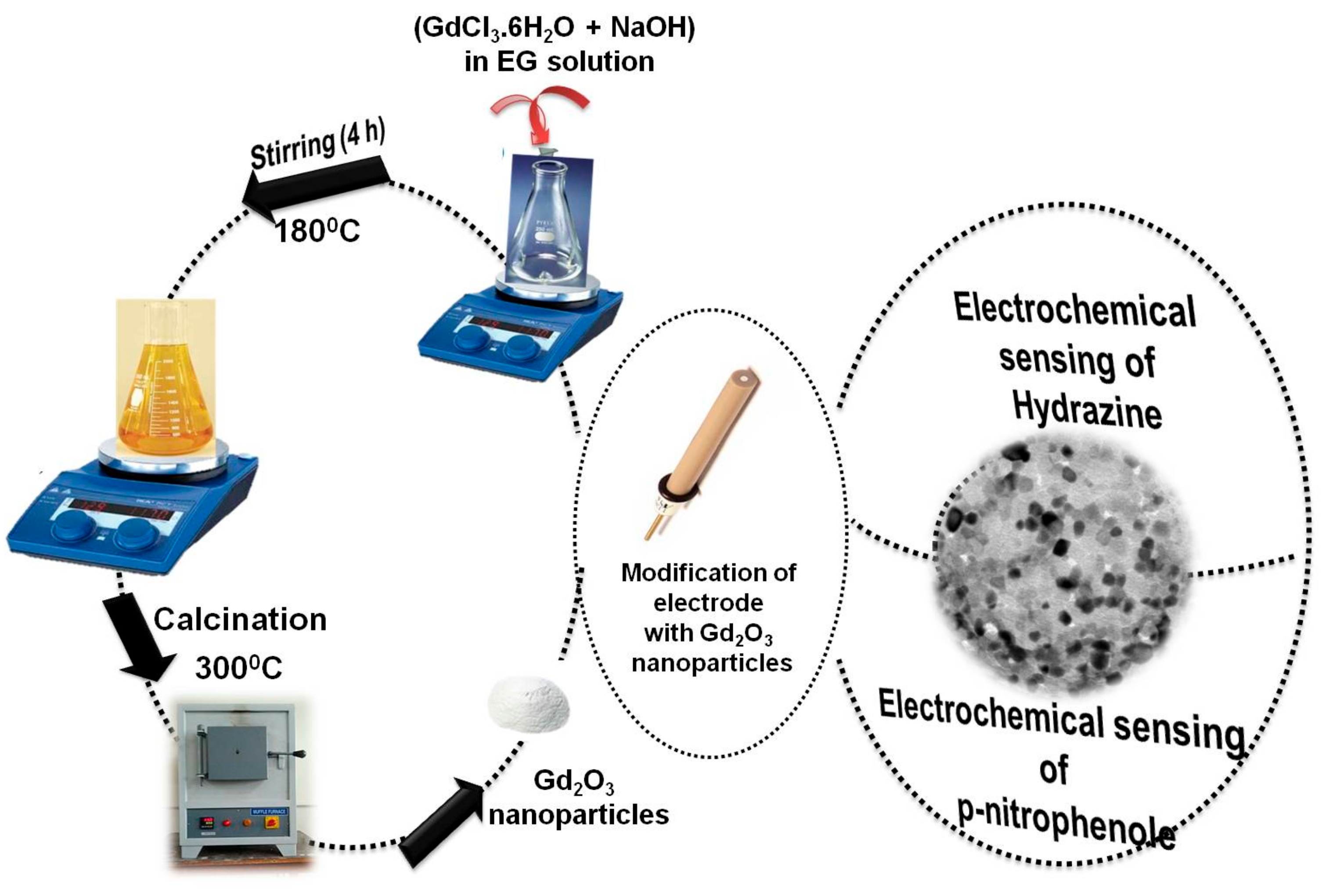
2.2. Synthesis of Gd2O3 Nanoparticles

The synthesis of Gd2O3 nanoparticles was done by using the hydrothermal method. In brief, 5 mM of GdCl3·6H2O was added to the 5-mL EG solution under stirring at 50 °C solution followed by the addition of 15 mM NaOH. The temperature of the reaction mixture was held constant at 140 °C for the first hour, and then raised to 180 °C for 4 h. The obtained solution was allowed to cool at room temperature. The obtained yellow precipitates of Gd2O3 nanoparticles were separated out from the reaction media. The obtained precipitates were subjected to calcinations for 3 h at 300 °C. The resulting particles were washed with water, acetone, and ethanol to remove the impurities. The corresponding separation was mainly performed by ultracentrifugation at 9000 rpm. The extracted particles were dried in an oven at 50 °C and further utilized for different analysis. For the optimization of synthetic parameters for the preparation of Gd2O3 nanoparticles, the respective concentration variations of GdCl3·6H2O have been carried out under respective reaction conditions. In the first instance, the concentration of variations of GdCl3·6H2O were done from 5 to 25 mM in all the reaction mixtures by keeping the concentration of the NaOH fixed at 0.015 M. A UV-visible spectral scan for each sample was taken from 230 to 400-nm wavelength to detect the optical properties of the formed nanoparticles. The optical band gap (Eg) of as-prepared nanoparticles was calculated as a function of the concentration variations of GdCl3·6H2O. The respective fluorescence emission spectra were also studied for the concentration variations of GdCl3·6H2O from 5 to 25 mM.

2.3. Electrode Preparation

The synthesized nanoparticles were further used to fabricate the electrochemical sensor for hydrazine and p-nitrophenol (Scheme 1). In order to form the modified electrode, the gold electrode with a surface area equivalent to 3.14 mm2 was first cleaned with alumina slurry and properly washed with distilled water under sonication. After drying the electrode at room temperature, the surface of the gold electrode was coated with the Gd2O3 nanoparticles by using butyl carbitol acetate (BCA) as the binding agent. The as-formed electrode was further dried at 60 ± 5 °C for 4–6 h to attain a homogeneous and dried layer of nanoparticles over the surface of electrode. All the electrochemical measurements were carried out on an μAutolab Type-III under neutral pH conditions. In all the analyses, a Gd2O3 customized gold electrode was acting as a working electrode, Ag/AgCl (sat. KCl) was acting as a reference, and Pt wire was acting as a counter electrode.

2.4. Physical Measurements

The obtained Gd2O3 nanoparticles were characterized with the help of an X-ray diffractometer from Panalytical D/Max-2500 (Malvern, UK), Hitachi (H-7500) Transmission electron microscope (Tokyo, Japan), Thermoscientific UV-vis. Spectrophotometer (Waltham, MA, USA), and FTIR spectrophotometer of Perkin-Elmer (RX1) (Waltham, MA, USA). The fluorescence measurements were carried out with a Hitachi F-7000 photoluminescence spectrophotometer. The photoluminescence analysis was carried out on an Edinburgh Instrument FLS 980 (Bain Square, UK). A JEOL (JSM-6610) scanning electron microscope (SEM) (Tokyo, Japan) with EDX analysis was carried out at 20 kV. The calcinations of the as-prepared Gd2O3 nanoparticles were done in an AICIL muffle furnace at 300 °C. Dynamic light scattering measurements were done on a Malvern Zen1690 instrument (Worcestershire, UK). Raman analysis was performed on a Renishaw inVia reflex micro-Raman spectrometer (Wotton-under-Edge, UK). The pH measurements were performed on a Mettler Toledo digital pH meter (Columbus, OH, USA). The surface area of EG-coated Gd2O3 nanoparticles was estimated by using Brunauer–Emmett–Teller (BET) analysis with an N2 adsorption analyzer (NOVA 2000e, Anton Par, Gurugram, India). The separation of the as-prepared Gd2O3 nanoparticles from aqueous media was done on a Remi R-24 centrifuge. Electrochemical measurements were carried out on an μAutolab Type-III cyclic voltammeter (Metrohm, Herisau, Switzerland). The gold electrode with a surface area of 3.14 mm2 was chosen for the analysis.

3. Results and Discussion

3.1. Characterization and Properties of Synthesized Gd2O3 Nanoparticles

The crystal structure of formed Gd2O3 nanoparticles has been further scrutinized by investigating the powdered XRD patterns of formed particles (Figure 1a). The absence of any peak related to impurity has confirmed the purity of the prepared nanoparticles [30]. The average crystallite size (D) of 15 nm has been estimated from the diffraction peaks by using the respective values of full width at half maximum (FWHM) via employing Debye–Scherrer’s formula [31,32]. The specific surface area and pore diameter of the obtained sample was found to be 15.3 m2·g−1 and 2.3 nm, which were respectively calculated by using the nitrogen sorption studies at 77 K in accordance with the BET (Brunauer–Emmett–Teller) process. The respective atomic content of Gd2O3 nanoparticles has been confirmed by using EDX analysis (Figure 1b). The obtained spectrum has only displayed the characteristic peaks of the Gd and O atoms in the synthesized sample, which verified the purity of the formed particles.
The SEM image of the Gd2O3 nanoparticles has been shown in Figure 1c, which has clearly shown the presence of agglomerated nanostructures of Gd2O3 nanoparticles. The presence of contacted particles has been mainly due to the existence of an EG template over the surface of the particles, which has further supported the presence of external electrostatic interactive forces generated from the templates over the surface of the nanoparticles. Detailed information regarding the morphology and structure of Gd2O3 nanoparticles has further been obtained from the HRTEM images presented in Figure 1d. It is clear from the images that the nanoparticles have shown crystallites with an irregular pseudo-spherical shape and a size distribution between 7–15 nm. The crystal spacing of 0.305 nm belongs to the (222) planes of Gd2O3 nanoparticles (Figure 1e,f). The observed result is in good agreement with the reported literature [33]. The presence of the connected nanocrystals has been attained due to the existence of a diverse range of forces (electrostatic, hydrogen bonding, and van der Waals forces) provided by the presence of external templates of EG coating over Gd2O3 nanoparticles.
The optical properties of formed Gd2O3 nanoparticles have been investigated by using UV-vis. and fluorescence analysis as a function of variation of the concentration of GdCl3·6H2O salt during the synthesis (Figure 2a,b). The formed particles have shown the characteristic peak between 255–262 nm. The respective peak has been associated with the electronic transition from 8S7/26I7/2. [34]. On interpreting the results, it has been found that the absorbance is dependent on the concentration of salt.
With increase in the concentration from 5 to 20 mM, there has an increment in the absorbance of around 63%. The change in the peak position was not so prominent with the concentration of the salt. On the other hand, a fluorescence emission peak was observed at 336 nm with λexc = 290 nm (Figure 2b). This peak has been associated with the emission from 6P7/28S7/2 in Gd(III) ions [35]. The enhancement in the concentration of the starting material has produced an increment of 50.7% in intensity value, whereas the peak position has only shown a change of 10 nm. This variation has displayed the similarity with UV-vis studies. The PL spectra of as-synthesized nanoparticles has shown the characteristic peaks at 390 nm and a broad peak between 400–500 nm with a center at 417 nm (λexc = 350 nm) (inset Figure 2b). The sharp peak at 390 nm has been associated with the radiative recombination of holes with electrons present at the oxygen vacant positions formed due to the photogeneration effect. The other peak was associated with the self-trapped exciton luminescence in formed particles [36]. The optical band-gap values (Eg) and agglomeration number for Gd2O3 nanoparticles (Figure 2c) have been estimated by using the application of the Brus method [37]. The results have clearly explained the behavioral variation of band-gap value with the concentration of GdCl3·6H2O salt during the synthesis. The decrease in the value of the band gap with the concentration has been associated with the variation of size of the formed particles with the concentration of the starting material. The particle size was comparatively higher for the nanoparticles prepared with 25 mM GdCl3·6H2O salt. These variations of starting material have directly influenced the size of the particles, and the respective agglomeration number of the particles has also varied in a similar manner. Figure 2d has displayed the respective assignment of their colors in the Commission Internationale de L’Eclairage (CIE) diagram for the Gd2O3 nanoparticles. The respective value of CIE chromaticity coordinates is found to be x = 0.3265, y = 0.4462, respectively. The outcomes have been associated with the green-shift effect in the formed particles. The formed particles have been further characterized by using the Raman and FTIR spectra of Gd2O3 nanoparticles (Figure 2e,f). On interpretation, Gd2O3 nanoparticles displayed four major Raman peaks at 444.1, 528, 637.8 and 767.4 cm−1, respectively (Figure 2e). These peaks are mainly associated with the Fg and Ag mode for cubic C-type Gd2O3 particles [38]. Moreover, other Raman active modes such as 4Ag, 4Eg, and 14Fg have also contributed toward the peaks in the spectra [39,40]. A sharp IR peak has been detected below 500 cm−1, which has been attributed to Gd–O [40]. The small peak at 1500 cm−1 has been associated with the δ(O–H) vibrations due to the water bound to the nanoparticles surface in the form of moisture [41,42,43,44].

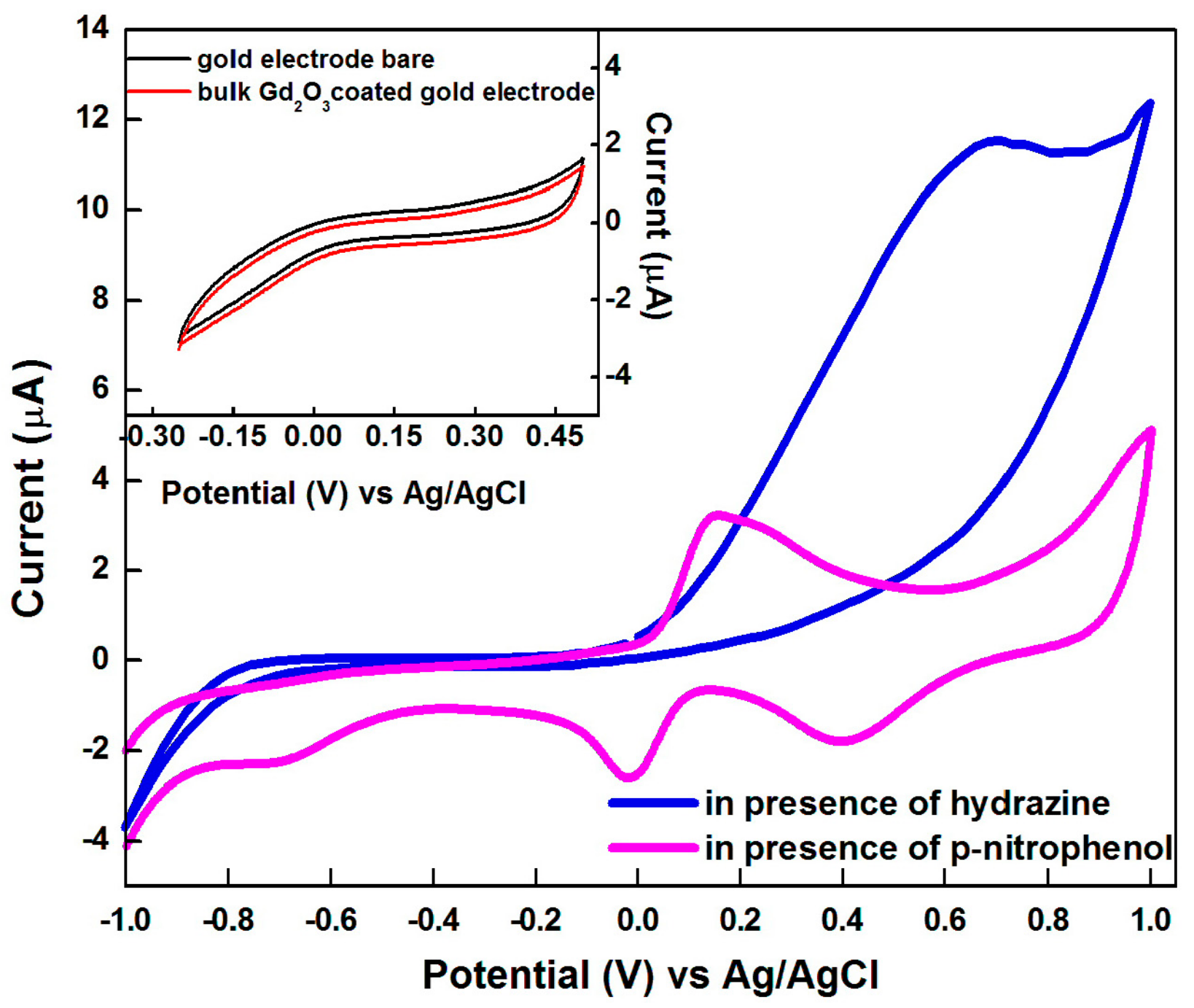
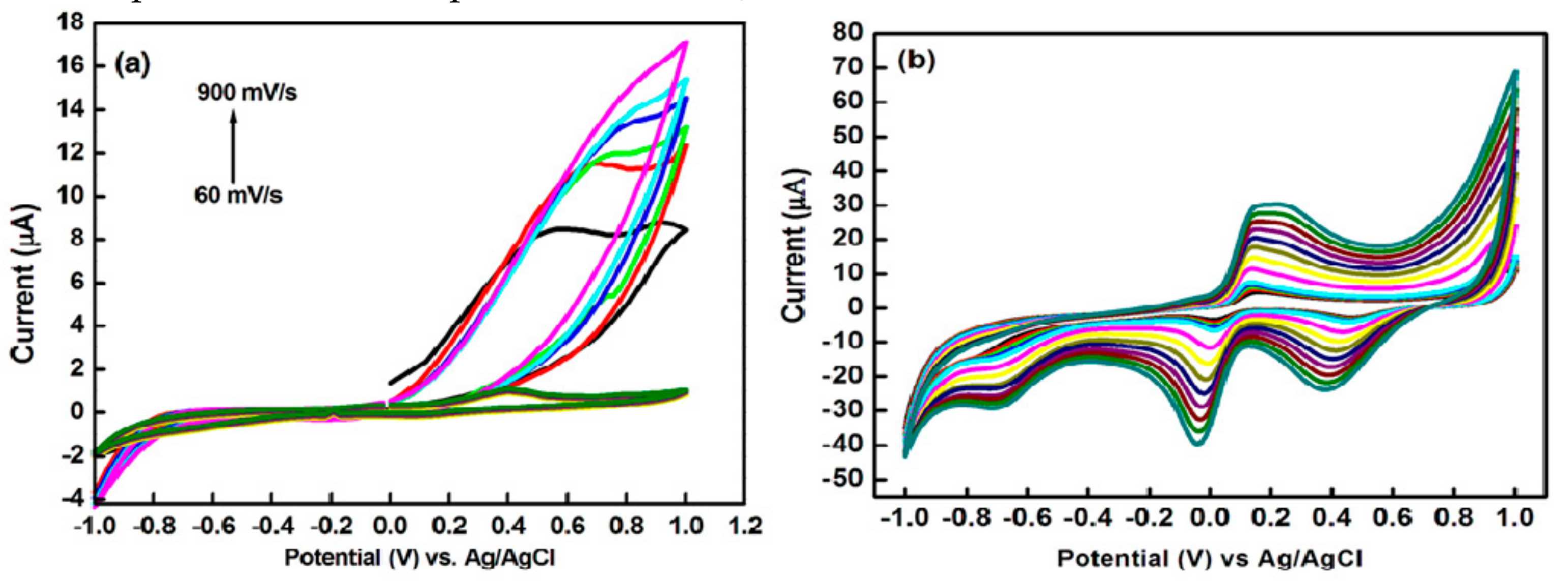
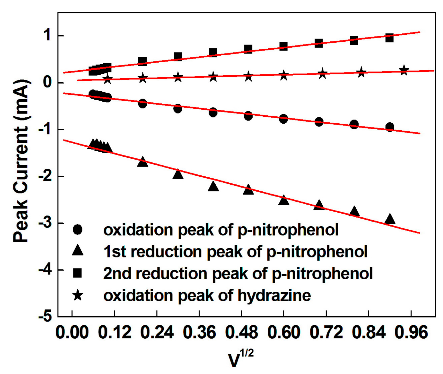
3.2. Electrochemical Behaviour of Hydrazine and p-Nitrophenol on Modified Electrode

The electrochemical action of formed Gd2O3 nanoparticles has been investigated toward the electrocatalysis of hydrazine and p-nitrophenol (PNP) via using cyclic voltammetric analysis. Figure 3 showed the cyclic voltammograms of a gold electrode in pH 7.0, phosphate buffer (PBS) under various electrode conditions. Interestingly, it has been found that the bare gold electrode does not show any signal in pH 7 PBS buffer. The response has been still negligible for bulk Gd2O3-coated gold electrodes in the presence of hydrazine and p-nitrophenol (Figure 3). On the other hand, well-defined voltammetric signals at 0.68 V have been obtained for the electrocatalysis of hydrazine in the presence of a Gd2O3 nanoparticles-coated gold electrode. In case of p-nitrophenol, one set of reversible redox peaks i.e., (GdR1), oxidation (GdO1) occurred at −0.021 and 0.163 V. Other irreversible reduction peaks (GdR2) at −0.694 V and (GdR3) at 0.4 V were also observed in phosphate buffer solution at pH 7 with a scan rate of 60 mV/s. The obtained results have clearly pointed out that the p-nitrophenol has displayed three types of electrochemical responses with two subsequent types of processes, including reduction and redox couple progression [21]. This might be aroused due to the two-electron oxidation reduction reaction in 4-aminophenol. Whereas, the reduction peaks were associated with the formation of a hydroxylamine group from the nitro group of p-nitrophenol.
From Figure 3, it was found that there has been no signal in the reverse sweep for hydrazine samples. The results have confirmed the irreversible nature of the oxidation process for hydrazine. On other hand, the samples of p-nitrophenol have displayed the redox peaks in both forward and backward directions, which make the analysis of p-nitrophenol reversible in nature [45]. In the presence of Gd2O3 nanoparticles, there has been found a significant increase in the peak current for respective analytes. This has clearly verified the utilities of formed nanoparticles for electro-analytical purposes. The enhancement of peak current has been mainly explained by the enhancement of conductivity of the electrode due to the functionalization of the gold electrode surface with Gd2O3 nanoparticles. These results have further validated that the formed Gd2O3 nanoparticles are capable as efficient electron transporters for the electrocatalysis of harmful pollutants.
The scan rate variations have also been carried out in order to investigate the electron transfer mechanism for a Gd2O3 modified gold electrode in the presence of hydrazine and p-nitrophenol. Figure 4a,b shows the typical voltammograms for a Gd2O3 nanoparticles-coated gold electrode with 1 mM solution of respective analyte (i.e., hydrazine and p-nitrophenol) in 0.1 M PBS solution with pH = 7.0 at different scan rates. The spectrum has revealed a regular enhancement of current response with varying the scan rate from 60 to 900 mV/S for both the analytes. The higher surface area of Gd2O3 nanoparticles has played a critical role for the improved electron transfer process for the catalytic performance toward the understudied analytes. The enhancement of peak current with the scan rate has clearly pointed out the electrocatalytic reaction of hydrazine and p-nitrophenol at the surface of the modified electrode. These current variations have been mainly explained by the surface adsorption to diffusion processes at a façade of modified electrodes [45].
At lower scan rates, the relative rate of diffusion is quick, and the adsorption of analyte has been found to be slowest during the mass transfer process. On the other hand, at higher scan rates, the diffusion step has been mainly controlling the rate of electrode reactions in the presence of external agents [46]. In the case of p-nitrophenol, both types of associated processes i.e., the reduction and redox couple processes, have shown the significant augmentation of current response as a function of scan rate. The respective calibration curve of peak current versus the square root of the scan rate has displayed a linear relation for hydrazine and p-nitrophenol (Figure 5). These obtained results have clearly pointed out that the oxidation of hydrazine is mainly a diffusion-controlled process at the surface of a Gd2O3 nanoparticles-modified gold electrode [47].
The number of electrons involved in the overall reaction (n) for hydrazine and p-nitrophenol has been calculated from the Randles–Sevcik equation mentioned below [45].
i p = 2.69 × 10 5 n 3 2 A D 1 2 v 1 2 C
where n is the number of electron equivalents exchanged during the redox process, A (cm2) is the active area of the working electrode, D (cm2·s−1) and C (mol·cm−3) are the diffusion coefficient and the bulk concentration of hydrazine, and v is the voltage scan rate (V·s−1). The obtained CV responses of Gd2O3/Au have clearly explained that the oxidation of N2H4 involves two electron changes, and the irreversible reaction of p-nitrophenol has undergone four electron changes, while the reversible reaction involves two processes—an electron redox process and an irreversible process—and gives a large reduction peak. (Figure 4b) shows the reduction of PNP to 4-(hydroxyamino) phenol. Two coupled redox peaks have indicated the oxidation of 4-(hydroxyamino) phenol to 4-nitrosophenol and the succeeding reversible reduction [48,49,50,51]. A schematic representation of the involved mechanism has been illustrated in Scheme 2.

3.3. Amperometric Responses for Hydrazine and p-Nitrophenol

The amperometric studies are one of the primary techniques to estimate the low concentration of analytes and carry out the relative studies in the presence of interfering analytes. Since the Gd2O3@Au-modified electrode has displayed the higher current response for hydrazine and p-nitrophenol as a model system in the cyclic voltammetric studies, therefore, it has been employed as the amperometric sensor for the detection of hydrazine and p-nitrophenol at low concentration levels.
Figure 6 and Figure 7 depict the amperometric response of Gd2O3@Au for the successive additions of hydrazine and p-nitrophenol at an applied potential of −0.694 and 0.640 V for p-nitrophenol and hydrazine, respectively, in 0.1 M buffer solution with pH = 7. The obtained values of the current response have been estimated after the consecutive injection of 1 µM concentration of hydrazine and p-nitrophenol at the time interval of 60 s in a continuously stirring condition. The Gd2O3@Au-modified electrode has exhibited a considerable and rapid amperometric reaction toward each addition of analyte.
The value of the current has reached its stable position within 3 s, demonstrating the fast electro-oxidation of the understudied analyte at the surface of the Gd2O3@Au-modified electrode. In addition, the response current has shown a linear increment for the subsequent additions of analyte over the wide range of concentrations. The respective regression plot of current response versus concentration of both the analytes has displayed a linear relation with the correlation coefficient values of 0.987 and 0.996 for p-nitrophenol and hydrazine, respectively (Figure 6b and Figure 7b). The limit of detection value has been calculated to be 1.527 and 0.704 µM for p-nitrophenol and hydrazine, respectively, by using the equation limit of detection (LOD) = 3σ/slope, where σ is the standard deviation for the particular system [52]. The sensitivity of the developed sensor has been found to be 0.33722 and 0.25734 mA·mM−1 from the slope of the linear regression for hydrazine and p-nitrophenol, respectively. The respective reusability, stability, and reproducibility of the as-prepared sensor has also been tested in the current work. The as-developed electrode was kept in the buffer media for one month, and its electrocatalytic efficiency has also been tested against the p-nitrophenol and hydrazine. The results have clearly verified that the formed sensor has displayed a reproducible performance with a decay rate of 5.7% and 6.3% in the oxidation peak current value for p-nitrophenol and hydrazine, respectively. This substantial constancy in results has been further attributed to the stability of Gd2O3 particles, which maintain the efficiency and performance of the electrode for a long period. Additionally, the reproducibility of the developed sensor has been confirmed by estimating the electrochemical response of the Gd2O3 particles as a working electrode for different electrodes in the solution media containing 1 mM of p-nitrophenol and hydrazine.
The obtained relative standard deviation (RSD) of peak currents has been found to be 5.3% and 4.7%, signifying the satisfactory report for the reproducibility of the modified electrode. In order to investigate the reusability of the developed sensor, the as-modified electrode has been rinsed with the respective sample solution. The obtained signal has been tested after the rinsing. It has been found that the obtained signal has maintained 94% of its original strength. The analytical performance of the as-modified electrode has been evaluated with some recent works in Table 1.
The data has further confirmed the authenticity of the developed sensor in a different range of concentrations with greater selectivity and sensitivity. In order to investigate the application of the formed sensor in real samples, the water samples from different sources have been taken, and the respective analyses have been made for the detection of hydrazine and p-nitrophenol. The relevant stock solutions of understudied analytes of known concentrations were made by using the water samples taken from different sources. The current response of the sensor has been examined for the different concentrations of hydrazine and p-nitrophenol by using amperometric studies. The outcomes of the measurements have shown the excellent recovery rate for the chosen analytes, as indicated in Table 2.
The sensitivity, selectivity, repeatability, recyclability, wide linear range, detection limit, and applicability in real water samples makes Gd2O3 Nps a favorable nanomaterial in the institution of the rapid and effectual scrutiny of harmful environmental pollutants. Thus, the prepared sensor appears to be a probable contender for preparing a simple, fast, and economical electrochemical sensor.

3.4. Selectivity Study

In order to apply the proposed sensor for the determination of p-nitrophenol and hydrazine in an aqueous system, the respective selectivity of the sensor has been investigated in the presence of 1 mM of various interfering compounds (benzaldehyde, benzoic acid, benzonitrile, phenol, ethanol, and aniline) at a fixed potential 0.67 V for hydrazine and −0.69 for PNP. Figure 8 has displayed the amperometric response of a Gd2O3/BCA/Au electrode for the same. From the data, it has been found that a negligible change to response current has been detected in the presence of different interfering compounds. However, significant and quick responses were observed in the presence of hydrazine and PNP. These outcomes have clearly explained the high selectivity of the fabricated sensor toward hydrazine and PNP, and enhanced the scope of the developed sensor.

4. Conclusions

In summary, the current work has reported the fabrication of the ethylene glycol-mediated synthesis of Gd2O3 Nps. The formation of the particles has been scrutinized by using sophisticated characterization techniques. The effects of synthetic parameters over the optical, photoluminescence, band-gap variation, and agglomeration number of the formed particles have been studied in detail. The fabricated particles have further been employed as a proficient electrocatalytic material for the enzyme-free detection of hydrazine and p-nitrophenol with great sensitivity and selectivity. The developed sensors hold a wider linear range of 1 to 10 µM with low detection limits of 1.527 µM and 0.704 µM for p-nitrophenol and hydrazine, respectively. The sensitivity, selectivity, repeatability, recyclability, wide linear range, and detection limit make Gd2O3 Nps a favorable nanomaterial in the institution of the rapid and effectual scrutiny of harmful environmental pollutants. The realistic application of the developed sensor has also been investigated by spiking known concentrations of hydrazine and p-nitrophenol in different water samples with good recoveries. Consequently, the successful synthesis of EG@Gd2O3 nanoparticles has immense potential for the design of highly effective electrochemical sensors, and is a probable way to provide momentum to the advancement of new electrode materials.

Author Contributions

S.C. for methodology, validation, writing; S.K. (Sandeep Kumar) and S.K. (Sushil Kumar) for formal analysis; A.U., G.R.C. and S.K.M. for data curation.

Funding

This research was funded by DST Inspire Faculty award [IFA-CH-17] and DST purse grant II. Sandeep Kumar is grateful to CSIR India for providing a senior research fellowship. Ahmad Umar would like to acknowledge the Ministry of Education, Saudi Arabia for this research through a grant (PCSED-013-18) under the Promising Centre for Sensors and Electronic Devices (PCSED) at Najran University, Kingdom of Saudi Arabia.

Conflicts of Interest

The authors declare no conflict of interest.

References

  1. Li, J.; Kuang, D.; Feng, Y.; Zhang, F.; Xu, Z.; Liu, M. A graphene oxide-based electrochemical sensor for sensitive determination of 4-nitrophenol. J. Hazard Mater. 2012, 201, 250–259. [Google Scholar] [CrossRef] [PubMed]
  2. Nam, G.; Leem, J.-Y. A new technique for growing ZnO nanorods over large surface areas using graphene oxide and their application in ultraviolet sensors. Sci. Adv. Mater. 2018, 10, 405–409. [Google Scholar] [CrossRef]
  3. Vernot, E.H.; MacEwen, J.D.; Bruner, R.H.; Haus, C.C.; Kinkead, E.R.; Prentice, D.E.; Hall, A., III; Schmidt, R.E.; Eason, R.L.; Hubbard, G.B.; et al. Long-term inhalation toxicity of hydrazine. Fundam. Appl. Toxicol. 1985, 5, 1050–1064. [Google Scholar] [CrossRef]
  4. Savafi, A.; Karimi, M.A. Flow injection chemiluminescence determination of hydrazine by oxidation with chlorinated isocyanurates. Talanta 2002, 58, 785–792. [Google Scholar] [CrossRef]
  5. Safavi, A.; Ensafi, A.A. Electrocatalytic oxidation of hydrazine with pyrogallol red as a mediator on glassy carbon electrode. Anal. Chem. Acta 1995, 300, 307–311. [Google Scholar] [CrossRef]
  6. Chen, H.; Li, D.; Ding, W. Template-less preparation of meso-MnO2 fibers by localized ostwald ripening and 2,4-dinitrophenol adsorption mechanism. Sci. Adv. Mater. 2018, 10, 1241–1249. [Google Scholar] [CrossRef]
  7. U.S. Department of Homeland Security. Homeland Security Information Bulletin; U.S. Department of Homeland Security: Washington, DC, USA, 2003.
  8. Shukla, S.; Chaudhary, S.; Umar, A.; Chaudhary, G.R.; Mehta, S.K. Dodecyl ethyl dimethyl ammonium bromide capped WO3 nanoparticles: Efficient scaffolds for chemical sensing and environmental remediation. Dalton Trans. 2015, 44, 17251–17260. [Google Scholar] [CrossRef]
  9. Collins, G.E.; Rose-Pehrsson, S.L. Sensitive, fluorescent detection of hydrazine via derivatization with 2,3-naphthalene dicarboxaldehyde. Analytica Chimica Acta 1993, 284, 207–215. [Google Scholar] [CrossRef]
  10. Liu, X.; Yang, Z.; Sheng, Q.; Zheng, J. One-pot synthesis of Au-Fe3O4-GO nanocomposites for enhanced electrochemical sensing of hydrazine. J. Electrochem. Soc. 2018, 165, B596–B602. [Google Scholar] [CrossRef]
  11. Gao, Y.; Zhang, S.; Hou, W.; Guo, H.; Li, Q.; Dong, D.; Wu, S.; Zhao, S.; Zhang, H. Perylene diimide derivative regulate the antimony sulfide morphology and electrochemical sensing for hydrazine. Appl. Surf. Sci. 2019, 491, 267–275. [Google Scholar] [CrossRef]
  12. Guo, W.; Xia, T.; Zhang, H.; Zhao, M.; Wang, L.; Pei, M.A. Molecularly imprinting electrosensor based on the novel nanocomposite for the detection of tryptamine. Sci. Adv. Mater. 2018, 10, 1805–1812. [Google Scholar] [CrossRef]
  13. Annalakshmi, M.; Balasubramanian, P.; Chen, S.-M.; Chen, T.-W. One pot synthesis of nanospheres-like trimetallic NiFeCo nanoalloy: A superior electrocatalyst for electrochemical sensing of hydrazine in water bodies. Sens. Actuators B 2019, 296, 126620. [Google Scholar] [CrossRef]
  14. Gu, D.; Dong, Y.-M.; Liu, H.; Ding, H.-Y.; Shang, S.-M. Efficient synthesis of highly fluorescence nitrogen doped carbon dots and its application for sensor and cell imaging. Sci. Adv. Mater. 2018, 10, 964–973. [Google Scholar] [CrossRef]
  15. Mishra, R.K.; Martin, A.; Nakagawa, T.; Barfidokht, A.; Lu, X.; Sempionatto, J.R.; Lyu, K.M.; Karajic, A.; Musameh, M.M.; Kyratzis, I.L.; et al. Detection of vapor-phase organophosphate threats using wearable conformable integrated epidermal and textile wireless biosensor systems. Biosens. Bioelectron. 2018, 101, 227–234. [Google Scholar] [CrossRef] [PubMed]
  16. Mishra, R.K.; Hubble, L.J.; Martin, A.; Kumar, R.; Barfidokht, A.; Kim, J.; Musameh, M.M.; Kyratzis, I.L.; Wang, J. Wearable flexible and stretchable glove biosensor for on-site detection of organophosphorus chemical threats. ACS Sen. 2017, 2, 553–561. [Google Scholar] [CrossRef] [PubMed]
  17. Wei, Y.; Kong, L.T.; Yang, R.; Wang, L.; Liu, H.; Huang, X.J. Single-walled carbon nanotube/pyrene cyclodextrin nanohybrids for ultrahighly sensitive and selective detection of p-nitrophenol. Langmuir 2011, 27, 10295–10301. [Google Scholar] [CrossRef]
  18. Karthik, R.; Chen, S.M.; Elangovan, A.; Muthukrishnan, P.; Shanmugam, R.; Lou, B.S. Phyto mediated biogenic synthesis of gold nanoparticles using Cerasus serrulata and its utility in detecting hydrazine, microbial activity and DFT studies. J. Colloid Int. Sci. 2016, 468, 163–175. [Google Scholar] [CrossRef]
  19. Zhang, R.; Sun, C.L.; Lu, Y.J.; Chen, W. Graphene nanoribbon-supported Pt-Pd concave nanocubes for electrochemical detection of TNT with high sensitivity and selectivity. Anal. Chem. 2015, 87, 12262–12269. [Google Scholar] [CrossRef]
  20. Chen, X.; Cheng, X.; Gooding, J.J. Detection of trace nitroaromatic isomers using Indium Tin oxide electrodes modified using β-cyclodextrin and silver nanoparticles. Anal. Chem. 2012, 84, 8557–8563. [Google Scholar] [CrossRef]
  21. Chaudhary, S.; Kumar, S.; Mehta, S.K. Glycol modified gadolinium oxide nanoparticles as a potential template for selective and sensitive detection of 4-nitrophenol. J. Mater. Chem. C 2015, 3, 8824–8833. [Google Scholar] [CrossRef]
  22. Du, G.; Tendeloo, G.V. Preparation and structure analysis of Gd(OH)3 nanorods. Nanotechnology 2015, 16, 595–597. [Google Scholar] [CrossRef]
  23. Mehta, S.K.; Chaudhary, S.; Bhasin, K.K. Understanding the role of hexadecyltrimethylammonium bromide in the preparation of selenium nanoparticles: A spectroscopic approach. J. Nanopart. Res. 2009, 11, 1759–1766. [Google Scholar] [CrossRef]
  24. Ballem, M.A.; Söderlind, F.; Nordblad, P.; Kall, P.O.; Oden, M. Growth of Gd2O3 nanoparticles inside mesoporous silica frameworks. Microporous Mesoporous Mater. 2013, 168, 221–224. [Google Scholar] [CrossRef]
  25. Rahman, A.T.M.; Vasilev, K.; Majewski, P. Ultra small Gd2O3 nanoparticles: Absorption and emission properties. J. Colloid Inter. Sci. 2011, 354, 592–596. [Google Scholar] [CrossRef]
  26. Jia, G.; You, H.P.; Liu, K.; Zheng, Y.H.; Guo, N.; Zhang, H.J. Highly uniform Gd2O3 hollow microspheres: Template-directed synthesis and luminescence properties. Langmuir 2010, 26, 5122–5128. [Google Scholar] [CrossRef]
  27. Hu, L.; Ma, R.; Ozawa, T.C.; Sasaki, T. Oriented monolayer film of Gd2O3:0.05Eu crystallites: Quasi-topotactic transformation of the hydroxide film and drastic enhancement of photoluminescence properties. Angew. Chem. Int. Ed. 2009, 48, 3846–3849. [Google Scholar] [CrossRef] [PubMed]
  28. Manigandan, R.; Giribabu, K.; Suresh, R.; Vijayalakshmi, L.; Stephen, A.; Narayanan, V. Structural, optical and magnetic properties of gadolinium sesquioxide nanobars synthesized via thermal decomposition of gadolinium oxalate. Mater. Res. Bull. 2013, 48, 4210–4215. [Google Scholar] [CrossRef]
  29. Abdullah, M.M.; Rahman, M.M.; Bouzid, H.; Faisal, M.; Khan, S.B.; Al-Sayari, S.A.; Ismail, A.A. Sensitive and fast response ethanol chemical sensor based on as-grown Gd2O3 nanostructures. J. Rare Earths 2015, 33, 214–220. [Google Scholar] [CrossRef]
  30. Chaudhary, S.; Kumar, S.; Umar, A.; Singh, J.; Rawat, M.; Mehta, S.K. Europium-doped gadolinium oxide nanoparticles: A potential photoluminescencent probe for highly selective and sensitive detection of Fe 3+ and Cr 3+ ions. Sens. Actuators B 2017, 243, 579–588. [Google Scholar] [CrossRef]
  31. Mehta, S.K.; Chaudhary, S.; Kumar, S.; Bhasin, K.K.; Torigoe, K.; Sakai, H.; Abe, M. Surfactant assisted synthesis and spectroscopic characterization of selenium nanoparticles in ambient condition. Nanotechnology 2008, 19, 295601–295612. [Google Scholar] [CrossRef]
  32. Chaudhary, S.; Sharma, P.; Kumar, R.; Mehta, S.K. Nanoscale surface designing of cerium oxide nanoparticles for controlling growth, stability, optical and thermal properties. Ceram. Int. 2015, 41, 10995–11003. [Google Scholar] [CrossRef]
  33. Maalej, N.M.; Qurashi1, A.; Assadi, A.A.; Maalej, R.; Shaikh, M.N.; Ilyas, M.; Gondal, M.A. Synthesis of Gd2O3:Eu nanoplatelets for MRI and fluorescence imaging. Nanoscale Res. Lett. 2015, 10, 215. [Google Scholar] [CrossRef] [PubMed]
  34. Mukherjee, S.; Dasgupta, P.; Jana, P.K. Size-dependent dielectric behaviour of magnetic Gd2O3 nanocrystals dispersed in a silica matrix. J. Phys. D Appl. Phys. 2008, 41, 215004–215015. [Google Scholar] [CrossRef]
  35. Rogow, D.L.; Swanson, C.H.; Oliver, A.G.; Oliver, S.R.J. Two related Gadolinium aquo carbonate 2-D and 3-D structures and their thermal, spectroscopic, and paramagnetic properties. Inorg. Chem. 2009, 48, 1533–1541. [Google Scholar] [CrossRef] [PubMed]
  36. Zhang, Y.; Han, K.; Cheng, T.; Fang, Z. Synthesis, characterization, and photoluminescence property of LaCO3OH microspheres. Inorg. Chem. 2007, 46, 4713–4717. [Google Scholar] [CrossRef] [PubMed]
  37. Chaudhary, S.; Umar, A. Glycols functionalized fluorescent Eu2O3 nanoparticles: Functionalization effect on the structural and optical properties. J. Alloys Compd. 2016, 682, 160–169. [Google Scholar] [CrossRef]
  38. Rajan, G.; Gopchandran, K.G. Enhanced luminescence from spontaneously ordered Gd2O3: Eu3+ based nanostructures. Appl. Surf. Sci. 2009, 255, 9112–9123. [Google Scholar] [CrossRef]
  39. White, W.B.; Keramidas, V.G. Vibrational spectra of oxides with the C-type rare earth oxide structure. Spectrochim. Acta Part A 1972, 28, 501–509. [Google Scholar] [CrossRef]
  40. Riri, M.; Benjjar, A.; Eljaddi, T.; Sefiani, N.; Touaj, K.; Cherif, A.; Hlaїbi, M. Characterization of two dinuclear complexes of the gadolinium ion by IR and Raman. J. Mater. Environ. Sci. 2013, 4, 961–966. [Google Scholar]
  41. Coats, A.W.; Redfern, J.P. Kinetic parameters from thermogravimetric data. Nature 1964, 201, 68–69. [Google Scholar] [CrossRef]
  42. Madhusudanan, P.M.; Krishnan, K.; Ninan, K.N. New approximation for the p(x) function in the evaluation of non-isothermal kinetic data. Thermochimica Acta 1986, 97, 189–201. [Google Scholar] [CrossRef]
  43. Tang, W.; Liu, Y.; Zhang, H.; Wang, Z.; Wang, C. New temperature integral approximate formula for non-isothermal kinetic analysis. J. Therm. Anal. Calorim. 2003, 74, 309–315. [Google Scholar] [CrossRef]
  44. Horowitz, H.H.; Metzger, G.A. A new analysis of thermogravimetric traces. Anal. Chem. 1963, 35, 1464–1468. [Google Scholar] [CrossRef]
  45. Singh, K.; Kaur, A.; Umar, A.; Chaudhary, G.R.; Singh, S.; Mehta, S.K. A comparison on the performance of zinc oxide and hematite nanoparticles for highly selective and sensitive detection of para-nitrophenol. J. Appl. Electrochem. 2015, 45, 253–261. [Google Scholar] [CrossRef]
  46. Raoof, J.B.; Ojani, R.; Beitollahi, H.; Hosseinzadeh, R. Electrocatalytic oxidation and highly selective voltammetric determination of L-cysteine at the surface of a 1-[4-(ferrocenyl ethynyl)phenyl]-1-ethanone modified carbon paste electrode. Anal. Sci. 2006, 22, 1213–1220. [Google Scholar] [CrossRef] [PubMed]
  47. Shahid, M.M.; Rameshkumar, P.; Basirunc, W.J.; Wijayantha, U.; Chiu, W.S.; Khiew, P.S.; Huang, N.M. An electrochemical sensing platform of cobalt oxide@gold nanocubes interleaved reduced graphene oxide for the selective determination of hydrazine. Electrochimica Acta 2018, 259, 606–616. [Google Scholar] [CrossRef]
  48. Liu, Z.; Du, J.; Qiu, C.; Huang, L.; Ma, H.; Shen, D.; Ding, Y. Electrochemical sensor for detection of p-nitrophenol based on nanoporous gold. Electrochem. Commun. 2009, 11, 1365–1368. [Google Scholar] [CrossRef]
  49. Ndlovu, T.; Arotiba, O.A.; Krause, R.W.; Mamba, B.B. Electrochemical detection of o-nitrophenol on a poly(propyleneimine)-gold nanocomposite modified glassy carbon electrode. Int. J. Electrochem. Sci. 2010, 5, 1179–1186. [Google Scholar]
  50. Shukla, S.; Chaudhary, S.; Umar, A.; Chaudhary, G.R.; Mehta, S.K. Tungsten oxide (WO3) nanoparticles as scaffold for the fabrication of hydrazine chemical sensor. Sens. Actuators B 2014, 196, 231–237. [Google Scholar] [CrossRef]
  51. De Oliveira, F.M.; Guedesa, T.d.; Lima, A.B.; da Silvaa, L.M.; dos Santos, W.T.P. Alternative method to obtain the Tafel plot for simple electrode reactions using batch injection analysis coupled with multiple-pulse amperometric detection. Electrochimica Acta 2017, 242, 180–186. [Google Scholar] [CrossRef]
  52. Wang, X.; Wang, Q.; Wang, Q.; Gao, F.; Gao, F.; Yang, Y.; Guo, H. Highly dispersible and stable copper terephthalate metal-organic framework-graphene oxide nanocomposite for an electrochemical sensing application. ACS Appl. Mater. Interfaces 2014, 6, 11573–11580. [Google Scholar] [CrossRef] [PubMed]
  53. Guerra, S.V.; Ubota, L.T.K.; Xavier, C.R.; Nakagaki, S. Experimental optimization of selective hydrazine detection in flow injection analysis using a carbon paste electrode modified with copper porphyrin occluded into zeolite cavity. Anal. Sci. 1999, 15, 1231–1234. [Google Scholar] [CrossRef]
  54. Salimi, A.; Miranzadeh, L.; Hallaj, R. Amperometric and voltammetric detection of hydrazine using glassy carbon electrodes modified with carbon nanotubes and catechol derivatives. Talanta 2008, 75, 147–156. [Google Scholar] [CrossRef] [PubMed]
  55. Salami, A.; Abdi, K. Enhancement of the analytical properties and catalytic activity of a nickel hexacyanoferrate modified carbon ceramic electrode prepared by two-step sol–gel technique: Application to amperometric detection of hydrazine and hydroxyl amine. Talanta 2004, 63, 475–483. [Google Scholar] [CrossRef]
  56. Zhao, Y.D.; Zhang, W.D.; Chen, H.; Luo, Q.M. Anodic oxidation of hydrazine at carbon nanotube powder microelectrode and its detection. Talanta 2002, 58, 529–534. [Google Scholar] [CrossRef]
  57. Umar, A.; Rahman, M.M.; Hahn, Y.B. ZnO nanorods based hydrazine sensors. J. Nanosci. Nanotechnol. 2009, 9, 4686–4691. [Google Scholar] [CrossRef]
Scheme 1. Schematic illustration of the electrochemical sensor for the detection of hydrazine and p-nitrophenol by using the modified electrode of gold with Gd2O3 nanoparticles.
Coatings 09 00633 sch001
Figure 1. Typical (a) XRD pattern, (b) EDS spectrum, (c) SEM image, (d) TEM image, and (e,f) HRTEM images of Gd2O3 nanoparticles.
Coatings 09 00633 g001
Figure 2. (a) UV-vis., (b) fluorescence and photoluminescence (PL) (inset) emission, (c) variation of bandgap and agglomeration number, (d) Commission Internationale de L’Eclairage (CIE) chromaticity analysis (e) Raman and (f) FTIR spectra of Gd2O3 nanoparticles.
Coatings 09 00633 g002
Figure 3. Cyclic voltammograms for a Gd2O3 nanoparticles-coated gold electrode under various electrode conditions in 0.1 M PBS (pH 7.0). The scan rate was 60 mV/s and the respective concentrations of analyte was kept constant at 1 mM.
Coatings 09 00633 g003
Figure 4. Cyclic voltammograms for a Gd2O3 nanoparticles-coated gold electrode for 1 mM (a) hydrazine and (b) p-nitrophenol at different scan rates ranging from 60 to 900 mV/s.
Coatings 09 00633 g004
Figure 5. The linear dependence of peak current versus square root of scan rate for 1 mM hydrazine and p-nitrophenol.
Coatings 09 00633 g005
Scheme 2. The pictorial representation and cyclic voltammetric sweep curves for the Gd2O3 nanoparticles/butyl carbitol acetate/gold (NPs/BCA/Au) electrode for the sensing of hydrazine and p-nitrophenol.
Coatings 09 00633 sch002
Figure 6. (a) Amperometric response of the Gd2O3/Au electrode with an increase in the concentration of hydrazine and (b) respective peak current vs. concentration plot of hydrazine in PBS at pH = 7.
Coatings 09 00633 g006
Figure 7. (a) Amperometric response of the Gd2O3/Au electrode with an increase in the concentration of p-nitrophenol and (b) respective peak current vs. concentration plot of p-nitrophenol in PBS at pH = 7.
Coatings 09 00633 g007
Figure 8. Amperometric response of the Gd2O3/Au electrode in the presence of different analytes with a concentration of 1 mM at pH 7 for (a) hydrazine and (b) PNP.
Coatings 09 00633 g008
Table 1. Comparison of detection limit and response time of different electrode materials.
Electrode MaterialsAnalyteDetection Limit/µMResponse Time/sRefs.
Copper tetraphenylporphyrin (CuTPP) onto zeolites cavity-modified carbon paste electrodeHydrazine, p-nitrophenol1[53]
Multi wall carbon nanotubes (MWCNT) and chlorogenic acidHydrazine8[54]
Single wall carbon nanotube (SWCNT) and catechin hydrateHydrazine and hydroxyl amine2.0[54]
Nickel hexacyanoferrate modified carbon ceramic electrodeHydrazine2.28<3[55]
Carbon nanotubes powder microelectrodeHydrazine<3[56]
ZnO nanorodsHydrazine, p-nitrophenol2.2<10[57]
Gd2O3 nanoparticles0.704<10This work
Table 2. Determination of hydrazine and p-nitrophenol real water samples. RSD: relative standard deviation.
SampleAdded Amount (µM)Hydrazinep-Nitrophenol
Recovery Mean ± RSD (%)
Tap water1.598.2 ± 2.997.6 ± 2.5
3.5100.2 ± 1.899.7 ± 3.9
7.5101.9 ± 1.5100.3 ± 3.7
Lake water1.596.6 ± 1.597.3 ± 1.1
3.596.9 ± 2.699.1 ± 2.4
7.598.4 ± 3.5100.1 ± 1.6
Water from Village Dhanas1.599.6 ± 2.397.4 ± 1.3
3.5100.2 ± 1.499.4 ± 2.8
7.5101.5 ± 1.2100.2 ± 1.2

