Article

Akkermansia muciniphila Ameliorates Alcoholic Liver Disease in Experimental Mice by Regulating Serum Metabolism and Improving Gut Dysbiosis

1
Laboratory of Brewing Microbiology and Applied Enzymology, School of Biotechnology, Jiangnan University, Wuxi 214122, China
2
Key Laboratory of Industrial Biotechnology, Ministry of Education, School of Biotechnology, Jiangnan University, Wuxi 214122, China
3
Center for Translational Medicine, Shanghai Jiao Tong University Affiliated Sixth People’s Hospital, 600 Yishan Road, Shanghai 200233, China
4
School of Chinese Medicine, Hong Kong Baptist University, Kowloon Tong, Hong Kong, China
*
Author to whom correspondence should be addressed.
These authors contributed equally to this work as first authors.
Metabolites 2023, 13(10), 1057; https://doi.org/10.3390/metabo13101057
Submission received: 15 September 2023 / Revised: 27 September 2023 / Accepted: 28 September 2023 / Published: 7 October 2023
(This article belongs to the Special Issue Dietary Strategies for Metabolic Syndrome)

Abstract

:
Alcoholic liver disease (ALD) represents a significant global health concern, yet the available treatment options remain limited. Numerous studies have shown that gut microbiota is a critical target for the treatment of ALD. Additionally, there is increasing evidence that host metabolism also plays a crucial role in the development of ALD. Akkermansia muciniphila has been demonstrated to ameliorate experimental ALD through its modulatory effects on the intestinal vascular barrier, enhancement of mucus layer thickness, and promotion of intestinal tight junction proteins. Nevertheless, there is a dearth of studies investigating the impact of A. muciniphila on host metabolism and gut microbiota. Here, C57BL/6 mice were utilized to establish a modified NIAAA model in order to investigate the impact of the oral administration of A. muciniphila during the development of ALD. Furthermore, we employed targeted metabolomics to analyze the serum metabolomic profiles of the mice and 2bRAD-M sequencing to comprehensively examine the underlying mechanisms of the efficacy of A. muciniphila on ALD. Our results illustrated that the oral administration of A. muciniphila alleviated alcohol-induced liver injury in conjunction with encouraged serum levels of ornithine and diminished the elevation of oxalic acid levels induced by alcohol intake. In addition, A. muciniphila also inhibited the proliferation of harmful bacteria, such as Escherichia coli and Helicobacter hepaticus, induced by alcohol consumption while promoting the growth of butyrate-producing and commensal bacteria, including Paramuribaculum intestinale and Bacteroides ovatus. In conclusion, this study suggests that A. muciniphila restores ALD by regulating the gut microbiota, and this corrective effect is associated with alterations in the serum metabolism. Our research supplies a theoretical basis for developing A. muciniphila as an innovative generation of probiotic for preventing and managing ALD.

1. Introduction

Alcoholic liver disease (ALD) is a spectrum of liver injury that occurs due to chronic alcohol consumption [1,2]. According to the World Health Organization’s Global Status Report on Alcohol and Health, there has been a notable rise in alcohol consumption across various regions except the WHO European Region since 2000. This upward trend in alcohol consumption has led to a significant increase in both the prevalence and risk of alcoholic liver disease. These findings highlight the need for heightened attention and interventions aimed at addressing the growing burden of alcohol-related liver diseases [3]. However, the prevention and treatment options for ALD remain limited [4]. Furthermore, the U.S. Food and Drug Administration (FDA) has yet to grant direct approval for specific drugs and treatments designed explicitly for the treatment of patients with ALD [5]. Therefore, there is an urgent need to develop preventive and therapeutic approaches to ALD.
The etiology of ALD is multifactorial, involving intricate mechanisms that encompass various factors including but not limited to hepatic toxicity induced by alcohol and its metabolites, cell membrane lipid peroxidation, immune dysregulation, endotoxemia, and genetic predisposition [6]. In the meantime, an increasing number of studies have indicated a strong association between host metabolism, the homeostasis of gut microbiota, and ALD [7,8]. Emerging evidence strongly suggests that gut microbes are a critical target for preventing and treating ALD [9].
Akkermansia muciniphila is a Gram-negative anaerobic bacterium and colonizes in the human gastrointestinal (GI) tract with high abundance [10]. It induces mucin production which is necessary for intestinal integrity. A. muciniphila was first discovered and named by Dutch scholar Willem M. de Vos in 2004 in human feces [11]. A. muciniphila has been considered a promising next-generation probiotic due to its various beneficial physiological functions, such as immune regulation, maintenance of intestinal barrier function, modulation of mucus layer thickness, and anti-inflammatory effects [12,13,14]. Surprisingly, in less than 15 years, the beneficial effects of A. muciniphila have been translated from preclinical observations to human intervention in the context of metabolic syndrome. This situation is unique and unparalleled by other next-generation microorganisms [15,16]. Hence, A. muciniphila has wide application prospects.
Currently, research on A. muciniphila in relation to liver diseases has primarily focused on improving nonalcoholic liver disease [17,18,19,20]. There are only a few ALD-related studies compared to NAFLD (nonalcoholic fatty liver disease)-related studies. A. muciniphila has been shown to ameliorate ALD, and its mechanism of action includes strengthening the intestinal vascular barrier, restoring the abundance of A. muciniphila in the gut, increasing the thickness of the intestinal mucus layer, and the expression of tight junction proteins [21,22]. However, none of the aforementioned studies have comprehensively evaluated the alterations in host serum metabolism and gut microbial composition following interventions with A. muciniphila in the context of ALD. The protective mechanism of A. muciniphila in ALD still needs to be further explored. The aim of our study is to verify the improvement effect of A. muciniphila on alcoholic liver disease, and to examine the alterations of host serum metabolism and intestinal microorganisms after A. muciniphila intervention. This study may enhance our comprehension of the possible mechanisms that underlie the efficacy of A. muciniphila in treating ALD, thus supplying a theoretical basis for developing A. muciniphila as an innovative generation of probiotic for preventing and managing ALD.

2. Materials and Methods

2.1. Cultivation of A. muciniphila

A. muciniphila ATCC BAA-835 was cultured in brain heart infusion broth under strict anaerobic conditions for 48 h. A representative culture stock was used to determine the CFU/mL under anaerobic conditions by plate counting using mucin media containing 1% agarose. After centrifugation at 12,000× g for 10 min at 4 °C, the medium was removed, and A. muciniphila cells were washed with phosphate-buffered saline (PBS). The A. muciniphila cells were resuspended in PBS to a concentration of 2 × 10⁸ CFU/200 µL [23].

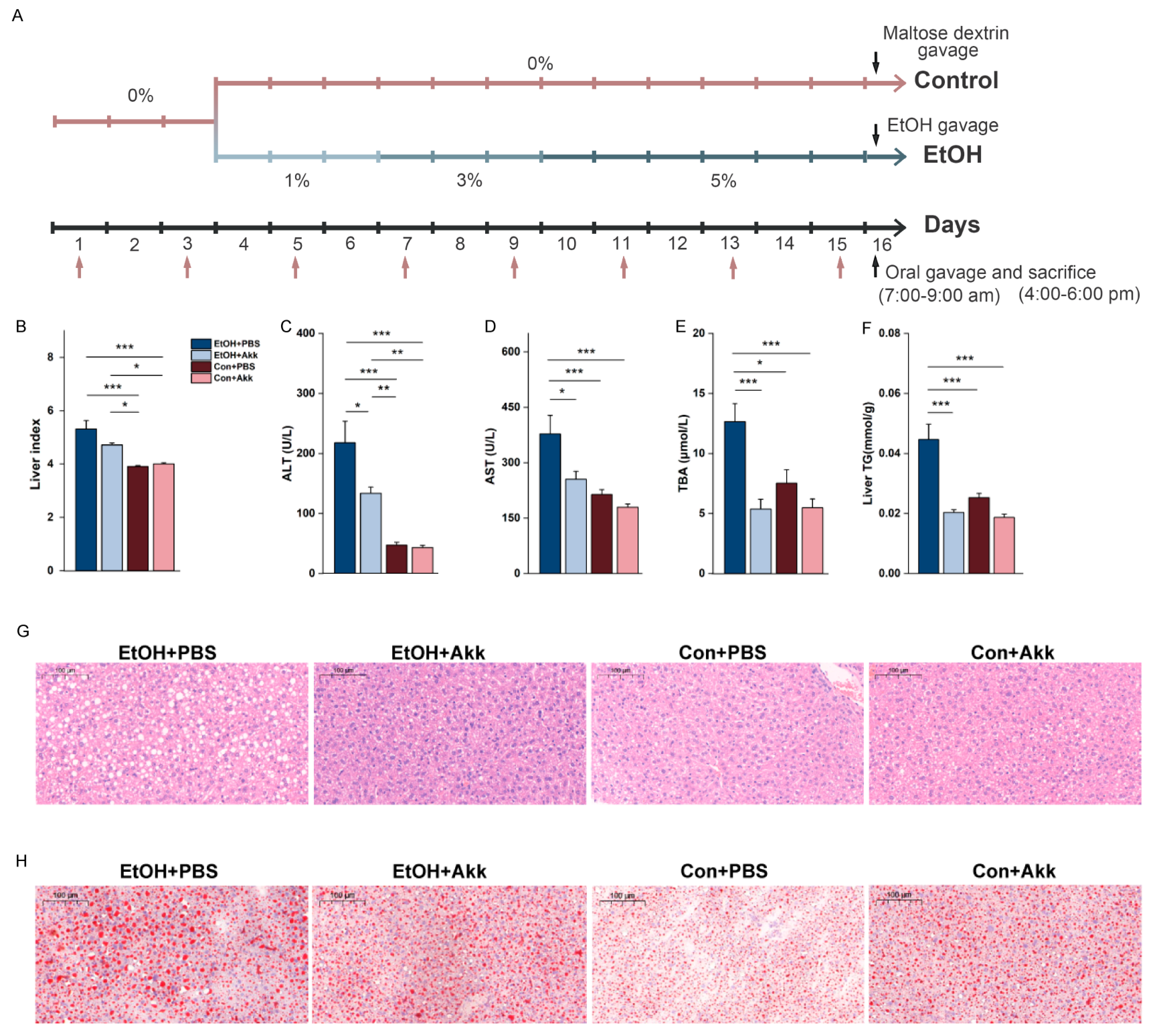
2.2. Animal Experiments

Specific pathogen-free C57BL/6 mice (female, age 10–12-week-old) were obtained from Shanghai Laboratory Animal Center (Shanghai, China). All experimental procedures commenced following the approval of the Institutional Animal Care and Use Committee of Shanghai Chedun Experimental Animal Breeding Farm (Approval number: ADO23003). All mice were housed in a controlled environment (20–22 °C, 40–60% humidity, 12 h light/dark cycle). Alcohol liver injury was induced in the mice using the NIAAA model with minor modifications [24]. After a 1-week acclimatization, the mice were fed a Lieber-DeCarli diet containing 1–5% vol% (TROPHIC Animal Feed High-tech Co., Ltd., Nantong, China) for 15 days [21,24]. The ethanol-fed or pair-fed mice were treated with A. muciniphila (2 × 10⁸ CFU/200 µL PBS) or PBS by intragastric infusion with a 24-gauge stainless steel free tube every other day, starting on day 1. On the last day of the experiment, the pair-fed mice received 5 g/kg maltose, and the ethanol-fed mice received 5 g/kg ethanol-water. Nine hours after the ethanol or maltose gavage, the mice were sacrificed. The blood was collected from the inferior vena cava and then centrifuged at 3000× g for 15 min at 4 °C for serum collection. Cecal contents and tissue samples of the cecum and colon were collected. Livers were carefully weighed and snap-frozen with liquid nitrogen. All the samples were stored at −80 °C immediately after collection for further analysis.

2.3. Biochemical Analyses

The biochemical markers, including serum alanine transaminase (ALT), aspartate aminotransferase (AST), and total bile acids (TBA), were measured by a fully automatic biochemical analyzer (Chemray 800, Rayto, Shenzhen, China). The liver tissues were pre-treated according to the instruction. The amounts of liver triglycerides were measured using a triglyceride quantification kit supplied by Nanjing Jiancheng Bioengineering Institute (Nanjing, China).

2.4. Histology and Immunohistochemistry (IHC)

The tissues of the liver samples were fixed in 10% neutral-buffered formalin for at least 24 h. Then, the tissues were embedded in paraffin and stained with hematoxylin and eosin (H&E) to assess the histologic features of steatosis and inflammation. Frozen liver tissues embedded in an optimal cutting temperature compound (Sakura, Torrance, CA, USA) were cut and stained with Oil Red O. Based on the observation using a laser-scanning confocal microscopy, the images were captured using a Nikon DSU3 camera (Nikon Corporation, Chiyoda Ward, Tokyo, Japan). For the immunofluorescence investigation of intestinal tight junction proteins, frozen intestinal sections were treated with anti-occludin and anti-claudin-2 antibodies (Thermo Fisher Scientific, Waltham, MA, USA), followed by labeling with a FITC-conjugated secondary antibody (Servicebio, Wuhan, China).

2.5. Serum Metabolites Assessment

The metabolomics study for plasma samples was based on the methods of our previous publication [25]. The serum metabolites were measured using an ultra-performance liquid chromatography linked to a tandem mass spectrometry (UPLC-MS/MS) system (ACQUITY UPLC-Xevo TQ-S, Waters Corp., Milford, MA, USA). The separation of the metabolites was achieved on a VanGuard pre-column (ACQUITY UPLC BEH C18, 2.1 mm × 5 mm, 1.7 μm) in combination with an analytical column (ACQUITY UPLC BEH C18, 2.1 mm × 100 mm, 1.7 μm). The column temperature and the manager temperature were set at 40 °C and 10 °C, respectively. Mobile phase A was water with formic acid (0.1%), and mobile phase B was acetonitrile/isopropanol (70:30). During gradient elution, the composition of the mobile phases changed in the following manner: keep 5% B for 1 min; increase to 78% B for 10 min; increase to 95% B for 2.5 min; increase to 100% B for 0.5 min; keep at 100% B for 2 min; rapidly drop to 5% B for 0.1 min; re-equilibrate at 5% B for 1.9 min. The injection volume was 5.0 μL, and the flow rate was 0.40 mL/min. Capillary voltages were 1.5 Kv in ESI+ and 2.0 Kv in ESI. The source temperature and desolvation temperature were set at 150 °C and 550 °C, respectively. The desolvation gas flow rate was 1000 L nitrogen per hour. The TMBQ program (v1.0, Metabo-Profile, Shanghai, China) was used to handle the raw data files produced by UPLC-MS/MS in order to perform peak integration, calibration, and quantitation for each metabolite. Discriminating metabolites between 2 classes of samples were identified using a statistically significant threshold of Variable Importance in Projection (VIP) value (VIP > 1), and further validated by a Student’s t-test analysis (p < 0.05).

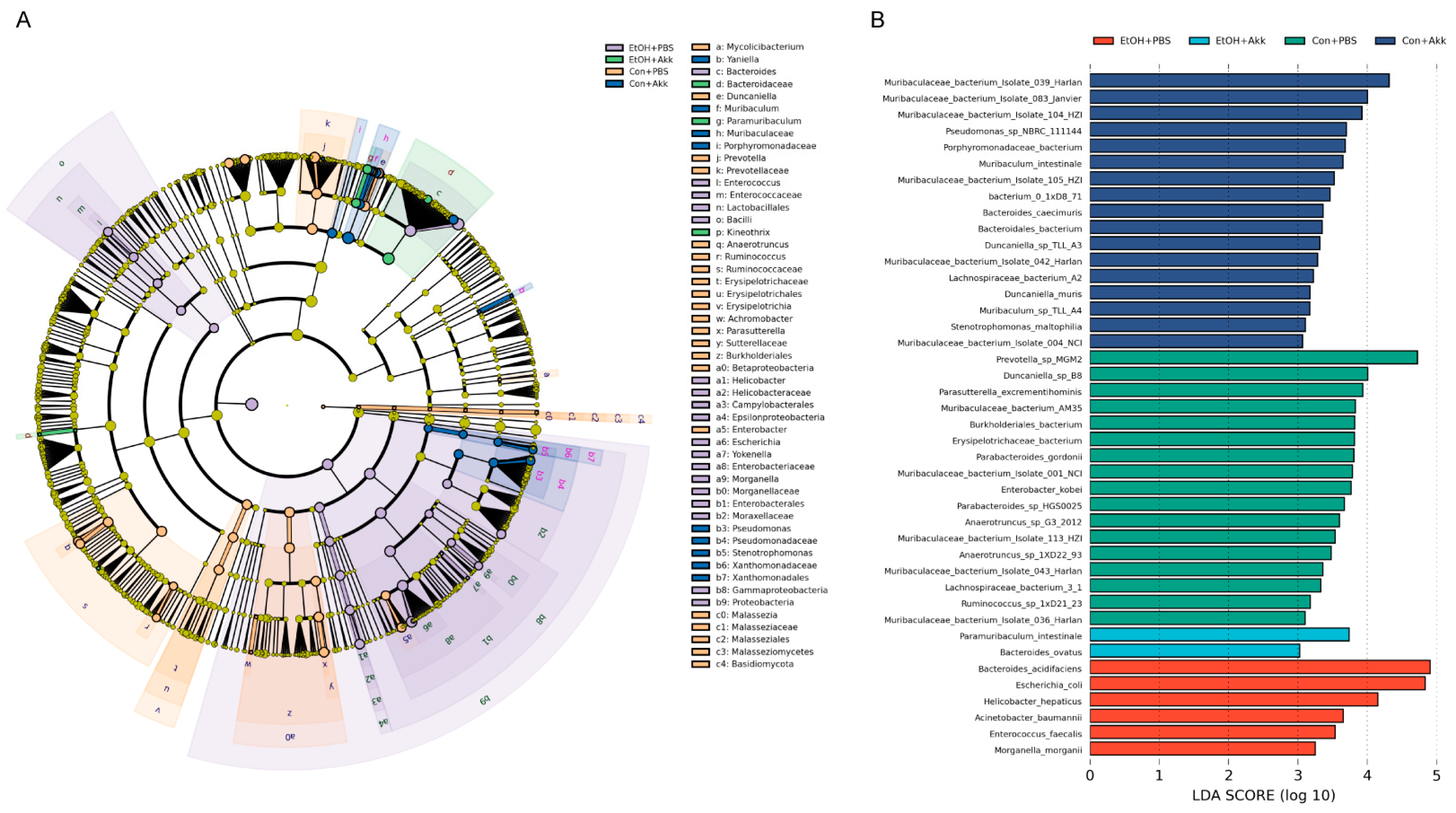
2.6. Gut Microbiota Analysis

The genomic DNA from the cecal content were extracted using a TIANamp Micro DNA Kit (Tiangen, China). The 2bRAD-M library preparation basically followed the original protocol developed by Wang et al. [26] with minor modifications, where the endonuclease enzyme was changed from BsaXI enzyme to BcgI enzyme. Then, 4 U of the BcgI (NEB) enzyme was used to digest DNA (1–200 ng) for 3 h at 37 °C. The adaptors were then joined to the DNA fragments. The ligation reaction was carried out by mixing 5 µL of digested DNA with 10 µL of a ligation master mix that contained 800 U of T4 DNA ligase (NEB), 0.2 µm of each adaptor, and 5 µL of digested DNA. Ligation was carried out at 4 °C for 12 h. After that, ligation products were amplified, and PCR products were subjected to 8% polyacrylamide gel. Bands of about 100 bp were removed from the polyacrylamide gel, and the DNA was then diffused from the gel for 12 h at 4 °C in nuclease-free water. Using platform-specific barcode-bearing primers, PCR was used to add sample-specific barcodes. Each 20 µL PCR contained 25 ng of a gel-extracted PCR product, 0.2 µm of each primer, 0.3 mm dNTP, 1× Phusion HF buffer, and 0.4 U Phusion high-fidelity DNA polymerase (NEB). QIAquick PCR purification kit (Qiagen, Hilden, Germany) was used to clean up the PCR products before sequencing them on the Illumina Nova PE150 platform (Illumina, San Diego, CA, USA). Then, 2bRAD-M was executed at the Qingdao OE Biotech Co., Ltd. (Qingdao, China).

2.7. Statistical Analysis

The metabolome data were analyzed using the partial least square-discriminate analysis (PLS-DA), principal components analysis (PCA), and orthogonal partial Least squares-discriminant analysis (OPLS-DA) in SIMCAP+ (v.13.0; Umetrics, Umeå, Sweden). The self-developed platform iMAP (v1.0, Metabo-Profile, Shanghai, China) was used for the Circos plot and heatmap making. Principal co-ordinates analysis (PCoA; Bray–Curtis distance) was performed using R programing language. Oebiotech (Shanghai, China, https://www.oebiotech.com/, accessed on 6 May 2023) carried out alpha diversity analysis, beta diversity analysis, the random forest analysis, Phylogenetic Investigation of Communities by Reconstruction of Unobserved States (PICRUSt) analysis, and linear discriminant analysis Effect Size (LEfSe). The Tukey–Kramer method was used to assess the metabolome profile differences between the four groups in SPSS software (v.19.0, IBM Corp., Armonk, NY, USA). Mean ± SEM was used to plot the data.

3. Results

3.1. A. miciniphila Intervention Alleviates Alcohol-Induced Liver Injury

To investigate the potential alleviating effect of A. muciniphila on alcohol-induced liver injury, we employed the NIAAA model with slight modifications and administered A. muciniphila through gavage every two days (Figure 1A). The intervention of A. miciniphila resulted in a decrease in the levels of the liver index, serum ALT, serum AST, serum TBA, and liver TG, which were all elevated due to ethanol treatment. Serum AST, serum TBA, and liver TG levels were even lower than those in the control group (Figure 1B–F). To further identify the liver injury, we conducted H&E staining of the liver tissue as well as Oil red O staining of the pathology sections for observation. By observing the H&E-stained sections, we found that EtOH+PBS mice exhibited infiltration of inflammatory cells in the liver tissue. In comparison to the Con+PBS group, the tissue showed enlarged vacuoles, indicating severe hepatic steatosis. However, the conditions were significantly improved by A. muciniphila (Figure 1G). Following Oil Red O staining, lipid droplets presented as red-stained areas. Ethanol treatment-induced enlargement of lipid droplets was suppressed by A. muciniphila, thereby inhibiting lipid deposition (Figure 1H). Collectively, these findings suggest that supplementation with A. muciniphila ameliorates alcohol-induced liver injury.

3.2. A. muciniphila Supplementation Alters Host Serum Metabolic Patterns

To study how A. muciniphila intervention affects host metabolism, we analyzed the compositions of plasma metabolites using targeted metabolomics. First, principal component analysis (PCA) and partial least squares discriminant analysis (PLS-DA) were conducted to reveal a clear separation between the ethanol-treated group and the control group (Figure 2A,B). PCA showed a trend of altered metabolic patterns in the absence or presence of A. muciniphila (Figure 2A). PLS-DA revealed distinct serum metabolic patterns resulting from A. muciniphila and PBS-treated mice (Figure 2B). The metabolomics data analysis, using a clustering-based heatmap approach, revealed significant differences among groups after ethanol treatment or supplementation with A. muciniphila (Figure 2C). Metabolites responsible for the differences were identified using variable significance projection (VIP > 1) and Student’s t test (p < 0.05). We observed a significant increase in ornithine content in the serum of the ethanol-fed mice after A. muciniphila administration, accompanied by a marked decrease in oxalic acid content (Figure 2D). Additionally, the levels of ornithine, serine, and lactic acid increased noticeably in the Con+Akk group compared to the Con+PBS group (Figure 3). These results indicated that supplementation with A. muciniphila induced changes in the serum metabolic patterns.

3.3. A. muciniphila Administration Restores Intestinal Homeostasis

The intestinal barrier is fundamental for modulating intestinal homeostasis and is the body’s first line of defense against pathogen invasion. Immunofluorescence staining was introduced to detect the expressions of occludin and Zo-1. In contrast to the mice fed ethanol as controls, we found recovered immunoreactivity of occludin and tight junction protein 1 (Zo-1) in colonic epithelial cells from A. muciniphila-treated mice (Figure 4A,B).
Gut microbiota plays an important role in the development of ALD. To examine the alterations in gut microbial composition in the mice with ALD following the ingestion of A. miciniphila, we analyzed the composition of gut microbiota in the cecal contents of the mice using 2bRAD-M. A total of 702 microorganism species were identified in all samples. At the phylum level, Bacteroidetes, Firmicutes, Proteobacteria, and Verrucomicrobia were the most predominant taxa (Figure 4C). The proportion of Proteobacteria was increased and the proportions of Bacteroides and Firmicutes were decreased compared to the Con+PBS group. But the changes were reversed by the treatment of A. muciniphila (Figure 4D). At the genus level, unclassified Bacteroides, Akkermansia, Escherichia, and Parabacteroides were the most abundant genera (Figure 4E). In addition, we assessed the effect of A. muciniphila administration on the diversity of gut microbiota. The alpha diversity was assessed by Chao1 index and Shannon index. Ethanol treatment significantly decreased the alpha diversity index (Chao1 and Shannon). Descriptively, there was an increase in alpha diversity with A. muciniphila, although this increase did not reach statistical significance (Figure 4F,G). Beta diversity was conducted using PCoA. As is shown in Figure 4F, the gut microbiota communities of the mice fed with ethanol diet were remarkably different from the control diet group. Meanwhile, PCoA revealed a clear pattern of separation between the gut microbiota communities in the mice treated with A. muciniphila or PBS (Figure 4H). Thus far, we have successfully demonstrated the ability of A. muciniphila to alter the composition of the gut microbiota.

3.4. Biomarkers of the Gut Microbiome in Different Groups

To identify the specific microorganisms responsible for these differences, we performed random forest and LEfSe analyses. The random forest algorithm was used to rank the microbial traits that contribute to the gut microbiota restructuring process. Using the random forest modeling, we obtained the top 30 most important microbial genera for determining microbiota structure with A. muciniphila administration (Figure 5). LEfSe was used to identify microbial taxa with significant differences in abundance between groups [27]. Ethanol feeding not only enriched Bacteroides and Enterococcus but also led to a marked enrichment of the pathogenic bacterial taxa Helicobacter, Escherichia, Yokenella, and Morganella in mice. Additionally, Paramuribaculum and Kineothrix were unique genera found only in the EtOH+Akk group (Figure 6A). At the species level, the abundance of Paramuribaculum intestinale and Bacteroides ovatus was significantly increased in the EtOH+Akk group, whereas the abundance of Bacteroides acidifaciens, Escherichia coli, Helicobacter hepaticus, Acinetobacter baumannii, Enterococcus faecalis, and Morganella morganii was significantly decreased (Figure 6B). Taken together, these results indicate that A. muciniphila has the ability to reduce the abundance of alcohol-induced harmful microbiota and increase the presence of intestinal commensal bacteria in mice with ALD.

3.5. A. muciniphila Administration Alters Intestinal Homeostasis

To further explore the biological functions, the most different top thirty KEGG pathways between EtOH+Akk and EtOH+PBS comparisons were generated by the PICRUSt algorithm (Figure 7). A. muciniphila administration upregulated many pathways, including arginine and signaling pathways, biofilm formation, butanoate metabolism, degradation of aromatic compounds, fatty acid degradation, etc. This result indicated that A. muciniphila altered the functional capabilities of the intestinal microbial community in mice fed with ethanol.

4. Discussion

Recent studies on ALD have found that several natural extracts or bioactive compounds exert their effects by augmenting the abundance of A. muciniphila [28,29,30,31,32]. This finding highlights the potential of A. muciniphila as a viable treatment option for alcoholic liver disease. A number of studies have demonstrated the association between A. muciniphila and ALD [21,22,33]. However, the influence of A. muciniphila supplementation in modulating serum metabolites and gut microbiota in mice with ALD remains unclear. In the present study, we validated the ameliorative effect of A. muciniphila on ALD. Furthermore, we explored potential mechanisms underlying the effects of A. muciniphila on ALD beyond its role in improving intestinal barrier function. Our study integrated the assessment of host serum metabolic patterns and gut microbiota, which have been overlooked in similar studies. Our study revealed that A. muciniphila ameliorates ALD during prolonged consumption by modulating host serum metabolism and remodeling the gut microbial community (Figure 8).
Phenotyping, biochemical parameters, and histopathological examination methods were used to confirm the effect of A. muciniphila. The consumption of A. muciniphila reduced the levels of liver index, serum ALT, AST, TBA, and liver TG. It also showed inhibitory effects on pathological changes in liver tissue, reducing the extent of fatty degeneration and lipid deposition. Alcohol consumption disrupts metabolic pathways in cells, leading to liver cell damage. ALT and AST are normally stored in hepatocytes, and damage to hepatocytes can result in elevated serum ALT and AST levels [34]. At the same time, the hepatocyte metabolism is disturbed, and insulin signaling transduction is impaired, resulting in increased TG storage in liver cells [35]. Changes in mouse serum TBA may also imply changes in host metabolism. Hepatic steatosis is one of the hallmarks of alcoholic liver disease. ALD often progresses from simple steatosis through various stages of steatohepatitis, fibrosis, cirrhosis, and ultimately liver failure, all of which manifest liver dysfunction [36]. Our study found that A. muciniphila intervention during persistent alcohol consumption effectively suppressed the development of alcoholic fatty liver.
To elucidate the underlying mechanisms responsible for the protective effects of A. muciniphila, we used targeted metabolomics to analyze the variation of serum metabolites in mice. We found that A. muciniphila could alter host serum metabolic patterns. PCA showed a trend of altered metabolic patterns in the absence or presence of A. muciniphila, and PLS-DA revealed distinct serum metabolic patterns resulting from A. muciniphila and PBS-treated mice. Two important differential metabolites, ornithine and oxalic acid, were screened using multidimensional statistics combined with unidimensional statistical analysis. Supplementation with A. muciniphila increased ornithine levels and decreased oxalic acid levels in mice with ALD. Ornithine is thought to be beneficial for gut heath [37]. Gut microbiota regulates the homeostasis of the intestinal mucosal layer via intestinal immune cells, which can influence the mucosal cell proliferation and mucin production [38]. L-Ornithine increases the levels of the aryl hydrocarbon receptor ligand L-kynurenine produced from tryptophan metabolism in gut epithelial cells, which in turn increases gut immune cells [39]. Ornithine aspartate is thought to have some hepatoprotective properties in patients with fatty liver disease [40]. Prolonged elevation of serum oxalic acid levels has been associated with the development of liver cirrhosis [41]. Therefore, elevated serum levels of oxalic acid are considered as a detrimental indicator of liver health deterioration. We believe that A. muciniphila probably exerts its protective effect against alcohol-induced damage by increasing the production of metabolites that promote intestinal homeostasis while reducing the levels of harmful substances present in the host serum.
Ethanol disrupts the intestinal barrier and dysregulates intestinal ecology [42]. Ethanol and its metabolite, acetaldehyde, can directly or indirectly damage intestinal epithelial cells, thereby consequently compromising the integrity of the intestinal mucosal barrier. Furthermore, chronic alcohol consumption induces a downregulation in the expression of proteins involved in the regulation of tight junction proteins, resulting in the decreased expression of tight junction proteins between intestinal epithelial cells [43]. Both animal models and clinical evidence suggest that alcohol consumption disrupts the composition and structure of the gut microbiota, leading to a decrease in the abundance of Firmicutes (Gram-positive bacteria) and Lactobacillus (lactic acid-producing bacteria) in the gut [44]. Ethanol consumption disrupts the intestinal epithelial barrier, resulting in the transfer of bacteria or bacterial components to the liver, leading to liver damage [45]. Although the pathogenesis of ALD is complex, the maintenance of intestinal barrier function and the composition of the gut microbiota are still recognized to play crucial roles in the pathogenesis of ALD [9].
Given the close physiological relationship between the intestine and liver, intestinal barrier function is critical for liver homeostasis, and disruption of intestinal barrier integrity can accelerate the development of liver disease [46]. To investigate how A. muciniphila affects intestinal permeability, we used immunofluorescence staining to detect the expression of occludin and Zo-1. Zo-1 is an important component of the intestinal barrier [47]. Occludin is a transmembrane protein that is depleted by ethanol in the colon. Occludin deficiency has been suggested to promote the ethanol-induced disruption of colonic epithelial junctions, intestinal barrier dysfunction, and liver damage in mice [48]. Sesamol and apigenin could increase the expression of Occludin, claudin-1, and Zo-1 to attenuate colitis [49,50]. A. muciniphila has been shown to attenuate ethanol-induced damage by restoring intestinal barrier function [21]. This finding was confirmed in our study. A. muciniphila alleviated the alcohol-induced reduction of the colonic tight junction proteins Occludin and ZO-1, and it reduced the degree of intestinal barrier damage. Our study confirmed this result [21].
Unsurprisingly, in our study, ethanol consumption led to gut microbiota dysbiosis in mice, but the supplementation with A. muciniphila inhibited the dysbiosis. Ethanol consumption significantly decreased the alpha diversity. Alpha diversity increased as A. muciniphila treatment increased, although it did not reach statistical significance. PCoA showed that the intestinal microbial community was altered following A. muciniphila administration.
We then used random forest and LEfSe analysis to identify the microbes responsible for these discrepancies. At the species level, the abundance of Paramuribaculum intestinale and Bacteroides ovatus was significantly increased in the EtOH+Akk group, while that of Bacteroides acidifaciens, Escherichia coli, Helicobacter hepaticus, Acinetobacter baumannii, Enterococcus faecalis, and Morganella morganii was significantly decreased. Interestingly, Escherichia coli, Helicobacter hepaticus, and Morganella morganii are considered to be pathogenic and belong to phyla Proteobacteria [51,52,53,54]. The expansion of Proteobacteria is a microbial signature of dysbiosis in the gut microbiota [55]. Both animal and clinical trials have shown a remarkable increase in the abundance of Proteobacteria during the process of ALD [56]. However, A. muciniphila supplementation decreased the relative abundance of Proteobacteria while promoting the relative abundance of Firmicutes and Bacteroidetes. Kineothrix belongs to Firmicutes phylum, a butyrate-producing bacterium [57]. An increase in the relative abundance of Kineothrix alysoides correlates with a decrease in anxiety-like behaviors [58]. Butyrate has anti-inflammatory effects and can improve intestinal barrier function and mucosal immunity [59]. Butyrate is a critical factor in maintaining gut health [60]. Butyrate is able to support colonocyte function, reduce inflammation, maintain the gut barrier, and promote a beneficial microbiome [61]. Moreover, butyrate has been reported to have the ability to inhibit liver injury in a mouse model of NAFLD [62]. However, its role in ALD remains unclear. A. muciniphila may be effective in the progression of ALD by increasing the abundance of the butyrate-producing microorganisms in the intestine, thereby increasing intestinal levels of butyrate. Unfortunately, our study lacks further experimental validation. There are few studies on Paramuribaculum intestinale. Existing studies have found a positive correlation between Paramuribaculum abundance and the severity of Epstein–Barr virus (EBV) infection and colitis, but there is a higher abundance in kidney injury [63,64,65]. There was also a significant enrichment of bacteria in the EtOH+Akk group, Bacteroides ovatus, which is characterized as one of the predominant species in intestinal commensal bacteria [66]. The results suggest that A. muciniphila may be a modulator in the development of ALD by inhibiting the ethanol-induced proliferation of pathogenic bacteria and promoting the abundance of commensal bacteria in the gut microbiota.

5. Conclusions

Overall, our study demonstrated the ability of A. muciniphila to ameliorate ethanol-induced injury in mice models of ALD, which differ from those employed in previous studies, and it established, for the first time, a correlation between the beneficial effect of A. muciniphila on ALD and alterations in host metabolism and gut microbiota composition. We found that the beneficial effect of A. muciniphila on ALD was associated with changes in host metabolism. Our study also revealed that A. muciniphila inhibited the proliferation of harmful bacteria and promoted beneficial effects, effectively regulating the gut microbiota and thereby alleviating ALD. This study adds to the mechanisms underlying the ameliorative effects of A. muciniphila on ALD and provides a theoretical basis for the future application of A. muciniphila as a therapeutic modality for ALD.

Author Contributions

Conceptualization, C.F. and Y.X.; methodology, C.F. and J.C.; software, J.C.; validation, W.J. and Y.X.; formal analysis, C.F. and J.C.; investigation, C.F. and J.C.; data curation, C.F. and J.C.; writing—original draft preparation, J.C.; writing—review and editing, C.F. and W.J.; visualization, J.C.; supervision, Y.X.; funding acquisition, C.F. All authors have read and agreed to the published version of the manuscript.

Funding

This work was supported by the National Natural Science Foundation of China (Grant No. 22108095).

Institutional Review Board Statement

The animal study and experimental protocol was examined and approved by the Institutional Animal Care and Use Committee (IACUC) of Jiangnan University.

Informed Consent Statement

Not applicable.

Data Availability Statement

All the data in this study are contained within the manuscript.

Conflicts of Interest

The authors declare no conflict of interest.

References

  1. Fulham, M.A.; Ratna, A.; Gerstein, R.M.; Kurt-Jones, E.A.; Mandrekar, P. Alcohol-induced adipose tissue macrophage phenotypic switching is independent of myeloid Toll-like receptor 4 expression. Am. J. Physiol. Physiol. 2019, 317, C687–C700. [Google Scholar] [CrossRef] [PubMed]
  2. Yuan, S.; Chen, J.; Ruan, X.; Sun, Y.; Zhang, K.; Wang, X.; Li, X.; Gill, D.; Burgess, S.; Giovannucci, E.; et al. Smoking, alcohol consumption, and 24 gastrointestinal diseases: Mendelian randomization analysis. eLife 2023, 12, e84051. [Google Scholar] [CrossRef] [PubMed]
  3. Tadokoro, T.; Morishita, A.; Himoto, T.; Masaki, T. Nutritional Support for Alcoholic Liver Disease. Nutrients 2023, 15, 1360. [Google Scholar] [CrossRef]
  4. Seitz, H.K.; Neuman, M.G. The History of Alcoholic Liver Disease: From an Unrecognized Disease to One of the Most Frequent Diseases in Hepatology. J. Clin. Med. 2021, 10, 858. [Google Scholar] [CrossRef]
  5. Wen, B.; Zhang, C.; Zhou, J.; Zhang, Z.; Che, Q.; Cao, H.; Bai, Y.; Guo, J.; Su, Z. Targeted treatment of alcoholic liver disease based on inflammatory signalling pathways. Pharmacol. Ther. 2021, 222, 107752. [Google Scholar] [CrossRef] [PubMed]
  6. Szabo, G. Gut–Liver Axis in Alcoholic Liver Disease. Gastroenterology 2015, 148, 30–36. [Google Scholar] [CrossRef]
  7. Hartmann, P.; Hochrath, K.; Horvath, A.; Chen, P.; Seebauer, C.T.; Llorente, C.; Wang, L.R.; Alnouti, Y.; Fouts, D.E.; Stärkel, P.; et al. Modulation of the intestinal bile acid/farnesoid X receptor/fibroblast growth factor 15 axis improves alcoholic liver disease in mice. Hepatology 2018, 67, 2150–2166. [Google Scholar] [CrossRef] [PubMed]
  8. Llopis, M.; Cassard, A.M.; Wrzosek, L.; Boschat, L.; Bruneau, A.; Ferrere, G.; Puchois, V.; Martin, J.C.; Lepage, P.; Le Roy, T.; et al. Intestinal microbiota contributes to individual susceptibility to alcoholic liver disease. Gut 2016, 65, 830–839. [Google Scholar] [CrossRef]
  9. Hsu, C.L.; Schnabl, B. The gut–liver axis and gut microbiota in health and liver disease. Nat. Rev. Microbiol. 2023; 21, 719–733. [Google Scholar] [CrossRef]
  10. Zhao, Q.; Yu, J.; Hao, Y.; Zhou, H.; Hu, Y.; Zhang, C.; Zheng, H.; Wang, X.; Zeng, F.; Hu, J.; et al. Akkermansia muciniphila plays critical roles in host health. Crit. Rev. Microbiol. 2023, 49, 82–100. [Google Scholar] [CrossRef]
  11. Derrien, M.; Vaughan, E.E.; Plugge, C.M.; de Vos, W.M. Akkermansia muciniphila gen. nov., sp. nov., a human intestinal mucin-degrading bacterium. Int. J. Syst. Evol. Microbiol. 2004, 54, 1469–1476. [Google Scholar] [CrossRef] [PubMed]
  12. Ghaffari, S.; Abbasi, A.; Somi, M.H.; Moaddab, S.Y.; Nikniaz, L.; Kafil, H.S.; Leylabadlo, H.E. Akkermansia muciniphila: From its critical role in human health to strategies for promoting its abundance in human gut microbiome. Crit. Rev. Food Sci. Nutr. 2022, 63, 7357–7377. [Google Scholar] [CrossRef] [PubMed]
  13. Derrien, M.; Belzer, C.; de Vos, W.M. Akkermansia muciniphila and its role in regulating host functions. Microb. Pathog. 2017, 106, 171–181. [Google Scholar] [CrossRef] [PubMed]
  14. Yan, J.; Sheng, L.; Li, H. Akkermansia muciniphila: Is it the Holy Grail for ameliorating metabolic diseases? Gut Microbes 2021, 13, 1984104. [Google Scholar] [CrossRef]
  15. Cani, P.D.; Depommier, C.; Derrien, M.; Everard, A.; de Vos, W.M. Akkermansia muciniphila: Paradigm for next-generation beneficial microorganisms. Nat. Rev. Gastroenterol. Hepatol. 2022, 19, 625–637. [Google Scholar] [CrossRef] [PubMed]
  16. Depommier, C.; Everard, A.; Druart, C.; Plovier, H.; Van Hul, M.; Vieira-Silva, S.; Falony, G.; Raes, J.; Maiter, D.; Delzenne, N.M.; et al. Supplementation with Akkermansia muciniphila in overweight and obese human volunteers: A proof-of-concept exploratory study. Nat. Med. 2019, 25, 1096–1103. [Google Scholar] [CrossRef]
  17. Han, Y.Q.; Ling, Q.; Wu, L.; Wang, X.S.; Wang, Z.F.; Chen, J.; Zheng, Z.P.; Zhou, Z.Y.; Jia, L.F.; Li, L.J.; et al. Akkermansia muciniphila inhibits nonalcoholic steatohepatitis by orchestrating TLR2-activated γδT17 cell and macrophage polarization. Gut Microbes 2023, 15, 2221485. [Google Scholar] [CrossRef]
  18. Kim, S.; Lee, Y.; Kim, Y.; Seo, Y.; Lee, H.; Ha, J.; Lee, J.; Choi, Y.; Oh, H.; Yoon, Y. Akkermansia muciniphila Prevents Fatty Liver Disease, Decreases Serum Triglycerides, and Maintains Gut Homeostasis. Appl. Environ. Microbiol. 2020, 86, e03004-19. [Google Scholar] [CrossRef]
  19. Liu, Y.W.; Xue, Y.; Zhang, Z.Q.; Ji, J.M.; Li, C.G.; Zheng, K.N.; Lu, J.L.; Gao, Y.T.; Gong, Y.; Zhang, Y.M.; et al. Wolfberry enhanced the abundance of Akkermansia muciniphila by YAP1 in mice with acetaminophen-induced liver injury. FASEB J. 2022, 37, e22689. [Google Scholar] [CrossRef]
  20. Shi, Z.J.; Lei, H.H.; Chen, G.; Yuan, P.H.; Cao, Z.; Ser, H.-L.; Zhu, X.H.; Wu, F.; Liu, C.X.; Dong, M.Y.; et al. Impaired Intestinal Akkermansia muciniphila and Aryl Hydrocarbon Receptor Ligands Contribute to Nonalcoholic Fatty Liver Disease in Mice. mSystems 2021, 6, 1. [Google Scholar] [CrossRef]
  21. Grander, C.; Adolph, T.E.; Wieser, V.; Lowe, P.; Wrzosek, L.; Gyongyosi, B.; Ward, D.V.; Grabherr, F.; Gerner, R.R.; Pfister, A.; et al. Recovery of ethanol-induced Akkermansia muciniphila depletion ameliorates alcoholic liver disease. Gut 2018, 67, 891–901. [Google Scholar] [CrossRef]
  22. Grander, C.; Grabherr, F.; Spadoni, I.; Enrich, B.; Oberhuber, G.; Rescigno, M.; Tilg, H. The role of gut vascular barrier in experimental alcoholic liver disease and A. muciniphila supplementation. Gut Microbes 2020, 12, 1851986. [Google Scholar] [CrossRef] [PubMed]
  23. Wang, L.J.; Tang, L.; Feng, Y.M.; Zhao, S.Y.; Han, M.; Zhang, C.; Yuan, G.H.; Zhu, J.; Cao, S.Y.; Wu, Q.; et al. A purified membrane protein from Akkermansia muciniphila or the pasteurised bacterium blunts colitis associated tumourigenesis by modulation of CD8+ T cells in mice. Gut 2020, 69, 1988–1997. [Google Scholar] [CrossRef] [PubMed]
  24. Bertola, A.; Mathews, S.; Ki, S.H.; Wang, H.; Gao, B. Mouse model of chronic and binge ethanol feeding (the NIAAA model). Nat. Protoc. 2013, 8, 627–637. [Google Scholar] [CrossRef] [PubMed]
  25. Fang, C.; Lu, W.; Liu, Q.; Chen, Y.; Jia, W.; Xu, Y. Comparative study between the effects of aged and fresh Chinese baijiu on gut microbiota and host metabolism. Food Biosci. 2022, 49, 101859. [Google Scholar] [CrossRef]
  26. Wang, S.; Meyer, E.; McKay, J.K.; Matz, M.V. 2b-RAD: A simple and flexible method for genome-wide genotyping. Nat. Methods 2012, 9, 808–810. [Google Scholar] [CrossRef]
  27. Huang, H.; Ren, Z.; Gao, X.; Hu, X.; Zhou, Y.; Jiang, J.; Lu, H.; Yin, S.; Ji, J.; Zhou, L.; et al. Integrated analysis of microbiome and host transcriptome reveals correlations between gut microbiota and clinical outcomes in HBV-related hepatocellular carcinoma. Genome Med. 2020, 12, 102. [Google Scholar] [CrossRef]
  28. Liu, H.R.; Meng, W.W.; Zhao, D.S.; Ma, Z.H.; Zhang, W.G.; Chen, Z.; Li, Z.G.; Zhao, P. Study on mechanism of action of total flavonoids from Cortex Juglandis Mandshuricae against alcoholic liver disease based on “gut-liver axis”. Front. Pharmacol. 2023, 13, 1074286. [Google Scholar] [CrossRef]
  29. Neyrinck, A.M.; Etxeberria, U.; Taminiau, B.; Daube, G.; Van Hul, M.; Everard, A.; Cani, P.D.; Bindels, L.B.; Delzenne, N.M. Rhubarb extract prevents hepatic inflammation induced by acute alcohol intake, an effect related to the modulation of the gut microbiota. Mol. Nutr. Food Res. 2017, 61, 1500899. [Google Scholar] [CrossRef]
  30. Li, S.; Wang, N.; Tan, H.Y.; Chueng, F.; Zhang, Z.J.; Yuen, M.F.; bin Feng, Y. Modulation of gut microbiota mediates berberine-induced expansion of immuno-suppressive cells to against alcoholic liver disease. Clin. Transl. Med. 2020, 10, e112. [Google Scholar] [CrossRef]
  31. Zhao, L.; Wang, S.X.; Zhang, N.H.; Zhou, J.X.; Mehmood, A.; Raka, R.N.; Zhou, F.; Zhao, L. The Beneficial Effects of Natural Extracts and Bioactive Compounds on the Gut-Liver Axis: A Promising Intervention for Alcoholic Liver Disease. Antioxidants 2022, 11, 1211. [Google Scholar] [CrossRef]
  32. Liu, H.; Liu, M.; Fu, X.; Zhang, Z.; Zhu, L.; Zheng, X.; Liu, J. Astaxanthin Prevents Alcoholic Fatty Liver Disease by Modulating Mouse Gut Microbiota. Nutrients 2018, 10, 1298. [Google Scholar] [CrossRef]
  33. Hao, L.Y.; Zhong, W.; Woo, J.; Wei, X.Y.; Ma, H.; Dong, H.B.; Guo, W.; Sun, X.G.; Yue, R.C.; Zhao, J.C.; et al. Conventional type 1 dendritic cells protect against gut barrier disruption via maintaining Akkermansia muciniphila in alcoholic steatohepatitis. Hepatology 2023, 78, 896–910. [Google Scholar] [CrossRef]
  34. Yu, J.Y.; Sun, J.G.; Sun, M.; Li, W.D.; Qi, D.M.; Zhang, Y.Q.; Han, C.C. Protective mechanism of Coprinus comatus polysaccharide on acute alcoholic liver injury in mice, the metabolomics and gut microbiota investigation. Food Sci. Hum. Wellness 2024, 13, 401–413. [Google Scholar] [CrossRef]
  35. Stapleton, J.; Kalk, N.; Heneghan, M.; Shah, N. Early diagnosis of alcohol-related liver disease and timely intervention: The role of alcohol care teams. J. Hepatol. 2020, 73, S188–S189. [Google Scholar] [CrossRef]
  36. Guo, R.; Xu, X.; Babcock, S.A.; Zhang, Y.; Ren, J. Aldehyde dedydrogenase-2 plays a beneficial role in ameliorating chronic alcohol-induced hepatic steatosis and inflammation through regulation of autophagy. J. Hepatol. 2015, 62, 647–656. [Google Scholar] [CrossRef] [PubMed]
  37. Cynober, L. Can arginine and ornithine support gut functions? Gut 1994, 35, S42–S45. [Google Scholar] [CrossRef] [PubMed]
  38. Hwang, J.; Jin, J.; Jeon, S.; Moon, S.H.; Park, M.Y.; Yum, D.-Y.; Kim, J.H.; Kang, J.-E.; Park, M.H.; Kim, E.-J.; et al. SOD1 suppresses pro-inflammatory immune responses by protecting against oxidative stress in colitis. Redox Biol. 2020, 37, 101760. [Google Scholar] [CrossRef]
  39. Qi, H.B.; Li, Y.Y.; Yun, H.; Zhang, T.; Huang, Y.G.; Zhou, J.; Yan, H.; Wei, J.M.; Liu, Y.Q.; Zhang, Z.Q.; et al. Lactobacillus maintains healthy gut mucosa by producing L-Ornithine. Commun. Biol. 2019, 2, 171. [Google Scholar] [CrossRef]
  40. Butterworth, R.F.; Canbay, A. Hepatoprotection by L-Ornithine L-Aspartate in Non-Alcoholic Fatty Liver Disease. Dig. Dis. 2019, 37, 63–68. [Google Scholar] [CrossRef]
  41. Kogiso, T.; Tokushige, K.; Hashimoto, E.; Miyakata, C.; Taniai, M.; Torii, N.; Omori, A.; Kotera, Y.; Egawa, H.; Yamamoto, M.; et al. Primary hyperoxaluria complicated with liver cirrhosis: A case report. Hepatol. Res. 2015, 45, 1251–1255. [Google Scholar] [CrossRef] [PubMed]
  42. Liu, T.T.; Yu, H.T.; Zhang, Z.M.; Xie, Y.F.; Yang, L.; You, F.P. Intestinal ELF4 Deletion Exacerbates Alcoholic Liver Disease by Disrupting Gut Homeostasis. Int. J. Mol. Sci. 2022, 23, 4825. [Google Scholar] [CrossRef]
  43. Wood, S.; Pithadia, R.; Rehman, T.; Zhang, L.; Plichta, J.; Radek, K.A.; Forsyth, C.; Keshavarzian, A.; Shafikhani, S.H. Chronic Alcohol Exposure Renders Epithelial Cells Vulnerable to Bacterial Infection. PLoS ONE 2013, 8, e54646. [Google Scholar] [CrossRef] [PubMed]
  44. Bull-Otterson, L.; Feng, W.; Kirpich, I.; Wang, Y.; Qin, X.; Liu, Y.; Gobejishvili, L.; Joshi-Barve, S.; Ayvaz, T.; Petrosino, J.; et al. Metagenomic Analyses of Alcohol Induced Pathogenic Alterations in the Intestinal Microbiome and the Effect of Lactobacillus rhamnosus GG Treatment. PLoS ONE 2013, 8, e53028. [Google Scholar] [CrossRef] [PubMed]
  45. Yan, A.W.; Fouts, D.E.; Brandl, J.; Stärkel, P.; Torralba, M.; Schott, E.; Tsukamoto, H.; Nelson, K.E.; Brenner, D.A.; Schnabl, B. Enteric dysbiosis associated with a mouse model of alcoholic liver disease. Hepatology 2011, 53, 96–105. [Google Scholar] [CrossRef]
  46. Li, D.K.; Chaudhari, S.N.; Lee, Y.; Sojoodi, M.; Adhikari, A.A.; Zukerberg, L.; Shroff, S.; Barrett, S.C.; Tanabe, K.; Chung, R.T.; et al. Inhibition of microbial deconjugation of micellar bile acids protects against intestinal permeability and liver injury. Sci. Adv. 2022, 8, eabo2794. [Google Scholar] [CrossRef]
  47. Fang, X.; Xu, R.-S. Protective effect of simvastatin on impaired intestine tight junction protein ZO-1 in a mouse model of Parkinson’s disease. J. Huazhong Univ. Sci. Technol.-Med. Sci. 2015, 35, 880–884. [Google Scholar] [CrossRef]
  48. Mir, H.; Meena, A.S.; Chaudhry, K.K.; Shukla, P.K.; Gangwar, R.; Manda, B.; Padala, M.K.; Shen, L.; Turner, J.R.; Dietrich, P.; et al. Occludin deficiency promotes ethanol-induced disruption of colonic epithelial junctions, gut barrier dysfunction and liver damage in mice. Biochim. Biophys. Acta (BBA)-Gen. Subj. 2016, 1860, 765–774. [Google Scholar] [CrossRef]
  49. Zhao, B.T.; Xia, B.; Li, X.H.; Zhang, L.; Liu, X.N.; Shi, R.J.; Kou, R.W.; Liu, Z.G.; Liu, X.B. Sesamol Supplementation Attenuates DSS-Induced Colitis via Mediating Gut Barrier Integrity, Inflammatory Responses, and Reshaping Gut Microbiome. J. Agric. Food Chem. 2020, 68, 10697–10708. [Google Scholar] [CrossRef]
  50. Fu, R.R.; Wang, L.C.; Meng, Y.; Xue, W.Q.; Liang, J.J.; Peng, Z.M.; Meng, J.; Zhang, M. Apigenin remodels the gut microbiota to ameliorate ulcerative colitis. Front. Nutr. 2022, 9, 1062961. [Google Scholar] [CrossRef]
  51. Lo, Y.-C.; Chuang, Y.-W.; Lin, Y.-H. Yokenella regensburgei in an immunocompromised host: A case report and review of the literature. Infection 2011, 39, 485–488. [Google Scholar] [CrossRef]
  52. Laupland, K.B.; Paterson, D.L.; Edwards, F.; Stewart, A.G.; Harris, P.N.A. Morganella morganii, an Emerging Cause of Bloodstream Infections. Microbiol. Spectr. 2022, 10, e0056922. [Google Scholar] [CrossRef]
  53. Jeffery, R.; Ilott, E.N.; Powrie, F. Genetic and environmental factors shape the host response to Helicobacter hepaticus: Insights into IBD pathogenesis. Curr. Opin. Microbiol. 2022, 65, 145–155. [Google Scholar] [CrossRef]
  54. Zhou, F.; Wang, D.; Hu, J.; Zhang, Y.; Tan, B.K.; Lin, S. Control Measurements of Escherichia coli Biofilm: A Review. Foods 2022, 11, 2469. [Google Scholar] [CrossRef]
  55. Shin, N.-R.; Whon, T.W.; Bae, J.-W. Proteobacteria: Microbial signature of dysbiosis in gut microbiota. Trends Biotechnol. 2015, 33, 496–503. [Google Scholar] [CrossRef] [PubMed]
  56. Ganesan, R.; Gupta, H.; Jeong, J.-J.; Sharma, S.P.; Won, S.-M.; Oh, K.-K.; Yoon, S.J.; Han, S.H.; Yang, Y.J.; Baik, G.H.; et al. Characteristics of microbiome-derived metabolomics according to the progression of alcoholic liver disease. Hepatol. Int. 2023; ahead of print. [Google Scholar] [CrossRef] [PubMed]
  57. Haas, K.N.; Blanchard, J.L. Kineothrix alysoides, gen. nov., sp. nov., a saccharolytic butyrate-producer within the family Lachnospiraceae. Int. J. Syst. Evol. Microbiol. 2017, 67, 402–410. [Google Scholar] [CrossRef] [PubMed]
  58. Liddicoat, C.; Sydnor, H.; Cando-Dumancela, C.; Dresken, R.; Liu, J.; Gellie, N.J.; Mills, J.G.; Young, J.M.; Weyrich, L.S.; Hutchinson, M.R.; et al. Naturally-diverse airborne environmental microbial exposures modulate the gut microbiome and may provide anxiolytic benefits in mice. Sci. Total Environ. 2020, 701, 134684. [Google Scholar] [CrossRef]
  59. Liu, H.; Wang, J.; He, T.; Becker, S.; Zhang, G.L.; Li, D.F.; Ma, X. Butyrate: A Double-Edged Sword for Health? Adv. Nutr. 2018, 9, 21–29. [Google Scholar] [CrossRef]
  60. Leonel, A.J.; Alvarez-Leite, J.I. Butyrate: Implications for intestinal function. Curr. Opin. Clin. Nutr. Metab. Care 2012, 15, 474–479. [Google Scholar] [CrossRef] [PubMed]
  61. Hodgkinson, K.; El Abbar, F.; Dobranowski, P.; Manoogian, J.; Butcher, J.; Figeys, D.; Mack, D.; Stintzi, A. Butyrate’s role in human health and the current progress towards its clinical application to treat gastrointestinal disease. Clin. Nutr. 2023, 42, 61–75. [Google Scholar] [CrossRef] [PubMed]
  62. Amiri, P.; Arefhosseini, S.; Bakhshimoghaddam, F.; Gurvan, H.J.; Hosseini, S.A. Mechanistic insights into the pleiotropic effects of butyrate as a potential therapeutic agent on NAFLD management: A systematic review. Front. Nutr. 2022, 9, 1037696. [Google Scholar] [CrossRef] [PubMed]
  63. Feng, P.; Li, Q.; Liu, L.; Wang, S.; Wu, Z.; Tao, Y.; Huang, P.; Wang, P. Crocetin Prolongs Recovery Period of DSS-Induced Colitis via Altering Intestinal Microbiome and Increasing Intestinal Permeability. Int. J. Mol. Sci. 2022, 23, 3832. [Google Scholar] [CrossRef] [PubMed]
  64. Wei, Y.-Y.; Zhang, Y.-N.; Wang, H.; Ga, Y.; Fan, Y.; Wang, Q.; Gu, J.-H.; Zhang, X.-Y.; Gong, X.-H.; Hao, Z.-H. Mori fructus aqueous extracts attenuate carbon tetrachloride-induced renal injury via the Nrf2 pathway and intestinal flora. Ecotoxicol. Environ. Saf. 2022, 245, 114118. [Google Scholar] [CrossRef] [PubMed]
  65. Xia, W.; Liu, L.; Shi, N.; Zhang, C.; Tang, A.; He, G. Epstein Barr virus infection in tree shrews alters the composition of gut microbiota and metabolome profile. Virol. J. 2023, 20, 177. [Google Scholar] [CrossRef]
  66. Wang, H.; Wang, Q.; Yang, C.; Guo, M.; Cui, X.; Jing, Z.; Liu, Y.; Qiao, W.; Qi, H.; Zhang, H.; et al. Bacteroides acidifaciens in the gut plays a protective role against CD95-mediated liver injury. Gut Microbes 2022, 14, 2027853. [Google Scholar] [CrossRef]
Figure 1. Effect of A. muciniphila on experimental ALD. (A) Experimental design diagram; pale pink arrows indicate A. muciniphila administration; (B) Liver index, i.e., liver-to-body-weight ratio; Levels of serum ALT (C), AST (D), and TBA (E); (F) Level of liver TG; (G) Representative photomicrographs of H&E liver sections; (H) Representative photomicrographs of Oil Red O-stained liver sections. Data are shown as mean ± SEM (n = 5–8 mice/group). * p < 0.05, ** p < 0.01, and *** p < 0.001.
Metabolites 13 01057 g001
Figure 2. Effects of A. muciniphila interventions on the serum metabolism. (A) The PCA score plot and (B) PLS-DA score plot of metabolites from EtOH+Akk, EtOH+PBS, Con+Akk, and Con+PBS groups; (C) Clustering heatmap of the metabolism of the four groups; (D) Differential metabolite ornithine and oxalic acid content in groups EtOH+PBS and EtOH+Akk. * p < 0.05, ** p < 0.01.
Metabolites 13 01057 g002
Figure 3. Comparison of differential metabolite content in E+A and E+P groups. (A) Content of ornithine; (B) Content of serine; (C) Content of lactic acid. * p < 0.05, ** p < 0.01.
Metabolites 13 01057 g003
Figure 4. Effects of A. muciniphila intervention on intestinal barrier and gut microbiota in mice. (A) Immunofluorescence staining of occludin in the colon; (B) Immunofluorescence staining of Zo-1 in the colon; (C) The Circos plot showing the composition of gut microbiota in each group; (D) Mean relative abundance of the most abundant phyla and (E) genera in the cohort population gut microbiome; (F) Alpha diversity analysis using Chao1 index and (G) Shannon index; (H) Beta diversity analysis using PCoA of the gut microbiota based on Bray–Curtis dissimilarities. * p < 0.05 and *** p < 0.001.
Metabolites 13 01057 g004
Figure 5. The top 30 most important microbial genera determination of microbiota structure with A. muciniphila administration were obtained by using random forest analysis.
Metabolites 13 01057 g005
Figure 6. LEfSe analysis of the key biomarkers in different groups. (A) LEfSe analysis from phylum to genus; (B) LDA histogram of significant microbial species (LDA score > 3).
Metabolites 13 01057 g006
Figure 7. Differences in the general functional characteristics of the gut microbiota. Bacterial functional pathways were estimated by using the PICRUSt algorithm.
Metabolites 13 01057 g007
Figure 8. Diagram of the mechanism by which A. muciniphila improves ALD. A. muciniphila improves ALD by altering host metabolism and regulating the gut microbiota. “↑” represents increase and “↓” represents decrease.
Metabolites 13 01057 g008
Disclaimer/Publisher’s Note: The statements, opinions and data contained in all publications are solely those of the individual author(s) and contributor(s) and not of MDPI and/or the editor(s). MDPI and/or the editor(s) disclaim responsibility for any injury to people or property resulting from any ideas, methods, instructions or products referred to in the content.

