Article

CFH (rs1061170, rs1410996), KDR (rs2071559, rs1870377) and KDR and CFH Serum Levels in AMD Development and Treatment Efficacy

by
Dzastina Cebatoriene
1,*,
Alvita Vilkeviciute
2,
Greta Gedvilaite
2,
Akvile Bruzaite
2,
Loresa Kriauciuniene
2,3,
Dalia Zaliuniene
3 and
Rasa Liutkeviciene
2,3
1
Medical Academy, Lithuanian University of Health Sciences, A. Mickeviciaus St. 9, LT-44307 Kaunas, Lithuania
2
Neuroscience Institute, Medical Academy, Lithuanian University of Health Sciences, Eiveniu St. 2, LT-50161 Kaunas, Lithuania
3
Department of Ophthalmology, Medical Academy, Lithuanian University of Health Sciences, Eiveniu St. 2, LT-50161 Kaunas, Lithuania
*
Author to whom correspondence should be addressed.
Biomedicines 2024, 12(5), 948; https://doi.org/10.3390/biomedicines12050948
Submission received: 15 March 2024 / Revised: 19 April 2024 / Accepted: 22 April 2024 / Published: 24 April 2024

Abstract

:
Background: Age-related macular degeneration (AMD) is a major global health problem as it is the leading cause of irreversible loss of central vision in the aging population. Av-vascular endothelial growth factor (anti-VEGF) therapies have been shown to be effective, but they do not respond optimally to all patients. Objective. This study investigates the genetic factors associated with susceptibility to AMD and response to treatment, focusing on key polymorphisms in the CFH (rs1061170, rs1410996) and KDR (rs2071559, rs1870377) genes and the association of CFH and KDR serum levels in patients with AMD. Results. A cohort of 255 patients with early AMD, 252 patients with exudative AMD, and 349 healthy controls underwent genotyping analysis, which revealed significant associations between CFH polymorphisms and the risk of exudative AMD. The CFH rs1061170 CC genotype was associated with an increased risk of early AMD (p = 0.046). For exudative AMD, the CFH rs1061170 TC + CC genotype increased odds (p < 0.001), while the rs1410996 GA + AA genotype decreased odds (p < 0.001). Haplotypes of CFH SNPs were associated with decreased odds of AMD. In terms of response to treatment, none of the SNPs were associated with the response to anti-VEGF treatment. We also found that both early and exudative AMD patients had lower CFH serum levels compared to the control group (p = 0.038 and p = 0.006, respectively). Exudative AMD patients with the CT genotype of CFH rs1061170 had lower CFH serum levels compared to the control group (p = 0.035). Exudative AMD patients with the GG genotype of CFH rs1410996 also had lower CFH serum levels compared to the control group (p = 0.021). Conclusions. CFH polymorphisms influence susceptibility to AMD but do not correlate with a response to anti-VEGF therapy. Further research is imperative to fully evaluate the developmental significance, treatment efficacy, and predictive role in influencing susceptibility to anti-VEGF therapy for KDR and CFH.

1. Introduction

Age-related macular degeneration (AMD) is the most common cause of irreversible loss of central vision in the older population of the industrialized world [1,2,3]. Accordingly, it increases the demands on the healthcare system as people live longer and treatment costs rise exponentially. AMD is responsible for around 9% of all blindness [4]. This disease affects the back of the eye and damages the macula; a part of the retina with many photoreceptor cells that are responsible for central vision [5]. Aging contributes significantly to AMD, and the age-adjusted prevalence is around 24% in people aged 65–74 years and more than 44% in 70–95-year-olds [6,7]. The human retina undergoes changes as a natural part of aging. This process results in the formation of visible focal yellow deposits called drusen, which consist of cellular debris between the retinal pigment epithelium and Bruch’s membrane. When these pathophysiological changes impair vision, and are accompanied by hypo- or hyper-pigmentation of retinal pigment epithelial (RPE) cells and enlargement or increasing confluence of drusen, this is diagnosed as AMD [8].
However, there is no treatment for the progression of dry AMD or GA so far [9]. The “wet” or neovascular form is less common but is responsible for 90% of acute blindness due to AMD [10], and the prevalence of advanced AMD is estimated at 1.6% worldwide [1]. Neovascular AMD (nAMD) is characterized by the formation of neovascular choroidal membranes, exudation, and fibrosis leading to acute vision loss [11].
Most therapies for the treatment of nAMD target the abnormal growth of blood vessels by inhibiting vascular endothelial growth factor (VEGF)-based antibodies, showing a range of effects. Significant strides have been made in the treatment of nAMD since the era of thermal laser photocoagulation and photodynamic therapy two to three decades ago [12]. The gold standard therapy for maintaining or improving visual acuity in most patients with nAMD is now intravitreal injection of anti-vascular endothelial growth factor. Many studies have shown positive results with anti-VEGF drugs, but there are also limitations to their use. Previous studies have shown that 20% of patients still lose their vision, and half of patients ultimately fail to achieve 20/40 visual acuity [13,14]. A large published meta-analysis showed that visual acuity with best-corrected visual acuity in 80-year-olds with nAMD has improved significantly since 2006 [15]. The improvement in visual acuity is thought to be related to a healthier lifestyle and the introduction of anti-vascular endothelial growth factor therapy. By default, the response to anti-VEGF therapy can be monitored by evaluating visual acuity and optical coherence tomography parameters such as central retinal thickness (CRT) or total macular volume, which reflect the extent of edema. Nevertheless, despite these therapies, most patients require indefinite treatment, do not regain their vision, or show progression of the disease [16]. However, there remains an unmet clinical need for new and improved therapies for nAMD, as many patients do not respond optimally to treatment, and the effect diminishes over time or has suboptimal durability, which compromises efficacy in practice. There is evidence that targeting VEGF-A alone, as has been the case with most agents until recently, may be insufficient and that agents targeting multiple signaling pathways (e.g., aflibercept, faricimab, and other agents in development) may be more effective.
Although genes may only have a minor impact on the total genetic variance of AMD, their effects do not consistently align with the significance of the disease’s pathogenesis and subsequent treatment [17]. It is important to note that the percentage of cases attributed to specific genetic variants does not necessarily indicate the genes’ role in the disease’s pathophysiology [17]. Therefore, the search for other existing loci that require clarification should be continued [18,19,20,21,22,23,24,25,26,27]. In the case of the GWAS experiment [28], genes were found that not only function in known AMD signaling pathways but also reveal the importance of additional pathways, including complement activation, collagen synthesis, lipid metabolism/cholesterol transport, receptor-mediated endocytosis, endodermal cell differentiation, and extracellular matrix organization. The VEGFR-2 receptor, encoded by the KDR gene, serves as a high-affinity receptor tyrosine kinase that is responsible for the majority of angiogenic and permeability-enhancing effects induced by VEGF-A. Consequently, KDR variants are considered potential candidates for influencing sensitivity to anti-VEGF therapy [29]. Research conducted by Lazzeri et al. [29,30,31] indicates that the KDR (VEGFR-2) genotype rs2071559 might serve as a predictive factor for both short- and long-term functional and anatomical outcomes in individuals with nAMD undergoing treatment with ranibizumab. In addition, Hermann et al. [30] concluded that genetic polymorphisms in the KDR gene play an important role in influencing the visual outcomes of patients treated with ranibizumab for nAMD. Ranibizumab binds VEGF-A and inhibits its effect on VEGFR-1 and -2. VEGFR-1 and -2 are encoded by FLT1 and KDR, respectively. In the context of ranibizumab binding to VEGFA, these variants may cause conformational changes or alter VEGFR1 delivery and expression. Consequently, these genetic variations may contribute to individual differences in response to ranibizumab. Recently, great progress has been made in the pathology and epidemiology of AMD, particularly in the field of genetics. Several genes have been identified that are associated with susceptibility to AMD, with the complement factor H (CFH) gene being one of the most important [32]. A meta-analysis of the Asian population found that the CFH polymorphisms rs1061170 and rs1410996 were associated with AMD risk, with both demonstrating higher susceptibility to AMD, particularly nAMD [33]. Whole genome sequencing identifies a significant association between CFH loci with AMD [34].
All in all, AMD is a multifactorial disease and identifying risk factors allows individuals to make lifestyle choices that can reduce their risk of developing the disease. In this study, we intentionally focused on investigating the association between specific polymorphisms and the response to AMD therapy. While diagnostic accuracy could provide valuable insights, our study was designed to prioritize understanding the genetic factors impacting therapeutic outcomes in AMD management. Therefore, our primary objective was to elucidate how genetic variations influence the effectiveness of AMD treatment modalities.

2. Materials and Methods

2.1. Study Design and Structure

The current study was conducted according to the guidelines of the Declaration of Helsinki, and the protocol was approved by the Kaunas Regional Biomedical Research Ethics Committee, Lithuanian University of Health Sciences (No. BE-2-/48). All study participants signed the informed consent form. An ophthalmological evaluation was performed for all the study subjects admitted for ophthalmological assessment at the ophthalmology department, Hospital of Lithuanian University of Health Sciences, from 2018 to 2023. Their health and other diseases were obtained during the general practitioner examination and gathered from medical records.
Our study involved 255 patients diagnosed with early AMD, 252 patients with exudative AMD, and 349 healthy controls. The control group was formed of 349 subjects that matched gender classification in the early and exudative AMD group structure; however, subjects of the control group were younger than exudative AMD patients (p < 0.001), and further analysis was performed adjusted by age (Table 1).
AMD classification is defined by the American Academy of Ophthalmology as follows:
  • Early AMD: Defined by the presence of numerous small (<63 microns, “hard”) or intermediate (≥63 microns but <125 microns, “soft”) drusen.
  • Intermediate AMD: Macular disease characterized by either extensive drusen of small or intermediate size, or any drusen of large size (≥125 microns).
  • Advanced AMD: Defined by the presence of either geographic atrophy or choroidal neovascular membrane (along with its sequelae, such as subretinal or sub-RPE hemorrhage or serous fluid, and subretinal fibrosis).
The AMD group consisted of subjects aged 55 years or older who underwent ophthalmological evaluation and were diagnosed with early, exudative AMD.
All patients underwent a comprehensive eye examination including best corrected visual acuity (BCVA) using the ETDRS visual chart, retinal photography of the fundus, structural OCT (Triton SS-OCT, Topcon, Tokyo, Japan), and OCT angiography (OCT-A). Fluorescein angiograms were performed if necessary.
Patients received a loading dose of 3-monthly injections of anti-VEGF and were then followed up monthly; treatment intervals (2 to 4 weeks) were shortened when disease activity recurred. The exudative AMD group consisted of patients with active subfoveal CNV lesions (SRS, IRS fluid, or macular thickening) caused by AMD. Juxtafoveal lesions with leakage affecting the fovea were also included.
The early AMD group consisted of patients with numerous small “hard” or intermediate “soft” drusen in the early stages of AMD.
AMD exclusion criteria consisted of the following:
  • Unrelated eye disorders; e.g., high refractive error, cloudy cornea, lens opacity (nuclear, cortical, or posterior subcapsular cataract) except minor opacities, keratitis, acute or chronic uveitis;
  • Systemic illnesses; e.g., diabetes mellitus, malignant tumors, systemic connective tissue disorders, chronic infectious and non-infectious diseases, coronary artery disease, stroke, or conditions following organ or tissue transplantation;
  • Ungraded color fundus photographs resulting from obscuring the ocular optic system or because of fundus photograph quality;
  • Use of antiepileptic or sedative drugs.
Based on the clinical OCT and BCVA data, patients with exudative AMD were categorized into one of two groups: responders and non-responders.
The efficacy of anti-VEGF treatment (ranibizumab, aflibercept, bevacizumab) was evaluated in patients with exudative AMD who had exudative or hemorrhagic features in the macula, but had not received a prior intravitreal anti-VEGF injection or other treatment and were followed up for at least 6 months after the first anti-VEGF injection. Central macular thickness (CMT) and best corrected visual acuity (BCVA) were measured before treatment and six months after the first intravitreal anti-VEGF injection.
Visual acuity was assessed before treatment and six months after the first intravitreal anti-VEGF injection. Deterioration in visual acuity was considered to have occurred if patients had lost one or more line (>5 letters) in the table. BCVA changes during the treatment period were calculated using the following formula: BCVA after six months minus BCVA before treatment.
Good response was defined when there was resolution of fluid according to OCT 6 months after the first injection, and/or improvement of >5 letters.
Non-response was defined as an increase in fluid of 100 μM (IRF, SRF and CRT), or increasing hemorrhage compared with the baseline and/or loss of >5 letters compared with the baseline or best corrected vision subsequently. CMT changes were calculated accordingly: CMT before treatment minus CMT after 6 months.
Subjects who underwent ophthalmological evaluation were involved in the control group.
Control group inclusion criteria consisted of the following:
  • Older than 18 years;
  • Patients after senile cataract surgeries (without any other ocular comorbidities);
  • Signed informed consent form.
Control group exclusion criteria consisted of the following:
  • Unrelated eye disorders; e.g., high refractive error, cloudy cornea, lens opacity (nuclear, cortical, or posterior subcapsular cataract) except minor opacities, keratitis, acute or chronic uveitis, glaucoma, or diseases of the optic nerve;
  • Systemic illnesses; e.g., diabetes mellitus, malignant tumors, systemic connective tissue disorders, chronic infectious and non-infectious diseases, hypertension, coronary artery disease, stroke, or conditions following organ or tissue transplantation;
  • Ungraded color fundus photographs resulting from obscuring the ocular optic system or because of fundus photograph quality;
  • Use of antiepileptic or sedative drugs.

2.2. SNP Selection

Our selection aimed to encompass variants with established relevance to AMD pathogenesis and treatment response.
In our study, the selection of SNPs was based on previous literature indicating their associations with exudative AMD occurrence and/or response to anti-VEGF injections. Specifically, we selected four SNPs from the CFH and KDR genes. From the CFH gene, we selected rs1061170 and rs1410996 based on their documented associations with response to anti-VEGF treatment in AMD patients [35].
KDR rs2071559 was chosen due to its association with AMD occurrence in previous research [29,30,31]. The KDR rs1870377 was selected as a potential biomarker for AMD treatment response even if the link between it and exudative AMD was not confirmed yet, but the associations with other diseases and their treatment were significant in several studies [36,37].

2.3. Deoxyribonucleic Acid Extraction from Peripheral Venous Blood and Genotyping

Deoxyribonucleic acid (DNA) extraction and genotyping of selected single nucleotide polymorphisms (SNPs), CFH (rs1061170, rs1410996) and KDR (rs2071559, rs1870377), were conducted at the Laboratory of Ophthalmology, Neuroscience Institute, Lithuanian University of Health Sciences. Utilizing predesigned TaqManTM genotyping assays from Thermo Fisher Scientific, Pleasanton, CA, USA, the procedures were executed in accordance with the manufacturer’s instructions, following established protocols. To ensure high-quality DNA samples, the DNA salting-out method was selected for DNA extraction.
SNPs were determined using TaqMan® genotyping assays (Applied Biosystems, New York, NY, USA; Thermo Fisher Scientific, Inc., Waltham, MA, USA), C___8355565_10, C___2530294_10, C__15869271_10 and C__11895315_20 according to manufacturer’s protocols by a StepOne Plus (Applied Biosystems, Waltham, MA, USA). For each reaction, 1 μL of genomic test DNA and 9 μL of PCR reaction mix were used. The composition of the PCR mixture and the PCR reaction conditions are given in Table 2 and Table 3, respectively.

2.4. Serum Protein Concentration Measurement

To prepare the serum, peripheral venous blood was collected and incubated for 30 min at room temperature before centrifugation. After centrifugation, the serum was separated from the pellet, transferred into 2 mL tubes, and then frozen at −80 °C until analysis. Serum KDR levels in AMD patients and control subjects were assessed following the manufacturer’s guidelines. The analysis utilized an KDR ELISA Kit (Human) (Aviva Systems Biology, San Diego, CA, USA), employing standard sandwich ELISA technology with a range of 0.78–50 ng/mL. Similarly, serum CFH levels were determined in AMD patients and control subjects using an Invitrogen Complement Factor H ELISA Kit (Human) (Thermo Fisher Scientific, United States), which operates on a sandwich-type principle with a range of 2–500 ng/mL, as per the manufacturer’s instructions.

2.5. Statistical Analysis

The statistical analysis was carried out using SPSS/W 29.0 software (Statistical Package for the Social Sciences for Windows, Inc., Chicago, IL, USA). Continuous data (age, BCVA and CMT) were tested for normality using the Shapiro–Wilk test. Continuous variables were presented as median with interquartile range (IQR) and compared using a non-parametric Mann–Whitney U test. Wilcoxon signed rank test was used to evaluate the BCVA and CRT changes after the treatment with anti-VEGF. Statistically significant differences were observed when p < 0.05.
Categorical data (sex and genotype distributions) are presented as absolute numbers with percentages in parentheses and compared between groups using the chi-squared test (χ2).
The effect of gene polymorphisms on early and exudative was assessed using binomial logistic regression analysis and presented as an odds ratio (OR) with a 95% confidence interval (CI) after adjusting for sex in early AMD and age in the exudative AMD groups. The results of the logistic regression analysis were presented as genetic models (codominant: heterozygotes versus wild-type homozygotes and minor allele homozygotes versus wild-type homozygotes; dominant: minor allele homozygotes and heterozygotes versus wild-type homozygotes; recessive: minor allele homozygotes versus wild-type homozygotes and heterozygotes; overdominant: heterozygotes versus wild-type homozygotes and minor allele homozygotes; the additive model was used to assess the effects of each minor allele on AMD). The selection of the best genetic model was based on the Akaike information criterion (AIC); therefore, the best genetic models had the lowest AIC values. Due to the multiple association calculations, we introduced a Bonferroni correction and applied an adjusted significance threshold for multiple comparisons α = 0.0125 (0.05/4, as we analyzed four different SNPs).

3. Results

3.1. Hardy–Weinberg Equilibrium Analysis

Our study quality assessment based on Hardy–Weinberg equilibrium (HWE) analysis showed that the distribution of genotypes of KDR (rs2071559, rs1870377), CFH (rs1061170, rs1410996) did not deviate from HWE in the control group (p < 0.05).

3.2. Analysis of KDR (rs2071559, rs1870377), CFH (rs1061170, rs1410996) in Early and Exudative AMD

Our study genotyping data showed that there were no statistically significant differences in genotype and allele distributions of KDR (rs2071559 and rs1870377) between early AMD and control groups, or between exudative AMD and controls, while the analysis of CFH rs1061170 and rs1410996 genotype and allele distributions revealed significant results. CFH rs1061170 genotypes (TT, TC, and CC) and rs1410996 genotypes (GG, GA and AA) were distributed significantly differently when comparing patients with exudative AMD and control subjects (15.1%, 51.6% and 33.3% vs. 39.3%, 45.3% and 15.5%, p < 0.001 and 64.3%, 32.5% and 3.2% vs. 37.8%, 48.1% and 14%, p < 0.001, respectively) (Table 4). Further analysis showed that the C allele at CFH rs1061170 was more frequent in the exudative AMD group than in the control group (59.1% vs. 38.1%, respectively, p < 0.001) and the A allele at rs1410996 was significantly less frequent in exudative AMD group than in controls (19.4% vs. 38.1%, respectively, p < 0.001) (Table 4).
Binary logistic regression analysis was performed to evaluate the impact of SNPs on early and exudative AMD. It showed that CFH rs1061170 TC + CC genotypes were associated with increased odds of early AMD under the dominant (OR = 1.414; CI: 1.005; 1.988; p = 0.046) genetic model, but these results did not survive after Bonferroni correction (Table 5). Also, CFH rs1061170 variant showed significant associations with increased odds of exudative AMD occurrence under the codominant (OR = 2.961; CI: 1.894; 4.630, p < 0.001 and OR = 5.578; CI: 3.319; 9.377; p < 0.001), dominant (OR = 3.629; CI: 2.372; 5.552; p < 0.001), recessive (OR = 2.704; CI: 1.796; 4.071; p < 0.001) and additive (OR = 2.354; CI: 1.820; 3.046; p < 0.001) genetic models after adjustment for age. Moreover, we found that CFH rs1410996 was associated with highly decreased odds of exudative AMD occurrence under the codominant (OR = 0.380; CI: 0.262; 0.550; p < 0.001 and OR = 0.119; CI: 0.053; 0.266; p < 0.001), dominant (OR = 0.318; CI: 0.223; 0.453; p < 0.001), recessive (OR = 0.184; CI: 0.084; 0.403; p < 0.001), overdominant (OR = 0.509; CI: 0.357; 0.725; p < 0.001) and additive (OR = 0.363; CI: 0.270; 0.488; p < 0.001) genetic models after the same adjustment for age (Table 5).

3.3. Analysis of KDR (rs2071559, rs1870377) and CFH (rs1061170, rs1410996) in Early and Exudative AMD in Male and Female Subgroups

We found that KDR rs2071559 GA genotype is associated with decreased odds of early AMD in men under the codominant genetic model (OR = 0.491; 95% CI: 0.254–0.946; p = 0.033).
Also, GA and AA genotypes together are associated with similarly decreased odds of early AMD in men under the dominant genetic model (OR = 0.491; 95% CI: 0.254–0.946; p = 0.033). Further analysis showed that the KDR rs2071550 GA genotype is associated with 2-fold decreased odds of exudative AMD in men under the codominant genetic model (OR = 0.500; 95% CI: 0.266–0.940; p = 0.031). Also, GA and AA genotypes together are associated with similarly decreased odds of exudative AMD in men under the dominant genetic model (OR = 0.508; 95% CI: 0.279–0.925; p = 0.027). However, the results remained insignificant after Bonferroni correction (Table 6).
Statistical analysis for CFH rs1061170 and rs1410996 in subgroups by gender showed the same results as in the overall group: CFH rs1061170 is associated with increased odds of exudative AMD and the CFH rs1410996 was associated with the decreased odds of exudative AMD in males and females, and these results remained significant even after strict Bonferroni correction (Table 6).

3.4. KDR and CFH Haplotype Associations with AMD

A strong pairwise linkage disequilibrium (LD) was observed between the polymorphisms CFH rs1061170 and rs1410996 (Table 7).
We identified KDR and CFH haplotypes and analyzed their frequencies between the early and exudative AMD and control. The results of frequencies of haplotypes have shown that haplotypes of CFH SNPs (rs1061170C-rs1410996G and rs1061170T-rs1410996A) are associated with the decreased odds of early (OR  =  0.76; 95% CI: 0.58–1.00; p = 0.049 and OR  =  0.60; 95% CI: 0.45–0.82; p = 0.0011, respectively) and exudative (OR  =  0.29; 95% CI: 0.20–0.40; p < 0.001 and OR  =  0.42; 95% CI: 0.30–0.58; p < 0.001, respectively) AMD. The CFH haplotype rs1061170C-rs1410996A showed an association with decreased odds of exudative AMD (OR = 0.03; 95% CI: 0.00–0.21; p < 0.001). Analysis of KDR haplotypes did not show any associations with early or exudative AMD (Table 8).

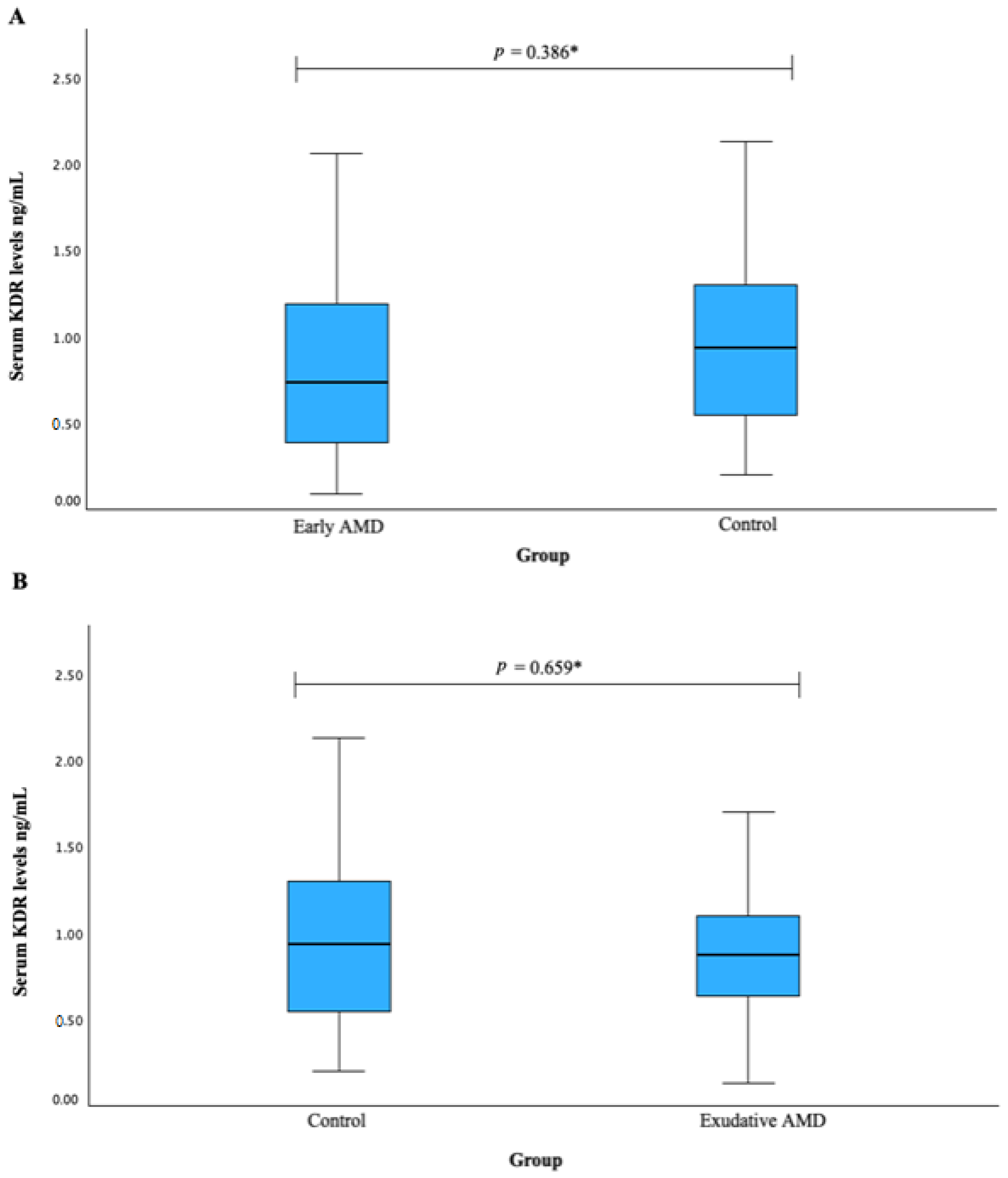
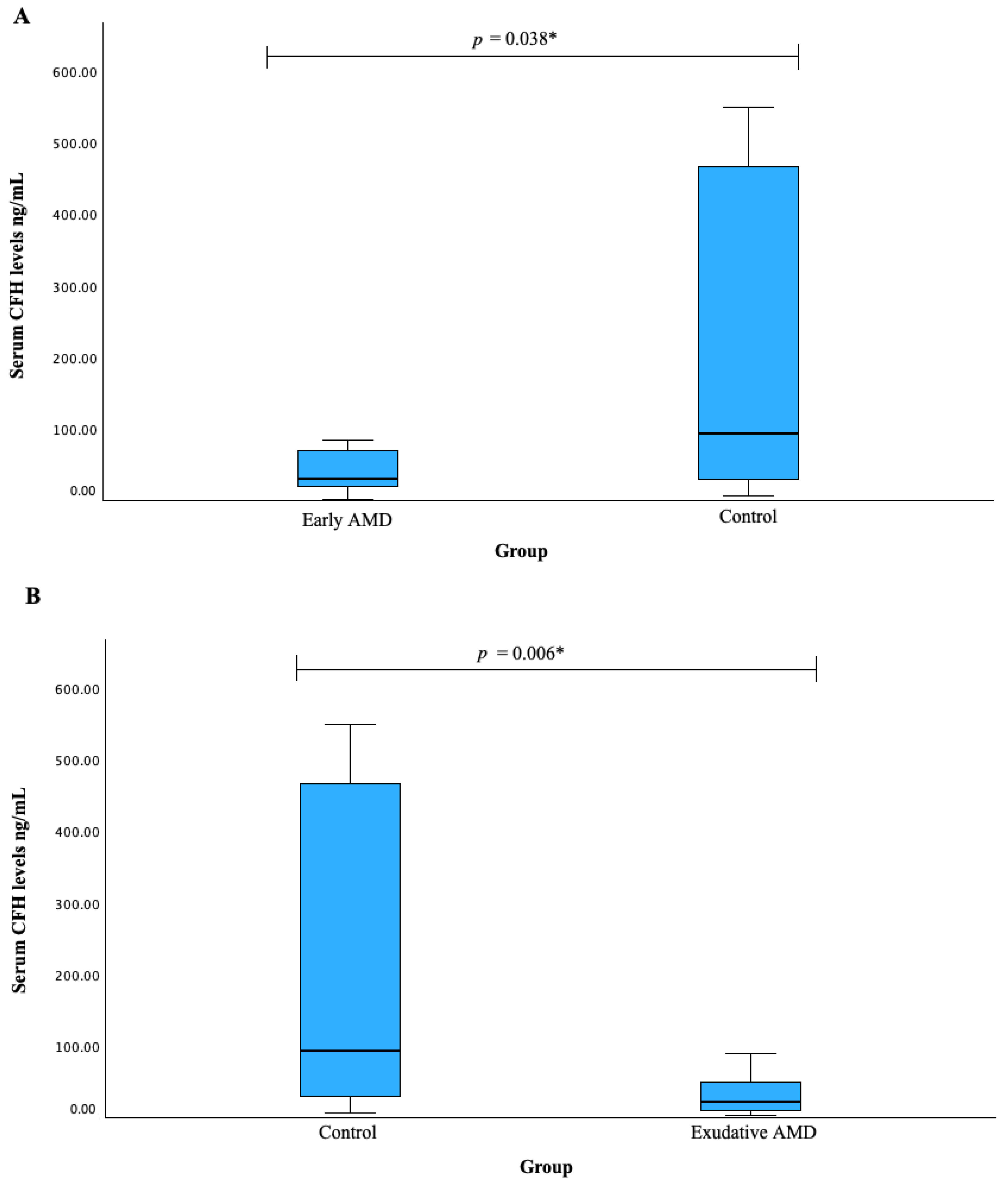
3.5. Serum KDR and CFH Associations with AMD

Serum KDR levels were measured in patients with early AMD vs. control group (A) and exudative AMD vs. control groups; however, no statistically significant difference was found (median (IQR): 0.732 (0.840) vs. 0.938 (0.771), p = 0.386; median (IQR): 0.871 (0.500) vs. 0.938 (0.771), p = 0.659, respectively). The results are shown in Figure 1.
Serum CFH levels were measured in patients with early AMD vs. control group (A) and exudative AMD vs. control group (B). We found that both early and exudative AMD patients had decreased CFH serum levels when compared to the control group subjects (median (IQR): 29.866 (53.707) vs. 93.550 (443.224), p = 0.038; median (IQR): 21.437 (42.549) vs. 93.550 (443.224), p = 0.006, respectively). The results are shown in Figure 2.

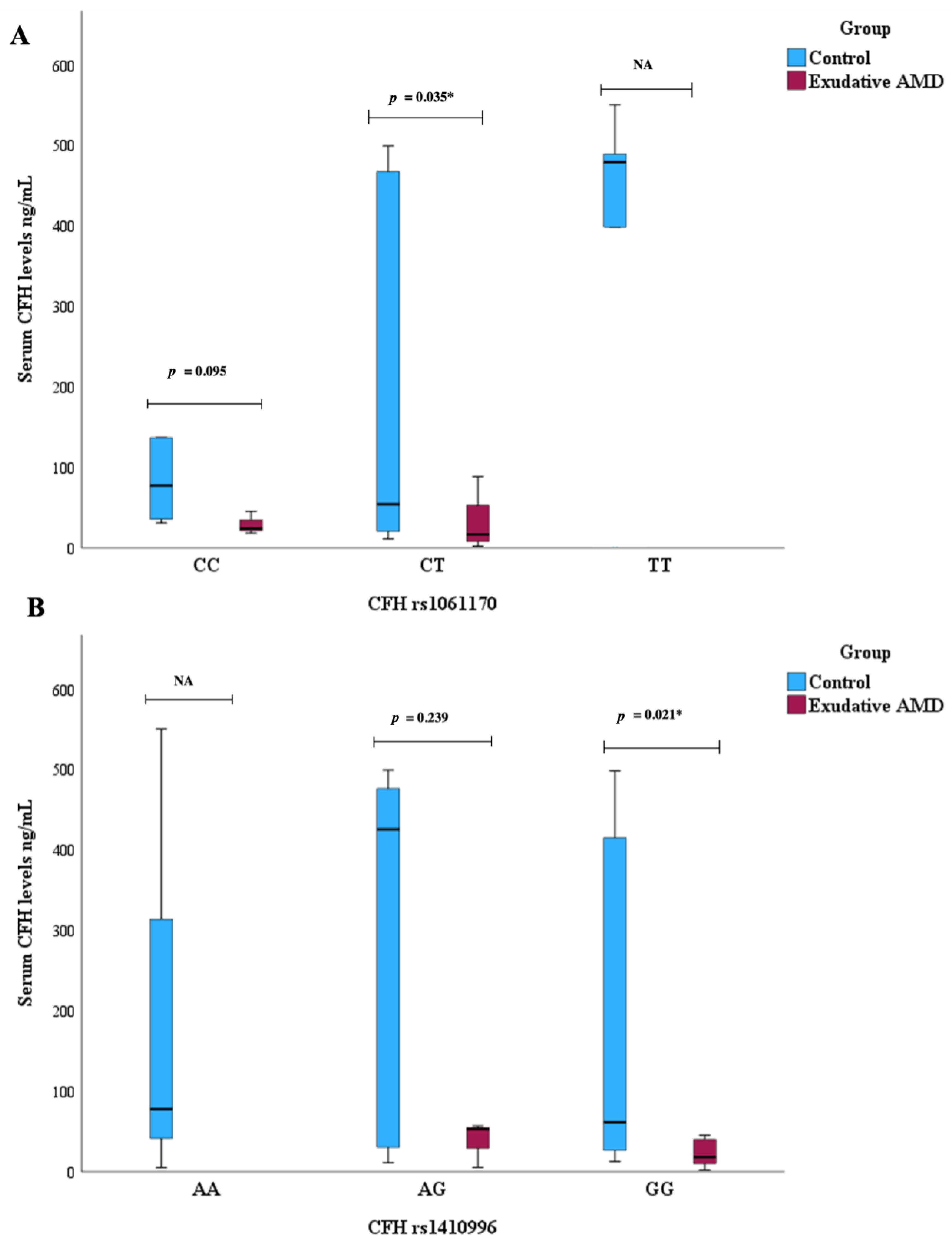
3.6. Serum CFH Levels and CFH SNPs Associations with AMD

A comparison of serum CFH levels was conducted among different genotypes for selected single nucleotide polymorphisms. Exudative AMD patients with the CT genotype of CFH rs1061170 exhibited lower serum CFH levels compared to the control group (median (IQR): 16.89 (48.22) vs. 54.32 (448.94), p = 0.035 (Figure 3A).
Exudative AMD patients with the GG genotype of CFH rs1410996 showed lower serum CFH levels compared to the control group, median (IQR): 18.48 (33.76) vs. 61.44 (417.34), p = 0.021) (Figure 3B).
No CFH level and CFH rs1061170 and rs1410996 genotype associations were revealed with early AMD occurrence.

3.7. Response to Exudative AMD Treatment with Anti-VEGF Therapy

The response to treatment was assessed in 121 individuals who had exudative age-related macular degeneration. The demographic and response parameters of the study population are summarized in Table 9. There was no difference in age or gender distribution between non-responders and responders.
The median visual acuity (VA) decreased in non-responders after treatment but increased in the responders group: (0.42 (0.45) vs. 0.31 (0.31), p = 0.020 and 0.26 (0.7) vs. 0.35 (0.32), p < 0.001, respectively). On the other hand, the CRT did not change statistically significantly in non-responders after treatment but was significantly thinner in responders after treatment (272.5 (86.5) vs. 314.5 (87.75) p = 0.386) and 321 (114) vs. 275 (94), p < 0.001, respectively) (Table 9).

3.8. Genetic Associations with Exudative AMD Response

Statistical analysis was performed to analyze the association between all four SNPs and treatment response. However, none of these SNPs were found to be linked to treatment response with anti-VEGF therapy.

3.9. Serum KDR and CFH Associations with Exudative AMD Response

A comparison of serum KDR and CFH levels was conducted among non-responders and responders, but no statistically significant differences were observed comparing these groups (p > 0.05).

4. Discussion

AMD affects around 170 million people worldwide, making it the third most common cause of visual impairment [35,38,39]. Clinically, AMD is divided into early stage (medium-sized drusen and pigmentary changes in the retina) and late stage (neovascular and atrophic) [5]. Early AMD is usually asymptomatic and can lead to a mild loss of visual acuity and function that delays the onset of night blindness [8]. In intermediate dry AMD, some people have no symptoms at first, but some notice mild symptoms such as a slight blurring of central vision or difficulty seeing in dim light. In late AMD (wet or dry type), many people complain of wavy or crooked lines, notice a blurred area near the center of vision, or see blank spots. Colors may also appear less vivid, and patients may have more difficulty seeing them in low-light conditions. Geographic atrophy is described as bilateral but not symmetrical. It is an area where there is a loss of the RPE and choriocapillaris with a corresponding loss or dysfunction of the overlying photoreceptors, and vision is dramatically impaired. GA affects more than 5 million people worldwide, and its prevalence in one eye is reported to be 0.6% [1].
It is known that Anti-VEGF treatment is one of the first therapies to benefit many AMD patients. Despite the efficacy of anti-VEGF drugs in many patients, some patients do not fully respond to treatment, and persistent intraretinal or subretinal fluid and vision loss occur. Patients with AMD have a reduced quality of life as several daily activities require functional central visual perception, such as driving and reading [40]. The findings from the 2-year analysis of AMD treatment studies revealed that 51.5% of eyes receiving monthly ranibizumab treatment and 67.4% of those receiving monthly bevacizumab treatment still exhibited persistent fluid on optical coherence tomography. Retrospective investigations into intravitreal ranibizumab therapy for neovascular AMD patients demonstrated recurrence rates ranging from 66% to 76% after 12 months of repeated treatment, and 74.8% after 24 months of treatment [41]. Recent research suggests that patients undergoing repeated intravitreal aflibercept injections for the treatment of neovascular AMD may experience disease recurrence in the range of 9% to 55%. This emphasizes the emergence of acquired resistance to anti-VEGF therapy [42]. An essential next step is to understand the functional consequences and downstream effects of AMD-associated genetic variants. Many genes that are considered risk factors for AMD are single nucleotide polymorphisms (SNPs), where one amino acid within the protein is replaced by another. SNPs can have different consequences. Those mutations in which a similar amino acid within a protein is replaced, e.g., valine by alanine, lead to a minor change in the protein [43]. Over the past decade, collaboration between geneticists and ophthalmologists has provided strong evidence that genetic factors are involved in AMD [44,45,46,47]; the changes have been found primarily in genes involved in immune modulation and the complement system—which plays an important role in the disease—along with other risk factors such as smoking [48,49,50,51], diet [52,53,54,55,56,57] and sun exposure [58,59]. Studies have shown that exposure to blue light emitted by smartphones and other devices damages vision and increases the risk of blindness [60]. Exposure of the retina to light from these devices promotes the formation of toxic molecules in the cells (photoreceptors or non-photoreceptors), which increases the risk of macular degeneration.
In our study, four SNPs in the CFH (rs1061170, rs1410996) and KDR (rs2071559, rs1870377) genes, and response to treatment of exudative AMD were investigated, but none of the SNPs were found to be associated with response to anti-VEGF therapy. Numerous clinical and observational studies have repeatedly reported that the retinal fluid only partially resolves in some patients after treatment [61,62]. Furthermore, as many as 10% of patients undergoing treatment exhibit a deterioration of symptoms [13,63,64]. It is the 20–40% of patients who may exhibit no response, and an additional subset who only partially respond, that necessitate alternative treatment strategies to optimize therapeutic outcomes. Due to the ambiguity surrounding these classifications, Amoaku et al. have endeavored to delineate non-responders and partial responders in nAMD based on both visual and anatomical responses [65]. Observing “non-responders” often raises the question of whether genetic variants could influence treatment response and consequently serve as biomarkers to discriminate or predict outcomes [66]. Previous studies have focused on the involvement of inflammation in both the development and progression of AMD. In particular, dysregulation of the complement system is an important factor in the pathogenesis of neovascular AMD. The CFH gene plays a significant role as an inhibitor of the complement cascade and has recently emerged as a key susceptibility gene for AMD. Studies investigating the genetics of AMD have identified susceptibility loci on chromosomes 1q31 and 10q26 [67], with the most compelling evidence of genetic risk associated with AMD observed in complement factor H (CFH) located on chromosome 1q31 [68]. CFH is composed of 20 modules of the complement control protein. The Y402H polymorphism (rs1061170) is located within a binding site for heparin and C-reactive protein, which has been implicated in the pathogenesis of AMD. Consequently, alterations in this specific region of the CFH protein may result in a dysfunctional CFH that is unable to adequately inhibit the complement cascade, potentially contributing to the pathophysiology of AMD. Wu et al. noted that the correlation strength between the rs1061170 polymorphism and AMD seems to diminish when studies transition from Western to Eastern populations. They found that the strong association observed in European cohorts did not translate to the same level of relevance for AMD risk in Asian ancestry populations [33]. KDR is a protein-coding gene that belongs to the VEGFR family and plays a critical role as a primary mediator of VEGF-induced processes such as endothelial proliferation, survival, migration, tubular morphogenesis, and sprouting. KDR is widely considered to play a crucial role in mediating VEGF-induced responses in angiogenesis [69]. Hagstrom et al. [70] studied patients with neovascular AMD who were genotyped for the SNP rs1061170 (CFH) to determine their response to treatment with ranibizumab or bevacizumab. In their study, no statistically significant differences in response by genotype were found for any of the clinical parameters analyzed. Specifically, no high-risk alleles were found to predict final visual acuity, changes in visual acuity, anatomical response (such as the presence of fluid on OCT or FA, retinal thickness, changes in total foveal thickness, or lesion size), or the number of injections required. Additionally, variations in response to anti-VEGF therapy were observed in terms of the frequency of injections needed. Five Genome-Wide Association Studies (GWAS) have been carried out, concerning the treatment of neovascular age-related macular degeneration (nAMD) [71,72,73,74,75], each comprising both a discovery and a replication cohort. However, none of these studies uncovered a variant with genome-wide significance. Notably, the p-values in the replication cohorts consistently showed less significance compared to the respective discovery studies, despite the replication cohort typically encompassing a larger sample size than the discovery study. The available data from meta-analyses allow a similar conclusion to be drawn, as the reported effects are often small or not reproducible. Of note, a study by Wang et al. [76] recalculated the results of 33 publications on genetic effects in response to anti-VEGF therapy in nAMD. In this study, SNP variants in nine genes were found to be significantly associated with response to anti-VEGF therapy; one of the samples NM is rs1410996 in the CFH gene. Further studies based on different ethnicities and large sample sizes are warranted to corroborate the findings found in the present study. The results of the ethnic subgroup analysis showed that rs800292-G and rs1061170-T in the CFH gene were associated with poor response to therapy in East Asians and Europeans, respectively. Although studies have attempted to characterize patients who either do not respond to anti-VEGF therapy or whose response declines over time [77], the mechanisms behind non-response and declining response remain unclear. The heterogeneity of response to anti-VEGF treatment in nAMD patients has led to increasing pharmacogenetic studies on the association of potential high-risk biomarkers associated with nAMD responding to anti-VEGF treatment.

5. Conclusions

CFH polymorphisms influence susceptibility to AMD but do not correlate with a response to anti-VEGF therapy. Further research is imperative to fully evaluate the developmental significance, treatment efficacy and predictive role in influencing susceptibility to anti-VEGF therapy for KDR and CFH.

Author Contributions

Conceptualization, D.C., A.V., G.G., A.B., R.L.; methodology, D.C., A.V., G.G., A.B.; software, A.V., G.G.; validation, D.C., R.L.; formal analysis, A.V., G.G.; investigation, D.C., A.V., G.G., A.B., R.L.; resources, L.K., D.Z., R.L.; data curation, D.C., A.V., G.G.; A.B.; writing—original draft preparation, D.C., A.V., G.G., R.L.; writing—review and editing, D.C., L.K., D.Z., R.L.; visualization, D.C., A.V., G.G., R.L.; supervision, R.L.; project administration, D.C., R.L.; funding acquisition, D.Z. All authors have read and agreed to the published version of the manuscript.

Funding

This study was funded by the Research Council of Lithuania under the initiative of Researcher Group Projects, grant No. Nr. S-MIP-23-96.

Institutional Review Board Statement

The study was conducted in accordance with the Declaration of Helsinki, and approved by the Biomedical Research Ethics Committee, Lithuanian University of Health Sciences (No. BE-2-/48). All study participants signed the informed consent form. An ophthalmological evaluation was performed for all the study subjects admitted to the ophthalmological assessment at the Ophthalmology Department, Hospital of Lithuanian University of Health Sciences, from 2018 to 2023. Their health and other diseases were obtained during the general practitioner examination and gathered from medical records. The study was conducted at the Laboratory of Ophthalmology, Neuroscience Institute, LUHS.

Informed Consent Statement

Informed consent was obtained from all subjects involved in the study.

Data Availability Statement

The datasets used and/or analyzed during the current study are available from the corresponding author on reasonable request.

Conflicts of Interest

The authors declare no conflicts of interest.

References

  1. Klein, R.; Klein, B.E.K.; Linton, K.L.P. Prevalence of age-related maculopathy, The Beaver Dam Eye Study. Ophthalmology 1992, 99, 933–943. [Google Scholar] [CrossRef]
  2. Mitchell, P.; Smith, W.; Attebo, K.; Wang, J.J. Prevalence of age-related maculopathy in Australia. Ophthalmology 1995, 102, 1450–1460. [Google Scholar] [CrossRef]
  3. Vingerling, J.R.; Dielemans, I.; Hofman, A.; Grobbee, D.E.; Hijmering, M.; Kramer, C.F.; de Jong, P.T. The prevalence of age-related maculopathy in the Rotterdam study. Ophthalmology 1995, 102, 205–210. [Google Scholar] [CrossRef]
  4. Wong, W.L.; Su, X.; Li, X.; Cheung, C.M.G.; Klein, R.; Cheng, C.Y.; Wong, T.Y. Global prevalence of age-related macular degeneration and disease burden projection for 2020 and 2040: A systematic review and meta-analysis. Lancet Glob. Health 2014, 2, e106–e116. [Google Scholar] [CrossRef]
  5. Age-Related Eye Disease Study Research Group. A randomized, placebo-controlled, clinical trial of high-dose supplementation with vitamins C and E, beta carotene, and zinc for age-related macular degeneration and vision loss: AREDS report no. 8. Arch. Ophthalmol. 2001, 119, 1417–1436. [Google Scholar] [CrossRef] [PubMed]
  6. Brandl, C.; Zimmermann, M.E.; Günther, F.; Barth, T.; Olden, M.; Schelter, S.C.; Kronenberg, F.; Loss, J.; Küchenhoff, H.; Helbig, H.; et al. On the impact of different approaches to classify age-related macular degeneration: Results from the German AugUR study. Sci. Rep. 2018, 8, 8675. [Google Scholar] [CrossRef]
  7. Korb, C.A.; Kottler, U.B.; Wolfram, C.; Hoehn, R.; Schulz, A.; Zwiener, I.; Wild, P.S.; Pfeiffer, N.; Mirshahi, A. Prevalence of age-related macular degeneration in a large European cohort: Results from the population-based Gutenberg Health Study. Graefe’s Arch. Clin. Exp. Ophthalmol. 2014, 252, 1403–1411. [Google Scholar] [CrossRef] [PubMed]
  8. Fernandes, A.R.; Zielińska, A.; Sanchez-Lopez, E.; dos Santos, T.; Garcia, M.L.; Silva, A.M.; Karczewski, J.; Souto, E.B. Exudative versus nonexudative age-related macular degeneration: Physiopathology and treatment options. Int. J. Mol. Sci. 2022, 23, 2592. [Google Scholar] [CrossRef] [PubMed]
  9. Fleckenstein, M.; Mitchell, P.; Freund, K.B.; Sadda, S.; Holz, F.G.; Brittain, C.; Henry, E.C.; Ferrara, D. The Progression of Geographic Atrophy Secondary to Age-Related Macular De-generation. Ophthalmology 2018, 125, 369–390. [Google Scholar] [CrossRef]
  10. Owen, C.G.; Jarrar, Z.; Wormald, R.; Cook, D.G.; Fletcher, A.E.; Rudnicka, A.R. The estimated prevalence and incidence of late stage age related macular degeneration in the UK. Br. J. Ophthalmol. 2012, 96, 752–756. [Google Scholar] [CrossRef]
  11. Gass, J.D. Pathogenesis of disciform detachment of the neuroepithelium. Am. J. Ophthalmol. 1967, 63, 1–139. [Google Scholar]
  12. Bakri, S.J.; Thorne, J.E.; Ho, A.C.; Ehlers, J.P.; Schoenberger, S.D.; Yeh, S.; Kim, S.J. Safety and Efficacy of Anti-Vascular Endothelial Growth Factor Therapies for Neovascular Age-Related Macular Degeneration: A Report by the American Academy of Ophthalmology. Ophthalmology 2019, 126, 55–63. [Google Scholar] [CrossRef]
  13. Rosenfeld, P.J.; Brown, D.M.; Heier, J.S.; Boyer, D.S.; Kaiser, P.K.; Chung, C.Y.; Kim, R.Y. Ranibizumab for neovascular age-related macular degeneration. N. Engl. J. Med. 2006, 355, 1419–1431. [Google Scholar] [CrossRef]
  14. Heier, J.S.; Brown, D.M.; Chong, V.; Korobelnik, J.-F.; Kaiser, P.K.; Nguyen, Q.D.; Kirchhof, B.; Ho, A.; Ogura, Y.; Yancopoulos, G.D.; et al. Intravitreal aflibercept (VEGF trap-eye) in wet age-related macular degeneration. Ophthalmology 2012, 119, 2537–2548. [Google Scholar] [CrossRef]
  15. Colijn, J.M.; Buitendijk, G.H.S.; Prokofyeva, E.; Alves, D.; Cachulo, M.L.; Khawaja, A.P.; Cougnard-Gregoire, A.; Merle, B.M.J.; Korb, C.; Erke, M.G.; et al. Prevalence of age-related macular degeneration in Europe. Ophthalmology 2017, 124, 1753–1763. [Google Scholar] [CrossRef]
  16. Leung, E.H.; Oh, D.J.; Alderson, S.E.; Bracy, J.; McLeod, M.; Perez, L.I.; Bottini, A.; Yee, D.C.; Mukkamala, K. Initial Real-World Experience with Faricimab in Treatment-Resistant Neovascular Age-Related Macular Degeneration. Clin. Ophthalmol. 2023, 17, 1287–1293. [Google Scholar] [CrossRef] [PubMed]
  17. Khachigian, L.M.; Liew, G.; Teo, K.Y.C.; Wong, T.Y.; Mitchell, P. Emerging therapeutic strategies for unmet need in neovascular age-related macular degeneration. J. Transl. Med. 2023, 21, 133. [Google Scholar] [CrossRef]
  18. Fisher, S.A.; Abecasis, G.R.; Yashar, B.M.; Zareparsi, S.; Swaroop, A.; Iyengar, S.K.; Klein, B.E.; Klein, R.; Lee, K.E.; Majewski, J.; et al. Meta-analysis of genome scans of age-related macular degeneration. Hum. Mol. Genet. 2005, 14, 2257–2264. [Google Scholar] [CrossRef] [PubMed]
  19. Klein, M.L.; Schultz, D.W.; Edwards, A.; Matise, T.C.; Rust, K.; Berselli, C.B.; Trzupek, K.; Weleber, R.G.; Ott, J.; Wirtz, M.K. Age-related macular degeneration. Clinical features in a large family and linkage to chromosome 1q. Arch. Ophthalmol. 1998, 116, 1082–1088. [Google Scholar] [CrossRef]
  20. Majewski, J.; Schultz, D.W.; Weleber, R.G.; Schain, M.B.; Edwards, A.O.; Matise, T.C.; Acott, T.S.; Ott, J.; Klein, M.L. Age-related macular degeneration—A genome scan in extended families. Am. J. Hum. Genet. 2003, 73, 540–550. [Google Scholar] [CrossRef]
  21. Schick, J.H.; Iyengar, S.K.; Klein, B.E.; Klein, R.; Reading, K.; Liptak, R.; Millard, C.; Lee, K.E.; Tomany, S.C.; Moore, E.L.; et al. A whole-genome screen of a quantitative trait of age-related maculopathy in sibships from the Beaver Dam Eye Study. Am. J. Hum. Genet. 2003, 72, 1412–1424. [Google Scholar] [CrossRef]
  22. Abecasis, G.R.; Yashar, B.M.; Zhao, Y.; Ghiasvand, N.M.; Zareparsi, S.; Branham, K.E.; Reddick, A.C.; Trager, E.H.; Yoshida, S.; Bahling, J.; et al. Age-related macular degeneration: A high-resolution genome scan for susceptibility loci in a population enriched for late-stage disease. Am. J. Hum. Genet. 2004, 74, 482–494. [Google Scholar] [CrossRef]
  23. Iyengar, S.K.; Song, D.; Klein, B.E.; Klein, R.; Schick, J.H.; Humphrey, J.; Millard, C.; Liptak, R.; Russo, K.; Jun, G.; et al. Dissection of genomewide-scan data in extended families reveals a major locus and oligogenic susceptibility for age-related macular degeneration. Am. J. Hum. Genet. 2004, 74, 20–39. [Google Scholar] [CrossRef]
  24. Schmidt, S.; Scott, W.K.; Postel, E.A.; Agarwal, A.; Hauser, E.R.; De La Paz, M.A.; Gilbert, J.R.; Weeks, D.E.; Gorin, M.B.; Haines, J.L.; et al. Ordered subset linkage analysis supports a susceptibility locus for age-related macular degeneration on chromosome 16p12. BMC Genet. 2004, 5, 18. [Google Scholar] [CrossRef] [PubMed]
  25. Kenealy, S.J.; Schmidt, S.; Agarwal, A.; Postel, E.A.; De La Paz, M.A.; Pericak-Vance, M.A.; Haines, J.L. Linkage analysis for age-related macular degeneration supports a gene on chromosome 10q26. Mol. Vis. 2004, 10, 57–61. [Google Scholar] [CrossRef]
  26. Jun, G.; Klein, B.E.K.; Klein, R.; Fox, K.; Millard, C.; Capriotti, J.; Russo, K.; Lee, K.E.; Elston, R.C.; Iyengar, S.K. Genome-wide analyses demonstrate novel loci that predispose to drusen formation. Investig. Opthalmol. Vis. Sci. 2005, 46, 3081–3088. [Google Scholar] [CrossRef] [PubMed]
  27. Silveira, A.C.; Morrison, M.A.; Ji, F.; Xu, H.; Reinecke, J.B.; Adams, S.M.; Arneberg, T.M.; Janssian, M.; Lee, J.-E.; Yuan, Y.; et al. Convergence of linkage, gene expression, and association data demonstrates the influence of the RAR-related orphan receptor alpha (RORA) gene on neovascular AMD: A systems biology-based approach. Vis. Res. 2010, 50, 698–715. [Google Scholar] [CrossRef] [PubMed]
  28. Fritsche, L.G.; Igl, W.; Bailey, J.N.C.; Grassmann, F.; Sengupta, S.; Bragg-Gresham, J.L.; Burdon, K.P.; Hebbring, S.J.; Wen, C.; Gorski, M.; et al. A large genome-wide association study of age-related macular degeneration highlights contributions of rare and common variants. Nat. Genet. 2016, 48, 134–143. [Google Scholar] [CrossRef]
  29. Lazzeri, S.; Orlandi, P.; Piaggi, P.; Sartini, M.S.; Casini, G.; Guidi, G.; Figus, M.; Fioravanti, A.; Di Desidero, T.; Ripandelli, G.; et al. IL-8 and VEGFR-2 polymorphisms modulate long-term functional response to intravitreal ranibizumab in exudative age-related macular degeneration. Pharmacogenomics 2016, 17, 35–39. [Google Scholar] [CrossRef]
  30. Hermann, M.M.; van Asten, F.; Muether, P.S.; Smailhodzic, D.; Lichtner, P.; Hoyng, C.B.; Kirchhof, B.; Grefkes, C.; den Hollander, A.I.; Fauser, S. Polymorphisms in vascular endothelial growth factor receptor 2 are associated with better response rates to ranibizumab treatment in age-related macular degeneration. Ophthalmology 2013, 121, 905–910. [Google Scholar] [CrossRef]
  31. Blánquez-Martínez, D.; Díaz-Villamarín, X.; Antúnez-Rodríguez, A.; Pozo-Agundo, A.; Muñoz-Ávila, J.I.; Martínez-González, L.J.; Dávila-Fajardo, C.L. Genetic Polymorphisms Affecting Ranibizumab Response in High Myopia Patients. Pharmaceutics 2021, 13, 1973. [Google Scholar] [CrossRef] [PubMed]
  32. Haines, J.L.; Hauser, M.A.; Schmidt, S.; Scott, W.K.; Olson, L.M.; Gallins, P.; Spencer, K.L.; Kwan, S.Y.; Noureddine, M.; Gilbert, J.R.; et al. Complement factor H variant increases the risk of age-related macular degeneration. Science 2005, 308, 419–421. [Google Scholar] [CrossRef] [PubMed]
  33. Wu, M.; Guo, Y.; Ma, Y.; Zheng, Z.; Wang, Q.; Zhou, X. Association of Two Polymorphisms, rs1061170 and rs1410996, in Complement Factor H with Age-Related Macular Degeneration in an Asian Population: A Meta-Analysis. Ophthalmic Res. 2016, 55, 135–144. [Google Scholar] [CrossRef] [PubMed]
  34. Acar, I.E.; Galesloot, T.E.; Luhmann, U.F.O.; Fauser, S.; Gayán, J.; den Hollander, A.I.; Nogoceke, E. Whole Genome Sequencing Identifies Novel Common and Low-Frequency Variants Associated with Age-Related Macular Degeneration. Investig. Opthalmol. Vis. Sci. 2023, 64, 24. [Google Scholar] [CrossRef] [PubMed]
  35. Hong, N.; Shen, Y.; Yu, C.-Y.; Wang, S.-Q.; Tong, J.-P. Association of the polymorphism Y402H in the CFH gene with response to anti-VEGF treatment in age-related macular degeneration: A systematic review and meta-analysis. Acta Ophthalmol. 2016, 94, 334–345. [Google Scholar] [CrossRef] [PubMed]
  36. Coltelli, L.; Allegrini, G.; Orlandi, P.; Finale, C.; Fontana, A.; Masini, L.C.; Scalese, M.; Arrighi, G.; Barletta, M.T.; De Maio, E.; et al. A pharmacogenetic interaction analysis of bevacizumab with paclitaxel in advanced breast cancer patients. NPJ Breast Cancer 2022, 8, 33. [Google Scholar] [CrossRef] [PubMed]
  37. Zheng, Y.-B.; Zhan, M.-X.; Zhao, W.; Liu, B.; Huang, J.-W.; He, X.; Fu, S.-R.; Zhao, Y.; Li, Y.; Hu, B.-S.; et al. The relationship of kinase insert domain receptor gene polymorphisms and clinical outcome in advanced hepatocellular carcinoma patients treated with sorafenib. Med. Oncol. 2014, 31, 209. [Google Scholar] [CrossRef] [PubMed]
  38. Stahl, A. The Diagnosis and Treatment of Age-Related Macular Degeneration. Dtsch. Aerzteblatt Online 2020, 117, 513–520. [Google Scholar] [CrossRef]
  39. Xu, X.; Wu, J.; Yu, X.; Tang, Y.; Tang, X.; Shentu, X. Regional differences in the global burden of age-related macular degeneration. BMC Public Health 2020, 20, 410. [Google Scholar] [CrossRef]
  40. Xu, K.; Gupta, V.; Bae, S.; Sharma, S. Metamorphopsia and vision-related quality of life among patients with age-related macular degeneration. Can. J. Ophthalmol. 2018, 53, 168–172. [Google Scholar] [CrossRef]
  41. Kuroda, Y.; Yamashiro, K.; Miyake, M.; Yoshikawa, M.; Nakanishi, H.; Oishi, A.; Tamura, H.; Ooto, S.; Tsujikawa, A.; Yoshimura, N. Factors associated with recurrence of age-related macular degeneration after an-ti-vascular endothelial growth factor treatment: A retrospective cohort study. Ophthalmology 2015, 122, 2303–2310. [Google Scholar] [CrossRef] [PubMed]
  42. Hara, C.; Wakabayashi, T.; Fukushima, Y.; Sayanagi, K.; Kawasaki, R.; Sato, S.; Sakaguchi, H.; Nishida, K. Tachyphylaxis during treatment of exudative age-related macular degeneration with aflibercept. Graefe’s Arch. Clin. Exp. Ophthalmol. 2019, 257, 2559–2569. [Google Scholar] [CrossRef] [PubMed]
  43. Mousavi, M.; Armstrong, R.A. Genetic risk factors and age-related macular degeneration (AMD). J. Optom. 2013, 6, 176–184. [Google Scholar] [CrossRef]
  44. Yoshida, A.; Yoshida, M.; Yoshida, S.; Shiose, S.; Hiroishi, G.; Ishibashi, T. Familial cases with age-related macular degeneration. Jpn. J. Ophthalmol. 2000, 44, 290–295. [Google Scholar] [CrossRef]
  45. Seddon, J.M.; Reynolds, R.; Maller, J.; Fagerness, J.A.; Daly, M.J.; Rosner, B. Prediction model for prevalence and incidence of advanced age-related macular degeneration based on genetic, demographic, and environmental variables. Investig. Ophthalmol. Vis. Sci. 2009, 50, 2044–2053. [Google Scholar] [CrossRef] [PubMed]
  46. Klein, M.L.; Francis, P.J.; Ferris, F.L.; Hamon, S.C.; Clemons, T.E. Risk assessment model for the development of advanced age-related macular degeneration. Arch. Ophthalmol. 2011, 129, 1543–1550. [Google Scholar] [CrossRef] [PubMed]
  47. Dietzel, M.; Farwick, A.; Hense, H.W. Genetic and risk factors for exudative AMD. Ophthalmology 2010, 107, 1103–1108. [Google Scholar]
  48. Chan, D. Cigarette smoking and age-related macular degeneration. Optom. Vis. Sci. 1998, 75, 476–484. [Google Scholar] [CrossRef] [PubMed]
  49. Bauer, P.; Barthelmes, D.; Kurz, M.; Fleischhauer, J.C.; Sutter, F.K. The potential effect of population development, smoking and antioxidant supplementation on the future epidemiology of age-related macular degeneration in Switzerland. Klin. Monatsblätter Augenheilkd. 2008, 225, 376–379. [Google Scholar] [CrossRef]
  50. Cackett, P.; Yeo, I.; Cheung, C.M.G. Relationship of smoking and cardiovascular risk factors with polypoidal choroidal vascu-lopathy and age-related macular degeneration in Chinese persons. Ophthalmology 2011, 118, 846–852. [Google Scholar] [CrossRef]
  51. Coleman, A.L.; Seitzman, R.L.; Cummings, S.R. The association of smoking and alcohol use with age-related macular degenera-tion in the oldest old: The study of osteoporotic fractures. Am. J. Ophthalmol. 2010, 149, 160–169. [Google Scholar] [CrossRef] [PubMed]
  52. Cho, E.; Hung, S.; Willett, W.C. Prospective study of dietary fat and the risk of age-related macular degeneration. Am. J. Clin. Nutr. 2001, 73, 209–218. [Google Scholar] [CrossRef] [PubMed]
  53. Chua, B.; Flood, V.; Rochtchina, E.; Wang, J.J.; Smith, W.; Mitchell, P. Dietary fatty acids and the 5-year incidence of age-related maculopathy. Arch. Ophthalmol. 2006, 124, 981–986. [Google Scholar] [CrossRef] [PubMed]
  54. Chiu, C.J.; Milton, R.C.; Klein, R.; Gensler, G.; Taylor, A. Dietary carbohydrate and the progression of age-related macular de-generation: A prospective study from the age-related eye disease study. Am. J. Clin. Nutr. 2007, 86, 1210–1218. [Google Scholar] [CrossRef] [PubMed]
  55. Chiu, C.-J.; Milton, R.C.; Gensler, G.; Taylor, A. Association between dietary glycemic index and age-related macular degeneration in nondiabetic participants in the age-related eye disease study. Am. J. Clin. Nutr. 2007, 86, 180–188. [Google Scholar] [CrossRef]
  56. Chiu, C.J.; Klein, R.; Milton, R.C.; Gensler, G.; Taylor, A. Does eating particular diets alter the risk of age-related macular degen-eration in users of the age-related eye disease study supplements. Br. J. Ophthalmol. 2009, 93, 1241–1246. [Google Scholar] [CrossRef] [PubMed]
  57. Adams, M.K.M.; Simpson, J.A.; Aung, K.Z.; Makeyeva, G.A.; Giles, G.G.; English, D.R.; Hopper, J.; Guymer, R.H.; Baird, P.N.; Robman, L.D. Abdominal obesity and age-related macular degeneration. Am. J. Epidemiol. 2011, 173, 1246–1255. [Google Scholar] [CrossRef] [PubMed]
  58. Delcourt, C.; Carrière, I.; Ponton-Sanchez, A.; Fourrey, S.; Lacroux, A.; Papoz, L. Light exposure and the risk of age-related macular degeneration. Arch. Ophthalmol. 2001, 119, 1463–1468. [Google Scholar] [CrossRef] [PubMed]
  59. Chalam, K.V.; Khetpal, V.; Rusovici, R.; Balaiya, S. A review: Role of ultraviolet radiation in age-related macular degeneration. Eye Contact Lens 2011, 37, 225–232. [Google Scholar] [CrossRef]
  60. Ratnayake, K.; Payton, J.L.; Lakmal, O.H.; Karunarathne, A. Blue light excited retinal intercepts cellular signaling. Sci. Rep. 2018, 8, 10207. [Google Scholar] [CrossRef]
  61. Simader, C.; Ritter, M.; Bolz, M.; Deák, G.G.; Mayr-Sponer, U.; Golbaz, I.; Kundi, M.; Schmidt-Erfurth, U.M. Morphologic Parameters Relevant for Visual Outcome During Anti-Angiogenic Therapy of Neovascular Age-Related Macular Degeneration. Ophthalmology 2014, 121, 1237–1245. [Google Scholar] [CrossRef] [PubMed]
  62. Chaudhary, V.M.; Matonti, F.; Zarranz-Ventura, J.M.; Stewart, M.W. Impact of fluid compartments on functional outcomes for patients with neovascular age-related macular degeneration. Retina 2021, 42, 589–606. [Google Scholar] [CrossRef] [PubMed]
  63. Tsilimbaris, M.K.; López-Gálvez, M.I.; Gallego-Pinazo, R.; Margaron, P.; Lambrou, G.N. Epidemiological and Clinical Baseline Characteristics as Predictive Biomarkers of Response to Anti-VEGF Treatment in Patients with Neovascular AMD. J. Ophthalmol. 2016, 2016, 4367631. [Google Scholar] [CrossRef] [PubMed]
  64. Ying, G.-S.; Kim, B.J.; Maguire, M.G.; Huang, J.; Daniel, E.; Jaffe, G.J.; Grunwald, J.E.; Blinder, K.J.; Flaxel, C.J.; Rahhal, F.; et al. Sustained Visual Acuity Loss in the Comparison of Age-Related Macular Degeneration Treatments Trials. JAMA Ophthalmol. 2014, 132, 915–921. [Google Scholar] [CrossRef] [PubMed]
  65. Amoaku, W.M.; Chakravarthy, U.; Gale, R.; Gavin, M.; Ghanchi, F.; Gibson, J.; Harding, S.; Johnston, R.L.; Kelly, S.P.; Lotery, A.; et al. Defining response to anti-VEGF therapies in neovascular AMD. Eye 2015, 29, 721–731. [Google Scholar] [CrossRef] [PubMed]
  66. Brantley, M.A.; Fang, A.M.; King, J.M.; Tewari, A.; Kymes, S.M.; Shiels, A. Association of Complement Factor H and LOC387715 Genotypes with Response of Exudative Age-Related Macular Degeneration to Intravitreal Bevacizumab. Ophthalmology 2007, 114, 2168–2173. [Google Scholar] [CrossRef]
  67. Cruz-González, F.; Cieza-Borrella, C.; Valverde, G.L.; Lorenzo-Pérez, R.; Hernández-Galilea, E.; González-Sarmiento, R. CFH (rs1410996), HTRA1 (rs112000638) and ARMS2 (rs10490923) gene polymorphisms are associated with AMD risk in Spanish patients. Ophthalmic Genet. 2013, 35, 68–73. [Google Scholar] [CrossRef]
  68. Liao, X.; Lan, C.-J.; Cheuk, I.-W.; Tan, Q.-Q. Four complement factor H gene polymorphisms in association with AMD: A meta-analysis. Arch. Gerontol. Geriatr. 2016, 64, 123–129. [Google Scholar] [CrossRef]
  69. Fu, Y.; Sun, W.; Xu, C.; Gu, S.; Li, Y.; Liu, Z.; Chen, J. Genetic variants in KDR transcriptional regulatory region affect promoter activity and intramuscular fat deposition in Erhualian pigs. Anim. Genet. 2014, 45, 373–380. [Google Scholar] [CrossRef]
  70. Hagstrom, S.A.; Ying, G.-S.; Pauer, G.J.; Sturgill-Short, G.M.; Huang, J.; Callanan, D.G.; Kim, I.K.; Klein, M.L.; Maguire, M.G.; Martin, D.F.; et al. Pharmacogenetics for Genes Associated with Age-related Macular Degeneration in the Comparison of AMD Treatments Trials (CATT). Ophthalmology 2013, 120, 593–599. [Google Scholar] [CrossRef]
  71. Riaz, M.; Lorés-Motta, L.; Richardson, A.J.; Lu, Y.; Montgomery, G.; Omar, A.; Koenekoop, R.K.; Chen, J.; Muether, P.; Altay, L.; et al. GWAS study using DNA pooling strategy identifies association of variant rs4910623 in OR52B4 gene with anti-VEGF treatment response in age-related macular degeneration. Sci. Rep. 2016, 6, 37924. [Google Scholar] [CrossRef] [PubMed]
  72. Yamashiro, K.; Mori, K.; Honda, S.; Kano, M.; Yanagi, Y.; Obana, A.; Sakurada, Y.; Sato, T.; Nagai, Y.; Hikichi, T.; et al. A prospective multicenter study on genome wide associations to ranibizumab treatment outcome for age-related macular degeneration. Sci. Rep. 2017, 7, 9196. [Google Scholar] [CrossRef] [PubMed]
  73. Akiyama, M.; Takahashi, A.; Momozawa, Y.; Arakawa, S.; Miya, F.; Tsunoda, T.; Ashikawa, K.; Oshima, Y.; Yasuda, M.; Yoshida, S.; et al. Genome-wide association study suggests four variants influencing outcomes with ranibizumab therapy in exudative age-related macular degeneration. J. Hum. Genet. 2018, 63, 1083–1091. [Google Scholar] [CrossRef] [PubMed]
  74. Lorés-Motta, L.; Riaz, M.; Grunin, M.; Corominas, J.; van Asten, F.; Pauper, M.; Leenders, M.; Richardson, A.J.; Muether, P.; Cree, A.J.; et al. Association of genetic variants with response to anti-vascular endothelial growth factor therapy in age-related macular degeneration. JAMA Ophthalmol. 2018, 136, 875–884. [Google Scholar] [CrossRef] [PubMed]
  75. Grunin, M.; Beykin, G.; Rahmani, E.; Schweiger, R.; Barel, G.; Hagbi-Levi, S.; Elbaz-Hayoun, S.; Rinsky, B.; Ganiel, M.; Carmi, S.; et al. Association of a variant in VWA3A with response to anti-vascular endothelial growth factor treatment in neovascular AMD. Investig. Ophthalmol. Vis. Sci. 2020, 61, 48. [Google Scholar] [CrossRef] [PubMed]
  76. Wang, Z.; Zou, M.; Chen, A.; Liu, Z.; Young, C.A.; Wang, S.B.; Zheng, D.; Jin, G. Genetic associations of anti-vascular endothelial growth factor therapy response in age-related macular degeneration: A systematic review and meta-analysis. Acta Ophthalmol. 2021, 100, e669–e680. [Google Scholar] [CrossRef]
  77. Yang, S.; Zhao, J.; Sun, X. Resistance to anti-VEGF therapy in neovascular age-related macular degeneration: A comprehensive review. Drug Des. Dev. Ther. 2016, 10, 1857–1867. [Google Scholar] [CrossRef]
Figure 1. Serum KDR levels were measured in patients with early AMD vs. control group (A) and exudative AMD vs. control groups (B). * Mann–Whitney U test was used.
Biomedicines 12 00948 g001
Figure 2. Serum CFH levels were measured in patients with early AMD vs. control group (A) and exudative AMD vs. control groups (B). * Mann–Whitney U test was used.
Biomedicines 12 00948 g002
Figure 3. Serum CFH levels were measured in patients with exudative AMD vs. control group and compared between CFH rs1061170 genotypes (A) and between CFH rs1420996 genotypes (B). * Mann–Whitney U test was used.
Biomedicines 12 00948 g003
Table 1. Demographic data of the study.
CharacteristicEarly AMD
n = 255
Exudative AMD
n = 252
Control
n = 349
p-Value
Gender
Males, n (%)
Females, n (%)
81 (31.8)
174 (68.2)
94 (37.3)
158 (62.7)
121 (34.7)
228 (65.3)
0.455 *
0.507 **
Age years; median (IQR)73 (12)77 (10)72 (11)0.119 *
<0.001 **
p—significance level, significance when p < 0.05; IQR—interquartile range; * early AMD vs. control group; ** exudative AMD vs. control group.
Table 2. PCR mixture.
Reagents1 Sample96 Samples
TaqMan Universal Master Mix II, no UNG (“Applied Biosystems”, Vilnius, Lithuania)
“Applied Biosystems” genotyping assay
C___8355565_10
C___2530294_10
C__15869271_10
C__11895315_20
H2O (“ZYMO RESEARCH”, Lithuania)
5 µL480 µL
0.5 µL48 µL
3.5 µL336 µL
Table 3. RT-PGR reaction conditions for CFH and KDR genes’ polymorphism.
Gene, SNPRT-PGR Reaction Conditions
CFH rs1061170 rs141099695 °C 10 min
45 cycles:
92 °C 15 s
60 °C 60 s
KDR rs2071559
KDR rs1870377
Table 4. Distributions of KDR and CFH SNPs genotypes and alleles in early, exudative AMD and control groups.
Gener/MarkerGenotype/
Allele
Groupp-Value *p-Value **
Early AMD
(n = 255)
n (%)
Exudative AMD
(n = 252)
n (%)
Control
(n = 349)
n (%)
KDR rs2071559GG
GA
AA

G
A
60 (23.2)
118 (46.3)
77 (30.2)

238 (46.7)
272 (53.3)
49 (19.4)
134 (53.2)
69 (27.4)

232 (46.0)
272 (54.0)
77 (22.1)
188 (53.9)
84 (24.1)

342 (49.0)
356 (51.0)

0.143


0.423

0.571


0.310
KDR rs1870377TT
TA
AA

T
A
127 (49.7)
101 (39.6)
27 (10.6)

355 (69.6)
155 (30.4)
125 (49.6)
99 (39.3)
28 (11.1)

349 (69.2)
155 (30.8)
172 (49.3)
145 (41.5)
32 (9.2)

489 (70.1)
209 (29.9)

0.799


0.866

0.691


0.763
CFH rs1061170TT
TC
CC

T
C
80 (31.4)
130 (51)
45 (17.6)

290 (56.9)
220 (43.1)
38 (15.1)
130 (51.6)
84 (33.3)

206 (40.9)
298 (59.1)
137 (39.3)
158 (45.3)
54 (15.5)

432 (61.9)
266 (38.1)

0.137


0.078

<0.001


<0.001
CFH rs1410996GG
GA
AA

G
A
114 (44.7)
108 (42.4)
33 (12.9)

336 (65.9)
174 (34.1)
162 (64.3)
82 (32.5)
8 (3.2)

406 (80.6)
98 (19.4)
132 (37.8)
168 (48.1)
49 (14)

432 (61.9)
266 (38.1)

0.232


0.155

<0.001


<0.001
p—significance level, Bonferroni corrected significance level p = 0.05/8. * Early AMD vs. control group; ** exudative AMD vs. control group.
Table 5. Associations between CFH rs1061170 and early AMD, and CFH rs1061170 and rs1410996 with exudative AMD.
Genetic ModelGenotype/AlleleOR * (95 %CI)p-ValueAIC
Early AMD
CFH rs1061170
DominantTC + CC vs. TT1.414 (1.005; 1.988)0.046820.629
Exudative AMD
CFH rs1061170
CodominantTC vs. TT
CC vs. TT
2.961 (1.894; 4.630)
5.578 (3.319; 9.377)
<0.001
<0.001
715.997
DominantTC + CC vs. TT3.629 (2.372; 5.552)<0.001722.380
RecessiveCC vs. TT + TC2.704 (1.796; 4.071)<0.001738.372
AdditiveC2.354 (1.820; 3.046)<0.001715.559
CFH rs1410996
CodominantGA vs. GG
AA vs. GG
0.380 (0.262; 0.550)
0.119 (0.053; 0.266)
<0.001
<0.001
712.575
DominantGA + AA vs. GG0.318 (0.223; 0.453)<0.001719.925
RecessiveAA vs. GG + GA0.184 (0.084; 0.403)<0.001737.824
OverdominantGA vs. GG + AA0.509 (0.357; 0.725)<0.001747.483
AdditiveA0.363 (0.270; 0.488)<0.001710.725
*—OR adjusted for age in exudative AMD group; OR—odds ratio; CI—confidence interval; p—significance level, Bonferroni corrected significance level p = 0.05/4; AIC—Akaike information criteria.
Table 6. Associations between KDR rs2071559, CFH rs1061170 and rs1410996 with early and exudative AMD in male and female groups.
Genetic ModelGenotype/AlleleOR * (95 %CI)p-ValueAIC
Males
Early AMD
KDR rs2071559
CodominantGA
AA
0.491 (0.254; 0.946)
0.657 (0.295; 1.463)
0.033
0.304
271.499
DominantGA + AA vs. TT0.536 (0.290; 0.991)0.047270.093
Exudative AMD
KDR rs2071559
CodominantGA
AA
0.500 (0.266; 0.940)
0.531 (0.239; 1.181)
0.031
0.121
292.937
DominantGA + AA vs. TT0.508 (0.279; 0.925)0.027290.964
CFH rs1061170
CodominantTC vs. TT
CC vs. TT
3.698 (1.775; 7.704)
8.457 (3.442; 20.780)
<0.001
<0.001
271.865
DominantTC + CC vs. TT4.620 (2.278; 9.369)<0.001274.871
RecessiveCC vs. TT + TC3.432 (1.687; 6.983)<0.001283.553
AdditiveC2.354 (1.820; 3.046)<0.001270.515
CFH rs1410996
CodominantGA vs. GG
AA vs. GG
0.400 (0.223; 0.719)
0.036 (0.036; 0.278)
0.002
0.001
270.037
DominantGA + AA vs. GG0.302 (0.171; 0.532)<0.001277.719
RecessiveAA vs. GG + GA0.053 (0.007; 0.406)0.005277.689
OverdominantGA vs. GG + AA0.566 (0.322; 0.995)0.048281.94\
AdditiveA0.314 (0.193; 0.510)<0.001270.282
Females
Exudative AMD
CFH rs1061170
CodominantTC vs. TT
CC vs. TT
2.608 (1.470; 4.629)
4.476 (2.346; 8.539)
0.001
<0.001
438.402
DominantTC + CC vs. TT3.163 (1.843; 5.427)<0.001440.214
RecessiveCC vs. TT + TC2.399 (1.440; 3.996)<0.001447.718
AdditiveC2.102 (1.562; 2.895)<0.001437.210
CFH rs1410996
CodominantGA vs. GG
AA vs. GG
0.359 (0.221; 0.583)
0.182 (0.072; 0.460)
<0.001
<0.001
434.805
DominantGA + AA vs. GG0.321 (0.202; 0.511)<0.001434.974
RecessiveAA vs. GG + GA0.291 (0.119; 0.713)0.007450.722
OverdominantGA vs. GG + AA0.464 (0.292; 0.736)<0.001448.285
AdditiveA0.392 (0.269; 0.573)<0.001433.132
*—OR adjusted for age in exudative AMD group; OR—odds ratio; CI—confidence interval; p—significance level, Bonferroni corrected significance level p = 0.05/4; AIC—Akaike information criteria.
Table 7. Linkage disequilibrium between KDR and CFH SNPs.
SNPsD’r2
Early AMD vs. Controls
rs2071550-rs18703770.22820.0207
rs1061170-rs14109960.80680.2510
Exudative AMD vs. Controls
rs2071550-rs18703770.23410.0216
rs1061170-rs14109960.79540.2430
SNPs—single nucleotide polymorphisms; D’ is the deviation between the expected haplotype frequency and the observed frequency [D’ scale: 0,1]. r2 is the squared correlation coefficient of the haplotype frequencies [r2 scale: 0,1]; p—significance level, significant when p < 0.05.
Table 8. KDR and CFH haplotype association with AMD.
Haplotypers2071559rs1870377rs1061170rs1410996FrequencyOR * (95% CI)p-Value
Haplotype associations with early AMD
1GT--0.36921.00-
2AT--0.32951.24 (0.91–1.69)0.17
3AA--0.19041.05 (0.76–1.46)0.77
4GA--0.11091.28 (0.80–2.03)0.3
5--CG0.37371.00-
6--TA0.33570.76 (0.58–1.00)0.049
7--TG0.26200.60 (0.45–0.82)0.0011
Haplotype associations with exudative AMD
1GT--0.36731.00-
2AT--0.32981.28 (0.93–1.77)0.13
3AA--0.19261.10 (0.78–1.53)0.59
4GA--0.11021.28 (0.81–2.03)0.3
5--CG0.44021.00-
6--TA0.27380.29 (0.20–0.40)<0.001
7--TG0.2570.42 (0.30–0.58)<0.001
8--CA0.02870.03 (0.00–0.21)<0.001
*—OR adjusted for age in exudative AMD group; s; OR—odds ratio; CI—confidence interval; p—significance level p < 0.05.
Table 9. Demographic and clinical parameter.
CharacteristicNon-Responders
n = 22
Responders
n = 99
p-Value
Gender7 (31.8)
15 (68.2)
32 (32.3)
67 (67.7)
0.963
Males, n (%)
Females, n (%)
Age years; median (IQR)77 (10)78 (10)0.184
Response parameter
VA, median (IQR)
Baseline0.42 (0.45)0.26 (0.26)0.020 *
Treated(final)0.31 (0.31)0.35 (0.32)<0.001 **
CRT (μm), median (IQR)
Baseline272.5 (86.5)321 (114)0.386 *
Treated (final)314.5 (87.75)274 (94)<0.001 **
p—significance level, significance when p < 0.05; IQR—interquartile range; VA—visual acuity; CRT—central macular thickness; * non-responders: baseline vs. treated; ** responders: baseline vs. treated.
Disclaimer/Publisher’s Note: The statements, opinions and data contained in all publications are solely those of the individual author(s) and contributor(s) and not of MDPI and/or the editor(s). MDPI and/or the editor(s) disclaim responsibility for any injury to people or property resulting from any ideas, methods, instructions or products referred to in the content.

