Article

Ponseti Technique for the Management of Congenital Talipes Equinovarus in a Rural Set-Up in India: Experience of 356 Patients

1
Department of Orthopaedics, ESIC Model Hospital, Baddi 173205, Himachal Pradesh, India
2
Department of Orthopaedics, Mohapatra Hospital, Ulhasnagar 421003, Maharashtra, India
3
Department of Anaesthesia, PGIMER, Chandigarh 160012, India
4
MBBS, Dr DY Patil Medical College and Hospital, Pimpri 411018, Maharashtra, India
5
Department of Orthopaedics, RDBP Jaipuria Hospital, Jaipur 302018, Rajasthan, India
6
Departmentof Orthopaedics, Pimprikar Hospital, Nasik 422009, Maharashtra, India
*
Author to whom correspondence should be addressed.
Children 2018, 5(4), 49; https://doi.org/10.3390/children5040049
Submission received: 22 December 2017 / Revised: 3 April 2018 / Accepted: 3 April 2018 / Published: 10 April 2018

Abstract

:
Congenital talipes equinovarus (CTEV), also known as clubfoot, is a complex congenital deformity of the foot that, left untreated, can limit a person’s mobility by making it difficult and painful to walk. Worldwide, 80% of children born with clubfoot are in low- and middle-income countries. The management of clubfoot has a long history. Non-operative management did not become popular, as an increasing number of orthopaedists started leaning towards surgical treatment. The late Dr. Ignacio Ponseti developed a method of clubfoot correction that successfully realigns clubfoot in infants without extensive and major surgery. The aim of the study was to assess the functional outcome of CTEV management by the Ponseti technique, to study the severity of CTEV deformity using the Pirani score, and to evaluate the cost-effectiveness of the technique. A total of 356 cases with 402 feet with CTEV were treated by the Ponseti method. The average age of the children and the number of casts applied before full correction were 4.03 months and 6.91, respectively. There was a good functional outcome in 95.45% of cases (score > 30) at the last follow up. The management of CTEV by the Ponseti technique provides a good functional and cosmetic outcome. In a developing country like India, this technique is a safe, easy, economical method of clubfoot management.

1. Introduction

Congenital talipes equinovarus (CTEV), also known as clubfoot, is a complex, congenital deformity of the foot. When untreated, children with clubfoot walk on the sides and/or tops of their feet, resulting in callus formation, potential skin and bone infections, inability to wear standard shoes, and substantial limitations in mobility and employment opportunities [1]. The incidence of congenital clubfoot is approximately 1 in every 1000 live births. It has a male predominance of 2:1 and an incidence of bilateralness estimated to be about 50% [2]. According to the Global Clubfoot Initiative report, the incidence of children born with clubfoot in India is 30,000 per year [3].
Worldwide, 80% of children born with clubfoot are in low- and middle-income countries (LMICs) [4]. A large proportion of these remain untreated or poorly treated, leaving them to face a life of disability. A neglected clubfoot causes crushing physical, social, psychological and financial burdens on the patients, their families and society [5]. Many of these cases are untreated or poorly treated, leading to a neglected clubfoot. These children undergo extensive corrective surgery, often with disturbing failures and complications. Revision surgeries are also, thus, more common. Although the foot looks better after surgery, it is stiff, weak and often painful. After adolescence, the pain increases and often becomes crippling [6]. There is nearly universal agreement that the initial treatment of clubfoot should be non-operative, regardless of the severity of the deformity. The late Dr. Ignacio Ponseti developed a method of clubfoot correction by manipulation and casting, based on the fundamentals of kinematics and pathoanatomy of the deformity, which successfully realigns clubfoot in infants without extensive and major surgery [7]. The Ponseti method has a success rate of up to 92–100% worldwide, with surgical rates decreasing by 7% per year after peaking in 2000–2001, and only 10% of cases requiring surgical intervention beyond a tenotomy to achieve a good functional outcome [8]. Over the years, there have been a number of scoring systems described for clubfoot. These include the Ponseti-Laaveg classification, the Dimeglio classification, etc. These are quite cumbersome to use and have not proved popular. In order to assess the level of severity of each of the components of clubfoot effectively, Shafique Pirani MD designed a convenient and easy tool known as the Pirani Score [9]. Widely implemented in high-income countries, the Ponseti method has been described as highly suitable for healthcare settings with scarce resources and is being increasingly used in low- and medium-income countries as well [10,11].
The present study was undertaken to assess the functional outcomes of CTEV management by the Ponseti technique, to study the severity of CTEV deformity using the Pirani score, and to assess the cost-effectiveness of the Ponseti technique in a rural set-up in a developing country like India.

2. Materials and Methods

2.1. Study Area

This was a prospective clinical study done in the orthopaedic and paediatrics department of a rural primary health centre in the state of Maharashtra, India, between 2013 and 2017. A total of 379 patients were selected based on inclusion and exclusion criteria. The parents of 13 patients initially agreed for the treatment but, after the whole study protocol was explained to them, backed out of the trial citing reasons such as inconvenience to the patients and duration of the study. This can be attributed to the fact that our study consisted of subjects coming from very low-income backgrounds like farmers, labourers, drivers and industrial workers who are illiterate and uneducated, and hence did not understand the importance and benefit of the procedure for their children. Surgical intervention was offered to all of the 13 patients, with four willing to undergo it while the remaining nine shunned it totally. In spite of our best efforts to explain the Ponseti procedure to them repeatedly, they were unwilling to proceed and hence these 13 patients were excluded from the study. The remaining 366 patients formed our study group. We lost 10 patients in follow up due to reasons like relocation of the patients, inconvenience to the subjects due to Ponseti casts, and transportation costs incurred by the parents. Hence, the final study consisted of 356 subjects with 402 CTEV feet.

2.2. Ethical Considerations

The clearance from the ethical committee (DY Patil Hospital, Pimpri research committee) was obtained before the start of study with the project code: IEC 0051/2013 dated 13/07/2013. The informed, written consent of each participant was obtained from the parents prior to the study by the issuance of a consent form.
All the subjects of the study were treated by the Ponseti method. Parents were educated about the condition, management technique and, more importantly, the course of the Ponseti method.

2.3. Inclusion and Exclusion Criteria

All patients aged <12 months with normal hips and spine having clubfoot, and with consent to participate, were included in the study. Patients of children >12 months of age having associated neurological defects, spine and hip conditions, and previously treated by other methods, were excluded from the study.
According to the Ponseti classification, children aged less than 2 years are considered as untreated clubfoot [12,13]. A thorough search of literature revealed that there have been many published studies done on children up to 2 years of age, but there have been few studies consisting of children for up to 1 year of age [14,15,16,17,18,19,20]. Hence, we decided to conduct our study on children aged ≤12 months. The age groups were divided as (a) ≤4 months (b) >4–8 months and (c) >8–12 months in order to provide a comprehensive and detailed review of the Ponseti method, as no study has been having a detailed age demarcation except one [20] has been previously undertaken.
A complete history of the patient’s condition was taken from the parents which included any other associated anomalies, any family history of the same condition or a history of consanguious marriage among the parents, and maternal obstetric history.
All patients were evaluated in detail about laterality, sex distribution, and severity of deformity. A general examination was undertaken to rule out any other congenital anomalies.

2.4. Evaluation of Final Outcome
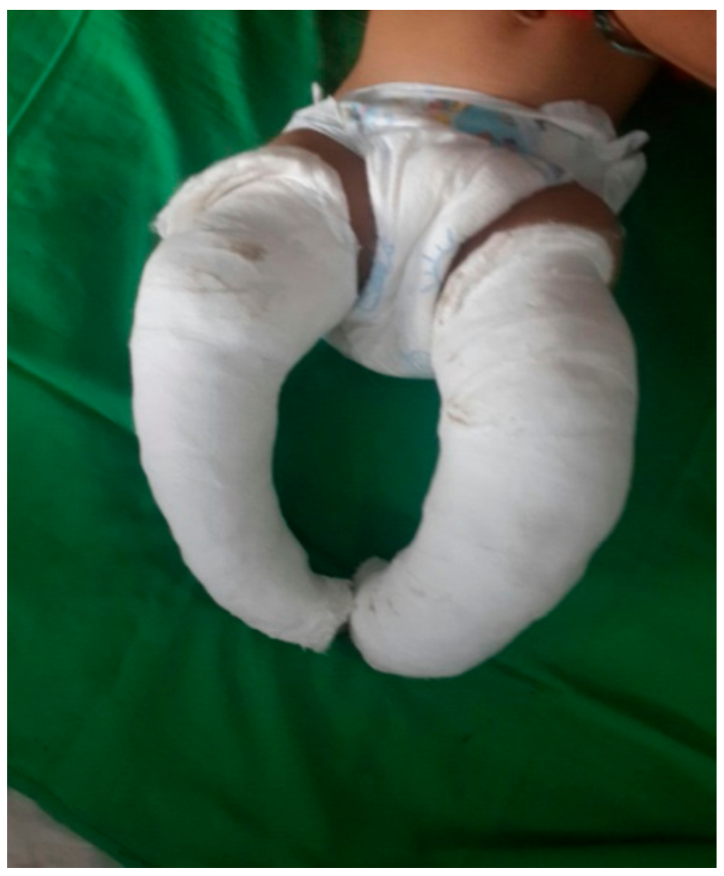
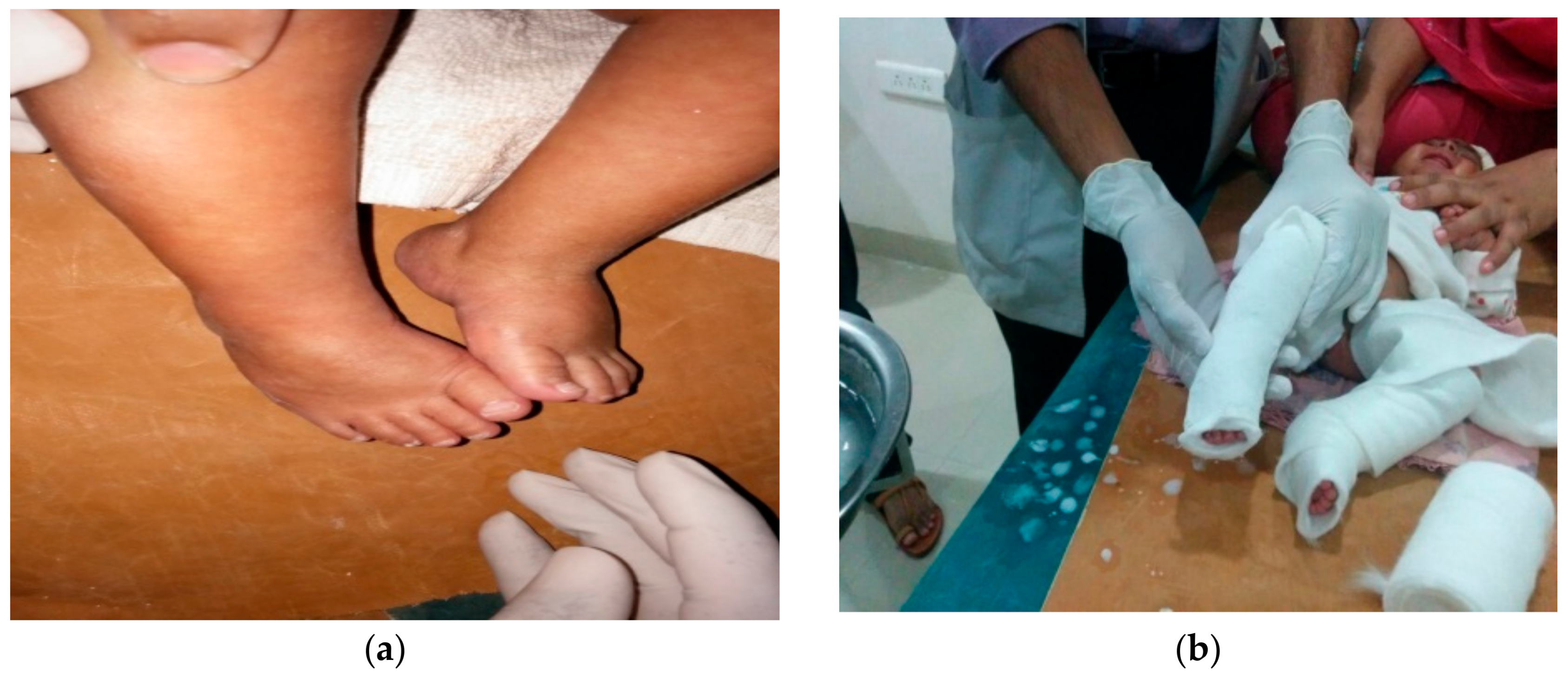
The severity of deformity and the functional outcome was graded according to the Pirani scoring system both at the beginning and at the end of the treatment. This detects the degree of correction. It scores according to 6 clinical signs: 3 for mid-foot, 3 for hind-foot. Three signs of mid-foot score (MS) and hind-foot score (HS) grade the amount of deformity between 0 and 3. The Pirani score 0 means a normal foot, score 3 means a moderately abnormal foot, score 6 means a severely abnormal foot [21]. We graded our results as excellent, good and poor with 0 to 0.4: excellent, 0.5 to 1: good and >1: poor. Excellent and good results were a reflection of the success of the treatment while the poor results were deemed as failures and offered surgical treatment.
Extensive counseling was given to the parents with regards to the plan and method of treatment, the duration of the treatment, the postoperative protocol, the importance of braces and compliance with them, and the goal of Ponseti method.
The treatment was done in 2 stages (Figure 1, Figure 2, Figure 3, Figure 4, Figure 5, Figure 6, Figure 7 and Figure 8):
(a)
correction of the deformity by weekly serial casting;
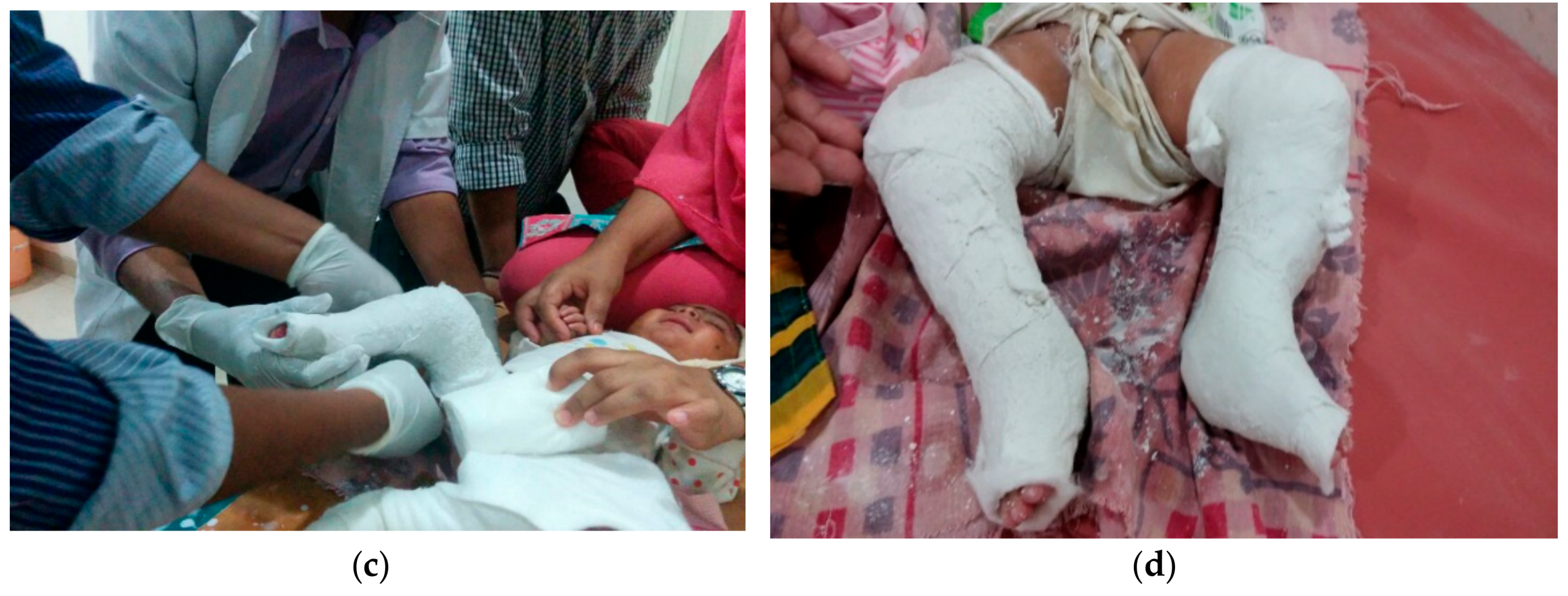
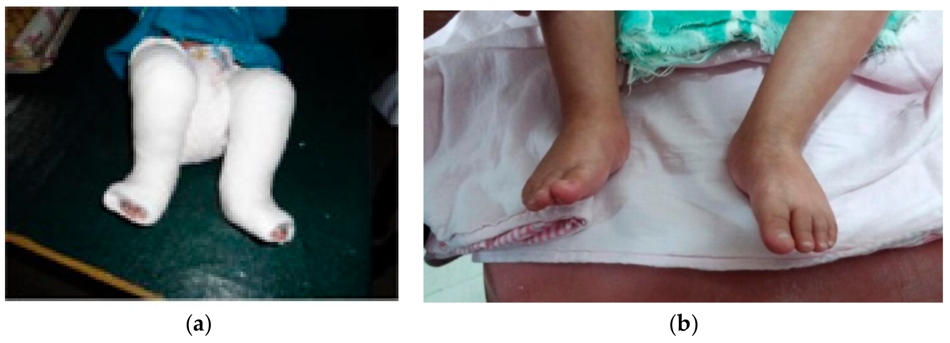
(b)
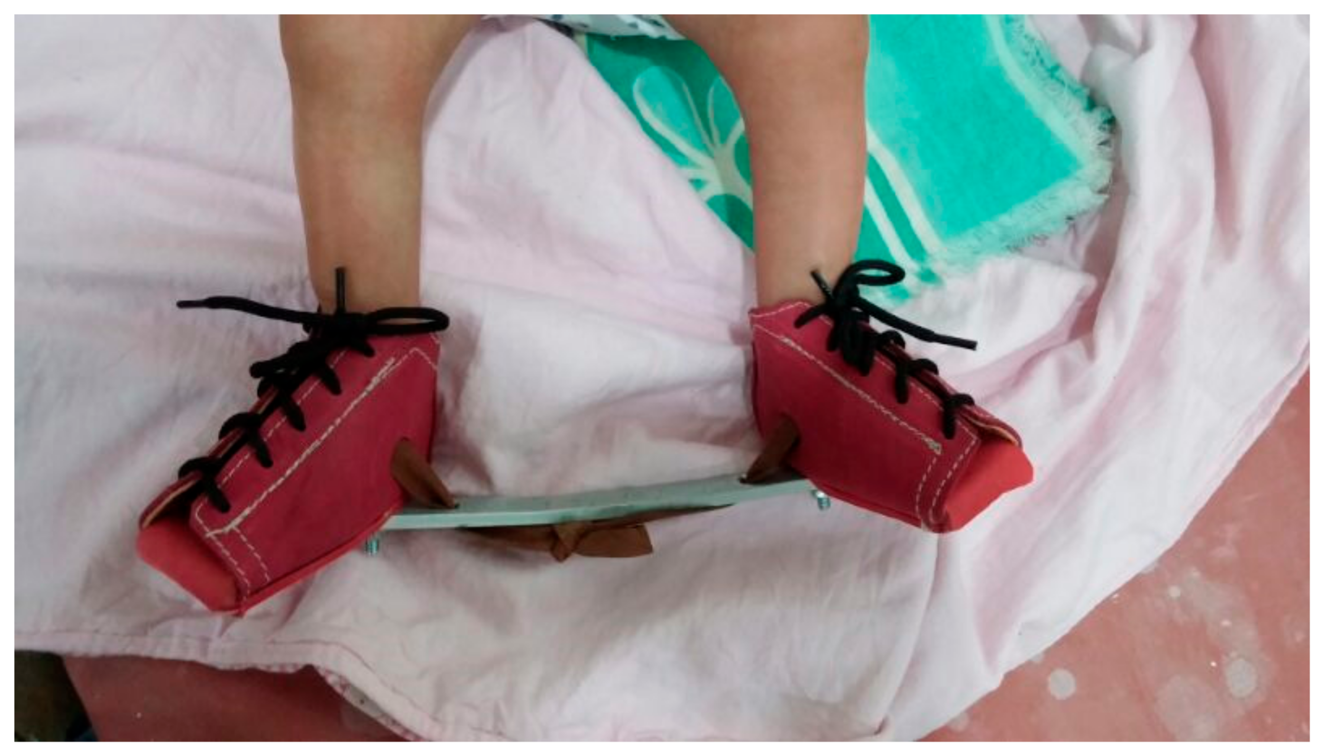
maintenance of that correction by bracing.
Casting was begun as soon as possible when the children came to us for treatment. In all the infants, the Pirani scoring was done to assess the initial severity. Weekly follow up were undertaken during the initial periods of bracing to ensure compliance and to periodically assure and educate the parents. Later, after the application of a Dennis Brown (DB) splint, a monthly follow up was advised for three months and then up to 4 years.

2.5. Statistical Analysis

Data analysis was undertaken using Statistical Package for the Social Science (SPSS) Version 17 [22] for Windows. The demographic variables and other variables were calculated by number and percentage. The Wilcoxon test was used to find a significant difference in Pirani score and functional rating before and after treatment, and a chi-square test was used to find an association between sex and laterality. Correlation and a test for correlation was used to find the significant correlation between the Pirani score and the total number of casts. A probability value of 0.05 was accepted as the level of statistical significance.

3. Results

3.1. Age

In our study, there were 180 males and 176 females. The mean age at presentation was 4.03 months, and the age-range from 0.5 to 12 months. The most common age group at presentation was ≤4 months i.e. 225 cases (63.33%); 83 cases (23.33%) were between the ages of >4–8 months; and 48 cases (13.33%) were more than 8 months and less than 12 months of age (Table 1).

3.2. Laterality

Among the 356 cases, 166 cases (46.67%) had bilateral clubfoot, and 190 cases (53.33%) had unilateral clubfoot out of which 106 (56%) were right-sided and 84 (44%) were left-sided.

3.3. Consaguinity and Family History
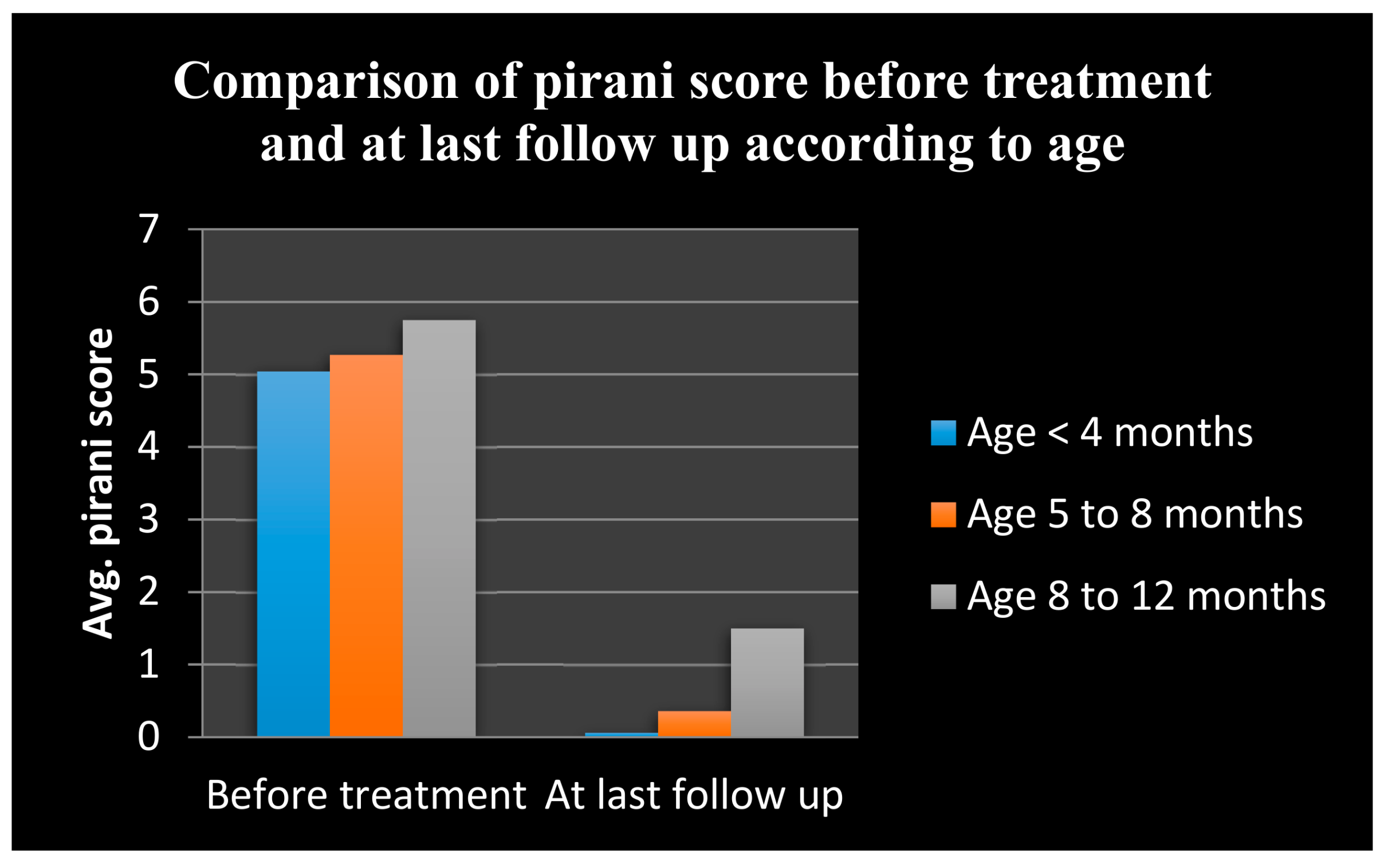
We found that 153 cases (43.33%) were born out of consanguineous marriages of parents and 203 cases (56.33%) had a positive family history of clubfoot.

3.4. Pirani Score

In our series, the mean initial Pirani score at the time of presentation of cases was 5.19 (range 3.5 to 6) and at last follow up was 0.33 (range 0 to 3.5) (Table 2 and Figure 9).
  • The mean + standard deviation (SD) at initial and final Pirani score in the age group of ≤4 months were 5.04 + 0. 85 and 0.06 + 0.21, respectively.
  • The mean + SD at initial and final Pirani score in the age group of >4–8 months were 5.27 + 0.65 and 0.36 + 0.39, respectively.
  • The mean + SD at initial and final Pirani score in the age group of >8–12 months were 5.75 + 0.61 and 1.50 + 1.41 respectively.

3.5. Number of Casts

The mean total number of casts required to correct the deformity was found to be 6.91, ranging from 5 to 10 (Table 3). In the age group of ≤4 months, the mean + SD total number of casts required was 6.56 + 0.75. In the age group of >4–8 months, the mean + SD total no. of casts required was 7 + 1. In the age group of >8 to 12 months, the mean + SD total number of casts required was 8.33 + 1.86.

3.6. Complications

In our series, a few minor complications were encountered during the casting procedure which included skin abrasions, cast saw injuries, cast loosening and cast breakage (Figure 10 and Figure 11).

3.7. Final Outcome

Based on the Pirani score, the final functional outcomes were graded as excellent, good and poor and consisted of 275, 41 and 40 patients, respectively (Table 4).

3.8. Comparison of Cost of Treatment

Total charges that we incurred for each patient were 71 USD, which was comparatively less than for the Turcos procedure charges of 96 USD and hence very affordable for each of the patients (Table 5).

4. Discussion

CTEV is one of the commonest congenital deformities. It is a complex deformity comprising equinus, varus, adductus and cavus, which are difficult to correct. It requires a meticulous and dedicated effort on the part of the treating physician and parents for the correction of the deformity The goal of treatment is to reduce or eliminate these deformities so that the patient has a functional, pain-free plantigrade foot with good mobility without calluses and does not need to wear modified shoes [15]. India is the second most-populous country in the world, with 25% of its people (about 375 million) living below the poverty line. Approximately 25,000 children are estimated to be born with idiopathic clubfoot every year in India. With such a large population living in poverty, non-invasive treatment of clubfoot with the Ponseti method has the potential to make a large impact on health outcomes for children who would otherwise be crippled by it [23]. Our study aims to evaluate the Ponseti method by using the Pirani score as a functional tool and to measure the cost-effectiveness of the Ponseti method.

4.1. Demographic and Etiological Variables

In our study, the average age of presentation was 4.03 months, with the most common age at presentation being ≤4 months i.e. 225 cases (63.33%), and with 131 cases (36.67%) presenting between >4 months–12 months (Table 1), which is comparable to M. Changulani et al. who treated 66 patients with 100 idiopathic clubfeet using the Ponseti method and reported the mean age at presentation of 12 weeks or three months (1 to 60 weeks) [24]. Our study had 180 cases (63.33%) in males and 176 (36.67%) in females, with a male to female ratio of 1.02, which is similar to the study of M. Changulani et al. who found that 50 patients (75.75%) were males and 16 patients (24.24%) were females [24]. As regards laterality, 166 cases (46.67%) had bilateral clubfoot and 190 cases (53.33%) had unilateral clubfoot (106 right-sided and 84 left-sided) which accords with other studies done by Ponseti et al., Changulani et al., Lehman et al., Christian et al. and Pavone et al. [7,24,25,26,27] (Table 6).
In our study, 153 cases (43.33%) had a history of consanguinous marriages of parents as compared to 203 cases (56.67%) who did not, which is comparable to the study by T. Sreenivas and A.R. Nataraj, which had, 54 (31%) out of 174 born of a consanguinous marriage [28]. This suggests a probable role of consanguinity as an etiological factor in the development of CTEV. Our study included 117 cases (33.33%) with a positive family history of clubfoot, which is similar to Morcuende et al. who evaluated 157 patients and reported that 22% of them had a positive family history of clubfoot deformity [6].

4.2. Pirani Score

The above findings signify that as the age of presentation increases the severity of the deformity increases. A comparison between the mean initial Pirani score and mean Pirani score at the last follow up shows that the Ponseti method was effective in treating the deformity in all the age groups of our study, as the p-value remains statistically significant in all age groups, although highly significant in the lower age group of <4 months (<0.0001) when compared with the higher age group >4–8 months (<0.005) and >8–12 months (<0.05). This clearly implies that the earlier the treatment begins, the better are the results. The available literature suggests that the results were better if this method of treatment was started as early as possible after birth [29,30].

4.3. Tenotomy and Number of Casts

In our study, 309 feet (77%) required tenotomy to correct the equinus deformity whereas 47 feet (23%) required only casting to the equinus deformity, which is comparable to studies by Changulani et al., Lehman et al., and Christian S. et al., Pavone et al. [24,25,26,27] (Table 7).
The mean total number of casts that we applied up to the final follow up to correct the deformity was 6.9 (ranged from 5 to 10) which was comparable to similar studies done by Changulani et al., Lehman et al., Christian et al. [24,25,26] (Table 8).

4.4. Complications

We encountered 4 types of complications among all the castings performed, which included the following: 57 cases (16%) had complications out of which 10 cases had skin abrasions, 36 cases had a cast breakage, 2 cases had a cast saw injury, and 9 cases had cast loosening and slippage. There were no major complications such as infection, skin necrosis, neurovascular compromise or profuse bleeding after tendoachilles tenotomy. Lehman et al. [25] reported having a 10.2% complication rate. The complications included cast saw injuries, abrasions, cast intolerence, maceration, blisters and slough.

4.5. Final Functional Outcome

We used the Pirani score as a functional tool for the assessment of the functional outcome. The results were graded as excellent, good and poor. Out of the 356 patients treated, excellent results were achieved in 275 cases (77.2%) while good and poor results were evenly distributed with 41 cases (11.2%) and 40 cases (11.1%), which is comparable to Sakale H et al. which had 92% excellent results [31]. Out of the 225 patients in the age group of ≤4 months, excellent results were achieved in 220 patients while two patients presented with poor results. These two patients did not follow the bracing protocol properly. The age group of >8–12 months had the maximum number of failures with 28 patients out of 48. This futher reinforced the belief that treatment begun earlier provides the best results. These 28 patients were offered surgery in the form of Turco’s procedure; 12 parents refused surgery citing the cost of the procedure as the main reason.

4.6. Cost-Effectiveness

Total charges that we incurred on each patient amounted to 71 USD, which was comparatively less than for the Turcos procedure charges of 96 USD and very affordable for the patients’ families (Table 6). Our findings in this regard were similar to the studies by Gadhok et al., Ferreira et al. and Hussain et al. [22,32,33]. Since our study group consisted of patients coming from low-income backgrounds in rural areas, cost was a very important variable for the success of the treatment.Grimes CT et al. [34] in their study also stated that the Ponseti method for the treatment of club foot is cost-effective and practical in a low-income country setting. The rural population in India was reported at 66.86% (% of total population) in 2016, according to World Bank development indicators [35], which makes the Ponseti method arguably the best for countering the menace of clubfoot.

5. Conclusions

Clubfoot or CTEV can be successfully graded by the Pirani score which is reproducible and does not show any inter-observer variation. This scoring system includes all the parameters in evaluating a clubfoot deformity and helps in decision-making as to whether to continue the ongoing management or opt for a surgical intervention. The Pirani scoring system also helps in documenting the progress of treatment and evaluating its results.
Management of CTEV by the Ponseti technique provides good functional and cosmetic outcomes when treated with strict adherence to the guidelines given by Ponseti that include serial casting, maintenance by bracing, and parental education, which can be started as soon as the child is born.
In a developing country like India, where there is a scarcity of resources and the referral system is poor, the Ponseti method is a very safe, efficient, cost-effective, economical treatment for the correction of clubfoot that radically decreases the need for extensive corrective surgery while also decreasing the social and financial burden of the parents, which can also be used successfully in rural contexts in other developing countries.

Acknowledgments

The authors would like to thank the support staff involved in the study. In addition, thanks are due to the superintendent of the primary health centre and to the Dean of Dnyandeo Yashwantrao Patil University. This project was funded by Dnyandeo Yashwantrao Patil research committee project no. IEC 0051/2013 who also helped to make the article open access.

Author Contributions

A.M. performed the analysis on all samples, interpreted the data, wrote the manuscript, and acted as corresponding author. R.M. performed data analysis, verified the analytical methods, and supervised the study. G.A. developed the analytical tools. P.C. helped in manuscript evaluation. H.J. and P.P. performed the experiments and analyzed the data.

Conflicts of Interest

The authors have no conflicts of interest.

References

  1. De Hoedt, A.M. Clubfoot Image Classification, University of Iowa, Iowa Research Online. 2013. Available online: http://ir.uiowa.edu/etd/4836 (accessed on 5 December 2017).
  2. Staheli, L. Clubfoot: Ponseti Management; Global HELP Publications: Seattle, WA, USA, 2003; pp. 4–5. [Google Scholar]
  3. Globalclubfoot. India | Global Clubfoot Initiative. 2018. Available online: http://globalclubfoot.com/countries/india/ (accessed on 31 March 2018).
  4. Africa Clubfoot Training Project. Chapter 2 Africa Clubfoot Training Basic & Advanced Clubfoot Treatment Provider Courses—Participant Manual; Africa Clubfoot Training Project; University of Oxford: Oxford, UK, 2017. [Google Scholar]
  5. Kelly, D.M. Congenital anomalies of the lower extremity. In Campbell’s Operative Orthopaedics, 12th ed.; Canale, S., Beaty, J., Eds.; Mosby Elsevier: Philadelphia, PA, USA, 2013; pp. 994–1012. [Google Scholar]
  6. Morcuende, J.; Dolan, L.; Dietz, F.; Ponseti, I. Radical reduction in the rate of extensive corrective surgery for clubfoot using the Ponseti method. Pediatrics 2004, 113, 376. [Google Scholar] [CrossRef] [PubMed]
  7. Ponseti, I.V.; Smoley, E.N. Congenital clubfoot: The results of treatment. J. Bone Joint Surg. 1963, 45, 261–344. [Google Scholar] [CrossRef]
  8. Morcuende, J. Keynote Address. In Proceedings of the 5th Australasian Ponseti Conference, Melbourne, Australia, 28–29 March 2014. [Google Scholar]
  9. Khan, M.A.; Chinoy, M.A.; Moosa, R.; Ahmed, S.K. Significance of Pirani score at bracing-implications for recognizing a corrected clubfoot. Iowa Orthop. J. 2017, 37, 151–156. [Google Scholar] [PubMed]
  10. Dobbs, M.B.; Nunley, R.; Schoenecker, P.L. Long term follow up of patients with clubfoot treated with extensive soft tissue releases. J. Bone Joint Surg. Am. 2006, 88, 986–996. [Google Scholar] [CrossRef] [PubMed]
  11. Cosma, D.; Vasilescu, D.; Vasilescu, D.; Valeanu, M. Comparative results of the conservative treatment in clubfoot by two different protocols. J. Pediatr. Orthop. B 2007, 16, 317–321. [Google Scholar] [CrossRef] [PubMed]
  12. Faizan, M.; Jilani, L.Z.; Abbas, M.; Zahid, M.; Asif, N. Management of idiopathic clubfoot by Ponseti technique in children presenting after one year of age. J. Foot Ankle Surg. 2015, 54, 967–972. [Google Scholar] [CrossRef] [PubMed]
  13. Staheli, L. Clubfoot: Ponseti Management, 3rd ed.; Global HELP Publications: Seattle, WA, USA, 2009. [Google Scholar]
  14. Pirani, S.; Outerbridge, H.K.; Sawatzky, B.; Stothers, K. A reliable method of clinically evaluating a virgin clubfoot evaluation. In Proceedings of the 21st World Congress of SICOT, Sydney, Australia, 11 October 1999. [Google Scholar]
  15. Ikeda, K. Conservative treatment of idiopathic clubfoot. J. Pediatr. Orthop. 1992, 12, 217–223. [Google Scholar] [CrossRef] [PubMed]
  16. Bensahel, H.; Guillaume, A.; Czukonyi, Z.; Desgrippes, Y. Results of physical therapy for idiopathic clubfoot: A long-term follow-up study. J. Pediatr. Orthop. 1990, 10, 189–192. [Google Scholar] [CrossRef] [PubMed]
  17. Nather, A.; Bose, K. Conservative and surgical treatment of clubfoot. J. Pediatr. Orthop. 1987, 7, 42–48. [Google Scholar] [CrossRef] [PubMed]
  18. Blakeslee, T.J. Congenital idiopathic talipes equinovarus (clubfoot). Current concepts. Clin. Podiatr. Med. Surg. 1997, 14, 9. [Google Scholar] [PubMed]
  19. Aronson, J.; Puskarich, C.L. Deformity and disability from treated clubfoot. J. Pediatr. Orthop. 1990, 10, 109–119. [Google Scholar] [CrossRef] [PubMed]
  20. Karski, T.; Wosko, I. Experience in the conservative treatment of congenital clubfoot in newborns and infants. J. Pediatr. Orthop. 1989, 9, 134–136. [Google Scholar] [CrossRef] [PubMed]
  21. Dobbs, M.B.; Rudzki, J.; Purcell, D.B.; Walton, T.; Porter, K.R.; Gurnett, C.A. Factors predictive of outcome after use of the Ponseti method for the treatment of idiopathic clubfeet. J. Bone Joint Surg. Am. 2004, 86, 22–27. [Google Scholar] [CrossRef] [PubMed]
  22. Bryman, A.; Cramer, D. Quantitative Data Analysis with IBM SPSS 17, 18 and 19: A Guide for Social Scientists; Routledge: New York, NY, USA, 2011. [Google Scholar]
  23. Gadhok, K.; Belthur, M.V.; Aroojis, A.J.; Cook, T.; Oprescu, F.; Ranade, A.S.; Morcuende, J.A. Qualitative assessment of the challenges to the treatment of idiopathic clubfoot by the Ponseti method in urban India. Iowa Orthop. J. 2012, 32, 135–140. [Google Scholar] [PubMed]
  24. Changulani, M.; Garg, N.K.; Rajagopal, T.S.; Bass, A.; Nayagam, S.N.; Sampath, J.; Bruce, C.E. Treatment of idiopathic club foot using the Ponseti method. Initial experience. J. Bone Joint Surg. Br. 2006, 88, 1385–1387. [Google Scholar] [CrossRef] [PubMed]
  25. Lehman, W.; Mohaideen, A.; Madan, S.; Scher, D.; Van Bosse, H.; Iannacone, M.; Bazzi, J.S.; Feldman, D.S. A method for the early evaluation of the Ponseti (Iowa) technique for the treatment of idiopathic clubfoot. J. Pediatr. Orthop. B 2003, 12, 133–140. [Google Scholar] [PubMed]
  26. Sætersdal, C.; Fevang, J.; Fosse, L.; Engesæter, L. Good results with the Ponseti method: A multicenter study of 162 clubfeet followed for 2–5 years. Acta Orthop. 2012, 83, 288–293. [Google Scholar] [CrossRef] [PubMed]
  27. Pavone, V.; Testa, G.; Costarella, L.; Pavone, P.; Sessa, G. Congenital idiopathic talipes equinovarus: An evaluation in infants treated by the Ponseti method. Eur. Rev. Med. Pharmacol. Sci. 2013, 17, 2675–2679. [Google Scholar] [PubMed]
  28. Sreenivas, T.; Nataraj, A. Parental consanguinity and associated factors in congenital talipes equinovarus. Foot 2012, 22, 2–5. [Google Scholar] [CrossRef] [PubMed]
  29. Cooper, D.M.; Dietz, F.R. Treatment of idiopathic clubfoot. A thirty-year follow-up note. J. Bone Joint Surg. Am. 1995, 77, 1477–1489. [Google Scholar] [CrossRef] [PubMed]
  30. Gupta, A.; Singh, S.; Patel, P.; Patel, J.; Varshney, M.K. Evaluation of the utility of the Ponseti method of correction of clubfoot deformity in a developing nation. Int. Orthop. 2008, 32, 75–79. [Google Scholar] [CrossRef] [PubMed]
  31. Sakale, H.; Agrawal, A.; Singh, A.; Sahoo, B.; Jain, A. Treatment of idiopathic clubfoot by Ponseti method. J. Orthop. Traumatol. Rehab. 2017, 9, 25. [Google Scholar] [CrossRef]
  32. Ferreira, L.F.A.; Nogueira, M.P.; Pereira, J.C.R.; Duarte, P.S. Comparison of hospital costs and duration of treatment with two different clubfoot protocols. Iowa Orthop. J. 2011, 31, 49–51. [Google Scholar] [PubMed]
  33. Hussain, H.; Burfat, A.M.; Samad, L.; Jawed, F.; Chinoy, M.A.; Khan, M.A. Cost-effectiveness of the Ponseti method for treatment of clubfoot in Pakistan. World J. Surg. 2014, 38, 2217–2222. [Google Scholar] [CrossRef] [PubMed]
  34. Grimes, C.E.; Holmer, H.; Maraka, J.; Ayana, B.; Hansen, L.; Lavy, C.B.D. Cost-effectiveness of club-foot treatment in low-income and middle-income countries by the Ponseti method. BMJ Glob. Health 2016, 1, e000023. [Google Scholar] [CrossRef] [PubMed]
  35. World Bank. Urban Population of Total Retrieved On. 2016. Available online: https://www.coursehero.com/file/p5tinva/World-Bank-2016-Urban-Population-of-total-Retrieved-on-March-23-2016/ (accessed on 3 April 2018).
Figure 1. Initial presentation.
Children 05 00049 g001
Figure 2. Manipulation.
Children 05 00049 g002
Figure 3. First cast for cavus correction.
Children 05 00049 g003
Figure 4. (ad) Subsequent steps in manipulation and casting (casts 2–4).
Children 05 00049 g004aChildren 05 00049 g004b
Figure 5. After removal of fourth cast.
Children 05 00049 g005
Figure 6. (a) Last cast (fifth cast) for equinus; (b) after removal of fifth cast.
Children 05 00049 g006
Figure 7. Dennis Brown (DB) splint and shoes.
Children 05 00049 g007
Figure 8. Clinical picture at last follow up.
Children 05 00049 g008
Figure 9. Bar diagram for Pirani score distribution.
Children 05 00049 g009
Figure 10. Skin abrasions.
Children 05 00049 g010
Figure 11. Cast saw injury.
Children 05 00049 g011
Table 1. Age distribution of patients.
Age of Patients (Months)Number of PatientsPercentage (%)
≤422563.33
>4–88323.33
>8–124813.33
Total356100
Table 2. Pirani score distribution pre- and post-treatment.
Age (Months)Pirani Score (Pre Treatment)Pirani Score (Last Follow Up)Wilcoxon Z Valuep Value
MeanSDMeanSD
≤45.040.850.060.215.61<0.0001
>4–85.270.650.360.392.99<0.005
>8–125.750.611.501.412.21<0.05
SD: Standard deviation
Table 3. Age distribution of casts.
Age (Months)Total Number of Casts
MeanSD
≤46.560.75
>4–871
>8–128.331.86
Table 4. Distribution of final outcome.
Age (Months) Functional Outcome
ExcellentGoodPoor
≤4220 (97.77%)03 (1.3%)02 (0.8%)
>4–853 (63.9%)20 (24%)10 (12%)
>8–1202 (4%)18 (3.7%)28 (58.33%)
Total275 (77.2%)41 (11.5%)40 (11.2%)
Table 5. Distribution of cost.
Ponseti Method (Variables)USDTurcos Method (Variables)USD
Cast and other consummables25Pre-operative antibiotics06
Tenotomy charges11Pre-operative investigations12
Dennis Brown (DB) Splint19Removal of stitches and k wires14
Other hospital charges16Post-operative antibiotics12
Hospital stay (3 days)26
DB splint and ankle foot orthosis26
Total71 96
Table 6. Comparison of laterality with other studies.
StudiesUnilateral (%)Bilateral (%)
Ponseti et al. [7]40 (60%)27 (40%)
Lehman et al. [25]15 (50%)15 (50%)
Changulani et al. [24]32 (48%)34 (52%)
Christian et al. [26]70 (60%)46 (40%)
Pavone et al. [27]50 (61%)32 (39%)
Our study190 (53.33%)166 (46.67%)
Table 7. Comparison of tenotomy with other studies.
StudiesPercentage of Feet Requiring Tenotomy
Changulani et al. [24]85% of feet
Pavone et al. [27]72% of feet
Lehman et al. [25]75% feet
Christian S. et al. [26]79% of feet
Our Study77% of feet
Table 8. Comparison of casts applied with other studies.
StudiesCasts
Lehman et al. [25]5.4 (range 4 to 9)
Changulani et al. [24]6 (range 2 to 12)
Christian S. et al. [26]7.2 (range 3 to 13)
Our Study6.9 (range 5 to 10)

