Chiral Recognition by DNA-Immobilized TLC Plate

Department of Chemistry, Faculty of Science, Okayama University of Science, Ridaicho 1-1, Kita-ku, Okayama 700-0005, Japan
*
Author to whom correspondence should be addressed.
Separations 2018, 5(1), 3; https://doi.org/10.3390/separations5010003
Submission received: 8 November 2017
/
Revised: 20 December 2017
/
Accepted: 22 December 2017
/
Published: 26 December 2017
Abstract
:DNA-immobilized silica gel TLC plate (DNA-TLC plate) by coating a mixture of double-stranded DNA and silica gel on a glass plate was stable in common organic solvents, such as alcohol, acetone, chloroform, and ethyl acetate. DNA-TLC plate showed different Rf values for the L- and D-form acidic amino acids, such as aspartic acid and glutamic acid. The chiral recognition of an acidic amino acid by the DNA-TLC was related to the interaction between the nucleic acid base, particularly guanine (G) and cytosine (C), and the amino acid. Furthermore, the DNA-TLC plate indicated the recognition of a chiral metal complex, such as the tris(1,10-phenanthroline)ruthenium(II) ([Ru(phen)3]2+) salt, which has Λ- and Δ-forms. Therefore, the double-stranded DNA-TLC plate may have the potential to be utilized as a chiral separation material for amino acids, peptides, and a metal complex with a chiral property.
1. Introduction
Chirality is ubiquitous in nature at the molecular level. Many essential biochemicals, such as the natural amino acids and monosaccharides, are chiral and, significantly, exist in only one enantiomeric form. Proteins or polysaccharides possess helical, sheeted, nematic, or turned structures with chiral properties [1,2,3,4,5]. These biopolymers have played an important role in biological processes. In the material science fields, these biopolymers with a chiral property have been used as a separation material of chiral substances [1], a sensing material of chiral drugs [6], a chiral catalyst for chemical reactions [7], or a nonlinear optical material [8]. Similarly, the double-stranded DNA, one of the most important biopolymers in living things, consists of two antiparallel polynucleotide strands intertwined with one another to form a right-handed double helix [9,10]. Therefore, double-stranded DNA can behave as a chiral polymer. As a result, double-stranded DNAs and these composite materials have been used as a chiral catalyst [11,12], chiral separation membrane [13,14], an affinity column or electrophoresis for chiral separation [15,16,17,18,19], and a nonlinear optical material [20]. We also recently reported the stereo-selective aggregation of an amyloidogenic peptide (AASIKVAVSADR) by the addition of double-stranded DNA [21]. Consequently, double-stranded DNA promoted aggregation of the amyloidogenic peptide, which consisted of only the L-form amino acid, and the amyloidogenic peptide/DNA matrix indicated a strong cell attachment activity [21].
Thin-layer chromatography (TLC) is a technique used to separate non-volatile mixtures [22]. TLC is performed on a glass plate, which is coated with a thin layer of an adsorbent material, such as silica gel or alumina. Generally, since silica gel does not possess a chiral property and cannot recognize a chiral molecule, TLC with a silica gel coating cannot separate chiral substances, such as L- and D-amino acids. On the other hand, silica and DNA show an affinity and form a DNA-silica composite material, such as an organic-inorganic hybrid material, through the sol-gel reaction [23,24]. Therefore, a double-stranded DNA-containing TLC might be used as a novel TLC plate with a chiral recognition property.
In this study, we prepared the DNA-immobilized silica gel TLC plate (DNA-TLC plate) by mixing double-stranded DNA and silica gel for use as a chiral separation material. As a result, the DNA-TLC plate showed different Rf values for an acidic amino acid, such as aspartic acid (Asp) and glutamic acid (Glu). The basic amino acid, such as arginine (Arg) and lysine (Lys), could almost not be separated. Additionally, we demonstrated the separation of a chiral metal complex, such as tris(1,10-phenanthroline)ruthenium(II) [Ru(phen)3]2+ with the Λ- and Δ-forms. Scheme 1 shows the molecular structures of Λ-[Ru(phen)3]2+ and Δ-[Ru(phen)3]2+. These metal complexes showed different Rf values for the Λ- and Δ-forms. These results suggested that these metal complexes stereoselectively interact with the double-stranded DNA. Therefore, the DNA-TLC plate may have the potential to be utilized as a chiral separation material for amino acids, peptides, drug molecules, and metal complexes.
2. Experimental Section
2.1. Materials
Double-stranded DNA (sodium salt from salmon milt, molecular weight; >5 × 10⁶) was obtained from Biochem Ltd. (Saitama, Japan). Wako gel® B-5, silica gel for TLC, was purchases from Wako Pure Chemical Industries, Ltd. (Osaka, Japan). This silica gel was contained the 5–7% CaSO4 in its. L-form amino acids of aspartic acid (Asp), glutamic acid (Glu), alanine (Ala), valine (Val), arginine (Arg), and lysine (Lys) were obtained from Wako Pure Chemical Industries. D-form amino acids of Asp, Glu, Ala, Val, Arg, and Lys were purchased from Peptide Institute, Inc. (Osaka, Japan). 1,10-Phenanthroline monohydrate, ruthenium(III) chloride hydrate, bis[(+)-tartrato]diantimonate(III) dipotassium trihydrate, sodium perchlorate monohydrate, perchloric acid, ninhydrin, and sodium hydrate were obtained from Wako Pure Chemical Industries, Nacalai Tesque, Inc. (Kyoto, Japan), and Kanto Chemical Co., Inc. (Tokyo, Japan). Other amino acids (histidine (His), proline (Pro), leucine (Leu), isoleucine (Ile), methionine (Met), phenylalanine (Phe), tryptophan (Trp), threonine (Thr), tyrosine (Tyr), cysteine (Cys), serine (Ser), asparagine (Asn), and glutamine (Gln)) were obtained from Merck KGaA (Darmstadt, Germany). Solvents were used an analytical grade in all the experiments described. Ultra-pure water (Merck KGaA, Darmstadt, Germany) was used in this experiment.
2.2. Preparation of DNA-TLC Plate
DNA-TLC plate was prepared as follows: Wako gel® B-5 (5 g) was put in mortar, added an aqueous DNA solution (15 mg/mL, 10 mL) in its, and then quickly mixed by pestle. This DNA-silica gel paste was spread uniformly on five glass plates (25.4 × 76.2 mm2, Matsunami Glass Ind., Ltd., Osaka, Japan). This glass plate was dried at room temperature for 1 h to evaporate the water, and heated at 140 °C for 5 h to be solidified. The thickness of silica gel layer in DNA-TLC plate was 0.8–1 mm.
2.3. Synthesis of Tris(1,10-phenanthroline)ruthenium(II)
The synthesis of tris(1,10-phenanthroline)ruthenium(II) dichloride (Ru(phen)3Cl2) was described in a previous report [25,26]. Ruthenium(III) chloride hydrate (1 g, 4 mmol) and 1,10-phenanthroline monohydrate (3.94 g, 20 mmol) were dissolved in ethanol (200 mL) and refluxed for 72 h. The resulting dark brown precipitate was filtered and dried overnight at room temperature. The solid was extracted in benzene to remove the non-reacted 1,10-phenanthroline. This remaining solid was recrystallized from water. The identification of the synthetic [Ru(phen)3]2+ was demonstrated by MALDI-TOF-MS (Autoflex speed, Bruker Corporation, Billerica, MA, USA).
Δ-Ru(phen)3(ClO4)2 was prepared as follows [26]: the synthetic rac-Ru(phen)3Cl2 (0.65 g) was dissolved in 50 mL of water and cooled. The bis[(+)-tartrato]diantimonate(III) dipotassium trihydrate (0.25 g) in 10 mL of water was slowly added to a cold Ru(phen)3Cl2 solution. The voluminous orange-yellow precipitate was washed several times with water and separated into the precipitate and filtrate. The precipitate was dissolved by shaking in an aqueous NaOH solution (50 mM, 37.5 mL). Dilute acetic acid was added until the acid and an orange precipitate were obtained. This precipitate was dried overnight at room temperature. This precipitate was dissolved in an aqueous NaOH solution (50 mM, 25 mL), then the solution was filtered. An aqueous sodium perchlorate solution was slowly added in excess, and the resulting orange-yellow precipitate filtered and washed with cold water contained a small amount of perchloric acid. This solid was twice recrystallized from water containing the perchloric acid. The specific rotation of Δ-Ru(phen)3(ClO4)2 was determined by a DIP-1000 digital polarimeter (Japan Spectroscopic Co., Tokyo, Japan): [α] = 1180°. The specific rotation value was similar to the reported value [26].
Λ-Ru(phen)3(ClO4)2 was prepared as follows [26]: bis[(+)-tartrato]diantimonate(III) dipotassium trihydrate (0.4 g) was added to the filtrate and cooled in ice to remove any traces of the Δ-Ru(phen)3(ClO4)2. After filtration, the solution was treated with a 3 M perchloric acid solution, and the resulting orange-yellow precipitate of the perchlorate was recrystallized twice from warm water containing a trace amount of perchloric acid. The obtained crystals were washed with cold methanol: diethyl ether (9:1, v/v). The specific rotation of Λ-Ru(phen)3(ClO4)2 was determined by the polarimeter: [α] = −1220°. This specific rotation value was similar to the reported value [26].
2.4. Separation of Amino Acid and Metal Complex on DNA-TLC Plate
The L- and D-form amino acids on the DNA-TLC plate were detected as follows: the L- and D-form amino acid solution was spotted on the DNA-TLC plate by a glass capillary, dried at room temperature for 10 min, and then developed for approximately 10 min using various mobile phases. Table 1 shows the various mobile phases. The amino acid spots were visualized by spraying the DNA-TLC plate with an aqueous ninhydrin solution in the presence of 0.2% ethanol, drying, and then heating on a hot plate at 120 °C for 1 min.
The metal complex on the DNA-TLC plate was detected as follows: the aqueous [Ru(phen)3]2+ solution was spotted on the DNA-TLC plate, dried at room temperature for 10 min, and then developed using various mobile phases. The DNA-TLC plate was dried at room temperature for 10 min. The [Ru(phen)3]2+ shows a strong fluorescence during the UV irradiation of 365 nm. Therefore, the spot of [Ru(phen)3]2+ on the DNA-TLC plate was detected by UV irradiation of 365 nm.
The Rf values are defined as the ratio of the distance developed by the eluted spot to that developed by the eluted front. All the Rf values represent the mean of three separate determinations.
2.5. IR Measurements of Amino Acid-Accumulated DNA Film
A water-insoluble UV-irradiated DNA film was prepared by a reported procedure [27]. The UV-irradiated DNA films were stored in ultrapure water for more than 1 day to remove the water-soluble DNA and used for further experiments. The L- and D-form Asp was dissolved in ultrapure water (5 mg/mL). The UV-irradiated DNA film was immersed in each aqueous amino acid solution (3 mL) for 24 h. The amino acid-accumulated DNA film was rinsed with ultrapure water (10 mL × 3 times) and dried overnight. The infrared (IR) absorption spectra of the amino acid-accumulated DNA film were measured by the KBr method using an FT-IR 8400 Fourier transform infrared spectrometer (Shimadzu Corp., Kyoto, Japan). The IR spectrum was measured with the resolution of 4 cm−1.
3. Results and Discussion
3.1. Chiral Recognition of Amino Acid on DNA-TLC Plate
Each of these DNA-TLC plates were immersed in common organic solvents, such as methanol, ethanol, butanol, acetone, chloroform, and ethyl acetate for 30 min, however, these plates did not show any elution of the DNA or silica components. Additionally, under basic conditions, such as entries 8 and 9 in Table 1, the DNA-TLC plate did not show any elution. These results suggested that the DNA-TLC plate is stable in common organic solvents or under basic conditions and can be used as an analytical tool for chromatography. Therefore, we used the DNA-TLC plate for the chiral separation of amino acids which have L- and D-forms. The separation of the amino acid by the DNA-TLC plate was demonstrated under various mobile phases (see Table 1). These amino acids on the DNA-TLC plate were detected by the ninhydrin reaction.
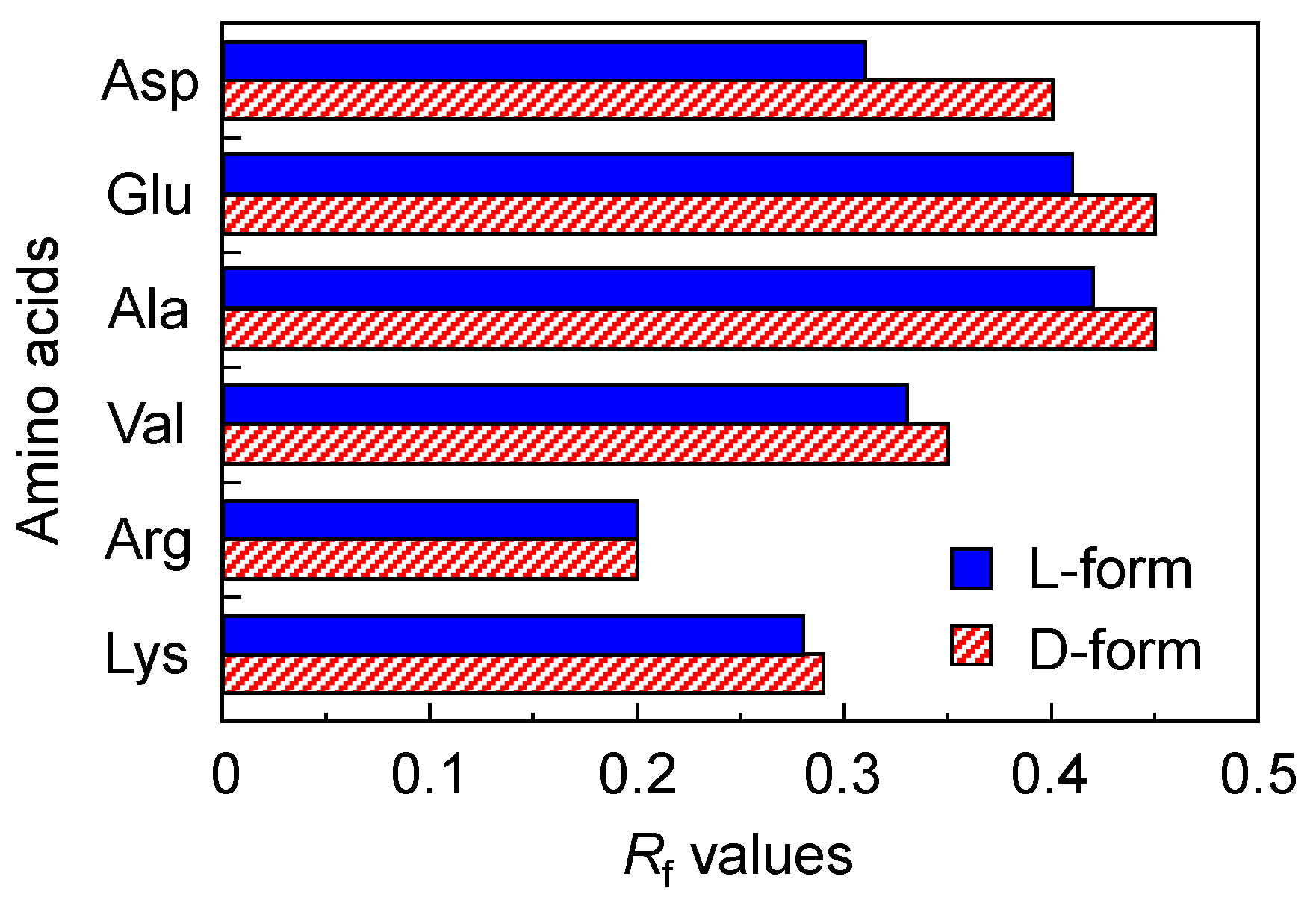
Figure 1 shows the Rf value of the L- and D-form amino acids (Asp, Glu, Ala, Val, Arg, and Lys) for the acidic mobile phase, such as entry 1 in Table 1. Additionally, Figure S1 in the Supplementary Materials shows the photograph of L- and D-form amino acids (Asp, Val, and Arg) on DNA-TLC plate. The Rf value of the L-form acidic and neutral amino acids showed a lower value than that of the D-form acidic and neutral amino acids. Especially, the differential Rf (ΔRf) value between the L- and D-forms of Asp was approximately 0.1. Therefore, we demonstrated the developments under other mobile phases. When the Asp and Glu, such as the acidic amino acids, were developed under the neutral mobile phase, such as 4–7 in Table 1, the ΔRf value became low (data not shown). Similar results were obtained for the neutral amino acids, such as Ala and Val. In contrast, the normal silica-TLC plate without the mixing of the double-stranded DNA did not show a ΔRf value. These results suggested that the chiral separation on the DNA-TLC plate is related to the electrostatic interaction between the double-stranded DNA and amino acid.
Additionally, we demonstrated the developments of other amino acids, such as His, Pro, Leu, Ile, Met, Phe, Trp, Thr, Tyr, Cys, Ser, Asn, and Gln, under similar acidic mobile phases, as shown in entry 1 in Table 1. The Rf values of the other L- and D-form amino acids are shown in Figure S1 in the Supplementary Materials. The ΔRf values were <0.03 and other amino acids did not also show a superior ΔRf value. These results suggested that the DNA-TLC plate possesses the chiral separation property of an acidic amino acid.
On the other hand, the basic amino acids, such as Arg and Lys, were developed under similar acidic mobile phases. However, the basic amino acid did not show a superior ΔRf value on the DNA-TLC plate using the acidic mobile phases (see Figure 1). Generally, under acidic conditions, the electric charge of basic amino acids, such as Arg, Lys, and His, is +2. Therefore, these amino acids strongly interacted with the negatively charged DNA and could not develop on the DNA-TLC plate. In fact, the Rf values of the basic amino acid were lower than that of other amino acids (see Figure 1 and Figure S1 in the Supplementary Materials). Additionally, a similar result has been reported for a DNA/polyvinyl alcohol interpenetrating polymer network coated TLC [28]. Therefore, we demonstrated the development of a basic amino acid using the basic mobile phase. However, the basic amino acids indicated a low Rf value of <0.05 (data not shown). These results suggested that the basic amino acid strongly interacts with not only the DNA, but also the silica gel on the TLC plate. Therefore, it is necessary for the separation of the basic amino acid to composite with a different inorganic components, such as alumina.
3.2. IR Spectrum of Amino Acid-Accumulated DNA Film
The interactions between the double-stranded DNA and amino acid were determined by IR spectrometry. However, the DNA-TLC plate showed a strong absorbance which was attributed to the silica and the absorption bands of the DNA and amino acid that were too weak to determine the molecular structure. Therefore, we used a water-insoluble UV-irradiated DNA film. The water-insoluble UV-irradiated DNA film was prepared by the cross-linking reaction by 254 nm UV irradiation [27].
Figure 2 shows the IR spectra of: (a) UV-irradiated DNA film; (b) L-Asp accumulated DNA film; (c) D-Asp accumulated DNA film; and (d) L-Asp. The D-Asp showed an IR spectrum similar to L-Asp (data not shown). The absorption band at 1228 cm−1, related to the antisymmetric vibration of the phosphate group [29,30,31,32], was shifted to a lower wavenumber with the accumulation of L-Asp and D-Asp (see the dashed line in Figure 2). This is due to the electrostatic interaction between the amino group of Asp and the phosphate group of DNA, and a similar interaction has been reported for the DNA-peptide biomatrix [21] and DNA-poly(allylamine) composite material [33]. Additionally, the absorption band at 1533 cm−1, related to the in-plane vibration of the cytosine (C) base and the stretching vibration of C = N of the guanine (G) base [29,30,31], was shifted to a higher wavenumber when the DNA-film accumulated the L-Asp (see the dashed line in Figure 2). A similar phenomenon, such as the shift to a higher wavenumber, did not occur by the accumulation of D-Asp. This result suggested that, although L-Asp interacts with not only the phosphate group, but also the nucleic acid base, D-Asp does not strongly interact with the nucleic acid base. Therefore, since L-Asp more strongly interacts with the double-stranded DNA than D-Asp, the Rf value of L-Asp on the DNA-TLC plate was lower than that of D-Asp.
3.3. Chiral Recognition of Metal Ion Complex on DNA-TLC Plate
The DNA-TLC plate showed the chiral recognition of the L- and D-amino acids. Therefore, we demonstrated the chiral recognition of a metal ion complex, such as [Ru(phen)3]2+. The [Ru(phen)3]2+ is one of chiral metal ion complexes that possesses the Λ- and Δ-forms (see molecular structures in Scheme 1). The interactions between Λ- and Δ-[Ru(phen)3]2+ and the double-stranded DNA in an aqueous solution were determined by the NMR, fluorescence spectrometry, circular dichroism spectrometry, cyclic voltammetry, and dialysis methods [34,35,36]. However, the interactive estimation between Λ- and Δ-[Ru(phen)3]2+ and the double-stranded DNA by thin layer chromatography has not been reported to the best of our knowledge.
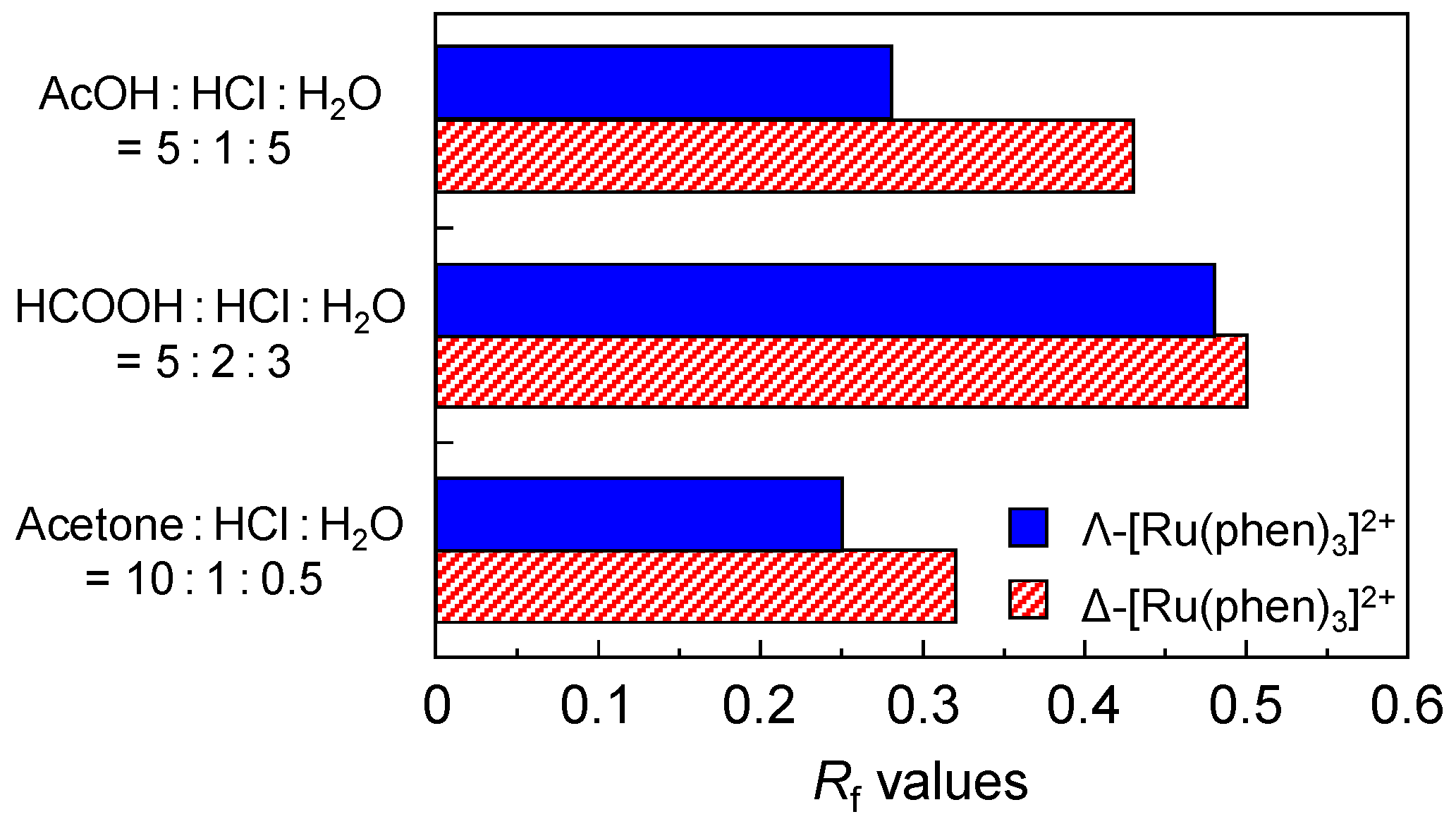
The [Ru(phen)3]2+ was developed under various acidic mobile phases and the fluorescence detected by 365 nm UV irradiation. Figure 3 shows the Rf value of the Δ- and Λ-form [Ru(phen)3]2+ when they were developed under various mobile phases. The [Ru(phen)3]2+ showed a ΔRf value for all the acidic mobile phases. Particularly, for AcOH:HCl:H2O = 5:1:5, the Rf value of Δ-[Ru(phen)3]2+ was higher than that of Λ-[Ru(phen)3]2+ and the ΔRf value was approximately 0.15. These results suggested that the Λ-[Ru(phen)3]2+ more strongly interacts with the double-stranded DNA on the DNA-TLC plate than Δ-[Ru(phen)3]2+. On the other hand, although we demonstrated the development under neutral and basic mobile phase conditions, the DNA-TLC plate did not show a superior ΔRf value (data not shown). Similar results were also obtained using the normal silica-TLC plate without the mixing of the double-stranded DNA. These results suggested that the chiral recognition of Λ-[Ru(phen)3]2+ and Δ-[Ru(phen)3]2+ by the double-stranded DNA on the DNA-TLC plate is due to not only an electrostatic interaction, but also a specific interaction mode, such as intercalation or groove binding.
Generally, the native double-stranded DNA possesses the B-formed structure in an aqueous solution [9]. Therefore, in an aqueous solution, it has been reported that the Δ-[Ru(phen)3]2+ mainly interacts with DNA during intercalation and base stacking, and the Λ-[Ru(phen)3]2+ binds to the surface of the DNA by an electrostatic interaction [34,35,36]. However, since these phenomena, such as the selective recognition of Δ-[Ru(phen)3]2+, occurred under neutral aqueous conditions, they do not correspond with the interaction on the DNA-TLC plate. In fact, we previously reported that the double-stranded DNA in a DNA-inorganic complex, which was prepared by mixing the double-stranded DNA and silane coupling agent, 3-aminopropyltrimethoxysilane, possessed the C-formed DNA, and this C-formed DNA interacted with Λ-[Ru(phen)3]2+ rather than Δ-[Ru(phen)3]2+ [37]. Therefore, we think that similar phenomena occurred on the DNA-TLC plate. As a result, Λ-[Ru(phen)3]2+ interacted with the double-stranded DNA on the DNA-TLC plate and indicated an Rf value lower than that of Δ-[Ru(phen)3]2+.
4. Conclusions
We prepared the DNA-immobilized silica gel TLC plate (DNA-TLC plate) by the coating of a double-stranded DNA and silica gel mixture as a chiral separation material. As a result, the DNA-TLC plate showed different Rf values for acidic amino acids, such as Asp and Glu. The basic amino acids, such as Arg and Lys, were almost not recognized. Additionally, we demonstrated the separation of a chiral metal ion complex, such as [Ru(phen)3]2+, with the Λ- and Δ-forms. As a result, Λ-[Ru(phen)3]2+ more strongly interacted with the double-stranded DNA on the DNA-TLC plate than Δ-[Ru(phen)3]2+. Therefore, the DNA-TLC plate may have the potential to be utilized as a chiral separation material for amino acids, peptides, drug molecules, and metal complexes with a chiral property.
Supplementary Materials
The following are available online at https://www.mdpi.com/2297-8739/5/1/3/s1, Figure S1: Photograph of L- and D-form amino acids on DNA-TLC plate. Figure S2: Rf values of L- and D-form amino acids on DNA-TLC plate.
Author Contributions
Masanori Yamada and Mami Inoue designed and performed the experiments and analyzed the data. Masanori Yamada wrote the paper.
Conflicts of Interest
The authors declare no conflict of interest.
References
- Zhang, M.; Qing, G.; Sun, T. Chiral biointerface materials. Chem. Soc. Rev. 2012, 41, 1972–1984. [Google Scholar] [CrossRef] [PubMed]
- Yashima, E.; Maeda, K. Chirality-responsive helical polymers. Macromolecules 2008, 41, 3–12. [Google Scholar] [CrossRef]
- Yashima, E.; Ousaka, N.; Taura, D.; Shimomura, K.; Ikai, T.; Maeda, K. Supramolecular helical systems: Helical assemblies of small molecules, foldamers, and polymers with chiral amplification and their functions. Chem. Rev. 2016, 116, 13752–13990. [Google Scholar] [CrossRef] [PubMed]
- Kane-Maguire, L.A.P.; Wallace, G.G. Chiral conducting polymers. Chem. Soc. Rev. 2010, 39, 2545–2576. [Google Scholar] [CrossRef] [PubMed]
- Lv, Z.; Chen, Z.; Shao, K.; Qing, G.; Sun, T. Stimuli-directed helical chirality inversion and bio-applications. Polymers 2016, 8, 310. [Google Scholar] [CrossRef]
- Bi, C.; Zheng, X.; Azaria, S.; Beeram, S.; Li, Z.; Hage, D.S. Chromatographic studies of protein-based chiral separations. Separations 2016, 3, 27. [Google Scholar] [CrossRef] [PubMed]
- Guibal, E. Heterogeneous catalysis on chitosan-based materials: A review. Prog. Polym. Sci. 2005, 30, 71–109. [Google Scholar] [CrossRef]
- Lee, B.; Kwon, H.; Kim, S.; Rotermund, F. Natural silk protein as a new broadband nonlinear optical material. Opt. Mater. Express 2016, 6, 993–1002. [Google Scholar] [CrossRef]
- Saenger, W. Principles of Nucleic Acid Structure; Springer-Verlag: Berlin, Germany, 1987. [Google Scholar]
- Bates, A.D.; Maxwell, A. DNA Topology; Oxford University Press: New York, NY, USA, 2005. [Google Scholar]
- Roelfes, G.; Boersma, A.J.; Feringa, B.L. Highly enantioselective DNA-based catalysis. Chem. Commun. 2006, 6, 635–637. [Google Scholar] [CrossRef] [PubMed]
- Park, S.; Sugiyama, H. DNA as a chiral scaffold for asymmetric synthesis. Molecules 2012, 17, 12792–12803. [Google Scholar] [CrossRef] [PubMed] [Green Version]
- Higuchia, A.; Hayashi, A.; Kanda, N.; Sanui, K.; Kitamura, H. Chiral separation of amino acids in ultrafiltration through DNA-immobilized cellulose membranes. J. Mol. Struct. 2005, 739, 145–152. [Google Scholar] [CrossRef]
- Yoshikawa, M.; Maruhashi, M.; Iwamoto, Y.; Ogata, N. Chiral separation of racemic amino acids through DNA. Macromol. Symp. 2007, 249–250, 557–561. [Google Scholar] [CrossRef]
- Michaud, M.; Jourdan, E.; Villet, A.; Ravel, A.; Grosset, C.; Peyrin, E. A DNA aptamer as a new target-specific chiral selector for HPLC. J. Am. Chem. Soc. 2003, 125, 8672–8679. [Google Scholar] [CrossRef] [PubMed]
- Michaud, M.; Jourdan, E.; Ravelet, C.; Villet, A.; Ravel, A.; Grosset, C.; Peyrin, E. Immobilized DNA aptamers as target-Specific chiral stationary phases for resolution of nucleoside and amino acid derivative enantiomers. Anal. Chem. 2004, 76, 1015–1020. [Google Scholar] [CrossRef] [PubMed]
- Tohala, L.; Oukacine, F.; Ravelet, C.; Peyrin, E. Chiral Resolution Capabilities of DNA Oligonucleotides. Anal. Chem. 2015, 87, 5491–5495. [Google Scholar] [CrossRef] [PubMed]
- Su, X.; Qin, F.; Kong, L.; Ou, J.; Xie, C.; Zou, H. Characterization of enantioselective binding of racemic natural tetrahydropalmatine to DNA by chromatographic methods. J. Chromatogr. B 2007, 845, 174–179. [Google Scholar] [CrossRef] [PubMed]
- Huang, R.; Xiong, W.; Wang, D.; Guo, L.; Lin, Z.; Yu, L.; Chu, K.; Qiu, B.; Chen, G. Label-free aptamer-based partial filling technique for enantioseparation and determination of dl-tryptophan with micellar electrokinetic chromatography. Electrophoresis 2013, 34, 254–259. [Google Scholar] [CrossRef] [PubMed]
- Grote, J.G.; Hagen, J.A.; Zetts, J.S.; Nelson, R.L.; Iggs, D.E.; Stone, D.M.O.; Yaney, P.P.; Heckman, E.; Zhang, C.; Steier, W.H.; et al. Investigation of polymers and marine-derived DNA in optoelectronics. J. Phys. Chem. B 2004, 108, 8584–8591. [Google Scholar] [CrossRef]
- Yamada, M.; Hara, S.; Yamada, T.; Katagiri, F.; Hozumi, K.; Nomizu, M. Double-stranded DNA stereoselectively promotes aggregation of amyloid-like fibrils and generates peptide/DNA matrices. Biopolymers 2014, 102, 465–472. [Google Scholar] [CrossRef] [PubMed]
- Randerath, K. Thin-Layer Chromatography; Verlag Chemie Academic Press: New York, NY, USA, 1968. [Google Scholar]
- Satoh, S.; Fugetsu, B.; Nomizu, M.; Nishi, N. Functional DNA-silica composite prepared by sol-gel method. Polym. J. 2005, 37, 94–101. [Google Scholar] [CrossRef]
- Yamada, M.; Aono, H. DNA-inorganic hybrid material as selective absorbent for harmful compounds. Polymer 2008, 49, 4658–4665. [Google Scholar] [CrossRef]
- Dwyer, F.P.; Humpolets, J.E.; Nyholm, R.S. The chemistry of ruthenium. Part I. The redox potential of the tris-orthophenanthroline ruthenous ion. J. Proc. R. Soc. N. S. W. 1946, 80, 212–216. [Google Scholar]
- Dwyer, F.P.; Gyarfas, E.C. The chemistry of ruthenium. Part VI. The existence of the tris-o-phenanthroline ruthenium II and the tris-o-phenanthroline ruthenium III ions in entantiomorphous forms. J. Proc. R. Soc. N. S. W. 1949, 83, 170–173. [Google Scholar]
- Yamada, M.; Kato, K.; Nomizu, M.; Sakairi, N.; Ohkawa, K.; Yamamoto, H.; Nishi, N. Preparation and characterization of DNA films induced by UV irradiation. Chem. Eur. J. 2002, 8, 1407–1412. [Google Scholar] [CrossRef]
- Liu, X.D.; Kubo, T.; Diao, H.Y.; Benjamas, J.; Yonemichi, T.; Nishi, N. DNA/polyvinyl alcohol interpenetrating polymer network as stationary phase for thin layer chromatography. Anal. Biochem. 2009, 393, 67–72. [Google Scholar] [CrossRef] [PubMed]
- Tajmir-Riahi, H.A.; Naoui, M.; Ahimad, R. The effects of Cu2+ and Pb2+ on the solution structure of calf thymus DNA: DNA condensation and denaturation studied by Fourier-transform IR difference spectroscopy. Biopolymers 1993, 33, 1819–1827. [Google Scholar] [CrossRef] [PubMed]
- Banyay, M.; Sarkaräslund, A. A library of IR bands of nucleic acids in solution. Biophys. Chem. 2003, 104, 477–488. [Google Scholar] [CrossRef]
- Silverstein, R.M.; Webster, F.X. Spectrometric Identification of Organic Compounds; John Wiley & Sons: New York, NY, USA, 1998. [Google Scholar]
- Yamada, M.; Goto, A. Proton conduction of DNA-imidazole composite material under anhydrous condition. Polym. J. 2012, 44, 415–420. [Google Scholar] [CrossRef]
- Yamada, M.; Hashimoto, K. DNA-cyclodextrin composite material for environmental applications. Biomacromolecules 2008, 9, 3341–3345. [Google Scholar] [CrossRef] [PubMed]
- Barton, J.K.; Danishefsky, A.T.; Goldberg, J.M. Tris(phenanthroline)ruthenium(II): Stereoselectivity in binding to DNA. J. Am. Chem. Soc. 1984, 106, 2172–2176. [Google Scholar] [CrossRef]
- Yamagishi, A. Electric dichroism evidence for stereospecific binding of optically active tris chelated complexes to DNA. J. Phys. Chem. 1984, 88, 5709–5713. [Google Scholar] [CrossRef]
- Eriksson, M.; Leijon, M.; Hiort, C.; Norden, B.; Graeslund, A. Binding of Δ- and Λ-[Ru(phen)3]2+ to [d(CGCGATCGCG)]2 Studied by NMR. Biochemistry 1994, 33, 5031–5040. [Google Scholar] [CrossRef] [PubMed]
- Yamada, M.; Nomizu, M.; Satoh, S.; Ohkawa, K.; Yamamoto, H.; Nishi, N. DNA with γ-Aminopropyltriethoxysilane switches the B- and C-form structures by thermal control. ChemBioChem 2003, 4, 232–234. [Google Scholar] [CrossRef] [PubMed]
Scheme 1.
Molecular structure of Λ-[Ru(phen)3]2+ and Δ-[Ru(phen)3]2+.
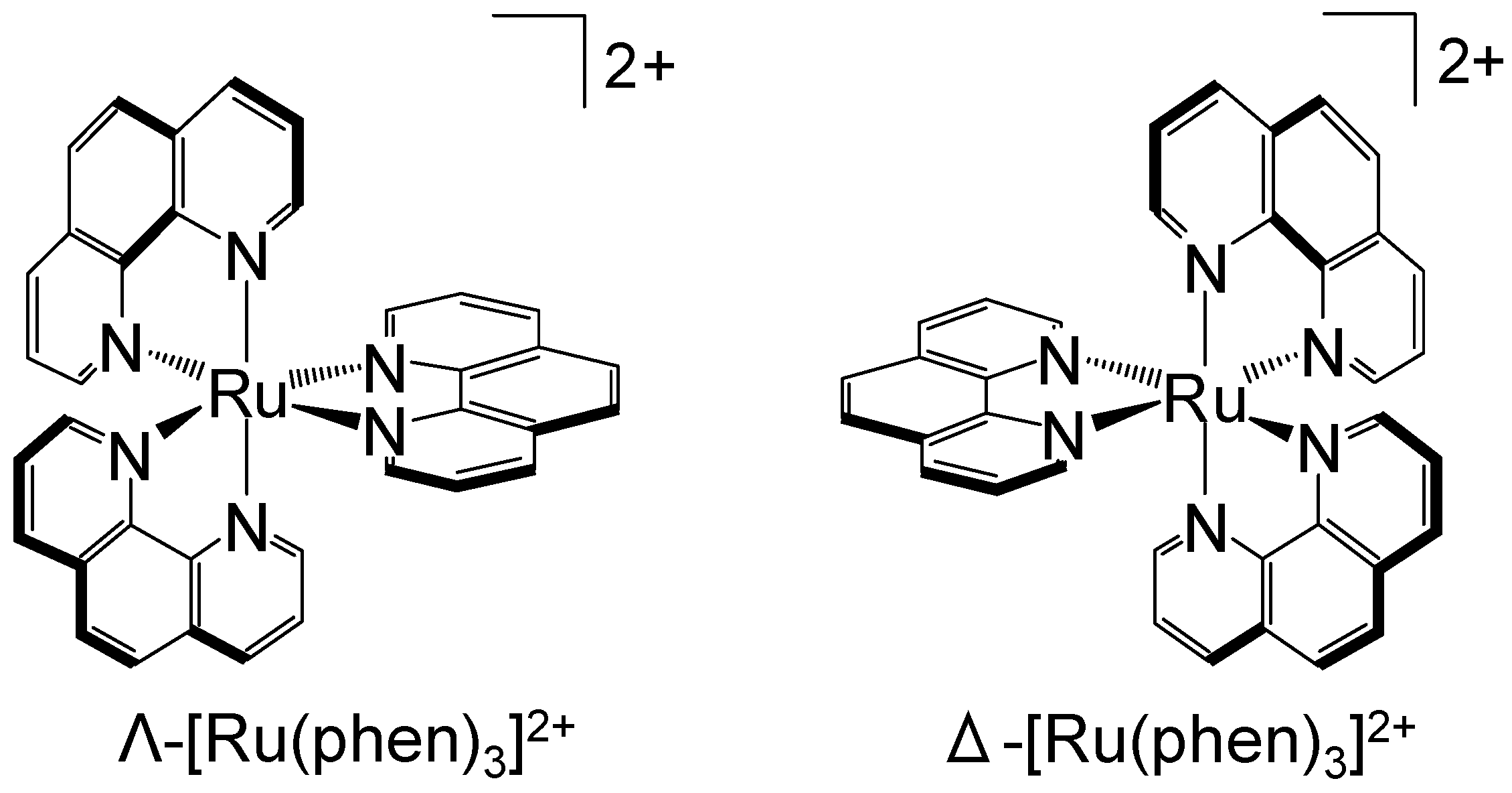
Figure 1.
Rf values of L- and D-form amino acids on DNA-TLC plate. The mobile phase is entry 1 in Table 1. The amino acid was detected by the ninhydrin reaction. Each of the values represents the mean of three separate determinations.
Figure 1.
Rf values of L- and D-form amino acids on DNA-TLC plate. The mobile phase is entry 1 in Table 1. The amino acid was detected by the ninhydrin reaction. Each of the values represents the mean of three separate determinations.

Figure 2.
IR spectra of: (a) UV-irradiated DNA film; (b) L-Asp accumulated DNA film; (c) D-Asp accumulated DNA film; and (d) L-Asp. Amino acid-accumulated DNA film was prepared by the immersion of the UV-irradiated DNA into an aqueous amino acid solution. Triplicate experiments gave similar results.
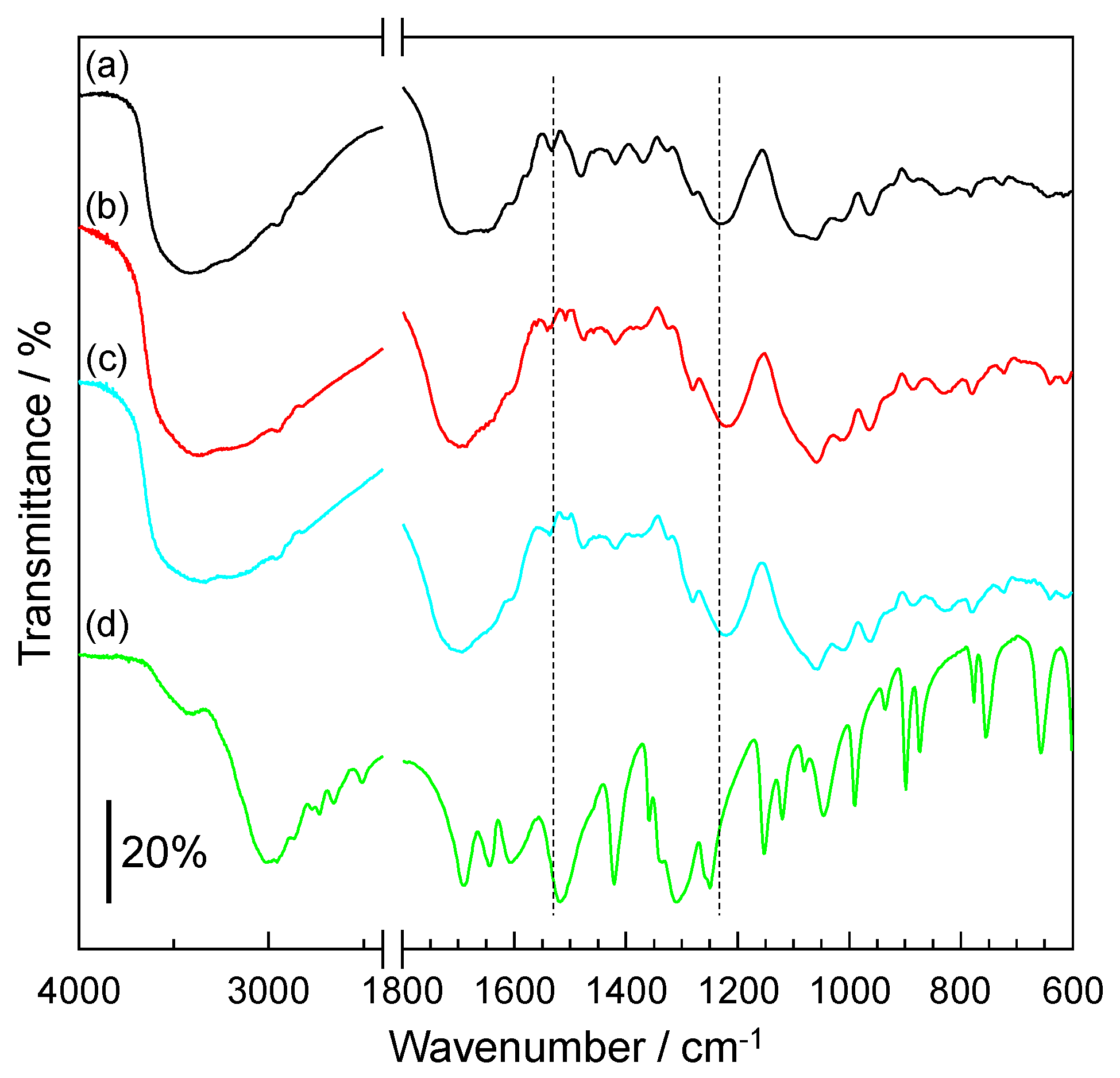
Figure 3.
Rf values of Λ- and Δ-[Ru(phen)3]2+ on the DNA-TLC plate. Each of the values represents the mean of three separate determinations.
Figure 3.
Rf values of Λ- and Δ-[Ru(phen)3]2+ on the DNA-TLC plate. Each of the values represents the mean of three separate determinations.

Table 1.
Various mobile phases which were used for the thin layer chromatography.
| Entry | Mobile Phases | Ratio/v/v |
|---|---|---|
| 1 | 1-BuOH:AcOH:H2O | 4:4:1 |
| 2 | 1-BuOH:AcOH:H2O | 4:2:1 |
| 3 | (CH3)2CO:AcOH:H2O | 70:30:7 |
| 4 | 1-BuOH:EtOH:H2O | 5:4:3 |
| 5 | EtOH:H2O | 1:1 |
| 6 | AcOEt:MeOH | 4:1 |
| 7 | MeOH:CHCl3:H2O | 12:5:3 |
| 8 | (CH3)2CO:1-BuOH:NH3(aq):H2O | 65:20:10:5 |
| 9 | NH3(aq):H2O | 17:3 |
© 2017 by the authors. Licensee MDPI, Basel, Switzerland. This article is an open access article distributed under the terms and conditions of the Creative Commons Attribution (CC BY) license (http://creativecommons.org/licenses/by/4.0/).
Share and Cite
MDPI and ACS Style
Yamada, M.; Inoue, M. Chiral Recognition by DNA-Immobilized TLC Plate. Separations 2018, 5, 3. https://doi.org/10.3390/separations5010003
AMA Style
Yamada M, Inoue M. Chiral Recognition by DNA-Immobilized TLC Plate. Separations. 2018; 5(1):3. https://doi.org/10.3390/separations5010003
Chicago/Turabian StyleYamada, Masanori, and Mami Inoue. 2018. "Chiral Recognition by DNA-Immobilized TLC Plate" Separations 5, no. 1: 3. https://doi.org/10.3390/separations5010003
Note that from the first issue of 2016, this journal uses article numbers instead of page numbers. See further details here.