Article

Influence of a Pulsed Electric Field on Charge Generation in a Flowing Protein Solution

1
Institute of Biomedical Chemistry, Pogodinskaya st., 10, Moscow 119121, Russia
2
Foundation of Perspective Technologies and Novations, Moscow 115682, Russia
3
Joint Institute for High Temperatures of the Russian Academy of Sciences, Moscow 125412, Russia
4
Institute of Bioorganic Chemistry, National Academy of Sciences of Belarus, Minsk 220141, Belarus
*
Author to whom correspondence should be addressed.
Separations 2018, 5(2), 29; https://doi.org/10.3390/separations5020029
Submission received: 27 December 2017 / Revised: 29 March 2018 / Accepted: 4 May 2018 / Published: 21 May 2018

Abstract

:
It is known that a charge is generated in water and protein solutions upon their motion; this phenomenon is also observed in analytical systems for atomic force microscopy (AFM)-based fishing. At that, the efficiency of protein fishing correlates with the value of charge, generated upon the motion of the analyzed solution. Earlier, we demonstrated that a pulsed electric field can well be used for the enhancement of the efficiency of AFM-based fishing of low-abundant protein. In this paper, we have demonstrated the influence of a pulsed electric field on the stimulation of the electric charge generation in a solution of low-abundant proteins observed in the injector part of an AFM-based fishing system at 23 °C and 38 °C. Taking this effect into account is important for the development of novel highly sensitive flow-based diagnostic systems, as well as for the development of models describing the influence of a pulsed electric field on pathological processes in the body, hemodynamics, and physicochemical properties of solutions.

1. Introduction

Development of novel approaches for the detection of low-abundant proteins is of high importance for highly sensitive proteomics. The motion of water and protein solutions through pipes in every analytical system is known to have an electrokinetic effect (usually named as the triboelectric effect), which manifests itself through charge generation [1]. We observed the occurrence of this effect in atomic force microscopy (AFM)-based fishing systems for the detection of low-abundant proteins upon flowing of an analyte solution through an injector into a measuring cell of this system [2,3,4,5]. Moreover, in the paper [2], it was reported that the efficiency of protein detection correlates with the amount of charge (in nanoCoulombs, nC). Studying this effect is very important for modeling hemodynamics and mechanisms of functioning of living systems.
The stimulating effect of a pulsed electric field on the efficiency of protein fishing in an AFM-based fishing system was demonstrated earlier with the example of human cytochrome b5 protein [5]. In the present study, we have used the solution of the same protein to determine the influence of pulsed electromagnetic fields on the charging of a protein solution at ultra-low protein concentrations (10−15 M). It is known that membrane cytochrome b5 is a hydrophobic protein, whose charge at pH = 6 is negative [6,7]. Experiments have been carried out at two temperatures: room temperature of 23 °C (which is typical for functioning of analytical systems) and an elevated temperature of 38 °C (which corresponds to the temperature of human body).
It has been demonstrated that the value of the charge generated in a flow of water or protein solution depends on temperature. The stimulating effect was ~40% for both water and protein solution at 23 °С, and for water and protein solution at 38 °С, this effect was ~40% and 75%, respectively.
Data obtained in this work should be taken into account when modeling the functioning of flow-based diagnostic systems employing AFM-based fishing, as well as other flow-based analytical systems: for instance, nanowire biosensors.
Moreover, taking into account the fact that living systems themselves generate electric fields and pulses, whereas external pulsed electric fields, in their turn, also affect living systems and enzymes [8,9,10], the data obtained in the present study can be interesting for use in modeling pathological processes in human body upon the impact of pulsed electric fields: for instance, in the development of hemodynamic models.

2. Materials and Methods

2.1. Materials

Deionized water (18 MΩ × cm resistivity) was obtained using Millipore Simplicity UV system (Molsheim, France).
Human cytochrome b5 protein cloned as described in [11] was provided by Prof. Usanov S.A. (Institute of Bioorganic Chemistry of the National Academy of Sciences of Belarus). 10−15 М protein solution was prepared by the serial ten-fold dilution of an initial 10−7 М solution.

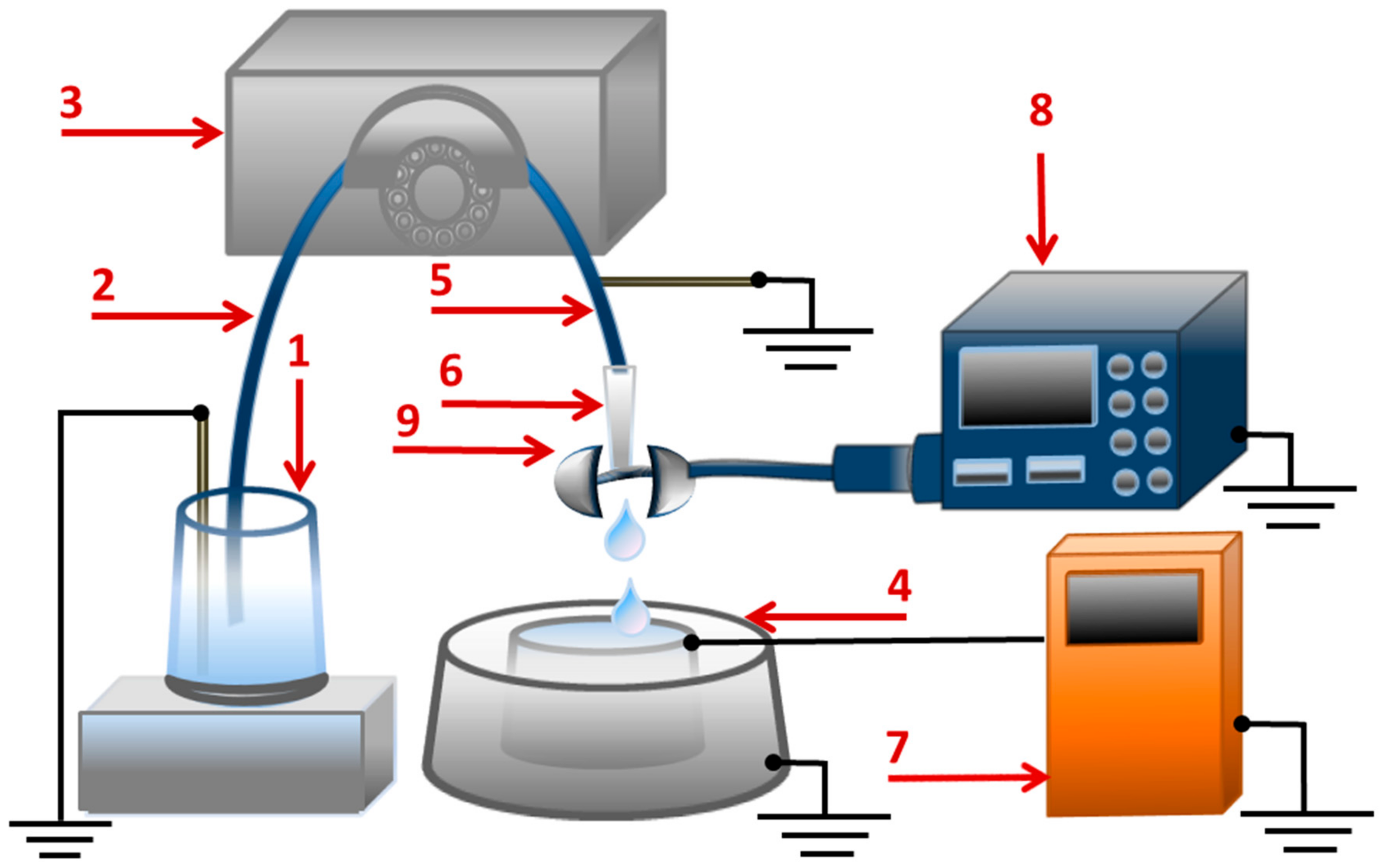
2.2. Charge Measurements

To study the effect of a pulsed electric field on the generation of charge in water and in protein solutions, we have used a system employing the AFM-based fishing device described in detail elsewhere [5]. The experiment setup is shown in Figure 1. The measurements of the electric charge were carried out with an electrometer coupled to the flow-based system for feeding the sample into the measuring cell [5] (Figure 1). The main elements of the sample supply system are: the peristaltic pump, the pipe with a tip for supplying the analyzed liquid, and the measuring cell. During measurements, water or protein solution from a 50-mL polypropylene tube (1) was continuously pumped through the tip (6) into the cell (4) using an Ismatec ISM 597D pump (IDEX Corp., Lake Forest, IL, USA) (3). A sterile silicone pipe (2, 5) (length 40 cm, inner diameter 2 mm) with a tip (6) was used to supply the analyzed liquid. The tip used was a standard disposable tip for an automatic pipette (inner diameter 0.4 mm, epT.I.P.S, 10 μL, Eppendorf, Germany). The flow rate (~15 μL/s) was selected so that droplets would form on the tip nozzle. The calculated drop volume was ~15 μL. To maintain the potential of the initial solution at a constant ground level, a ground electrode was inserted into the analyzed liquid in the polypropylene tube (1). The stainless steel cell (4) served as an internal cylinder in a system coupled to an electrometer (7). The dimensions of this cell were as follows: height 90 mm, inner diameter 75 mm, outer diameter 77 mm, and thickness of the polypropylene insert between the outer and the inner plates was 0.2 mm. In general, the system for charge measurements was similar to the one used in [5]. The charge in the cell was registered using an electrometer (7) developed in Institute of Biomedical Chemistry (IBMC). The charge registration accuracy was 0.1 nC. The effect of a pulsed electric field on the liquid was produced using the “homemade” pulse generator (8) developed in IBMC. The parameters of the generator were as follows: pulse repetition time can be selected in the range from 0.5 μs to 9900 ms; pulse width: 50 ns; pulse amplitude: 100 V; pulse rise time: 10 ns; output resistance: 9 kΩ. The tip of the liquid-supply pipe (6) was placed between parallel metal plates (9). The dimensions of the plates were 30 mm × 14 mm, and the distance between the plates was 10 mm. A pulsed voltage from the pulse generator (8) was applied to the plates, and the parameters of this voltage were as follows: 100 V, front rise time 10 ns, pulse width 50 ns, pulse repetition time 2 µs. The temperature of water and protein solution was maintained using a thermostat, where the tube (1) containing the analyzed liquid was placed. The experiments were performed at 23 °C and 38 °C. Prior to the measurements, the system was equilibrated under the experimental conditions for at least 2 h. The duration of one measurement was 7 min. The air humidity was 45%. The experimental series for each set of parameters included at least three repetitions.
The sequence of the experiments was as follows. Firstly, experiments were conducted in the absence of a pulsed electric field (with the generator turned off). The system was filled with water from the tube (1) and washed with ~10 mL of analyzed liquid (water or protein solution). After washing, the liquid supply system remained filled with the analyzed liquid. Then, the analyzed liquid was removed from the measuring cell with a pipette, and subsequent control measurements of the baseline signal for the unfilled cell (3 repetitions) were conducted. After that, the pump was turned on, and either water or protein solution was dropped into the measuring cell. The registration began at the moment when the pump was turned on. The data were recorded every 15 s for a duration of 7 min. After the measurements within one experiment were completed, the electrometer indications were reset. The obtained data were presented in the form of a time dependence of the value of charge entering the measuring cell Δq(t). After that, experiments were conducted using a similar procedure in the presence of a pulsed electric field with the generator turned on, the voltage from which was applied to the metal plates (9).

3. Results

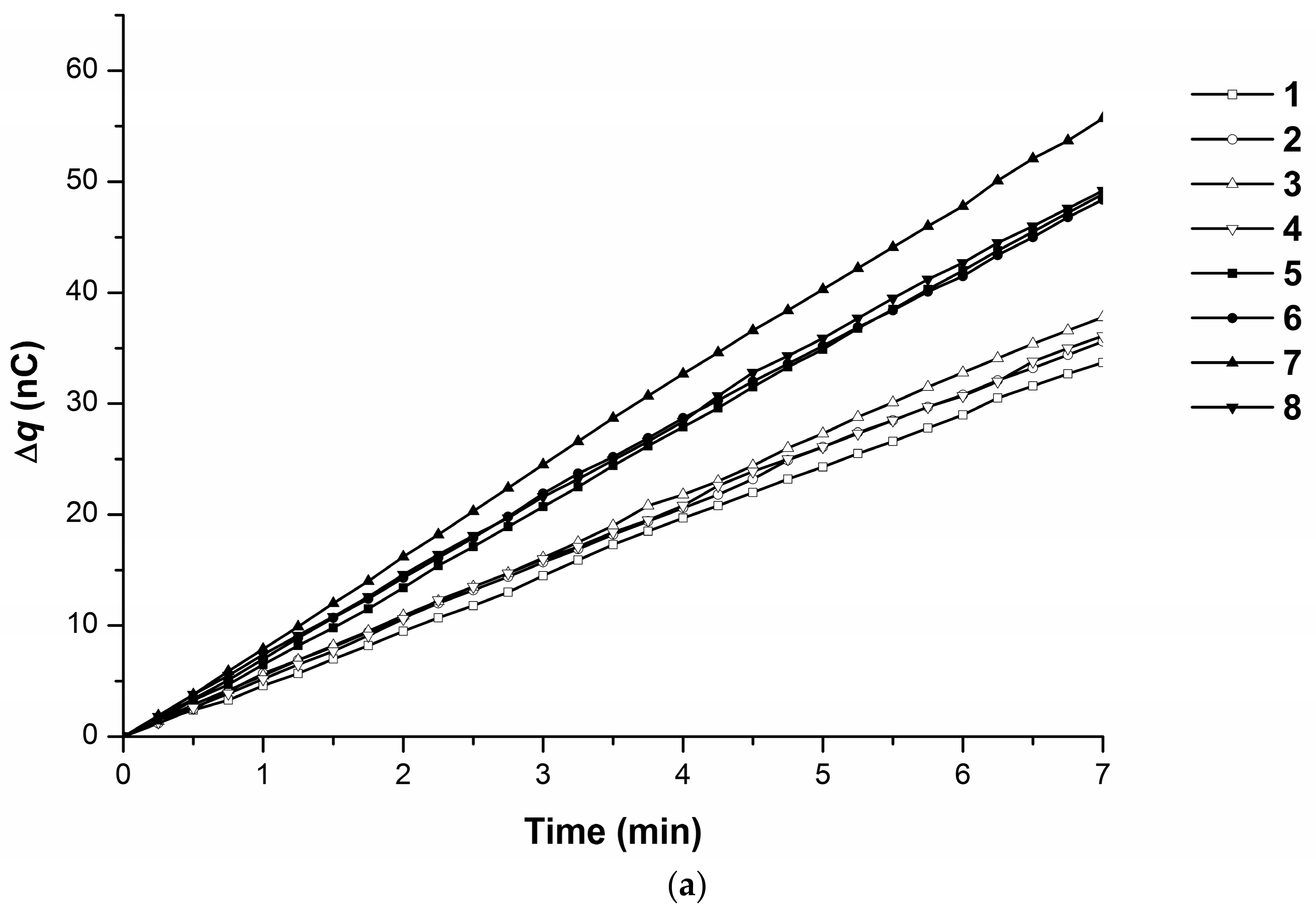
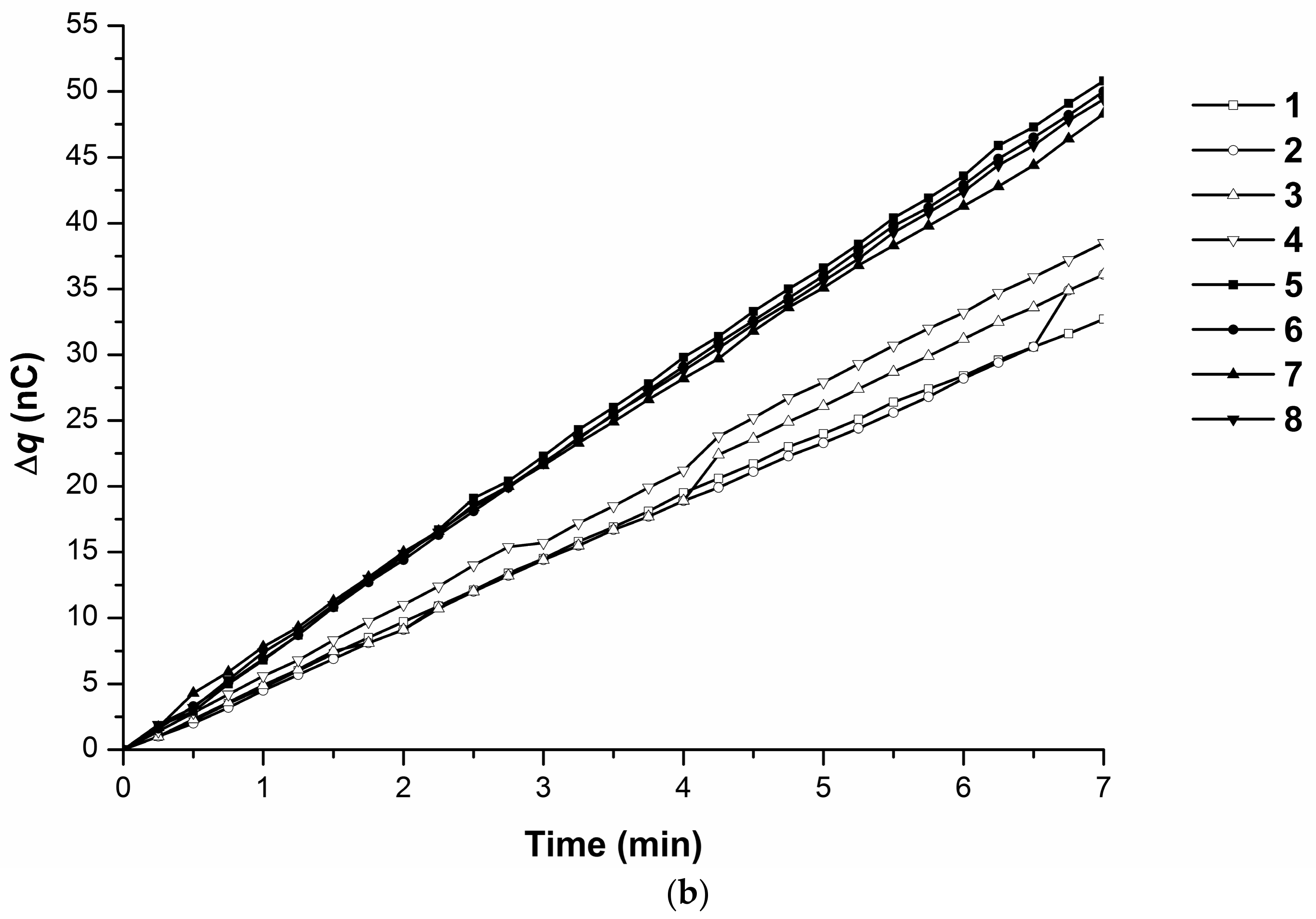
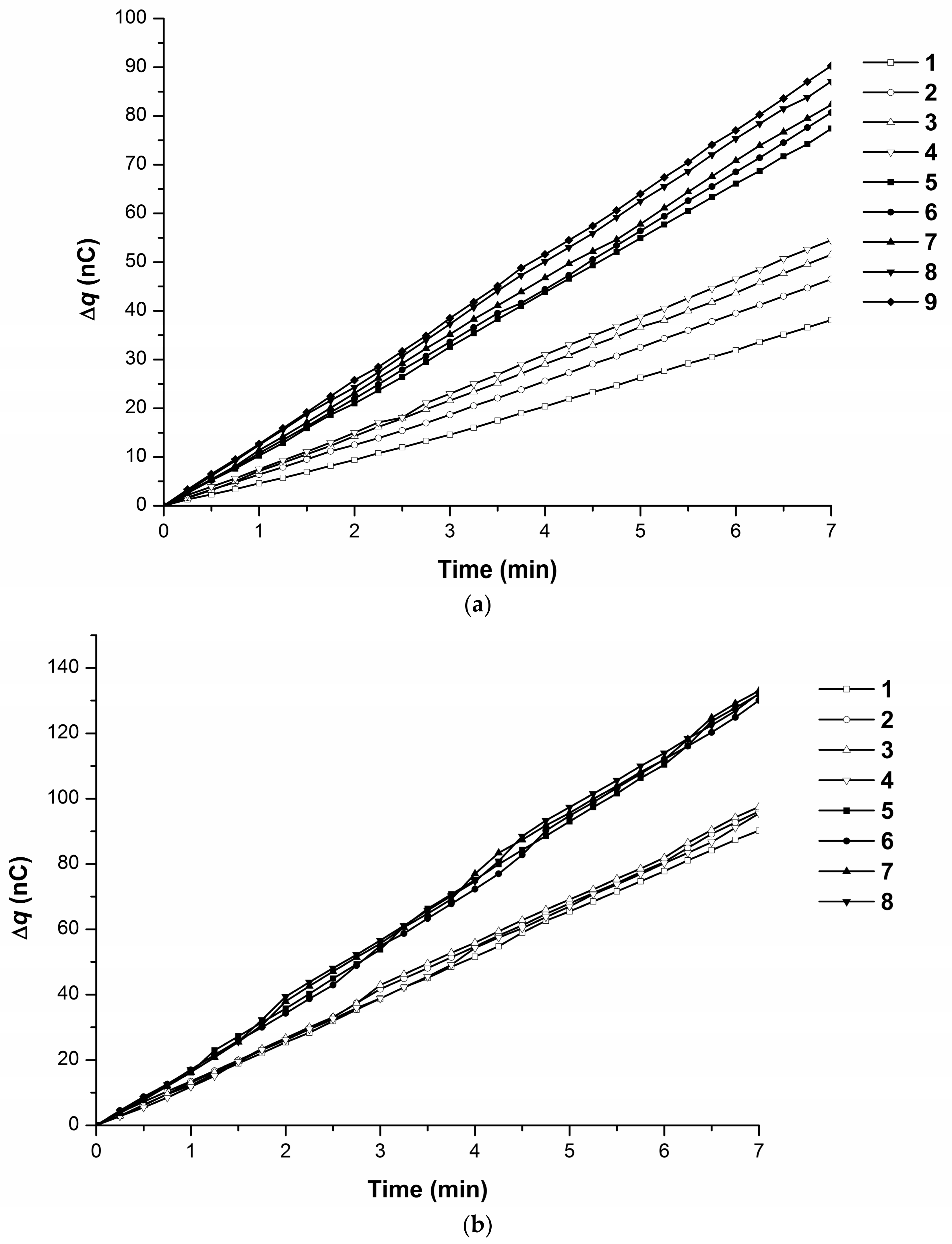
3.1. Effect of the Presence of a Low-Abundant Protein on the Charging of Liquid

In this control series of experiments, the impact of protein on the value of the charge generated in the analyzed liquid has been determined. Figure 2 displays an example of typical time dependencies of the value of the charge Δq accumulated in the measuring cell upon pumping water in the absence and in the presence of the protein at 23 °C (Figure 2a) and 38 °C (Figure 2b). These data are also summarized in Table 1.
As shown in Figure 2a, at 23 °C, a linear time dependence of the generated charge accumulated in the measuring cell was observed for both water and protein solution. For water, the value of the charge accumulated in 7 min was Δq = (31 ± 4) nC, and for water containing low-abundant protein, this value was Δq = (36 ± 2) nC. Thus, the presence of protein in solution at this temperature had no significant effect on the amount of charge accumulated in the measuring cell.
As shown in Figure 2b, at 38 °C, a linear time dependence of the accumulation of the generated charge in the measuring cell was observed for both water and protein solution, but the value of charge generated during the motion of the protein solution was higher than in the case of water. For water, the value of charge accumulated in 7 min was (74 ± 6) nC, and for the low-abundant protein solution, this value was (95 ± 3) nC. Thus, the presence of low-abundant protein at 38 °C caused a significant increase in the amount of charge accumulated in the measuring cell.

3.2. Effect of the Pulsed Electric Field on the Charging of Water and Protein Solution

The results of the generation of charge and its accumulation in the measuring cell upon the flowing of analyzed liquid in an external electric field are presented in Figure 3 and Figure 4 and Table 2. Figure 3a displays typical time dependencies of the value of the charge Δq accumulated in the measuring cell upon pumping of water in the absence and in the presence of a pulsed electric field at 23 °C. As shown in Figure 3a, in the absence of an electric field, the accumulation of the generated charge in the measuring cell was linearly dependent on time. The charge accumulation rate was 34–38 nC in 7 min, and the average value was (36 ± 2) nC in 7 min (Table 2). In the presence of an electric field, an increased level of charge accumulation (48–55 nC) was observed, with an average value of (51 ± 3) nC in 7 min, and the time dependence was linear.
Figure 3b displays typical time dependencies of the value of charge Δq accumulated in the measuring cell upon pumping of 10−15 M cytochrome b5 solution in the absence and presence of a pulsed electric field at 23 °C. As shown in Figure 3b, for the protein solution, accumulation of the generated charge in the measuring cell in the absence of an external pulsed electric field was observed in the form of a linear time dependence with an average value of Δq = (36 ± 2) nC in 7 min. In the presence of an electric field, there was an increased level of charge accumulation rate: Δq = (50 ± 1) nC in 7 min.
Figure 4a displays typical time dependencies of the value of charge accumulated in the measuring cell upon the pumping of water in the presence and absence of the pulsed electric field at 38 °C. As shown in Figure 4a (curves 1–4), in water in the absence of the pulsed electric field, accumulation of the generated charge in a measuring cell was observed with a rate of 38–53 nC in 7 min. The Δq(t) dependence was linear. The average value of charge accumulated in the cell in 7 min was Δq = (48 ± 7) nC.
In the presence of an electric field (Figure 4a, curves 5–9), there was an increase in the level of accumulated charge: 77–93 nC in 7 min; the Δq(t) dependence was linear. The average value of charge accumulated in the cell in 7 min was Δq = (84 ± 5) nC.
Figure 4b displays typical time dependencies of the value of charge accumulated in the measuring cell upon the pumping of cytochrome b5 solution with and without the pulsed electric field at 38 °C. As shown in Figure 4b (curves 1–4), at 38 °C in the absence of the external pulsed electric field, charge generation in the protein solution and its accumulation in the measuring cell with a rate of 90–98 nC in 7 min were observed. The Δq(t) dependence was linear. The average value of the charge accumulated in the cell in 7 min was Δq = (95 ± 3) nC. In the presence of the electric field (Figure 4b, curves 5–8), an increase in the level of charge accumulated in the cell (130–134 nC in 7 min) was observed. As with the absence of the field, there was a short-term increase in the charge of Δq = 5 nC for 15–30 s, superimposed on the linear dependence. The average value of the charge accumulated in the cell in 7 min was Δq = (132 ± 2) nC.

4. Discussion

As is known, upon the motion of liquid through polymer pipes, triboelectrization takes place, involving water ions including water clusters [12]. This leads to the spontaneous generation of charge in droplets flowing out of these pipes.
The data obtained in our present study describe the stimulating effect of an external pulsed electric field on the generation of charge in water and protein solution upon their motion through the injection part of an AFM-based fishing system.
We will first consider the effect of the presence of protein at a femtomolar concentration on the charge generation in the absence of an electric field. In the case of 23 °C, a similar charge generation rate in water and protein solution is observed. According to the data obtained, at 38 °C, the presence of protein (as compared to water) increases the level of charge accumulated in the cell from 74 nC (water) to 95 nC (protein); that is, the increase makes up ~30%. Thus, we have observed the temperature effect of the presence of protein in water on the charge generation, which is much more pronounced with the increase of temperature. It should be noted that with increasing the temperature from 23 °C to 38 °C, the resistivity of water decreases from 20 to 10 MΩ × cm [13]. One of the factors causing the unusual behavior of water is its complex structure, which represents a mixture of the solid ice-like and liquid phases (para- and ortho-states of water); these phases are characterized by different physicochemical properties: particularly surface tension, viscosity, and so forth [14,15], which determine the efficiency of the triboelectric effect and the electrohydrodynamics of the flow of water from the tip nozzle. The fact that a change in the proportion of the ortho-state should be observed in water, and that a change in this fraction has a resonant character at approximately 37 °C, was noted in [14,15]. According to the approximation of data on the electrical conductivity of a solution estimated in [16], the presence of cytochrome b5 at a femtomolar concentration slightly (by about 0.1%) changes the resistivity of water. However, probably, in this case, protein molecules at this low concentration serve as the “catalyst” of the process of water transfer between the para- and the ortho-state, which leads to a significant difference in the accumulation of charge in the measuring cell upon the pumping of water and protein solution through the tip.
With regard to the effect of the pulsed electric field, the obtained data indicate that such a field stimulates an increase in the value of charge accumulated in the measuring cell when water and the protein solution flow through the tip at both 23 °C and 38 °C. In this way, for water at 23 °C, the charge accumulated in the measuring cell is shown to increase by ~42% (from 36 to 51 nC in 7 min), and for the protein solution by ~39% (from 36 to 50 nC) in the presence of an electric field. Thus, in this case, the effect of the electric field in water and in protein solution is approximately equal.
More significant changes are observed at 38 °C. For water at 38 °C, the value of charge accumulated in the measuring cell increases from 48 nC (in the absence of the electric field) to 84 nC (in the presence of the electric field): that is, by 36 nC (i.e., by ~75%). For the protein solution at 38 °C, the value of charge accumulated in the measuring cell increases from 95 nC (in the absence of the electric field) to 130 nC (in the presence of the electric field): that is, by 35 nC (i.e., by ~37%) in 7 min. Thus, in this case, the effect of the electric field on charge generation in water was higher than that observed for the protein solution.
Thus, we have observed a more significant effect of the external pulsed electric field on the generation of the charge in moving water. In the case of cytochrome b5 solution, the observed stimulation of charge generation is less significant, being comparable with the measurement error. The physical nature of the stimulation of the charge generation in water moving inside the polymer pipe can be explained as follows. As noted above, water represents a complex structure, which represents a mixture of the solid ice-like and liquid phases (para- and ortho-states of water). These phases are characterized by different physicochemical properties: particularly surface tension, viscosity, and so forth [16,17]. With electrical excitation in a pulsed electric field with 10 ns front rise time, an increased conversion of the water para-state into the ortho-state can occur; the possibility of the occurrence of this phenomenon in microwave fields was reported in [17]. Thus, one of the important causes of the observed stimulation of charge generation in water in a pulsed electric field can be a change in the fraction of ice-like water structures.
It should be noted that blood circulation through blood vessels and capillaries is the basis of the functioning of the human body, and a change in the electrohydrodynamics of blood in a pulsed electric field can lead to various pathologies. In this way, pulsed electric fields of medium intensity (~100 V/cm) stimulate blood coagulation, as was demonstrated in [8]. Therefore, this effect should be taken into account in the development of hemodynamic models.

5. Conclusions

The subject of our study was the effect of a pulsed electric field on charge generation in femtomolar protein solutions in the injection part of the AFM-based fishing system. The stimulating influence of an external pulsed electric field on the charge generation in both water and protein solution at 23 °C and 38 °C has been observed. It has been found that the stimulating effect on charge generation in the AFM-based fishing system was ~40–70%. Accounting for the observed effect is important for the development of novel highly sensitive diagnostic and analytical systems, operating in the range of ultra-low protein concentrations. The results obtained can be of interest for the development of models describing the physicochemical properties of water and aqueous solutions, and also thermodynamic models of human pathological states.

Author Contributions

Conceptualization, Yu.D.I.; Methodology, Yu.D.I. and T.O.P.; Validation, R.A.G., V.Yu.T. and V.S.Z.; Formal Analysis, V.S.Z.; Investigation, A.F.K.; Resources, S.A.U.; Data Curation, V.Yu.T.; Writing-Original Draft Preparation, Yu.D.I., A.F.K. and R.A.G.; Writing-Review & Editing, T.O.P.; Visualization, A.F.K. and R.A.G.; Supervision, T.O.P.; Project Administration, Yu.D.I.; Funding Acquisition, Yu.D.I.

Funding

This research was funded by the Russian Science Foundation grant number14-25-00132.

Acknowledgments

This study was supported by the Russian Science Foundation (Grant No. 14-25-00132).

Conflicts of Interest

The authors declare no conflicts of interest.

References

  1. Whitesides, G.M.; McCarty, L.S. Electrostatic Charging Due to Separation of Ions at Interfaces: Contact Electrification of Ionic Electrets. Angew. Chem. Int. Ed. 2008, 47, 2188–2207. [Google Scholar]
  2. Ivanov, Y.D.; Pleshakova, T.; Malsagova, K.; Kozlov, A.; Kaysheva, A.; Kopylov, A.; Izotov, A.; Andreeva, E.; Kanashenko, S.; Usanov, S.; et al. Highly sensitive protein detection by combination of atomic force microscopy fishing with charge generation and mass spectrometry analysis. FEBS J. 2014, 281, 4705–4717. [Google Scholar] [CrossRef] [PubMed]
  3. Pleshakova, T.O.; Malsagova, K.A.; Kozlov, A.F.; Kanashenko, S.L.; Ivanova, N.D.; Sadovskaya, T.A.; Archakov, A.I.; Ivanov, Y.D. Highly sensitive AFM-fishing of albumin. Pathogenesis 2016, 14, 23–30. (In Russian) [Google Scholar]
  4. Pleshakova, T.O.; Malsagova, K.A.; Kaysheva, A.L.; Kopylov, A.T.; Tatur, V.Y.; Ziborov, V.S.; Kanashenko, S.L.; Galiullin, R.A.; Ivanov, Y.D. Highly sensitive protein detection by biospecific AFM-based fishing with pulsed electrical stimulation. FEBS Open Bio 2017, 7, 1186–1195. [Google Scholar] [CrossRef] [PubMed]
  5. Ivanov, Y.D.; Pleshakova, T.O.; Malsagova, K.A.; Kaysheva, A.L.; Kopylov, A.T.; Izotov, A.A.; Tatur, V.Y.; Vesnin, S.G.; Ivanova, N.D.; Ziborov, V.S.; et al. AFM-based protein fishing in the pulsed electric field. Biochem. (Moscow) Suppl. Ser. B Biomed. Chem. 2015, 9, 121–129. [Google Scholar] [CrossRef]
  6. UniProtKB—P00167 (CYB5_HUMAN). Available online: http://www.uniprot.org/uniprot/P00167 (accessed on 29 March 2018).
  7. Kiyoshi, A.; Yoshiki, S. Properties of cytochrome b5, and methemoglobin reduction in human erythrocyte. Eur. J. Biochem. 1979, 101, 423–428. [Google Scholar]
  8. Filipič, B.; Kovacs, K.; Somogyvari, F.; Ihan, A.; Ocsovszky, I.; Koren, S.; Toth, S. The effects of medium-strength electric impulses on human blood. Bioelectrochemistry 2000, 52, 29–36. [Google Scholar] [CrossRef]
  9. Blank, M.; Findl, E. Mechanistic Approaches to Interactions of Electric and Electromagnetic Fields with Living System; Plenum Press: New York, NY, USA, 1987; Volume 9, pp. 365–387. [Google Scholar]
  10. Ivanov, Y.D.; Kozlov, A.F.; Malsagova, К.А.; Pleshakova, Т.О.; Vesnin, S.G.; Tatur, V.Y.; Ivanova, N.D.; Ziborov, V.S. Monitoring of microwave emission of HRP system during the enzyme functioning. Biochem. Biophys. Rep. 2016, 7, 20–25. [Google Scholar] [CrossRef] [PubMed]
  11. Duarte, M.P.; Palma, B.B.; Gilep, A.A.; Laires, A.; Oliveira, J.S.; Usanov, S.A.; Rueff, J.; Kranendonk, M. The stimulatory role of human cytochrome b5 in the bioactivation activities of human CYP1A2, 2A6, and 2E1: A new cell expression system to study cytochrome P450 mediated biotransformation. Mutagenesis 2005, 20, 93–100. [Google Scholar] [CrossRef] [PubMed]
  12. Burgo, T.A.; Galembeck, F.; Pollack, G.H. Where is water in the triboelectric series? J. Electrostat. 2016, 80, 30–33. [Google Scholar] [CrossRef]
  13. Bushuev, E.N. The calculation of temperature dependence of water ion production, specific conductance and extremely diluted electrolytic solution. Vestnik IGAU 2007, 2, 49–52. (In Russian) [Google Scholar]
  14. Pershin, S. Conversion of ortho-para H2O isomers in water and a jump in erythrocyte fluidity through a microcapillary at a temperature of 36.6 ± 0.3 °C. Phys. Wave Phenom. 2009, 17, 241. [Google Scholar] [CrossRef]
  15. Kholmanskiy, A.S. Two types of anomalous thermodynamics of water. Apriori. Ser. Iestestv. I Tekh. Nauki 2015, 1, 1–17. [Google Scholar]
  16. Ivanov, Y.D.; Kozlov, A.F.; Galiullin, R.A.; Kanashenko, S.L.; Usanov, S.A.; Ivanova, N.D.; Ziborov, V.S.; Pleshakova, T.O. Spontaneous generation of charge in the flow-based AFM fishing system. J. Electrostat. 2018, 91, 16–20. [Google Scholar] [CrossRef]
  17. Pershin, S.M. New Conception of Electromagnetic Field Influence on Water and Water Solutions with Considering Quantum Distinct Ortho-/Para-Spin Isomers of H2O. 2013. Available online: www.biophys.ru/archive/sarov2013/proc-p17.pdf (accessed on 29 March 2018).
Figure 1. Experimental setup. (1) Tube with the analyzed liquid; (2) silicone pipe (incoming section); (3) peristaltic pump; (4) measuring cell coupled with electrometer (7); (5) silicone tube (outgoing section); (6) tip; (7) electrometer; (8) pulse generator; (9) parallel metal plates.
Separations 05 00029 g001
Figure 2. Typical Δq(t) dependencies obtained upon pumping of water or protein solution through the tip. Experiment conditions: (a) T = 23 °C, water (curves 1–3), protein solution (curves 4–7); (b) Т = 38 °С, water (curves 1–3), protein solution (curves 4–8). The flow rate was 15 μL/s. The data of one and the same experimental series obtained using the same tube-pipe-tip set (see Materials and Methods) are presented.
Separations 05 00029 g002
Figure 3. Typical Δq(t) dependencies obtained in the absence (white markers) and in the presence (dark markers) of an electric field for water (a) and protein solution (b) at 23 °C. The data of one and the same experimental series obtained using one and the same tube-pipe-tip set (see “Materials and Methods” section) are presented.
Separations 05 00029 g003aSeparations 05 00029 g003b
Figure 4. Typical Δq(t) dependencies obtained in the absence (white markers) and in the presence (dark markers) of an electric field for water (a) and protein solution (b) at 38 °C. The data of one and the same experimental series obtained using one and the same tube-pipe-tip set (see “Materials and Methods” section) are presented.
Separations 05 00029 g004
Table 1. Value of charge accumulated in the cell without an electric field, in nC.
Analyzed liquid23 °C38 °C
Water31 ± 4 74 ± 6
Protein solution36 ± 295 ± 3
Table 2. Value of charge accumulated in the cell with the electric field present, in nC.
Analyzed liquidTemperatureWithout Electric FieldWith Pulsed Electric Field
Water23 °C36 ± 251 ± 3
38 °C48 ± 784 ± 5
Protein solution23 °C36 ± 250 ± 1
38 °C95 ± 3132 ± 2

