Article

Nylon Membrane-Based Electromembrane Extraction Coupled with Matrix-Assisted Laser Desorption/Ionization Mass Spectrometry for the Determination of Insulin

1
Department of Chemistry, National Kaohsiung Normal University, Yanchao, Kaohsiung 824, Taiwan
2
Division of Gastroenterology and Hepatology, Department of Medicine, Kaohsiung Veterans General Hospital, Kaohsiung 813, Taiwan
3
Department of Post-Baccalaureate Medicine, College of Medicine, National Sun Yat-sen University, Kaohsiung 804, Taiwan
4
School of Medicine, National Yang Ming Chiao Tung University, Taipei 112, Taiwan
*
Author to whom correspondence should be addressed.
Separations 2022, 9(10), 286; https://doi.org/10.3390/separations9100286
Submission received: 13 August 2022 / Revised: 8 September 2022 / Accepted: 29 September 2022 / Published: 4 October 2022

Abstract

:
A rapid and sensitive protein determination method that uses electromembrane extraction (EME) and is coupled with matrix-assisted laser desorption/ionization mass spectrometry (MALDI/MS) is developed. A flat nylon membrane is used to collect proteins from an aqueous solution and is directly analyzed by MALDI/MS after the addition of the MALDI matrix. Insulin is used as a model protein to investigate the optimum extraction of the parameters. The optimum EME conditions are obtained at 12 V of voltage, 10 min of extraction time, 12 mL sample volume, and 400 rpm agitation rate. The linear dynamic range (LDR) of insulin in an aqueous solution is in the range of 1.0–100.0 nM. The limit of detection (LOD) for insulin in an aqueous solution is 0.3 nM with 103-fold signal-to-noise (S/N) ratio enhancement. Furthermore, the applicability of this method to determine insulin in complicated sample matrices is also investigated. The LDR of insulin in human urine samples is in the range of 5.0–100.0 nM, and the LOD of insulin in urine samples is calculated to be 1.5 nM. The precision and accuracy of this method are evaluated at three different concentration levels, and the coefficient of variation (CV) and relative error are less than 6%. This approach is time-efficient and economical, as the flat membrane mode of EME coupled with MALDI/MS is suitable.

Graphical Abstract

1. Introduction

Electromembrane extraction (EME) has attracted much interest since it was first studied by Pedersen-Bjergaards and Rasmussa in 2006 [1]. In EME, charged analytes are extracted under an electric field, from an aqueous sample, through the supported liquid membrane (SLM) in the pores of a follow fiber, and into the aqueous acceptor phase located inside the lumen of the hollow fiber [2]. Due to the ease of operation, rapidity, low cost, and high enrichment factor, EME has become a popular technique to extract target analytes from different complicated sample matrices [3,4].
In traditional EME, porous polypropylene (PP) hollow fibers [5,6] and flat membranes [7,8] have been the most widely used membranes in the EME method. The low-cost and disposable nature of the PP material prevents undesired carryover between the extracted samples. Additionally, PP membranes are usually impregnated with 2-nitrophenyl octyl ether (NPOE) and serve as the SLM for the extraction of nonpolar and medium polar compounds. To expand the applicability of EME extraction to a variety of compounds with different polarities, the use of various kinds of membranes has been proposed. Agarose gel [9,10,11], polyacrylamide gel [12], and silver nanoparticle-tragacanth gel [13] were used as green membranes for extracting basic drugs [12], amino acids [11], and peptides [10] without using any organic solvent. The gel membranes also function as a cleanup filter that obstructs the transportation of neutral and high molecular mass compounds such as humic acid and proteins. For highly polar analytes, a nanostructured sheet (Tiss-OH) that consists of acrylic nanofibers and contains a high density of OH groups was used as the EME support. The flat membrane that is permeated with Aliquat 335 in 1-octanol successfully extracted highly polar acidic compounds [14]. Polymer inclusion membranes (PIMs) have also been proposed as supports in EME. PIMs can be easily prepared by mixing carriers, base polymers, and plasticizers [15,16,17]. Carriers are usually ion exchangers or complexing agents that bind with analytes through electrostatic attraction or complex formation. After these membranes become infused with organic solvents, PIMs act as SLMs for the extraction of basic drugs [17], acidic drugs [15], and herbicides [16]. In addition, polyacrylonitrile (PAN)/polydimethylsiloxane (PDMS) membranes [18], chitosan membranes [19], and polyvinylidene difluoride (PVDF) membranes [20,21] have been proposed as supports to extract phthalate compounds, acidic drugs, and peptides.
Matrix-assisted laser desorption/ionization (MALDI) mass spectrometry (MS) has become the most powerful tool for the determination of proteins [22,23]. Usually, an acquired mass spectrum of protein is the average of 200 spectra, which can be obtained within 10 s. However, complicated sample matrices can cause the suppression of protein signals [24]. A sample preparation technique prior to MALDI/MS is needed to obtain the mass spectrum of the analyte, which is usually present at low concentrations in complicated sample matrices. In this study, we present a flat-mode EME method that enables the fast migration of charged proteins from an aqueous solution onto a nylon membrane by applying an electric field between two electrodes. After extraction, the nylon membrane was directly analyzed by MALDI/MS after the addition of MALDI matrices. Insulin was used as the model protein. Furthermore, the applicability of the method for the determination of insulin in complicated sample matrices was also investigated. To the best of our knowledge, no work on the use of nylon membranes in EME has been reported. In addition, this is the first report that demonstrates the use of EME for the extraction of protein.

2. Materials and Methods

2.1. Chemicals and Solutions
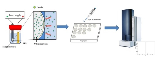
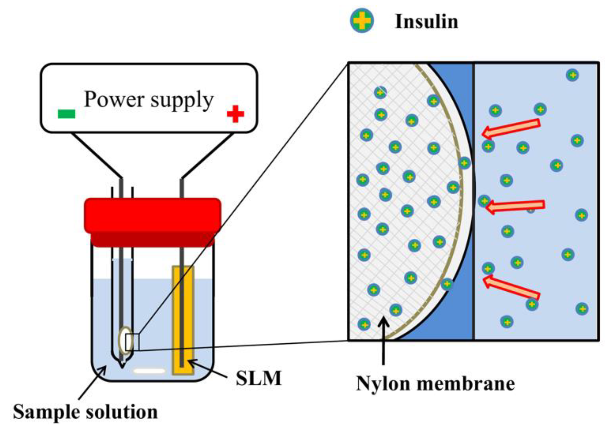
Insulin (from the porcine pancreas), NPOE, and sinapinic acid (SA) were purchased from Sigma–Aldrich (St. Louis, MO, USA). Trichloroacetic acid (TFA) was obtained from Alfa-Aesar (Ward Hill, MA, USA). Hydrophilic polyvinylidene difluoride (PVDF), nylon, polytetrafluoroethylene (PTFE), mixed cellulose ester (MCE), and hydrophobic polypropylene (PP) sheet membranes with a thickness of 200 μm and pore size of 0.22 μm were purchased from Greattech Technology Ltd. (Taipei, Taiwan). Deionized water was obtained using a Millipore Synergy water purification system (Billerica, MA, USA).
A stock standard solution (1 mg/mL) of insulin was prepared in 10 mM trifluoroacetic acid (TFA) aqueous solution and diluted to the desired concentrations with 10 mM TFA solution (pH 2.2). The insulin solution was stored at −20 °C, and the SA solution (10 mg/mL) was freshly prepared in a 40% acetonitrile/water solution containing 0.1% TFA.

2.2. EME Procedure

The EME device followed the design of the previously reported device, as shown in Figure 1 [25], which consists of a glass tube with a nylon membrane (diameter of 4.7 mm) and a 3.0 cm piece of PP hollow fiber (diameter 3.0 mm). The PP hollow fiber was dipped in NPOE for 10 s and utilized as the SLM. Then, 50 μL of 10 mM TFA solution was pipetted into the lumen of the hollow fiber. Previously, an EME device without the use of PP hollow fiber was developed in our laboratory [21]. However, the system was operated with a high current of up to 5 mA, which is substantially higher than that in a typical EME device (1–400 μA). The high system current resulted in the breakdown of the EME system for biological samples with a complicated sample matrix. Therefore, the PP hollow fiber was used to decrease the system current and stabilize the EME system. A 12 mL aliquot of insulin standard solution was loaded into the sample compartment and stirred at 400 rpm throughout the experiments. Two platinum wires (diameters of 0.5 mm and lengths of 6.8 cm) were inserted into the lumen of the hollow fiber and glass tube as the cathode and anode, respectively. The two electrodes were connected to the DC power supply (P-200, Hila International Inc., Hsinchu, Taiwan). The EME system current was c monitored with a computer connected to the Peak-ABC chromatographic data handling system (Great Tide Instrument Company, Taipei, Taiwan).

2.3. MALDI/MS Measurements

An overlay technique was used to prepare the insulin-enriched nylon membrane. After extraction, this membrane was deposited onto a MALDI sample target with double-sided tape. Then, 3 μL of SA solution was applied onto the top of a nylon membrane prior to the MALDI/MS analysis. For the regular MALDI sample, the dried droplet method was used by depositing the mixture solution containing 1 μL of insulin solution and 1 μL of SA solution onto the MALDI sample target.
The MALDI/MS experiment was performed on a Microflex time-of-flight mass spectrometer (Bruker Daltonics, Bremen, Germany) equipped with a flight length of 1.05 m and a nitrogen laser (337 nm). The insulin-enriched membrane was irradiated with the laser using laser energy that slightly exceeded the threshold. The generated positive ions were accelerated at a voltage of 19 kV. Each mass spectrum was acquired from an average of 200 laser pulses.

2.4. Preparation of Human Urine Sample

The urine samples in this work were collected from one healthy volunteer. Written informed consent was obtained from the participant. The urine samples were stored at −20 °C until analysis. The urine samples were thawed prior to preparation of the biological samples, and 11.8 mL was transferred to the EME sample compartment and further spiked with 100 μL of insulin standard solution and 100 μL of TFA solution. The resulting urine sample contained 10 mM TFA with a total volume of 12 mL. The urine samples with various analyte concentrations were prepared by spiking the urine with the desired amounts of analytes. Finally, the urine samples were subjected to EME according to the procedure described above.

3. Results

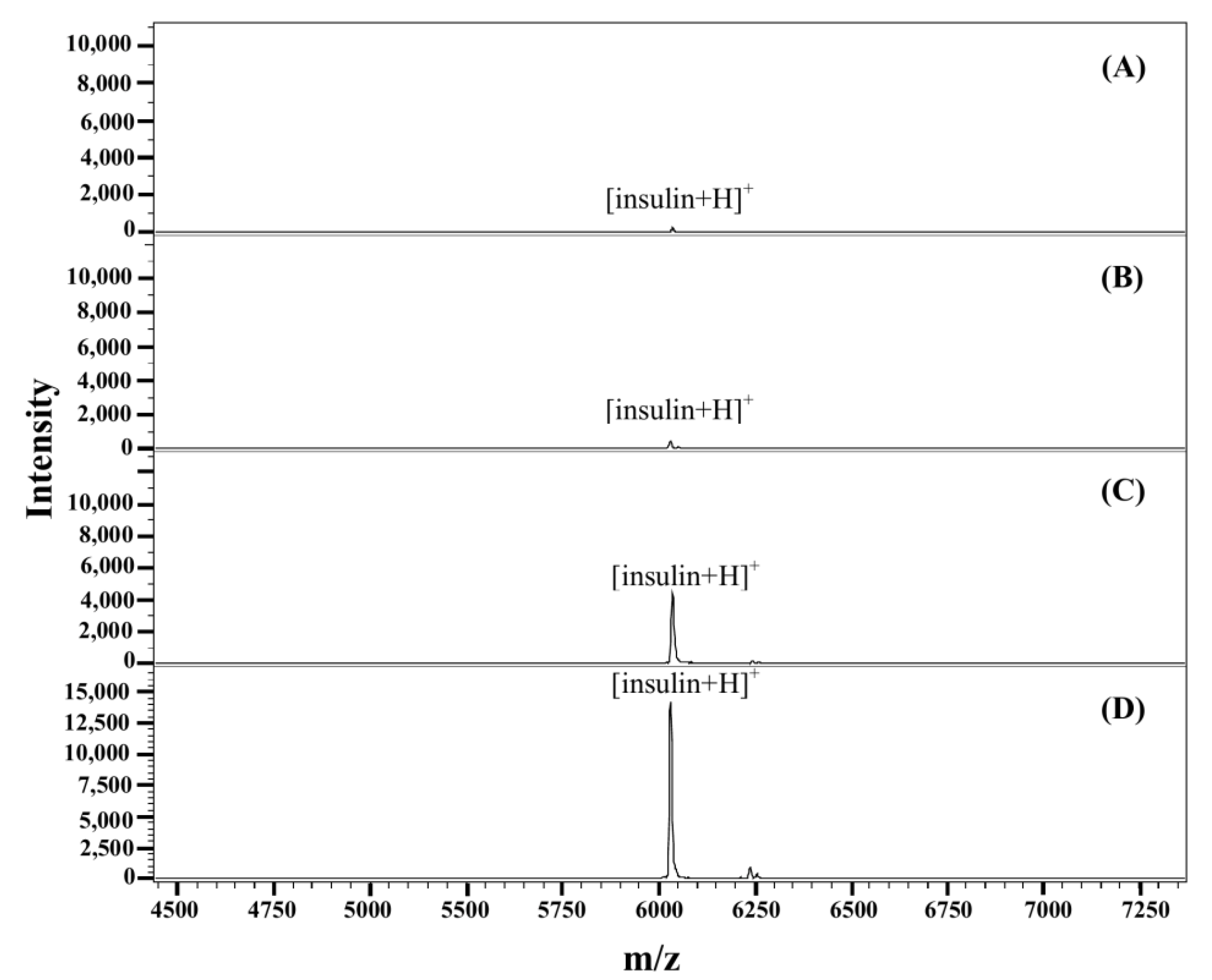
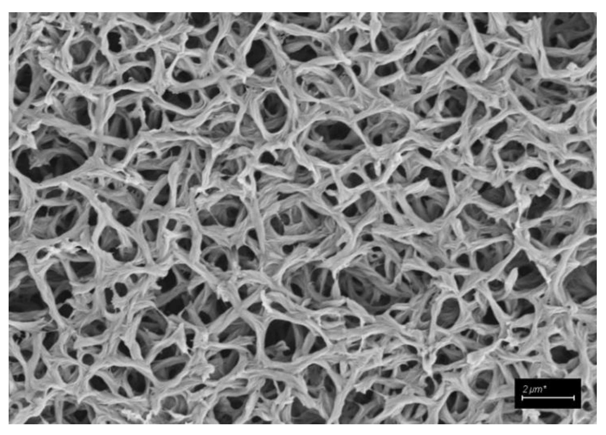
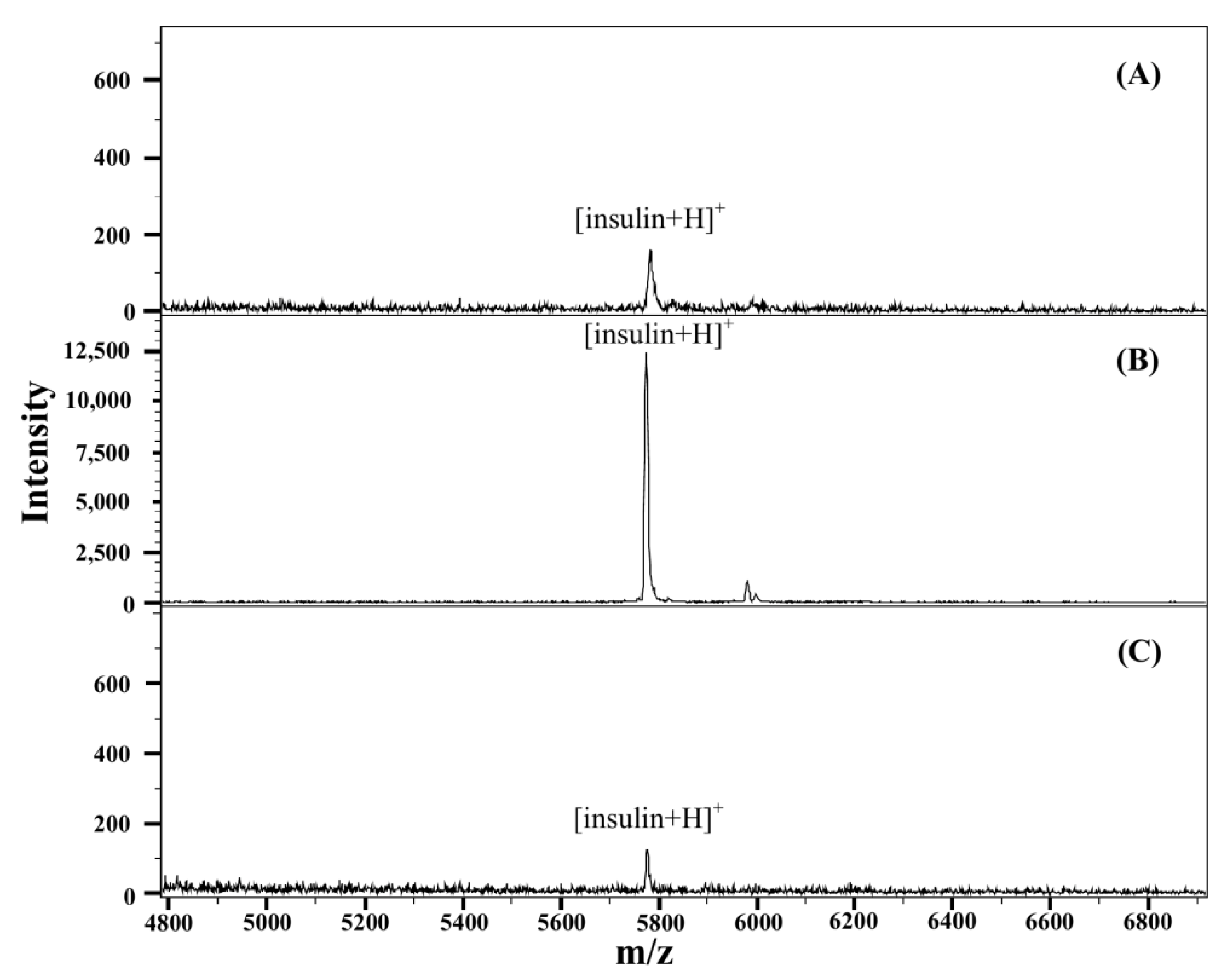
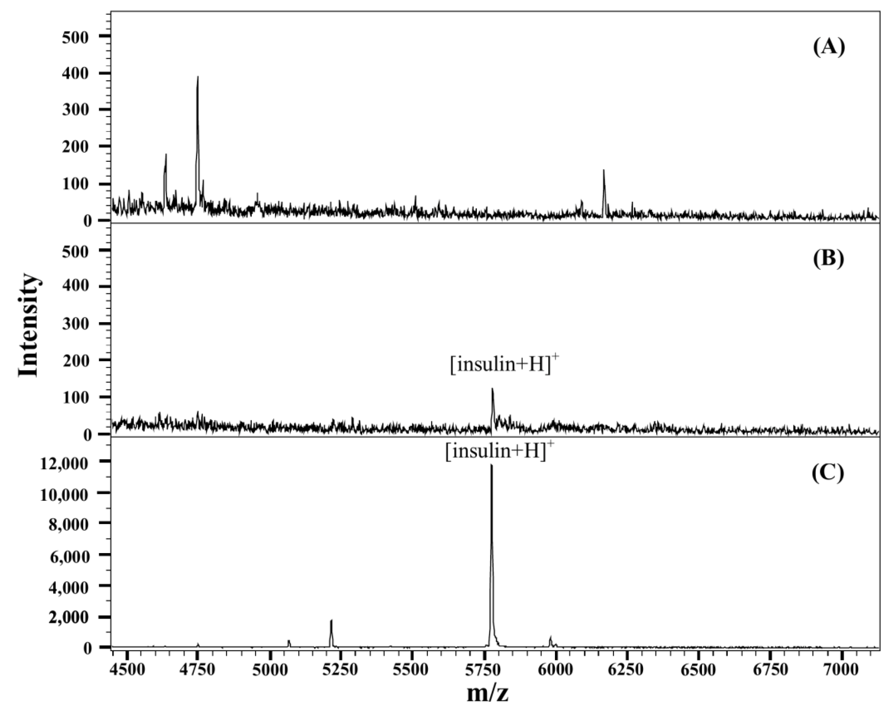
3.1. Detection of Insulin on Nylon Membrane by MALDI/MS

In traditional EME, large proteins are obstructed by the small pore size of the PP hollow fiber. To date, no paper has reported using EME to extract proteins. Previously, a solvent-free EME device coupled with MALDI/MS for the determination of peptides was developed in our laboratory. Based on previous results, the PVDF is an efficient membrane for the detection of peptides by using an α-cyano-4-hydroxycinnamic acid (CHCA) solution as a matrix [21]. To test if insulin can be desorbed and ionized from the PVDF membrane, 3 μL of insulin solution was pipetted onto the PVDF membrane and allowed to dry at room temperature. Then, 3 μL of SA solution in 30% acetonitrile/water solution that contains 0.1% TFA was applied on top of the membrane. Insulin on the PVDF membrane was successfully desorbed and ionized, as shown in Figure 2A, and the ions observed at m/z = 5778 were assigned as [insulin+H]+. However, the signal intensity of the insulin was weak. To promote the detection efficiency of insulin, several commercially available hydrophilic membranes were evaluated. Hydrophilic PTFE, MCE, and nylon membranes were tested. When applying the SA solution onto the membrane, the insulin redissolved in the SA solution and underwent crystallization. As shown in Figure 2, the nylon membrane provided the highest signal intensity of insulin. The better desorption/ionization efficiency that was obtained with the nylon membrane might be caused by the homogeneous crystallization of insulin and SA solution on the nylon membrane. Figure 3 shows an image of the nylon membrane that was obtained by scanning electron microscopy (SEM), where the surface structure of the membrane can be observed. The homogeneity of crystallization and signal intensity of insulin will also be affected by the composition of the SA solution. Since insulin is a hydrophobic protein, the effect of the acetonitrile content in the SA solution was also investigated at levels of 30%, 40%, 50%, 60%, and 70%. As shown in Figure 4, the results indicated that SA in a 40% acetonitrile/water solution provided the highest signal intensity of insulin. Using this sample preparation technique, the spot-to-spot reproducibility was less than 7.6% from 21 sample spots of 3 samples. Therefore, SA in a 40% acetonitrile/water solution was selected for the subsequent experiments.

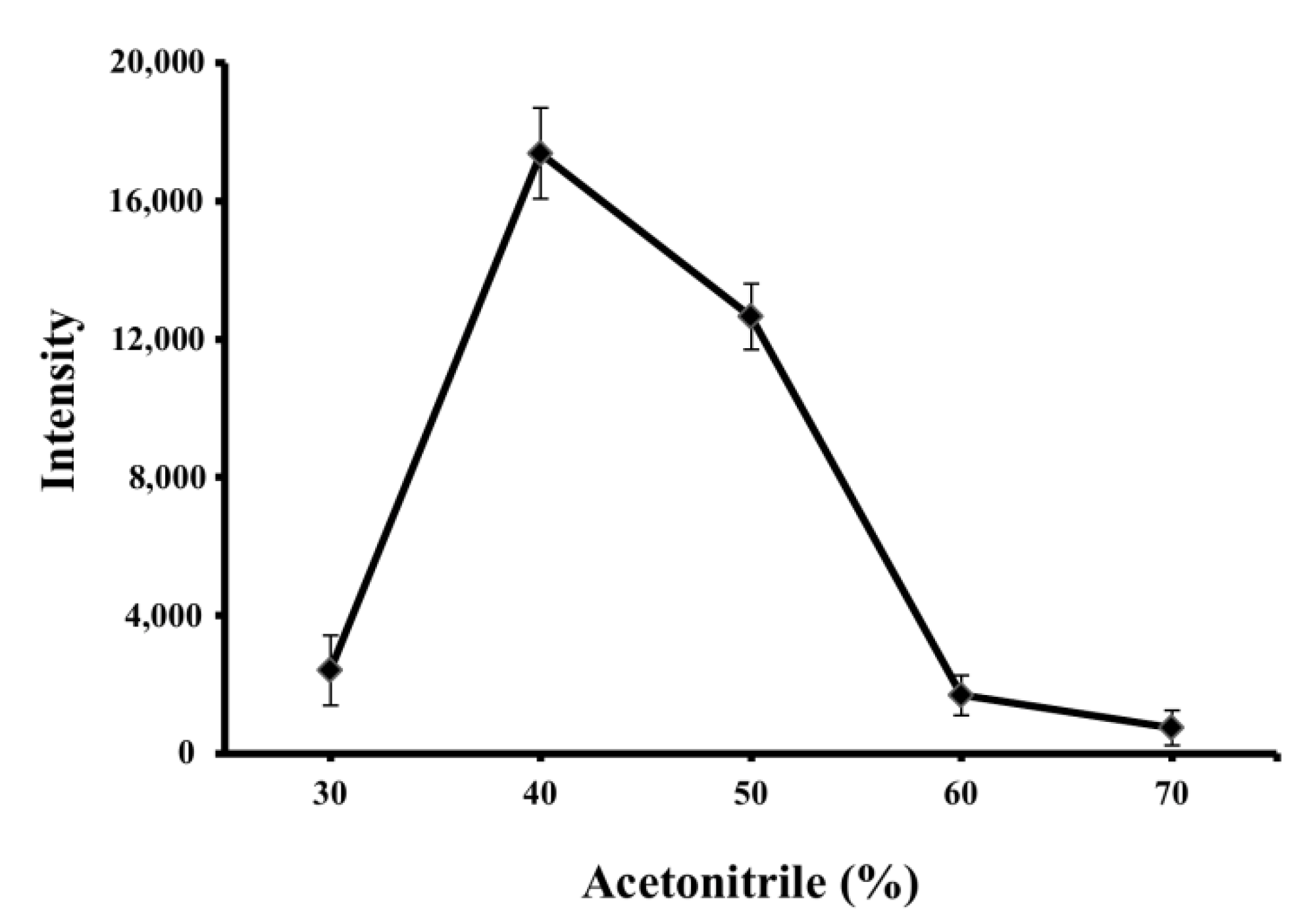
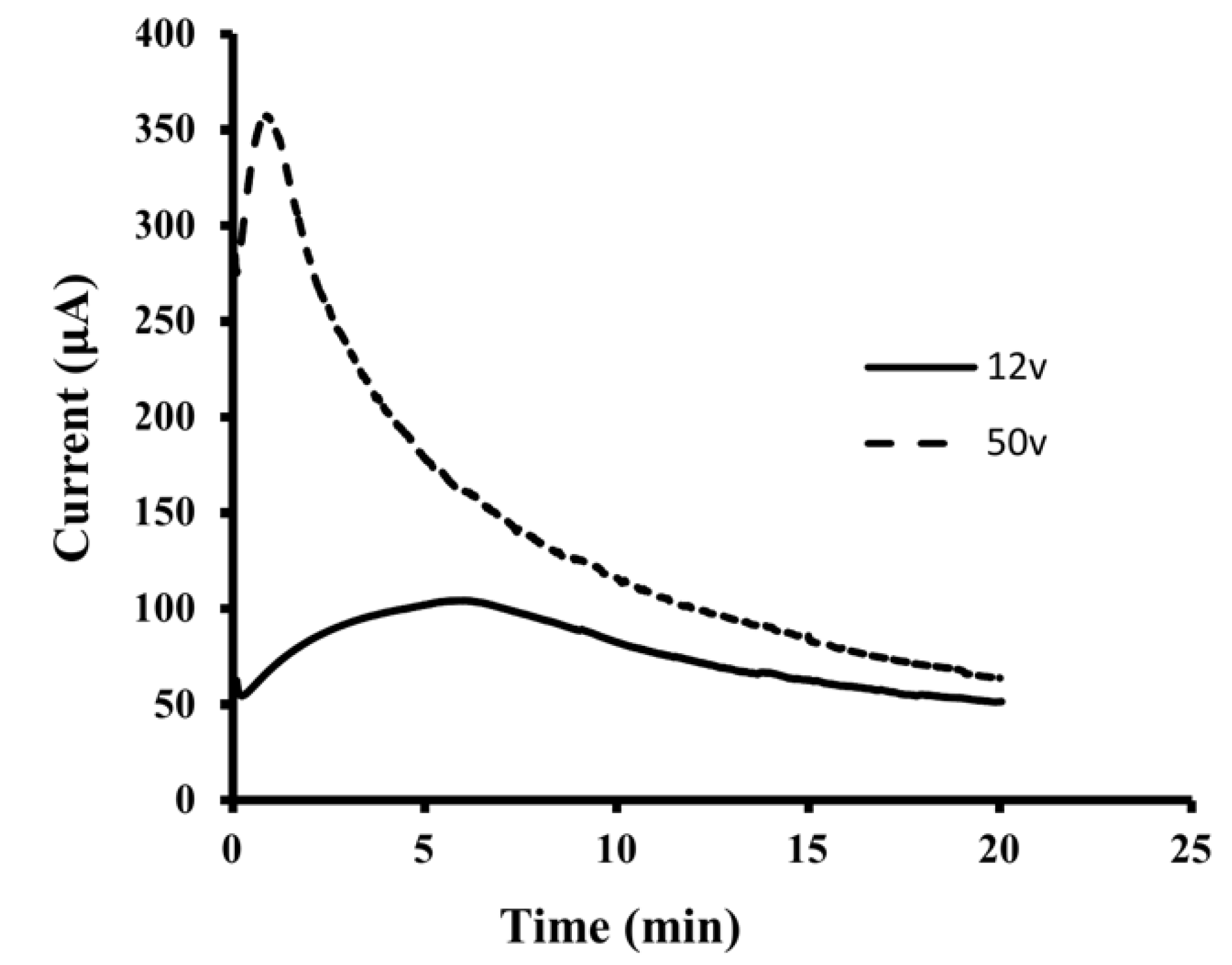
3.2. Optimization of EME Conditions

For the EME, the pH in the sample solution must be lower than the pI of the protein to ensure sufficient cationization of proteins in the sample solution. According to the literature, the pI value of insulin is 5.4 [26]. To ensure the cationization of insulin, 10 mM trifluoroacetic acid solution (pH 2.2) was utilized to prepare the sample solution. Upon the application of an electrical potential, positive insulin ions migrated toward the cathode inside the glass tube and were trapped by the nylon membrane at the side of the glass tube. After 10 min of extraction, the insulin-enriched nylon membrane was analyzed by MALDI/MS for insulin content. The effect of the applied voltages was initially investigated by applying 12 and 50 V at an extraction time of 10 min. However, when a higher voltage (50 V) was applied, lower extraction efficiency was obtained. With an applied voltage of 50 V, the system current was in the range of 100–350 μA, as shown in Figure 5. A drastic decrease in the current magnitude was observed within the first 2 min of the process; this drastic decrease was caused by the formation of an electrical double layer [27]. The system current that was obtained with 12 V was lower than 100 μA and stable throughout the extraction period. EME at low voltage provides the advantages of low system current and high system stability [28]. Therefore, a low voltage was selected for subsequent experiments. The extraction efficiency of insulin was evaluated by the signal intensity of [insulin+H]+ ions at m/z = 5778 for 21 sample spots of 3 samples. The effect of the applied voltage on the extraction efficiency was investigated in the range of 3 to 15 V at an extraction time of 10 min. As shown in Figure 6A, the signal intensity of insulin increased when the applied voltage was increased from 3 to 12 V, reaching a plateau. The effect of the sample volume on the signal intensity of insulin was further investigated in the range of 6–14 mL. As shown in Figure 6B, the signal intensity of insulin increased from 6 to 12 mL and then reached a plateau. Therefore, a sample volume of 12 mL was used in the following experiments.
The effect of the agitation rate on the signal intensity was investigated with varying stirring rates ranging from 100 to 500 rpm. The result is shown in Figure 6C. The signal intensities of insulin increased when the stirring rate increased from 100 to 400 rpm, and then the signal intensities of insulin decreased with an increasing agitation rate. The effect of the extraction time on the signal intensities of insulin was investigated by varying the extraction time from 2 to 12 min. The signal intensities of insulin increased when the extraction time increased from 2 to 10 min and then reached a plateau, as shown in Figure 6D. Therefore, the extraction time was set to 10 min for subsequent experiments.

3.3. Analytical Characteristics

A linear dynamic range (LDR) for insulin in an aqueous solution was obtained in the range of 1.0–100.0 nM, and the results are summarized in Table 1. The linear equation was y = 253.7x + 5.4, with a correlation coefficient of 0.9991. Figure 7A shows the mass spectrum of 50.0 nM insulin obtained by MALDI/MS without the EME. The S/N ratio of insulin was determined to be 11. With the use of EME to preconcentrate insulin, the signal intensities were greatly enhanced, with an S/N ratio of 1133, as shown in Figure 7B. A 103-fold enhancement of the S/N ratio of insulin can be obtained by using the EME in contrast to that of direct analysis. The mass spectrum of 1.0 nM insulin in an aqueous solution with the EME is illustrated in Figure 7C. Based on a signal-to-noise (S/N) ratio of 3, the limit of detection (LOD) was calculated as 0.3 nM for insulin. The recovery of the insulin in a standard solution (50.0 nM) was determined by a standard addition method. Based on triplicate measurements, the mean recovery of the insulin in a standard solution was found to be 65.1%.

3.4. Analysis of Insulin in Complicated Sample Matrices

The EME procedure with the new nylon membrane has also been evaluated for the determination of insulin in human urine samples. The system current of the urine sample was in the range of 40–110 μA and stable throughout the extraction period. The mass spectrum of the blank urine sample is shown in Figure 8A, in which no insulin signal was detected. The direct analysis (without the EME) of insulin-spiked urine samples was performed. Without the EME, an S/N ratio of 5 was obtained for insulin, as shown in Figure 8B. Weak signal intensity obtained resulted from the suppression effect in MALDI/MS, which was caused by the presence of salt and complicated matrices in samples. When using the EME to extract insulin from the insulin-spiked urine sample, the insulin signal was significantly enhanced, with an S/N ratio of 383, as shown in Figure 8C. A 77-fold enhancement of the S/N ratio of insulin can be obtained using the EME in contrast to that of the direct analysis. The LDR of insulin in urine samples was in the range of 5.0–100.0 nM. The linear equation was y = 253.2x + 45.6 with a correlation coefficient of 0.9979 for insulin in urine. Moreover, the results are summarized in Table 1. The LOD for insulin in urine was calculated to be 1.5 nM. The within-day and day-to-day accuracy and precision of the method were evaluated using urine samples that were spiked with low (8.0 nM), medium (50.0 nM), and high (80.0 nM) amounts of insulin. The results are presented in Table 2. Both the within-day and day-to-day accuracies for the three spiked levels of insulin in urine samples were expressed as relative error and found to be less than 6.0%. The within-day and day-to-day precisions for the three spiked levels of insulin were expressed as the coefficients of variations (CVs) and found to be less than 3.5% and 5.5%, respectively. The recovery of insulin from urine samples (50.0 nM) was determined by a standard addition method. Based on triplicate measurements, the mean recovery of insulin in urine samples was found to be 57.2%. The low recovery might be caused by penetration of the analyte through the pores of the nylon membrane into the solution of the glass tube.
A comparison between the proposed method and other published techniques for analyzing insulin is presented in Table 3. The LOD of insulin that was obtained with this method was comparable to that reported using gold-nanoparticle (NP)-decorated target plates [29] and NP-based solid phase extraction (SPE) [30]. The LOD of insulin is two orders of magnitude lower than those of NP-assisted single drop microextraction (SPME) [31] and NP-assisted liquid microextraction (LLME) methods [32]. This developed method provides a shorter extraction time, ease of operation, and higher enhancement of the S/N ratio. Furthermore, there is no need to have an elaborated synthesis of NP or modification of the target plate surface.

4. Conclusions

A nylon membrane was successfully evaluated for the electromembrane extraction of protein from aqueous solutions coupled with MALDI/MS analysis. Insulin was extracted and preconcentrated when the pH of the solution was lower than the pI through electromigration. For the first time, EME, in combination with MALDI/MS, was utilized for the determination of insulin. The incorporation of EME and MALDI/MS improved the S/N ratio by more than two orders of magnitude. The analysis of insulin in aqueous solution can be performed in less than 15 min. This approach is simple, time-efficient, and economical, as the flat membrane mode of EME coupled with MALDI/MS is suitable. In addition, this method was successfully applied to the determination of insulin in human urine samples. In the future, this developed method will be suitable for the screening of protein levels in human urine samples.

Author Contributions

Conceptualization, S.Y.C.; methodology, S.Y.C.; validation, J.-K.H. and Y.-W.H.; formal analysis, J.-K.H. and Y.-W.H.; investigation, S.Y.C., J.-K.H. and Y.-W.H.; resources, W.-C.C.; data curation, S.Y.C. and J.-K.H.; writing—original draft preparation, S.Y.C.; writing—review and editing, S.Y.C.; supervision, S.Y.C.; project administration, S.Y.C. and W.-C.C.; funding acquisition, S.Y.C. All authors have read and agreed to the published version of the manuscript.

Funding

This research was funded by the Ministry of Science and Technology of Taiwan, MOST 110-2113-M-017-001.

Institutional Review Board Statement

All subjects gave their informed consent for inclusion before they participated in the study. The study was conducted in accordance with the Declaration of Helsinki and approved by the Institutional Review Board (IRB) of Kaohsiung Veterans General Hospital (KSVGH20-CT8-13) for studies involving human urine samples.

Informed Consent Statement

Informed consent was obtained from all subjects involved in the study.

Data Availability Statement

Not applicable.

Acknowledgments

This work was financially supported by the Ministry of Science and Technology (MOST 110-2113-M-017-001) of Taiwan.

Conflicts of Interest

The authors declare no conflict of interest.

References

  1. Pedersen-Bjergaard, S.; Rasmussen, K.E. Electrokinetic migration across artificial liquid membranes new concept for rapid sample preparation of biological fluids. J. Chromatogr. A 2006, 1109, 183–190. [Google Scholar] [CrossRef] [PubMed]
  2. Balchen, M.; Halvorsen, T.G.; Reubsaet, L.; Pedersen-Bjergaard, S. Rapid isolation of angiotensin peptides from plasma by electromembrane extraction. J. Chromatogr. A 2009, 1216, 6900–6905. [Google Scholar] [CrossRef] [PubMed]
  3. Drouin, N.; Kubáň, P.; Rudaz, S.; Pedersen-Bjergaard, S. Elecromembrane extraction: Overview of the last decade. Trends Anal. Chem. 2019, 113, 357–363. [Google Scholar] [CrossRef]
  4. Huang, C.; Chen, Z.; Gjestad, A.; Pedersen-Bjergaard, S.; Shen, X. Elecromembrane extraction. Trends Anal. Chem. 2017, 95, 47–56. [Google Scholar] [CrossRef]
  5. Balchen, M.; Hatterud, A.G.; Reubsaet, L.; Pedersen-Bjergaard, S. Fundamental studies on the electrokinetic transfer of net cationic peptides across supported liquid membranes. J. Sep. Sci. 2011, 34, 186–195. [Google Scholar] [CrossRef]
  6. Fashi, A.; Khanban, F.; Yaftian, M.R.; Zamani, A. The cooperative effect of reduced grapheme oxide and Triton X-114 on the electromembrane microextraction efficiency of Pramipexole as a model analyte in urine samples. Talanta 2017, 162, 210–217. [Google Scholar] [CrossRef] [PubMed]
  7. Huang, C.; Gjelstad, A.; Pedersen-Bjergaard, S. Exhaustive extraction of peptides by electromembrane extraction. Anal. Chim. Acta 2015, 853, 328–334. [Google Scholar] [CrossRef]
  8. Huang, C.; Gjelstad, A.; Pedersen-Bjergaard, S. Selective electromembrane extraction based on isoelectric point: Fundamental studies with angiotensin II antipeptide as model analyte. J. Membr. Sci. 2015, 481, 115–123. [Google Scholar] [CrossRef]
  9. Abbasi, S.; Haeri, S.A. Enrichment of psychotropic drugs using rhamnolipid bioaggregates after electromembrane extraction based on an agarose gel using a rotating electrode as a green and organic solvent-free strategy. J. Chromatogr. A 2021, 1655, 462500. [Google Scholar] [CrossRef]
  10. Pourahadi, A.; Nojavan, S.; Davarani, S.S.H. Gel-electromembrane extraction of peptides: Determination of five hypothalamic agents in human plasma samples. Talanta 2020, 217, 121025. [Google Scholar] [CrossRef]
  11. Sedehi, S.; Tabani, H.; Nojavan, S. Electro-driven extraction of polar compounds using agarose gel as a new membrane: Determination of amino acids in fruit juice and human plasma samples. Talanta 2018, 179, 318–325. [Google Scholar] [CrossRef] [PubMed]
  12. Asadi, S.; Tabani, H.; Nojavan, S. Application of polyacrylamide gel as a new membrane in electromembrane extraction for the quantification of basic drugs in breast milk and wasterwater samples. J. Pharm. Biomed. Anal. 2018, 151, 178–185. [Google Scholar] [CrossRef] [PubMed]
  13. Hajizadeh, S.; Farhadi, K.; Molaei, R.; Forough, M. Silver nanoparticles-tragacanth gel as a green membrane for effective extraction and determination of capecitabine. J. Sep. Sci. 2020, 43, 2666–2674. [Google Scholar] [CrossRef]
  14. Román-Hidalgo, C.; Martín-Valero, M.J.; Fernández-Torres, R.; Callejón-Mochón, M.; Bello-López, M.A. New nanostructured support for carrier-mediated electromembrane extraction of high polar compounds. Talanta 2017, 162, 32–37. [Google Scholar] [CrossRef] [PubMed]
  15. Cristina, R.-H.; Jesús, M.-V.M.; Rut, F.-T.; Ángel, B.-L.M. Use of polymer inclusion membranes (PIMs) as support for electromembrane extraction of non-steroidal anti-inflammatory drugs and highly polar acidic drugs. Talanta 2018, 179, 601–607. [Google Scholar] [CrossRef] [PubMed]
  16. Mamat, N.A.; See, H.H. Simultaneous electromembrane extraction of cationic and anionic herbicides across hollow polymer inclusion membranes with a bubbleless electrode. J. Chromatogr. A 2017, 1504, 9–16. [Google Scholar] [CrossRef] [PubMed]
  17. Mamat, N.A.; See, H.H. Development and evaluation of electromembrane extraction across a hollow polymer inclusion membrane. J. Chromatogr. A 2015, 1406, 34–39. [Google Scholar] [CrossRef]
  18. Mollahosseini, A.; Elyasi, Y.; Rastegari, M. An improvement of electrospun membrane reusability via titanium dioxide nanoparticles and silane compounds for the electromembrane extraction. Anal. Chim. Acta 2019, 1088, 168–177. [Google Scholar] [CrossRef]
  19. Román-Hidalgo, C.; López-Perez, G.; Martín-Valero, M.J.; Bello-López, M.A. Chitosan tailor-made membranes as biopolymeric support for electromembrane extraction. Talanta 2019, 199, 290–295. [Google Scholar] [CrossRef]
  20. Hansen, F.A.; Pedersen-Bjergaard, S. Electromembrane extraction of streptomycin from biological fluids. J. Chromatogr. A 2021, 1639, 461915. [Google Scholar] [CrossRef]
  21. Yeh, C.-S.; Cheng, P.-S.; Chang, S.Y. Solvent-free electromembrane extraction: A new concept in electro-driven extraction. Talanta 2019, 199, 296–302. [Google Scholar] [CrossRef] [PubMed]
  22. Tseng, C.H.; Ho, K.C.; Lin, Y.S.; Chen, Y.C. Gold Nanoparticles as Selective and Concentrating Probes for Samples in MALDI MS Analysis. Anal. Chem. 2004, 76, 4337–4342. [Google Scholar]
  23. Kong, X.L.; Huang, L.C.L.; Hsu, C.M.; Chen, W.H.; Han, C.C.; Chang, H.C. High-Affinity Capture of Proteins by Diamond Nanoparticles for Mass Spectrometric Analysis. Anal. Chem. 2005, 77, 259–265. [Google Scholar] [CrossRef] [PubMed]
  24. Chandler, J.; Haslam, C.; Hardy, N.; Leveridge, M.; Marshall, P. A systematic investigation of the best buffers for use in screening by MALDI-MASS spectromstry. Slas Discov. Adv. Life Sci. RD 2017, 22, 1262–1269. [Google Scholar] [CrossRef] [PubMed] [Green Version]
  25. Chen, C.S.; Chen, W.C.; Chang, S.Y. Electromembrane extraction of posaconazole for matrix-assisted laser desorption/ionization mass spectrometric detection. Membranes 2022, 12, 620. [Google Scholar] [CrossRef] [PubMed]
  26. Askari, N.; Beheshti-Marnani, A.; Askari, M.B.; Rohani, T. Detection of ultra-trace levels of insulin by Fe3O4@MoS2/γGO-GCE a sensor based on isoelectric points. J. Mater. Sci. Mater. Electron. 2019, 30, 9652–9662. [Google Scholar] [CrossRef]
  27. Rahmani, T.; Rahimi, A.; Nojavan, S. Study on electrical current variations in electromembrane extraction process: Relation between extraction recovery and magnitude of electrical current. Anal. Chim. Acta 2016, 903, 81–90. [Google Scholar] [CrossRef]
  28. Ara, K.M.; Raofie, F. Low-voltage electrochemically stimulated stir membrane liquid-liquid microextraction as a novel technique for the determination of methadone. Talanta 2017, 168, 105–112. [Google Scholar] [CrossRef]
  29. Liang, K.; Wu, H.; Li, Y. Immune-enrichment of insulin in bio-fluids on gold-nanoparticle decorated target plate and in situ detection by MALDI MS. Clin. Proteom. 2017, 14, 5. [Google Scholar] [CrossRef] [Green Version]
  30. Chang, S.Y.; Zhang, N.Y.; Chen, C.S.; Chen, C.D.; Chen, Y.Y.; Wang, C.R.C. Analysis of peptides and proteins affinity-bound to iron oxide nanoparticles by MALDI MS. J. Am. Soc. Mass Spectrom. 2007, 18, 910–918. [Google Scholar] [CrossRef] [Green Version]
  31. Shastri, L.; Abdelhamid, H.N.; Nawaz, M.; Wu, H.F. Synthesis, characterization and bifunctional applications of bidentate silver nanoparticle assisted single drop microextraction as a highly sensitive preconcentrating probe for protein analysis. RSC Adv. 2015, 5, 41595–41603. [Google Scholar] [CrossRef]
  32. Shrivas, K.; Wu, H.F. Rapid and highly sensitive protein extraction via cobalt oxide nanoparticle-based liquid-liquid microextraction coupled with MALDI mass spectrometry. Analyst 2012, 137, 890–895. [Google Scholar] [CrossRef] [PubMed]
Figure 1. Schematic illustration of the EME setup.
Separations 09 00286 g001
Figure 2. Mass spectra of insulin obtained with different membranes: (A) PVDF, (B) PTFE, (C) MCE, and (D) nylon. Experimental conditions: insulin volume, 3 μL; SA solution volume, 3 μL; and insulin concentration, 5.0 μM. The mass spectrum was obtained by averaging 200 mass spectra using laser energy that slightly exceeded the threshold.
Separations 09 00286 g002
Figure 3. SEM image of the nylon membrane. The SEM operated at an accelerating voltage of 20 kV.
Separations 09 00286 g003
Figure 4. Mass spectra of insulin with an SA solution in 0.1% aqueous TFA containing different amount of acetonitrile. Experimental conditions: sample volume, 3 μL; SA solution volume, 3 μL; and insulin concentration, 5.0 μM. The signal intensities of insulin were evaluated by the peak height of [insulin+H]+ ions at m/z = 5778 for 21 sample spots of 3 samples.
Separations 09 00286 g004
Figure 5. The system current, as a function of extraction time, obtained with EME at an applied voltage of 50 V and 12 V.
Separations 09 00286 g005
Figure 6. Effect of (A) the applied voltage, (B) the sample volume, (C) the agitation rate, and (D) the extraction time on the signal intensity of insulin obtained from the EME. Experimental conditions: insulin concentration, 50.0 nM. All other conditions were identical to those outlined in Figure 4.
Separations 09 00286 g006
Figure 7. Mass spectra of (A) insulin (50.0 nM) obtained without the EME, (B) insulin (50.0 nM) obtained with the EME, and (C) insulin (1.0 nM) obtained with the EME. Experimental conditions: sample volume, 12 mL; applied voltage, 12 V; agitation rate, 400 rpm; and extraction time, 10 min.
Separations 09 00286 g007
Figure 8. Mass spectra of human urine samples. (A) blank urine without the EME, (B) insulin-spiked urine without the EME, and (C) insulin-spiked urine with the EME. Experimental conditions: sample volume, 12 mL; applied voltage, 12 V; stirring rate, 400 rpm; extraction time, 10 min; and insulin, 50.0 nM.
Separations 09 00286 g008
Table 1. Analytical characteristics for the determination of insulin.
InsulinLinear Range (nM)Regression Equation aR bLOD c (nM)EFRecovery
Standard solution 1.0–100.0y = 253.7x + 5.40.99910.310365.1%
Urine samples5.0–100.0y = 253.2x + 45.60.99791.57757.2%
a y, signal intensity of insulin; x, insulin concentration. b Correlation coefficient. c The LOD was calculated as three times the S/N ratio.
Table 2. Within-day and day-to-day precision and accuracy for the EME-MALDI/MS analysis of insulin in urine samples (n = 5).
AnalyteConcentration (nM)Within-DayDay-to-Day
Precision a
(%)
Accuracy b
(%)
Precision a
(%)
Accuracy b
(%)
Insulin8.02.3−5.85.1−5.3
50.03.30.63.53.0
80.02.00.63.6−1.1
a Precision expressed as CV. b Accuracy expressed as relative error.
Table 3. Comparison between the proposed method and other reported methods for the determination of insulin in aqueous solution.
Analytical MethodSample PreparationExtraction Time
(min)
S/N Ratio EnhancementLOD
(nM)
Reference
MALDI/MSNP-LLMS1012.55.0[32]
MALDI/MSNP-SDME10not available80.0[31]
MALDI/MSNP-based SPE60150.1[30]
MALDI/MSNP-decorated target plate30not available0.8[29]
MALDI/MSEME101030.3This work
Publisher’s Note: MDPI stays neutral with regard to jurisdictional claims in published maps and institutional affiliations.

