Article

Synthesis, Structures, and Photoluminescence of Two Novel Zinc(II) Compounds Containing 2-Acetylpyridine-aminoguanidine

by
Mirjana M. Radanović
1,
Ljiljana S. Vojinović-Ješić
1,
Miodrag G. Jelić
2,
Elias Sakellis
3,
Berta Barta Holló
1,*,
Vukadin M. Leovac
1 and
Marko V. Rodić
1
1
Faculty of Sciences, University of Novi Sad, Trg Dositeja Obradovića 3, 21000 Novi Sad, Serbia
2
NTT Data Global IT Innovator, 300000 Timisoara, Romania
3
National Centre for Scientific Research “Demokritos”, Institute of Nanoscience and Nanotechnology, Terma (End) Patriarhou Gregoriou Str, P.O. Box 60228 Aghia Paraskevi, 15310 Athens, Greece
*
Author to whom correspondence should be addressed.
Inorganics 2022, 10(10), 147; https://doi.org/10.3390/inorganics10100147
Submission received: 29 August 2022 / Revised: 15 September 2022 / Accepted: 16 September 2022 / Published: 20 September 2022
(This article belongs to the Special Issue Metal Complexes with N-donor Ligands)

Abstract

:
In the reaction of zinc(II) sulfate and the chloride salt of 2-acetylpyridine-aminoguanidine, two types of complex were obtained, i.e., [Zn(H2O)6](H2L)2(SO4)3·3H2O and [Zn(L)H2O(SO4)]·H2O, depending on the presence of LiOAc as the deprotonating agent. The physicochemical, structural, and photoluminescence properties of the complexes were examined. In the first complex, obtained in the absence of LiOAc, the Schiff base had the role of a counter-ion in its doubly protonated form, while in the presence of LiOAc, upon deprotonation, coordination takes place, and thus the Schiff base acts as a tridentate N3 ligand. In the latter complex, the ligand is coordinated through pyridine, azomethine, and the imino nitrogen of the aminoguanidine residue, leading to formation of two fused five-membered chelate rings. Both the examined complexes, as well as the ligand itself, show high photoluminescence.

1. Introduction

Metal complexes with Schiff base ligands have been intensively studied due to their promising biological activities [1], catalytic behavior [2], magnetism [3], and, particularly, their optoelectronic properties [4,5,6]. Among these, the complexes of Zn(II) stand out owing to their luminescence and potential application in OLED [7,8]. Although Zn(II) has no optical spectroscopic signature (3d10 configuration), it can improve the luminescence of the Schiff bases [9], and also enables a wide range of symmetries and coordination numbers [10]. For preparing complexes with photoluminescence properties, metal centers with zero crystal field stabilization energy and with the absence of d–d transitions are the most suitable. Metals with d10 configuration fulfill these requirements [11,12]. Zn(II), as a d10 metal with good bioavailability, is a promising candidate for preparing complexes with photoluminescent properties for biological applications.
Due to the evident biological importance of aminoguanidine and its Schiff bases [13], interest in the synthesis and full characterization of their metal complexes has risen [14]. On the other hand, the newest research results have shown that metal complexes with Schiff bases of 2-acetylpyridine exhibit significant cytotoxic activity against some solid tumor lines and leukemia cells [15]. On the basis of the abovementioned points, it is undoubtedly of high interest to make some further investigations in this field, i.e., to examine the structure and properties of metal complexes with aminoguanidine derivatives. Recently, the photoluminescence of an aminoguanidine Schiff base with pyridoxal and its metal complexes have been thoroughly examined [16,17,18,19]. One of these compounds was used as a new electron-injecting layer for PLED, which led to higher luminous efficiency a reduction in the turn-on voltage, and an increase in the brightness [18].
Here, we describe the synthesis of two novel Zn(II) complexes with the Schiff base of 2-acetylpyridine and aminoguanidine (Figure 1), and their physicochemical and structural characteristics. The photoluminescence of the chloride ligand salt was also examined.

2. Results and Discussion

2.1. Synthesis and Physicochemical Properties of the Complexes

In the reaction of the warm aqueous solutions of the chloride salt of 2-acetylpyridine-aminoguanidine, [H2L]Cl2 and zinc(II) sulfate at a molar ratio of 1:1, two Zn(II) complexes were obtained, viz. [Zn(H2O)6](H2L)2(SO4)3·3H2O (Complex 1) and [Zn(L)H2O(SO4)]·H2O (Complex 2). During the synthesis of Complex 2, in the reaction mixture, LiOAc was added in order to deprotonate the Schiff base and enable the coordination of its neutral form. Similar behavior of this ligand has been described recently [20].
The complexes are white crystalline substances that are stable in air. Complex 1 is very soluble in H2O, slightly soluble in MeOH and EtOH, and insoluble in DMF. On the contrary, Complex 2 shows good solubility only in DMF, while it is poorly soluble in H2O and alcohols.
The molar conductivity of a H2O solution of Complex 1 is in accordance with its proposed coordination formula (i.e., it corresponds to the presence of six ions). The result was additionally proved by molar conductivity measurements of the H2O solution of the starting compounds. The molar conductivity of Complex 1 (720 S cm2 mol−1) is proportionally higher than the molar conductivity values of the precursors (205 S cm2 mol−1 for ZnSO4 and 320 S cm2 mol−1 for the chloride salt of the ligand), which show the presence of two and three ions in the solution of ZnSO4 and [H2L]Cl2, respectively [21].
As the coordination formula shows, and X-ray analysis has confirmed (vide infra), in Complex 1, the coordination of the Schiff base, a potentially tridentate ligand, did not take place. This could be assumed on the basis of the presence of the characteristic bands in the IR spectrum of Complex 1 at the nearly same position as in the spectrum of the dichloride salt of this Schiff base [22]. Weak and wide ν(NH+) bands in the 3100–2800 cm−1 region suggested the protonation of the nitrogen atom of the pyridine ring, while the bands at 1683 cm−1 and 1631 cm−1 correspond to the ν(C=N) vibrations of the azomethine and imino group of aminoguanidine residue, respectively [22]. Due to coordination, in the IR spectrum of Complex 2, the latter two bands underwent a negative shift by ~30 cm−1 and could be found at 1654 and 1599 cm−1.
Finally, in the IR spectra of both complexes, some additional bands of sulfate anion were present. Therefore, in the spectrum of Complex 1, strong bands at 1120 cm−1 and 618 cm−1 could be ascribed to non-coordinated SO42– ions, while a broad band in the region of 1160–1112 cm−1, as well as a sharp band at 620 cm−1 in the spectrum of Complex 2 points to the monodentate coordination of sulfate ions [23].

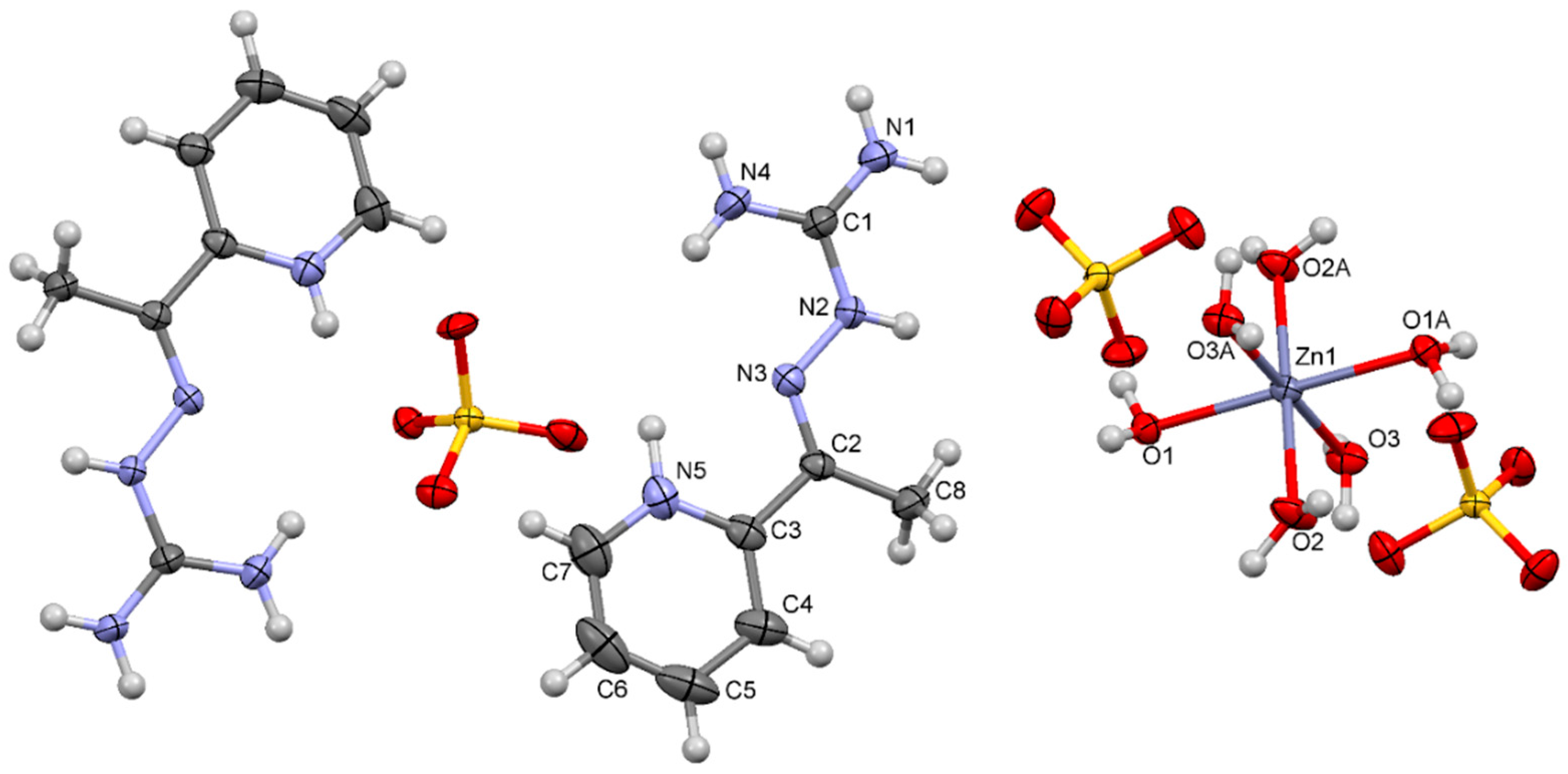
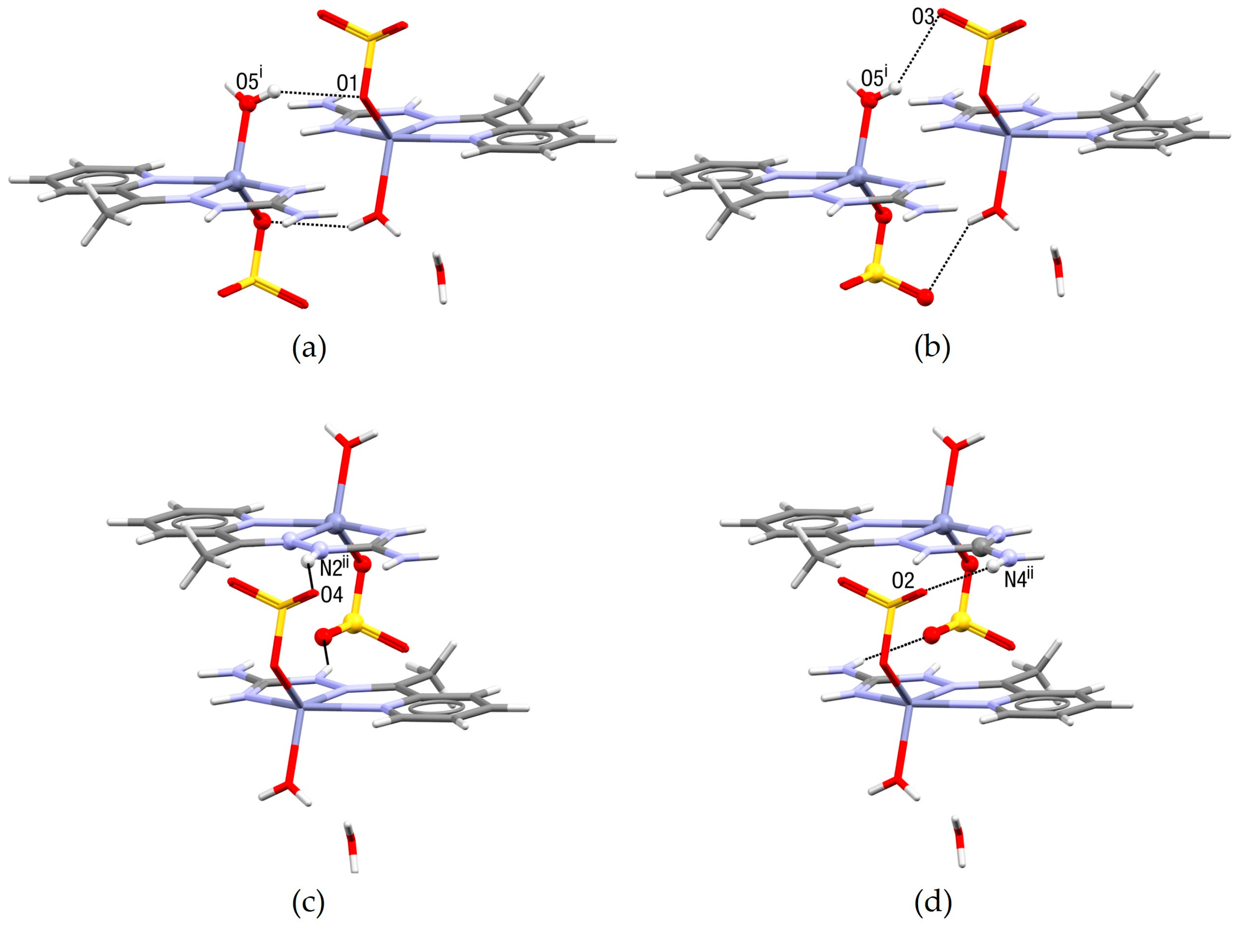
2.2. Crystal Structure

Figure 2 and Figure 3 show the molecular structures of the complexes including the numbering schemes, while Table 1 contains selected structural data for these structures.
The asymmetric unit of Complex 1 consists of 1/2 [Zn(H2O)6]2+ ions, a doubly protonated molecule of the Schiff base, 3/2 sulfate anions, and 3/2 water molecules. Namely, the [Zn(H2O)]2+ ion is located at the inversion center, while one sulfate ion and one water molecule are disordered around another inversion center. In the present cation of Schiff base, both aminoguanidine residue and pyridine ring are protonated. In Complex 2, the chelating ligand is coordinated in its neutral form, namely in the usual tridentate N3 mode, through pyridine (N5), azomethine (N3), and the imino (N1) nitrogen of aminoguanidine moiety. In this way, two planar, fused, five-membered chelate rings are formed, with the Zn(II) ion being coplanar with the nitrogen ligators of the Schiff base. Besides the ligand molecule, the square-pyramidal environment of Zn(II) in Complex 2 is formed by monodentate coordination of one sulfate anion in the equatorial position and one water molecule in the apical position. Deviation from the ideal square-pyramidal geometry is described by the τ5 parameter, which has the value of 0.22 [24]. The asymmetric unit of this complex contains another, non-coordinated, water molecule as well.
The crystal packing of Complex 1 is presented in Figure 4. The crystal structure can be described as layered, where the cation and anion layers are stacked in the ab plane. Namely, [Zn(H2O)6]2+ octahedra are situated at z = 0, SO42− tetrahedra approximately at z = 1/8, [H2L]2+ cations at approximately z = 3/8, and disordered SO42− tetrahedra at z = 0.5. Water molecules additionally connect adjacent [Zn(H2O)6]2+ octahedra and SO42− tetrahedra through hydrogen bonding, while another set of disordered water molecules (not shown in Figure 4) is hydrogen-bonded to SO42− tetrahedra at z = 0.5.
The presence of the Schiff base dication in Complex 1 and its coordinated neutral form in Complex 2, allow good insight into the effects of deprotonation and coordination. In previous work, a similar comparison between the structural parameters of the ligands and complexes was made [25]. From the values of bond lengths given in Table 1, it can be seen that the C1–N1 and N2–N3 bonds are shorter (by ~0.03 and 0.02 Å, respectively), while the C1–N2 and C1–N4 bonds (by ~0.03 Å) are longer in Complex 2 than in Complex 1, which could be explained as a consequence of deprotonation of the aminoguanidine fragment. However, wide delocalization in this part of the structure is present in both the compounds, thus the values of the mentioned bond lengths are between those characteristics for single and double C–N/N–N bonds. The C2–N3 bond length has the value of a double bond in both compounds. On the other hand, the effects of coordination could be seen from the angular changes, i.e., widening of N1–C1–N4 angle and shrinkage of the N3–N2–C1 and N2–C1–N1 angles. Another difference important to note is the value of the C3–N5–C7 angle, which is, due to protonation of the pyridine nitrogen atom, larger than 120° in Complex 1, but in Complex 2, upon deprotonation, has a value lower than 120°.
In Complex 2, the metal–Schiff base bond lengths are in the range of 2.0400 (19)–2.1712 (17) Å, the one with the pyridine nitrogen atom (N5) being the longest, and the one with the imino nitrogen atom of the aminoguanidine moiety (N1) being the shortest. The same trend was observed in the structure of other complexes with this Schiff base [20,22]. Among all the ligators in the coordination sphere, the oxygen atom of the sulfate anion is the closest to the metal ion.
The structure is stabilized by the H-bond network made by all potential H-donors of the Schiff base moiety, except for the imino nitrogen atom N1 of the aminoguanidine residue in Complex 2. The details of the H-bond parameters are given in Table 2 and Table 3. It is interesting to note that the extensive hydrogen bonding between complex molecules in Complex 2 is mainly realized through the formation of ring-type motifs of different sizes between inversion-related molecules (Figure 5).

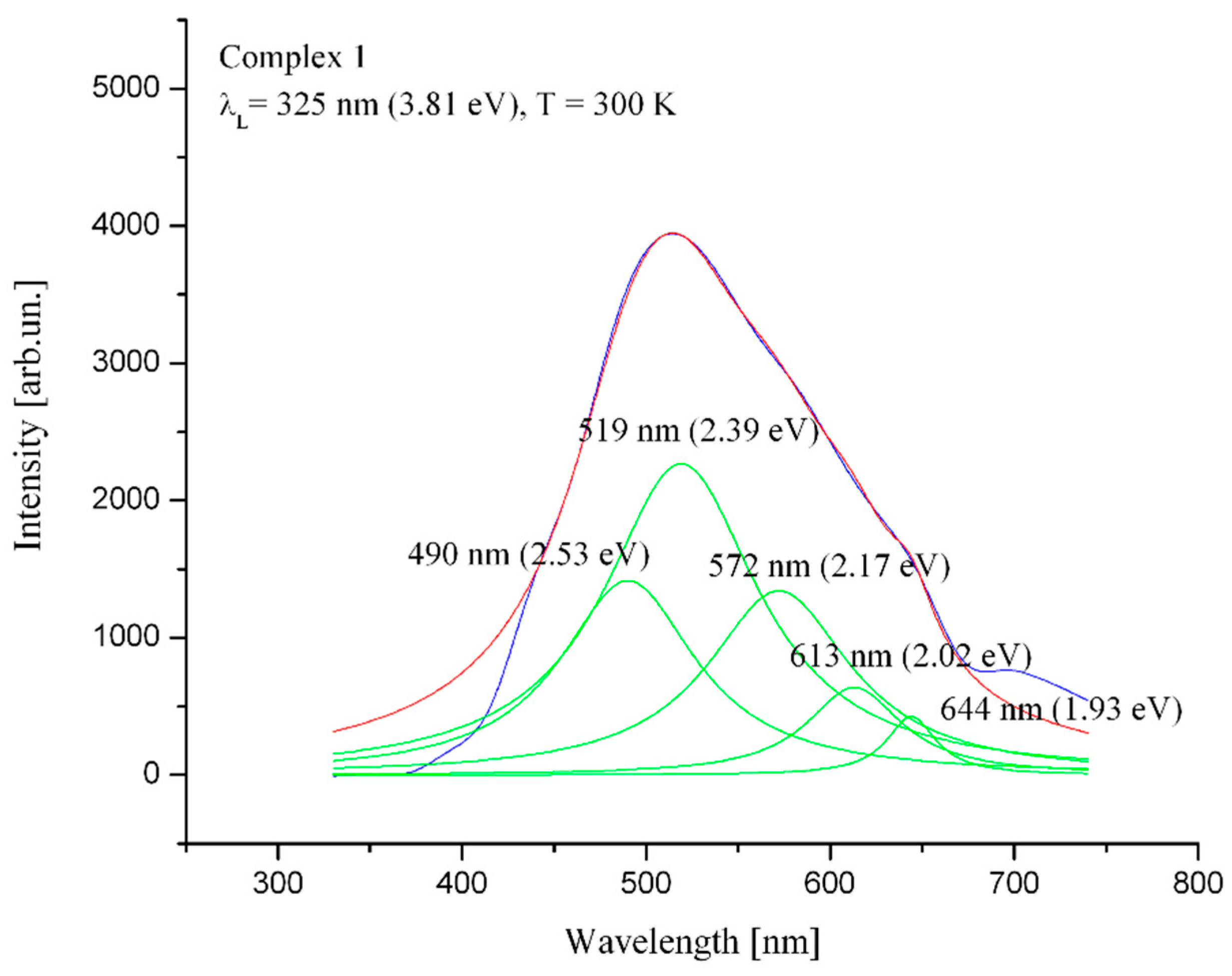
2.3. Photoluminescence Measurement

Three graphs were utilized, based on the measurements of the photoluminescence spectra of the materials. For each material, one measurement was made with the uncertainty of ~50 counts.
The photoluminescence characteristics of Complex 1 are presented in Figure 6. The width of the entrance slit was 500 µm and the exposure time was 2 s. Five optical peaks (green curves) in the complex photoluminescence spectrum were distinguished after a Lorentzian multipeak fitting of the characteristics. Through use of a 325 nm laser, experimental setup peak values of 1413, 2268, 1341, 640, and 424 a.u. were detected at the wavelength values of 490 nm (2.53 eV), 519 nm (2.39 eV) and 572 nm (2.17 eV), 613 nm (2.02 eV), and 644 nm (1.93 eV), respectively. It can be concluded that this complex shows high photoluminescence.
The second graph (Figure 7) displays the photoluminescence characteristics of Complex 2. The entrance slit was 100 µm wide and the exposure time was 3 s. The same fitting technique as used in the previous case revealed the five different peaks again (green curves). For this setup, the intensity peaks appeared at almost the same wavelength values as in the previous complex: 493 nm (2.51 eV), 520 nm (2.38 eV), 571 nm (2.17 eV), 614 nm (2.02 eV), and 644 nm (1.93 eV). Their intensity levels were 3055, 25,830, 11,821, 2482, and 1308 a.u., respectively. This complex shows very high photoluminescence, with intensities that are one order higher compared with Complex 1.
In the last graph (Figure 8), the photoluminescence characteristics of the ligand used in both complexes is displayed. The width of the entrance slit was 300 µm and the exposure time was 3 s. It was noticed that four different peaks appeared. For this setup, intensity peaks appeared at the following wavelength values: 465 nm (2.67 eV), 489 nm (2.53 eV) and 504 nm (2.46 eV), 526 nm (2.36 eV). Their intensity levels were 24721, 19773, 4634, and 6469 a.u., respectively. The ligand itself shows photoluminescence of very high intensity and a narrow spectrum.
The intensity values of the peaks of Complex 1 are similar to those of the complex (PLAG)2[ZnCl4] [18], in which the organic part is also a counter-ion, as in Complex 1. A comparison of Complex 2 with (PLAG)2[ZnCl4] gave almost the same result as the comparison of Complex 2 with Complex 1, where Complex 2 exhibited higher photoluminescence than (PLAG)2[ZnCl4] [18]. However, compared with the photoluminescent complex of Ru(II) [17], it can be concluded that tris(2,2′-bipyridine)ruthenium(II) shows significantly higher photoluminescence than the studied zinc (II) complexes. The free ligand shows the highest photoluminescence, but its coordination with Zn(II) leads to the spectrum shifting to higher wavelengths and significant spectrum widening. The photoluminescent properties of the ligand and Complexes 1 and 2 are promising for future applications in OLED/PLED development [18].

3. Materials and Methods

3.1. Materials and Physical Measurements

All chemicals used were commercial products of analytical reagent grade, except for the ligand 2-acetylpyridine-aminoguanidine dihydrogenchloride, [H2L]Cl2, which was obtained by the reaction described in [22].
Elemental analyses (C, H, N, S) of the air-dried complexes were carried out by standard micromethods in the Center for Instrumental Analyses, ICTM in Belgrade. The molar conductivities of freshly prepared complex solutions (c = 1 × 10⁻³ mol dm−3) were measured on a Jenway 4010 conductivity meter. IR spectra were recorded using KBr pellets on a NEXUS 670 FTIR spectrophotometer (Thermo Nicolet) in the range of 4000–400 cm−1. Melting points were measured on a Nagema melting point microscope (Rapido).

3.2. Synthesis of Complexes

[Zn(H2O)6](H2L)2(SO4)3·3H2O (Complex1)
The ligand (0.125 g (0.5 mmol)) [H2L]Cl2 was dissolved in hot H2O (5 mL), and 0.15 g (0.5 mmol) ZnSO4 was added. This mixture was heated for several minutes and left at room temperature. Eight days later, white rod-like single crystals were filtered, and washed with H2O and EtOH. The yield was 0.092 g (42%). The calculated composition for C16H44N10O21S3Zn was: C, 21.98; H, 5.07; N, 16.08; S, 11.01. The amounts found were: C, 21.86; H, 4.98; N, 16.00; S, 10.79. IR bands (cm−1) were seen at 3379, 3318, 1683, 1631, 1120, 780, and 618. The M.p. was > 250 °C. The molar conductivity was ΛM (S cm2 mol−1): 720 (H2O).
[Zn(L)H2O(SO4)]·H2O (Complex2)
The ligand (0.125 g (0.5 mmol)) [H2L]Cl2 was dissolved in hot H2O (5 mL), and 0.1 g (1 mmol) LiOAc was added. After the addition of 0.15 g (0.5 mmol) ZnSO4 dissolved in H2O (2 mL), the mixture was heated until complete dissolution and left at room temperature. A week later, white rod-like single crystals were filtered, and washed with H2O and EtOH. The yield was 0.09 g (48%). The calculated composition for C8H15N5O6SZn was: C, 25.65; H, 4.04; N, 18.69; S, 8.56. The amounts found were: C, 25.51; H, 4.00; N, 18.60; S, 8.32. IR bands (cm−1) were seen at: 3464, 3362, 1654, 1599, 1112, 789, and 620. The M.p. was >250 °C. The molar conductivity was ΛM (S cm2 mol−1): 4.2 (DMF).

3.3. Single Crystal X-ray Diffraction

Diffraction data were collected on a Gemini S κ-geometry diffractometer (Oxford Diffraction) equipped with a Sapphire3 CCD area detector, using graphite-monochromated Mo Kα radiation (λ = 0.71073 Å) at room temperature. Data collection and reduction were performed with the CrysAlisPRO [26]. Structures were solved by SHELXT [27] and refined with the SHELXL program [28]. Hydrogen atoms bonded to carbon atoms were introduced in idealized positions and refined using a riding model. The positions of hydrogen atoms bonded to hetero atoms were taken from residual electron density maps and treated with a mixture of free and constrained refinement. The programs SHELXLE [29] and PLATON [30] were used to prepare material for publication.
A summary of the crystallographic data for the crystal structures is given in Table 4. CCDC 1875361–1875362 contain the supplementary crystallographic data for this paper. These data can be obtained free of charge from The Cambridge Crystallographic Data Centre via https://summary.ccdc.cam.ac.uk/structure-summary-form (accessed on 28 August 2022).

3.4. Photoluminescence Measurements

The powdered Complexes 1 and 2, as well as the chloride salt of the ligand [22], were pressed to pellets at 1 GPa. The pellets’ diameter was 10 mm. In this experimental setup, the thickness of the samples within a few millimeters was not important for the results.
A Horiba Jobin Yvon 320 iHR model of monochromator was utilized for recording the spectrum. The monochromator’s entrance slit width was 1000 µm, the exposure time was set to 2–3 s, and both the CCD subpixel and CCD binning values were 1. A 325 nm laser source was used for detection of the signal in the Peltier-cooled Horiba Jobin Yvon Synapse CCD. The laser output power was 15 mW for the 325 nm continuous wave He-Cd laser source. The laser beam’s width was 50–100 µm. A visible light optimized grating was used (1200 lines/mm). The photoluminescence spectra depended on the laser intensity, slit widths, acquisition times, the efficiency of the gratings, the optical components, and the detectors. It was possible to compare the photoluminescence intensities obtained with the same experimental setup. The compared photoluminescence intensities were obtained under the same experimental conditions.

4. Conclusions

Here, we present the synthesis, and a structural and optical characterization of two novel complexes of Zn(II) containing 2-acetylpyridine-aminoguanidine. SC-XRD analysis revealed that in the first complex, the Schiff base is not coordinated and has the role of a counter-ion, while in the second complex, it is neutral and coordinated in the expected tridentate N3 manner. Optical measurements revealed the high values of photoluminescence for both complexes and also the ligand. The chloride ligand salt, as well as the complex in which it was chelated, showed higher photoluminescence than the complex with the cation of the Schiff base. These findings are promising for future investigations of the use of these compounds for producing efficient OLED/PLED materials.

Author Contributions

Conceptualization, M.M.R., M.G.J. and M.V.R.; investigation, M.M.R., M.G.J., E.S. and M.V.R.; resources, B.B.H.; writing—original draft preparation, M.M.R., M.G.J., E.S. and M.V.R.; writing—review and editing, B.B.H. and L.S.V.-J.; visualization, M.V.R. and E.S.; supervision, L.S.V.-J. and V.M.L.; funding acquisition, B.B.H. All authors have read and agreed to the published version of the manuscript.

Funding

This work was supported by the Ministry of Education, Science and Technological Development of the Republic of Serbia (Grant No. 451-03-68/2022-14/200125).

Institutional Review Board Statement

Not applicable.

Informed Consent Statement

Not applicable.

Data Availability Statement

Not applicable.

Acknowledgments

The authors acknowledge Branko Kordić for recording the FTIR spectra.

Conflicts of Interest

The authors declare no conflict of interest.

References

  1. Hernández-Molina, R.; Mederos, A. Acyclic and Macrocyclic Schiff Base Ligands. Compr. Coord. Chem. II 2003, 1, 411–446. [Google Scholar] [CrossRef]
  2. Gupta, K.C.; Sutar, A.K. Catalytic Activities of Schiff Base Transition Metal Complexes. Coord. Chem. Rev. 2008, 252, 1420–1450. [Google Scholar] [CrossRef]
  3. Zhang, J.; Xu, L.; Wong, W.Y. Energy Materials Based on Metal Schiff Base Complexes. Coord. Chem. Rev. 2018, 355, 180–198. [Google Scholar] [CrossRef]
  4. Di Bella, S.; Colombo, A.; Dragonetti, C.; Righetto, S.; Roberto, D. Zinc(II) as a Versatile Template for Efficient Dipolar and Octupolar Second-Order Nonlinear Optical Molecular Materials. Inorganics 2018, 6, 133. [Google Scholar] [CrossRef]
  5. Matozzo, P.; Colombo, A.; Dragonetti, C.; Righetto, S.; Roberto, D.; Biagini, P.; Fantacci, S.; Marinotto, D. Communication A Chiral Bis(Salicylaldiminato)Zinc(II) Complex with Second-Order Nonlinear Optical and Luminescent Properties in Solution. Inorganics 2020, 8, 25. [Google Scholar] [CrossRef]
  6. Hossain, A.M.S.; Méndez-Arriaga, J.M.; Xia, C.; Xie, J.; Gómez-Ruiz, S. Metal Complexes with ONS Donor Schiff Bases. A Review. Polyhedron 2022, 217, 115692. [Google Scholar] [CrossRef]
  7. Gao, X.S.; Ni, C.C.; Ren, X.M. Syntheses, Crystal Structures, Photoluminescent and Magnetic Properties of Complexes of Zinc(II) and Copper(II) with Schiff-Base Ligands Derived from 2,6-Diacetylpyridine. Polyhedron 2017, 138, 225–231. [Google Scholar] [CrossRef]
  8. Bizzarri, C.; Spuling, E.; Knoll, D.M.; Volz, D.; Bräse, S. Sustainable Metal Complexes for Organic Light-Emitting Diodes (OLEDs). Coord. Chem. Rev. 2018, 373, 49–82. [Google Scholar] [CrossRef]
  9. Ji, Y.F.; Wang, R.; Ding, S.; Du, C.F.; Liu, Z.L. Synthesis, Crystal Structures and Fluorescence Studies of Three New Zn(II) Complexes with Multidentate Schiff Base Ligands. Inorg. Chem. Commun. 2012, 16, 47–50. [Google Scholar] [CrossRef]
  10. Basak, S.; Sen, S.; Banerjee, S.; Mitra, S.; Rosair, G.; Rodriguez, M.T.G. Three New Pseudohalide Bridged Dinuclear Zn(II) Schiff Base Complexes: Synthesis, Crystal Structures and Fluorescence Studies. Polyhedron 2007, 26, 5104–5112. [Google Scholar] [CrossRef]
  11. Araškov, J.B.; Višnjevac, A.; Popović, J.; Blagojević, V.; Fernandes, H.S.; Sousa, S.F.; Novaković, I.; Padrón, J.M.; Holló, B.B.; Monge, M.; et al. Zn(Ii) Complexes with Thiazolyl-Hydrazones: Structure, Intermolecular Interactions, Photophysical Properties, Computational Study and Anticancer Activity. CrystEngComm 2022, 24, 5194–5214. [Google Scholar] [CrossRef]
  12. Ejarque, D.; Calvet, T.; Font-Bardia, M.; Pons, J. Steric Crowding of a Series of Pyridine Based Ligands Influencing the Photophysical Properties of Zn(II) Complexes. CrystEngComm 2021, 23, 6199–6213. [Google Scholar] [CrossRef]
  13. Taguchi, T.; Sugiura, M.; Hamada, Y.; Miwa, I. In Vivo Formation of a Schiff Base of Aminoguanidine with Pyridoxal Phosphate. Biochem. Pharm. 1998, 55, 1667–1671. [Google Scholar] [CrossRef]
  14. Vojinović-Ješić, L.S.; Radanović, M.M. Koordinaciona Hemija Aminogvanidina i Njegovih Šifovih Baza; Faculty of Sciences, University of Novi Sad: Novi Sad, Serbia, 2017; ISBN 9788670314542. [Google Scholar]
  15. Garcia, C.V.; Parrilha, G.L.; Rodrigues, B.L.; Barbeira, P.J.S.; Clarke, R.M.; Storr, T.; Beraldo, H. Cobalt(III) Complexes with 2-Acetylpyridine-Derived Schiff Bases: Studies Investigating Ligand Release upon Reduction. Polyhedron 2017, 124, 86–95. [Google Scholar] [CrossRef]
  16. Jelić, M.G.; Boukos, N.; Lalović, M.M.; Romčević, N.Ž.; Leovac, V.M.; Hadžić, B.B.; Baloš, S.S.; Jovanović, L.S.; Slankamenac, M.P.; Živanov, M.B.; et al. Synthesis, Structure and Photoluminescence Properties of Copper(II) and Cobalt(III) Complexes with Pyridoxalaminoguanidine. Opt. Mater. 2013, 35, 2728–2735. [Google Scholar] [CrossRef]
  17. Radanović, M.M.; Jelić, M.G.; Romčević, N.; Boukos, N.; Vojinović-Ješić, L.S.; Leovac, V.M.; Hadžić, B.B.; Bajac, B.M.; Nad, L.F.; Chandrinou, C.; et al. Synthesis, Structure and Photoluminescence of (PLAGH)2[ZnCl4] and Comparative Analysis of Photoluminescence Properties with Tris(2,2′-Bipyridine)Ruthenium(II). Mater. Res. Bull. 2015, 70, 951–957. [Google Scholar] [CrossRef]
  18. Jelić, M.G.; Georgiadou, D.G.; Radanović, M.M.; Romčević, N.Z.; Giannakopoulos, K.P.; Leovac, V.M.; Nađ, L.F.; Vojinović-ješić, L.S.; Edited by Jelena Radovanovic, G.; Stepić, M.; et al. Efficient Electron Injecting Layer for PLEDs Based on (PLAGH)2[ZnCl4]. Opt. Quant. Electron. 2016, 48, 276. [Google Scholar] [CrossRef]
  19. Jelić, M.G.; Romčević, N.Z.; Hadžić, B.B.; Lalović, M.M.; Slankamenac, M.P.; Živanov, M.B. Photoluminescence Study of Cobalt (III) and Copper (II) Complexes with the Schiff Base of Pyridoxal and Aminoguanidine. Phys. Scr. Top. Issues 2014, T162, 014010. [Google Scholar] [CrossRef]
  20. Radanović, M.M.; Rodić, M.V.; Vojinović-Ješić, L.S.; Armaković, S.; Armaković, S.J.; Leovac, V.M. Complexes of Zn(II) and Cd(II) with 2-Acetylpyridine -Aminoguanidine—Syntheses, Structures and DFT Calculations. Inorg. Chim. Acta 2018, 473, 160–168. [Google Scholar] [CrossRef]
  21. Geary, W.J. The Use of Conductivity Measurements in Organic Solvents for the Characterisation of Coordination Compounds. Coord. Chem. Rev. 1971, 7, 81–122. [Google Scholar] [CrossRef]
  22. Vojinović-Ješić, L.S.; Radanović, M.M.; Rodić, M.V.; Živković-Radovanović, V.; Jovanović, L.S.; Leovac, V.M. Syntheses and Characterization of 2-Acetylpyridine-Aminoguanidine and Its Copper(II) Complexes: Crystallographic and Antimicrobial Study. Polyhedron 2016, 117, 526–534. [Google Scholar] [CrossRef]
  23. Nakamoto, K. Infrared and Raman Spectra of Inorganic and Coordination Compounds; John Wiley & Sons, Inc.: Hoboken, NJ, USA, 2009; ISBN 978-0-471-74493-1. [Google Scholar] [CrossRef]
  24. Addison, A.W.; Rao, T.N.; Reedijk, J.; van Rijn, J.; Verschoor, G.C. Synthesis, Structure, and Spectroscopic Properties of Copper(II) Compounds Containing Nitrogen–Sulphur Donor Ligands; the Crystal and Molecular Structure of Aqua[1,7-Bis(N-Methylbenzimidazol-2′-Yl)-2,6-Dithiaheptane]Copper(II) Perchlorate. J. Chem. Soc. Dalton Trans. 1984, 7, 1349–1356. [Google Scholar] [CrossRef]
  25. Radanović, M.M.; Novaković, S.B.; Vojinović-ješić, L.S.; Rodić, M.V.; Leovac, V.M. 2-Acetylpyrydine-Aminoguanidine Schiff Base-Novel Ligand Salt and Zinc(II) Complex Containing Thiocyanate. J. Serb. Chem. Soc. 2018, 83, 157–166. [Google Scholar] [CrossRef]
  26. Rigaku Oxford Diffraction. CrysAlisPro Software System; Rigaku Corporation: Oxford, UK, 2015. [Google Scholar]
  27. Sheldrick, G.M. Foundations and Advances SHELXT-Integrated Space-Group and Crystal-Structure Determination. Acta Crystallogr. Sect. A 2015, 71, 3–8. [Google Scholar] [CrossRef] [PubMed]
  28. Sheldrick, G.M. Crystal Structure Refinement with SHELXL. Acta Crystallogr. Sect. C 2015, C71, 3–8. [Google Scholar] [CrossRef]
  29. Hübschle, C.B.; Sheldrick, G.M.; Dittrich, B. ShelXle: A Qt Graphical User Interface for SHELXL. J. Appl. Cryst. 2011, 44, 1281–1284. [Google Scholar] [CrossRef]
  30. Spek, A.L. Biological Crystallography Structure Validation in Chemical Crystallography. Acta Crystallogr. Sect. D 2009, D65, 148–155. [Google Scholar] [CrossRef] [Green Version]
Figure 1. Structural formula of the chloride salt of the 2-acetylpyridine and aminoguanidine Schiff base, [H2L]Cl2.
Inorganics 10 00147 g001
Figure 2. Molecular structure of Complex 1. Color code: H—white; C—gray, N—violet, O—red, S—yellow, Zn—dark violet.
Inorganics 10 00147 g002
Figure 3. Molecular structure of Complex 2. Color code: H—white; C—gray, N—violet, O—red, S—yellow, Zn—dark violet.
Inorganics 10 00147 g003
Figure 4. Perspective view of the crystal structure of Complex 1 perpendicular to the bc plane. Hydrogen atoms are omitted for the sake of clarity. Only one set of disordered sulfate ions at approximately z = 0.5 is shown.
Inorganics 10 00147 g004
Figure 5. Ring-type graph sets describing the hydrogen bonds between [Zn(L)H2O(SO4)] molecules: (a) R 2 2 ( 8 ) ; (b) R 2 2 ( 12 ) ; (c) R 2 2 ( 14 ) ; (d) R 2 2 ( 16 ) . Symmetry codes (i) −x + 1, −y + 1, −z + 1; (ii) −x, −y + 1, −z + 1.
Inorganics 10 00147 g005
Figure 6. Photoluminescence characteristics of Complex 1. Blue, curve of the experimental intensity; red, curve of the fitted total intensity; green, curve of the fitted individual peaks.
Inorganics 10 00147 g006
Figure 7. Photoluminescence characteristics of Complex 2. Blue, curve of the experimental intensity; red, curve of the fitted total intensity; green, curve of the fitted individual peaks.
Inorganics 10 00147 g007
Figure 8. Photoluminescence characteristics of the chloride ligand salt. Blue, curve of the experimental intensity; red, curve of the fitted total intensity; green, curve of the fitted individual peaks.
Inorganics 10 00147 g008
Table 1. Selected structural data (Å, °) for the complexes.
BondsDistance (Å) BondsAngle (°)
12 12
M–O12.1644 (14)1.9913 (15)N3–N2–C1117.28 (17)115.11 (17)
M–O1 i2.1644 (14)N1–C1–N4122.1 (2)127.2 (2)
M–O22.0466 (15)N2–C1–N1120.24 (19)118.20 (18)
M–O2 i2.0466 (15)C3–N5–C7122.6 (3)118.98 (19)
M–O32.0603 (15)
M–O3 i2.0603 (15)
M–O52.0135 (18)
M–N12.0400 (19)
M–N32.1115 (17)
M–N52.1712 (17)
C1–N11.316 (3)1.286 (3)
C1–N21.350 (3)1.376 (3)
C1–N41.309 (3)1.337 (3)
N2–N31.372 (2)1.349 (2)
C2–N31.281 (3)1.278 (3)
Symmetry code: (i) −x, −y + 2, −z.
Table 2. Hydrogen bonding geometry parameters for Complex 1.
BondDistances (Å)Angles (°)
D—H···AH···AD···AD–H···A
O1—H1B···S13.02 (3)3.7856 (16)156 (2)
O1—H1B···O51.93 (3)2.756 (2)175 (3)
O1—H1A···S1 i2.89 (3)3.5686 (15)152 (3)
O1—H1A···O8 i2.01 (3)2.755 (2)172 (3)
O2—H2A···S1 ii2.89 (3)3.6713 (18)155 (2)
O2—H2A···O8 ii1.83 (3)2.674 (2)178 (3)
O2—H2B···O4 iii2.01 (3)2.727 (2)175 (3)
O3—H3B···O7 iv1.91 (3)2.713 (2)172 (3)
O3—H3A···O4 ii1.90 (3)2.707 (2)173 (3)
N3—H3···S12.95 (2)3.7332 (18)151 (2)
N3—H3···O52.07 (3)2.913 (2)164 (2)
N4—H4A···S2 v2.833.635 (2)158
N4—H4A···O11 v1.972.825 (4)172
N4—H4A···O12 vi1.952.757 (4)155
N4—H4B···S12.863.6809 (19)161
N4—H4B···O61.942.760 (2)158
N5—H5A···S2 v2.953.730 (2)152
N5—H5A···O10 v1.882.715 (4)163
N5—H5A···O12 vi2.483.144 (4)135
N5—H5B···O9 vii2.333.160 (4)164
N5—H5B···O132.002.814 (4)157
N1—H1···S2 vii2.863.458 (2)128
N1—H1···O9 vii1.732.506 (4)149
N1—H1···O132.203.019 (5)159
O4—H4C···S1 ii2.82 (3)3.5505 (19)154 (3)
O4—H4C···O7 ii2.02 (3)2.807 (2)176 (3)
O4—H4D···O1 viii2.05 (3)2.831 (2)171 (3)
Symmetry codes: (i) x + 1, y, z; (ii) −x, −y + 2, −z; (iii) −x + 1, −y + 2, −z; (iv) −x, −y + 1, −z; (v) x − 1, y + 1, z; (vi) −x, −y + 1, −z + 1; (vii) −x + 1, −y, −z + 1; (viii) x, y + 1, z.
Table 3. Hydrogen bonding geometry parameters for Complex 2.
Distances (Å)Angles (°)
D–H∙∙∙ADAH∙∙∙AD–H∙∙∙A
O5–H5A∙∙∙O61.840 (18)2.652 (3)175 (3)
O5–H5B∙∙∙O1 i2.31 (2)3.026 (3)150 (3)
O5–H5B∙∙∙O3 i2.34 (2)3.025 (3)146 (3)
N2–H2∙∙∙O2 ii1.976 (18) 2.737 (2)154 (2)
N4–H4A∙∙∙O2 iii2.66 (2)3.295 (3)134 (2)
N4–H4A∙∙∙O4 iii2.145 (18)2.974 (3)168 (2)
N4–H4A∙∙∙O2 ii2.16 (2)2.914 (3)147 (2)
Symmetry codes: (i) −x + 1, −y + 1, −z + 1; (ii) –x, –y + 1, –z + 1; (iii) x, y + 1, z.
Table 4. Crystal data and refinement parameters for the complexes.
12
Chemical formula C16H44N10O21S3ZnC8H15N5O6SZn
Formula weight 874.16374.68
Temperature, K293 (2)294 (2)
Wavelength, Å0.71073 0.71073
Crystal system TriclinicMonoclinic
Space group Inorganics 10 00147 i001P21/n
a6.3640 (2)9.7385 (7)
b7.2609 (3)7.8010 (7)
c19.9173 (12)17.6698 (15)
α87.989 (4)90
β88.451 (5)93.153 (7)
γ68.731 (4)90
V/Å3857.03 (7)1340.34 (19)
Z14
Dc/g cm−31.6941.857
µ/mm−10.9972.025
Absorption correctionAnalyticalAnalytical
Tmax, Tmin0.885, 0.7320.764, 0.432
Crystal size, mm0.46 × 0.38 × 0.160.11 × 0.25 × 0.65
θ range, °3.01–29.12.9–29.0
Reflections collected84728954
Independent reflections38183112
Rint0.0230.022
Restraints, parameters0. 2968, 215
Goodness of fit on F21.0861.050
R1 (I > 2σ (I))0.0320.029
R1 (all data)0.0360.035
wR2 (I > 2σ (I))0.0780.068
wR2 (all data)0.0810.072
Δρmax, Δρmin/e A−30.38, −0.410.49, −0.35
Publisher’s Note: MDPI stays neutral with regard to jurisdictional claims in published maps and institutional affiliations.

