Review

The Importance of Being Casiopeina as Polypharmacologycal Profile (Mixed Chelate–Copper (II) Complexes and Their In Vitro and In Vivo Activities)

by
Zenayda Aguilar-Jiménez
1,
Adrián Espinoza-Guillén
1,
Karen Resendiz-Acevedo
1,
Inés Fuentes-Noriega
2,
Carmen Mejía
3 and
Lena Ruiz-Azuara
1,*
1
Inorganic Chemistry Department, Faculty of Chemistry, UNAM, Av. Universidad 3000, Circuito Exterior s/n, CU, Mexico City 04510, Mexico
2
Pharmacy Departament, UNAM, Av. Universidad 3000, Circuito Exterior s/n, CU, Mexico City 04510, Mexico
3
Interdisciplinary Biomedicine Laboraty, Faculty of Natural Sciencies, University of Queretaro, Juriquilla, Queretaro 76230, Mexico
*
Author to whom correspondence should be addressed.
Inorganics 2023, 11(10), 394; https://doi.org/10.3390/inorganics11100394
Submission received: 3 September 2023 / Revised: 23 September 2023 / Accepted: 4 October 2023 / Published: 7 October 2023

Abstract

:
In this review, we present a timeline that shows the origin of mixed chelate copper (II) complexes, registered as Mark Title Casiopeínas®, as the first copper (II) compounds proposed as anticancer drugs in 1988 and 1992. In the late twentieth century, the use of essential metals as anticancer agents was not even considered, except for their antifungal or antibacterial effects; also, copper, as gold salts, was used for arthritis problems. The use of essential metals as anticancer drugs to diminish the secondary toxic effects of Cisplatin was our driving force: to find less toxic and even more economical compounds under the rational design of metal chelate complexes. Due to their chemical properties, copper compounds were the choice to continue anticancer drug development. In this order of ideas, the rational designs of mixed chelate–copper (II) complexes (Casiopeínas, (Cas) homoleptic or heteroleptic, depending on the nature of the secondary ligand) were synthesized and fully characterized. In the search for new, more effective, and less toxic drugs, Casiopeína® (Cas) emerged as a family of approximately 100 compounds synthesized from coordinated Cu(II) complexes with proven antineoplastic potential through cytotoxic action. The Cas have the general formula [Cu(N–N)(N–O)]NO3 and [Cu(N–N)(O–O)]NO3, where N–N is an aromatic substituted diimine (1,10-phenanthroline or 2,2′-bipyridine), and the oxygen donor (O–O) is acetylacetonate or salicylaldehyde. Lately, some similar compounds have been developed by other research groups considering a similar hypothesis after Casiopeína’s discoveries had been published, as described herein. As an example of translational medicine criteria, we have covered each step of the established normative process for drug development, and consequently, one of the molecules (Casiopeína III ia (CasIIIia)) has reached the clinical phase I. For these copper compounds, other activities, such as antibacterial, antiparasitic and antiviral, have been discovered.

Graphical Abstract

1. Introduction

Metals such as copper, silver and gold, and even minerals containing those metal ions, have been used since early civilizations. Egyptians, Greeks and Romans used them for curative applications and for purifying water [1,2,3]; however, the use of inorganic compounds for medicinal purposes had not occurred until the beginning of the Twentieth Century, when Paul Ehrlich developed Salvarsan (Arsfenamina, C12H13N2ClO2As2) for the treatment of syphilis, and coined the term chemotherapy [4] for the use of a compound to treat some type of disease [5,6]. Anticancer drugs were mainly derived from organic molecules until some years later, when Barnett Rosenberg discovered the cytotoxic activity of the pure inorganic molecule Cisplatin cis-[Pt(NH3)2Cl2], which was approved by the FDA in 1978 as an anticancer drug [7,8,9,10]. The success of Cisplatin as the pioneering anticancer metallodrug is due to its activity mainly against esophageal, neck, lung, and ovarian cancer [11,12,13]. Due to its secondary toxic effects on the kidneys (ototoxicity), some analogues were developed—carboplatin, oxaliplatin, among others—with the purpose of reducing those toxic effects. Despite being useful in therapies, this compound presents several secondary toxic effects on the kidneys, as well as myelotoxicity and neuropathy [14,15,16,17,18]. Another very serious limitation of using Cisplatin in cancer chemotherapy is drug resistance [19,20]. After Cisplatin was accepted for use in anticancer therapy, the research turned to focus on platinum metal compounds, and several reviews have been dedicated to such compounds [1,2,5,6,7].
To overcome the side effects and drug resistance of Cisplatin and its derivatives, some new approaches have been considered, including the use of elements from the platinum family, such as palladium [21], ruthenium [22], and iridium [23]. A more innovative approach was designing drugs based on endogenous trace metals. Among the first transitional metal species, copper is a trace element involved in a series of fundamental biological processes associated with metalloproteins and -enzymes, as well as several related to mitochondrial metabolism [24,25] and acting towards cellular oxidant species protection [26,27]. Even essential metals, metal compounds, or organic compounds at higher doses can present side effects; therefore, copper is essential in healthy cells, and there are homeostatic natural control processes acting to stabilize the level of copper in organisms, but which at higher concentrations can produce tumor growth, angiogenesis, and metastasis [28,29,30,31]. In accordance with these characteristics of copper, it has been considered as a promising component of metallodrugs used in cancer therapy [32], and in the late 1980′s, new mixed chelates–copper (II) complexes were designed and then patented a few years later [33]. Afterwards, copper complexes were synthesized with ligands containing principally N, O, S and P as donor atoms (phenanthroline, disulfiram, and thiosemicarbazones) [34,35,36]. These complexes have been shown to be multitargeting and multifunctional agents, and their anticancer activity involves several mechanisms. The presence of ligands leads to a minor concentration of the metal, and reduces the growth of malignant cells and angiogenesis. Compounds containing different types of ligands interact with proteasomes, inhibiting their function [37]. The Cu–esclemol complex can interact with the ferredoxin function, producing cuproptosis, a mode of independent cell death that occurs in pathways parallel to apoptosis [38].
The use of essential metals as anticancer agents in the late Twentieth Century was not even considered, except for their antifungal and antibacterial purposes. Also, copper in the form of gold salts was used for arthritis problems [39,40]. In the search for new anticancer drugs with fewer side effects, Casiopeínas®, comprising mixed chelate–copper complexes, was synthesized, reported, and patented [33,41,42]. The use of essential metals as anticancer drugs to diminish the secondary toxic effects of Cisplatin was the driving force of this research, as we sought less toxic and more economical compounds, incorporating the rational design of metal chelate complexes. The chemical properties of copper compounds were favored in continuing anticancer drug development. In this order of ideas, the rational design of mixed chelate–copper (II) complexes, such as Casiopeínas, as either homoleptic or heteroleptic depending on the nature of the secondary ligand, was synthesized and fully characterized. In the search for new, more effective, and less toxic drugs, Casiopeína® (Cas) emerged [43]. A family of approximately 100 compounds has been synthesized from coordinated Cu(II) complexes with antineoplastic potential proven through cytotoxic action [44,45,46] (Figure 1).
Casiopeínas® are well-known copper compounds with proven potent anticancer activity. Due to the solubility of this compound in polar solvents, several X-ray structures have been reported [47,48,49] (Figure 2). Their general formula is [Cu(N–N)(L-L)]n+ (n = 1, 2), where N–N = 4,7-dimethyl-1,10-phenanthroline or 4,4′-dimethyl-2,2′-bipyridine and L-L = different secondary ligands, that is, different bidentate chelate ligands (Table 1). One such ligand (CasIIIia:[Cu(44′dmbipy)(acac)]+) is now being tested in clinical trials.
The second generation of Casiopeínas possess a neutral L-L ligand, such as ethylenediamine and substituted benzimidazoles, as well as 1,2-dianilines, with a general formula [Cu(N–N)(N1–N2)](NO3)2. The third generation of Casiopeínas have a formula of [Cu(N–N)(O1–O1)]NO3, where the secondary ligand is a more hydrophilic mono-charged molecule, and they have been previously used as commercial drugs, such as indometacinate and several curcuminates [48,50]. The fourth generation was conceived using tetra- and tridentated Shiff bases derived from salen ligand types. Tetradentate Shiff bases coordinated towards metal (II) ions produce neutral compounds that are generally insoluble in polar solvents such as water. Therefore, some tridentated ligands have been designed, synthesized and fully characterized. The mixed chelate compounds have the formula [Cu(NNO)(N–N)]+, where NNO corresponds to asymmetric salen ligands (E)-2-((2-(methylamino)(ethylimino)methyl)phenolate (L1) and (E)-3-((2-(methylamino)ethylimino)methyl)naphthalenolate (LN1). The fifth generation contain hydrogenated derivatives 2-((2-(methylamino)ethylamino)methyl)phenolate (LH1) and 3-((2-(methylamino)ethylamino)methyl)naphthalenolate (LNH1); N–N corresponds to 4,4′-dimethyl-2,2′-bipiridyne (dmbpy) or 1,10-phenanthroline (phen). The insertion of this type of secondary ligand may modulate and increase the anticancer activity, as well as the anti-inflammatory activity, leading to an enhanced antitumor activity. However, the main mechanism of action of the first generation of Casiopeínas would not be different in the third generation, because of their main weak oxidant properties, and their ROS generation and apoptosis induction are similarly enacted through the reduction of Cu(II) to Cu(I). The mechanism of action has been studied in more than 20 compounds, and ROS generation has been identified, leading towards the induction of apoptosis and interaction with DNA, as well as nuclease action [51,52]. These compounds represent a viable, attractive, and available alternative for cancer treatment, including lung, cervix, and breast cancer. Additionally, Casiopeínas® show low toxicity against normal cells, which suggests high selectivity [53]. It has also been demonstrated that Casiopeínas® possess a multitarget cytotoxicity mechanism that converges in cellular apoptosis induction [54,55]. In addition, in silico studies suggest that all three generations of Casiopeínas® can act as antiviral agents via the inhibition of the main protease of SARS-CoV-2 [56].
Figure 2. Structures of Casiopeínas and the generation to which they belong. X-ray structure of (A) CasIIIia, (B) CasIIgly, (D) [Cu(4,7dmphen)(dmethoxcur)]NO3 and (E) [Cu(4,4′dmbpy)(L1)]NO3, and the optimized M06/LanL2DZ structure of (C) [Cu(4,7dmphen)(ambz)]2+ and (F) [Cu(4,4′dmbpy)(LH1)]+ (Adapted from data from [47,48,49,53,56]).
Inorganics 11 00394 g002

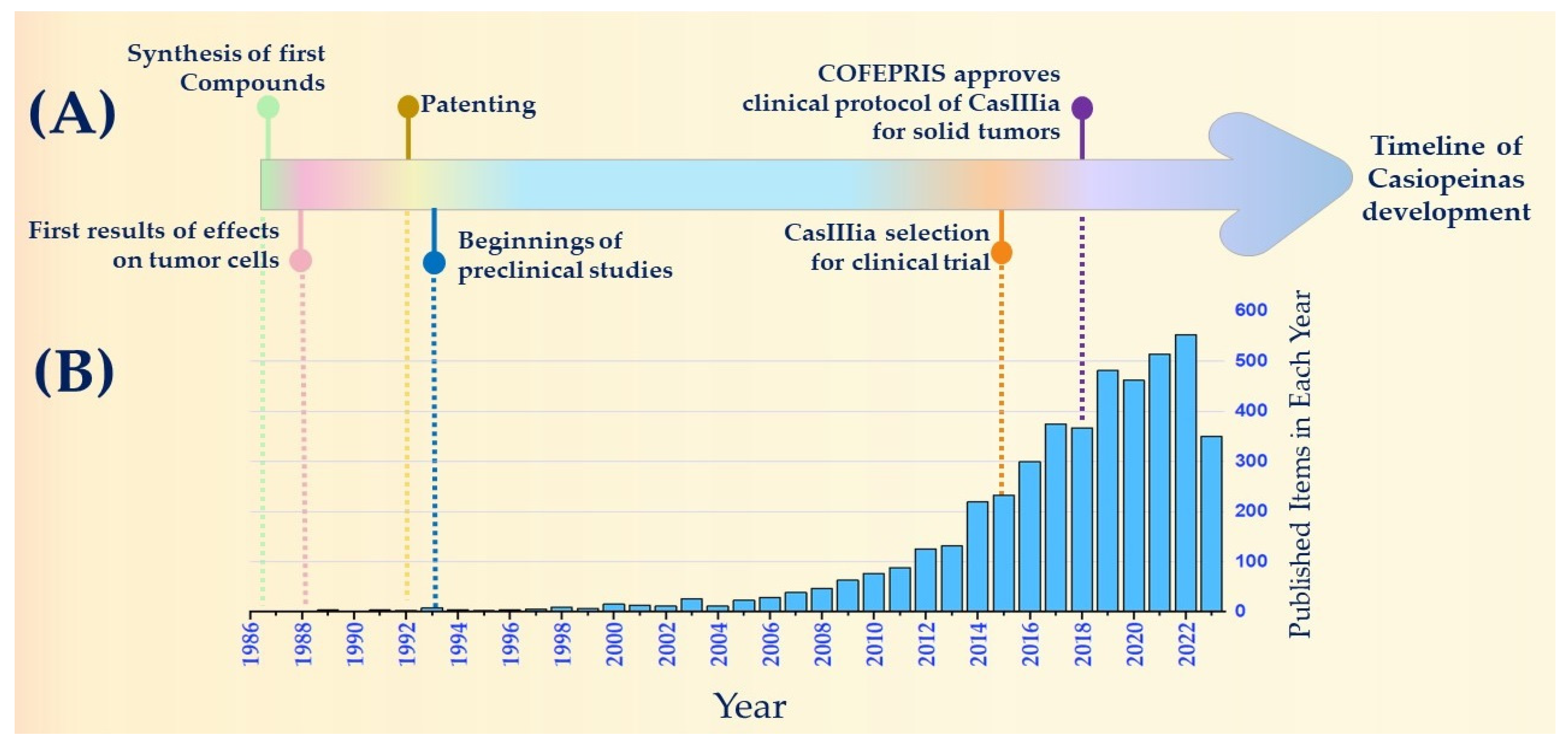
2. Historical Timeline of Anticancer Drugs: Casiopeínas®

The use of metals in healthcare is an activity that has been described since ancient civilizations, who used metals such as copper, gold, or silver to disinfect water, food, or wounds [24,57,58].
In 1786, Thomas Fowler proposed a solution of As2O3 with potassium bicarbonate for treating periodic fever [59]. In subsequent years, arsenic trioxide was used to treat diseases such as psoriasis, malaria, asthma, and rheumatism [60]. However, because of its adverse effects and the emergence of new treatments, its use in treating these diseases has been avoided. Nonetheless, arsenic trioxide remains relevant, as in 2000, the FDA approved [61] this compound for use in the treatment of acute promyelocytic leukemia [34,62]. Paul Ehrlich, who is the “father of chemotherapy”, having introduced the concepts of specific targets when developing new drugs, introduced Salvarsan in 1910 as an effective treatment for syphilis [35]. Salvarsan is an organic compound containing arsenic, which has led to the development of modern medicinal inorganic chemistry.
During the 1970s, the understanding of the molecular biology of cancer was limited, thus consequently constraining the identification of specific molecular targets when designing novel drugs [36,37]. Investigations of the mechanism of action of certain antitumor agents have piqued the interest in studying the characteristics of DNA when developing new anticancer molecules [38]. In 1978, the FDA approved Cisplatin, which was the first inorganic agent available for the treatment of cancer [11]. This spurred inorganic chemists to devise novel oncological medicines. As a result, some research groups involved in coordination chemistry have primarily focused on catalysis, and have shifted their focus to coordination compounds of the platinum metal group, which exhibit promising anticancer activities [2,41].
In the 1980s, Dr. Lena Ruiz Azuara, driven by the pursuit of novel cancer treatments, proposed a family of inorganic compounds with endogenous metals based on the following overarching criteria:
  • Given DNA’s status as a cancer target at the time, the synthesis of compounds that induce DNA damage was sought. Thus, molecules were designed with ligands situated in the equatorial plane of the metal’s center to facilitate intercalation interactions with DNA;
  • Molecules were devised to incorporate a copper atom, which, being an endogenous metal, was expected to mitigate compound toxicity. In addition, we explored whether the redox properties of the metal could be relevant in tumor cells;
  • Small cationic molecules were proposed to enhance solubility in biological environments.
The research working group focused on the synthesis and characterization of the first copper ternary compounds to be designed, which were tested in 1986, whereby their potential anticancer activity was reported. With the patenting of the molecules in the early 1990s, the name Casiopeínas emerged, and preclinical studies of these compounds were initiated.
After more than 30 years since the design of Casiopeínas, there is solid evidence regarding the mechanism of action of these compounds. They are characterized as multitarget molecules that can be used for cancer treatment, inducing mitochondrial damage, increasing reactive oxygen species, and causing DNA damage. This cooperative action reinforces their effects on tumor cells. Their polypharmacological profiles ensure that the design of these molecules remains up to date as an alternative in the treatment of diseases like cancer. The relevance of polypharmaceutical activities of compounds, determined either by the nature of their ligands [63] or their oncological biomarkers, was later indicated [64].
The research group’s expertise and preclinical results led to the selection of Casiopeína III ia (CasIIIia) for submission to the Mexican regulatory agency (COFEPRIS), marking the initiation of the first Phase I clinical trial of a copper-based anticancer compound in México. Recently, these molecules have garnered significant interest within the scientific community, due to their chemical characteristics and biological activities.

3. Activity and Properties

The anticancer activity of these compounds (Casiopeínas) depends mainly on the three descriptors obtained by QSAR study (Figure 3). Halfwave potential (E1/2) is important in oxidant agents; aromaticity refers to the number of aromatic conjugated rings, and has a minor influence on the lopP of mono-charged cationic copper (II) complexes [42], as corroborated by analyses of the properties of the other compounds derived in the other generations [48,50], except for the second generation of Casiopeínas, wherein the mixed chelate complexes are bi-charged cationic complexes. These findings suggest that greater hydrophilicity in the cation complex decreases the membrane crossing efficiency in cancer cells. Structure–activity correlation analyses have been performed, and in vitro assays of IC50 have shown lower values against, mainly, E1/2 and LogD [42,48,49,50,65].
Casiopeinas and acetylacetonate analogues have been synthesized and studied by EPR and ENDOR spectroscopy. It has been found that small variations towards a square planar geometry can be detected by advanced EPR techniques [46,66].
It is important to mention that Casiopeínas of the first generation contain essential amino acids (aa); a large number of aa are used as secondary ligands, indicating that the amino acids produce very small variations in the activity compared with a change in the diimine; therefore, glycine was chosen to continue the studies [67].
Regarding the in vivo assays, the xenografting of several human cell lines into nu/nu mice models diminished the tumor volume, and even achieved an important increase in the activity towards the control drug Cisplatin. For example, when compared with the IC50 of cisplatin over HCT-15, CasIIIia has greater value, however, the in vivo value for the delay of specific growth as a function of the doses applied, the activity of CasIIIia is more effective than cisplatin [42,68] (Table 2, Figure 4).
The tetramethylated phenanthroline analogues CasVIIIgly [Cu(3,4,7,8tm-phen)(gly)]NO3 and CasIIIJa, [Cu(3,4,7,8tm-phen)(acac)]NO3 were tested in an in vivo assay; when the HCT-15 (human colon adenocarcinoma) cell line was xenografted onto nu/nu mice, the acac analogue was more active than the IIgly one, which coheres with the prediction from the QSAR study. Then, the secondary ligand plays an important role in distribution in vivo. It is because of the above that there is a balance between two pharmacological properties—the tumor response of the drug and the access to the tumor—and this gives Casiopeínas its effect as a potent antitumor drug.
A lower cytotoxicity against non-cancer cell Lymphocytes, or stem cells, has been reported, and shows the safety of these compounds compared with commercial platinum drugs [69].
Pharmacological interactions between Casiopeínas analogues and Cisplatin were evaluated in HeLa cells (human cervical adenocarcinoma). An isobolographic analysis revealed that one of the combinations increased the antiproliferative activity from 50 to 77% when the cells were exposed to 4.59 and 9.70 µM of CasIIIia and Cisplatin, respectively. The results indicate that those compound analogous to tetraphenanthroline and Cisplatin had a synergistic effect with CasIIIia. The synergistic combinations assessed may be useful in future in vivo analyses with the aim of reducing the toxicity of Casiopeínas containing phenanthroline in their structure [70]. CasIIgly in combination with the 17 first-line antineoplastic drugs used at the National Institute of Cancer of México induces a synergistic response in HeLa cells (human cervical adenocarcinoma). The drugs combined with CasIIgly diminish the viability and proliferation rates at nanomolar doses, without any apparent effect on the normal proliferation of cells [71].

4. Toxicity

Lethal dose response was evaluated in twenty mongrel dogs. CasIIIia and CasIIgly were infused intravenously for 30 min. The reported LD99 was 10 mg/kg (200 mg/m2) for CasIIIia and 8 mg/kg (160 mg/m2) for CasIIgly. The therapeutic safety margin (TSM) and true therapeutic safety margin (TTSM) are observed in Figure 5. The authors recommend doses of 33.3 mg/m2 for CasIIIia and 26.6 mg/m2 for CasIIgly. At DL99, the Casiopeínas caused acute dog death by pulmonary edema after a latency time of 30 to 50 min. Death due to pulmonary edema in dogs was confirmed by histopathological studies. In addition, it was found that the heart did not suffer direct cardiac toxicity; however, transmission electron microscopy studies revealed mitochondrial damage in myocardial cells [72]. CasIIgly and IIIEa were two and seven times more potent as inhibitors, respectively, than CasIIIia on respiratory activity. This security range was considered when proposing CasIIIia for use in clinical trial. Also, the effects of CasIIIia, CasIIgly, and CasIIIEa treatments on cardiac mitochondria-isolated cardiomyocytes were evaluated. It is proposed that phenanthroline, present in the structure of CasIIIEa and CasIIgly, may act as a transporter in the cellular uptake of copper compounds, meaning Casiopeínas provoke a loss of membrane potential, which increases the opening of the mitochondrial permeability transition pore (mPTP), and this could be associated with cardiotoxicity [73,74,75].

Hematoxicity

Hematoxicity assays of Casiopeínas were carried out in Wistar rats, via IV in singular doses. The results show that CasIIIia (3 mg/kg), CasIIgly (5 mg/kg), and CasIIIEa (4 mg/kg) caused hemolytic anemia at 24, 12, and 48 h, respectively. The restoration of hematic parameters was observed after 21 days (CasIIIia) and 15 days (CasIIgly). It seems that the evaluated Casiopeínas produce an early hematological effect with no delayed or permanent toxic effect. For CasIIIia and CasIIgly, hemolytic anemia was presented without any effect on biochemical parameters, but this was not the case for CasIIIEa, wherein alterations were observed in the concentrations of urea, bilirubin, albumin, and total proteins; however, recovery was observed at 96 h after the biochemical parameters were measured, whereat reversible renal damage was shown [76,77].

5. Pharmacokinetics

To determine the half-life (t1/2) and distribution of Casiopeínas in a preclinical in vivo model, pharmacokinetic studies were carried out in species such as rats, dogs, and rabbits. A pharmacokinetic study was performed on CasIIgly in dog blood at two doses (1.5 and 3 mg/kg, n = 2 for each dose) given via intravenous infusion. The results obtained indicate that CasIIgly has a high elimination rate and a wide distribution for both tested doses. The reported half-life in dogs is slightly higher than that reported in male Wistar rats (8 mg/kg, n = 10). The results in dogs and rats suggest that CasIIgly is eliminated in a short time in animals [78]. Regarding CasIIIia, pharmacokinetic studies have been carried out in rats and rabbits. The volume of distribution and the half-life are both greater in rats than in rabbits, and the clearance is slower in rats than in rabbits. In addition, the half-life (3.9 h) and plasma concentration profile suggest that CasIIIia is distributed in several organs, with an apparent half-life of 13.4 µg/mL (administration dose was 4.5 mg/kg) [79].
Isomeric Casiopeínas have been synthesized. In order to study the differences in half-life, pharmacokinetic studies of CasIIILa (1 mg/kg) and CasIIIEa (4 mg/kg) in male Wistar rats (n = 6) were performed. The elimination of CasIIIEa was observed to be slower than that of CasIIILa. This compound had a greater distribution, even in organs with low blood perfusion. The differences found with respect to the elimination and distribution of both isomeric compounds are suggested to be related to the way they interact with cell membranes. When observing the structures of both isomeric compounds, it is possible to see that when the lipophilic character of the substituents increases, a decrease in the distribution and elimination rate of these compounds is also observed [69].
Plasma protein binding studies of CasIIIia have shown that the percentage of binding to plasma proteins is approximately 80% at the concentrations of 12.25 and 50 (μg/mL), which leads to the inference that CasIIIia is found in greater proportions within the blood circulation, which thus acts as a deposit at the concentrations tested. Correia et al. evaluated the binding of CasIIIia, CasIIgly, and CasIIIEa to human serum albumin (HSA). According to their results, the transport of Casiopeínas may occur through HSA, because they form 1:1 adducts within the physiological range of concentrations. The values were determined by circular dichroism, and the fluorescence emission spectra indicate that this binding takes place close to the Trp214 residue [80].
The data obtained from each study indicate that there is great interspecies variation, which is related to body weight and the physiological processes of each species. Therefore, further studies are required in these species in order to allow allometric scaling. Knowing the pharmacokinetic parameters in different animal species will help us in the future when designing dosing intervals, as well as in making an appropriate selection of dose levels for pharmacokinetic studies in future clinical stages.

6. Mechanism of Action

The biological activities of Casiopeínas have been evaluated both in vitro and in vivo, and they have demonstrated antiproliferative, cytotoxic and genotoxic effects, which has led to the clinical evaluation of several members of this family [68,80]. Research has focused on three main derivatives: CasIIgly, CasIIIia and CasIIIEa [53,54]. Various studies have been carried out on Casiopeínas in a wide variety of human and animal models, highlighting the antiproliferative influence in cell cultures of breast cancer [51], cervical cancer [81,82], lung cancer [83], rat C6 glioma [84], medulloblastoma and neuroblastoma [85]. On the other hand, non-tumor cells with accelerated growth, such as 3T3-L1 (healthy mice fibroblasts) treated with CasIIgly and CasIIIEa, presented low toxicity. As Casiopeínas have been shown to induce damage in healthy human peripheral lymphocytes, it is necessary to apply a higher mean inhibitory concentration (IC50) (1 mM) with respect to CHP-212 neuroblastoma tumor cells (31.5 μΜ, 47.5 μΜ and 18.6 μΜ of CasIIgly, CasIIIia, and CasIIIEa, respectively) [53].
These experiments elucidate the tumor selectivity of Casiopeínas, as well as their low toxicity in healthy systems compared to most standard treatments used for different neoplasias.
In vivo, CasIIgly reduced tumor volume, mitosis, and cell proliferation, as well as promoting increased apoptosis in a model of C6 glioma cells xenografted in Wistar rats (doses of 0.4 and 0.8 mg/kg, injected intraperitoneally). The antineoplastic effect of CasIIgly occurred without leukopenia [86] or toxicity in the hepatobiliary or renal system, and furthermore, it did not induce animal mortality [84].
At high concentrations, CasIIIia underwent binding to proteins, specifically to the alpha-acid glycoprotein, thus intervening in binding to red blood cells [87]. On the other hand, the distribution of the whole blood assayed in Wistar rats showed a Cblood/Cplasma ratio > 2 (where C = concentration), which implies the binding of CasIIIia to erythrocytes. All the tests indicate a pattern in the mechanism of action of Casiopeínas based on the overproduction of reactive oxygen species (ROS) and their coupling to DNA, as well as the alteration of energy metabolism, thus promoting the induction of mitochondrial apoptosis [54,81,82,83,84,85].
From the energetic point of view, Casiopeínas can intervene in different levels of cellular respiration. Glycolysis is the main source of ATP in solid tumors, before even oxidative phosphorylation (OxPhos), known as the Warburg effect [88]. The protein responsible for initiating glycolysis is hexokinase (HK) [89], and so selective HK inhibitors such as 3-bromopyruvate (3-BrPyr) are important chemotherapeutic candidates [90,91]. The influence of CasIIgly on glycolysis was determined, whereby the inhibition of HK activity to a degree 1.3 to 21 times greater than that achieved with 3BrPy causes a drop in ATP production [92]. In a study in cardiac cells, it was determined that CasIIgy, CasIIIia, and CasIIIEa induce the loss of mitochondrial membrane potential (Δψm) due to increased ROS. In addition, the inhibition of the opening of the mitochondrial permeability transition pore (mPTP) by cyclosporin-A (CsA) prevents the depolarization of cells when treated with Casiopeínas, thus protecting cells from apoptosis [93], which suggests that the PTPm participates in the mechanism of action of Casiopeínas [73] (Figure 6).
In cancer cells, it has been reported that there is a high oxidative environment, which makes them susceptible to increased ROS [94]. In order to understand the participation of copper in oxidative stress processes, studies have described the oxidation of cysteine-containing peptides in the presence of copper [95]. Particularly, the redox pairs GSSG/GSH and Cystine/Cysteine have very similar oxidation-reduction potentials (−263 and 220 mV vs. NHE), and CasIIIia and CasIIgly show potentials of 62 mV and 90 mV vs. NHE, respectively. Therefore, the oxidation of cysteine and glutathione may be favored by the presence of Casiopeínas [83,96].
Cancer cells can adapt to these oxidant species by increasing concentrations of antioxidants, such as glutathione (GSH). Ref. [97] demonstrated that CasIIgly induced ROS overproduction by catalyzing the Fenton-type reaction and using GSH as an electron source in lung cancer cell lines (H157 and A549), inducing cell cytotoxicity via a rapid drop in GSH levels due to the delay in the cells’ ability to initially control the levels of ROS. The hypothesis suggests that glutathione reacts with CasIIgly, which leads to the reduction of Cu(II) to Cu(I) and the formation of the glutaryl radical (GS•), which can react with another GS• product via the same reaction or via the superoxide (•-O2)-mediated oxidation of GSH, which in turn causes GS• and •-O2 to form oxidized glutathione (GSSG). GSSG is a compound composed of two glutathione molecules linked by a disulfide (S-S) bond. SOD (superoxide dismutase) catalyzes O2 to form H2O2, which in turn reacts with Cu(I) to return to its oxidized Cu(II) state and produces the OH radical, which initiates mitochondrial DNA damage. This impairment promotes a drop in the expression of complex I proteins from the mitochondrial respiratory chain, causing their uncoupling, which is associated with the formation of O2. The regression to oxidized CasIIgly restarts the GHS oxidation cycle, and consequently the subsequent reactions [83].
On the other hand, bioinformatic analysis has shown that Casiopeínas have the characteristics required to intercalate between A and T bases, which redirects the bases towards the major groove of DNA. Experimental studies of the interaction of the same 21 Casiopeínas with pBR322 plasmid DNA via three different analytical techniques (circular dichroism, UV-Vis, and gel electrophoresis) have shown that, depending on the main diamine and the secondary ligand, Casiopeínas can be classified as intercalators with minor groove interaction, as well as with Ct-DNA/Cas and associated contents of the order of Kb (M−1) 105 [54].
In a complete transcriptome mapping study by Espinal-Enríquez et al. in 2016, it was determined that Casiopeínas block the cell cycle during the transition from the G1 to the S phase. Thus, in cervical cancer (HeLa) and neuroblastoma (CHP-212) cells treated with CasIIgly, the expressions of different apoptotic molecules (at 6 h and 2 h, respectively) were observed. The overexpression of the BAX, CIT-C (cytochrome-C), CAS9 (caspase-9), and CAS3 (caspase-3) genes, and the under-expression of the CAS8 (caspase-8) and BCL2 genes, identified the mitochondrial pathway as the preferred pathway by which Casiopeínas act. In turn, a high concentration of ROS was observed. Apoptosis was potentiated by the interruption of the cell cycle, which increased the expression of BAX, TP53, and P21 genes [82]

7. Metabolomics: CasIIgly and Its Effect on Triple-Negative Breast Cancer (TNBC) Metabolism

Metabolomics is an important tool that measures metabolic biology in response to systemic changes [98]. In cancer research, metabolomics provides information on complex changes in the molecular phenotype during malignant progression, whereby, in addition, the concentration of the metabolites can be associated with cancer state, and thus be used to complement other omics studies (genomic, proteomic, and transcriptomic). Thanks to this, the development of new treatments and patient stratification is possible [99,100,101].
The metabolomic studies that have been undertaken using coordination compounds are very few in number; therefore, they could be considered an emergent area. Until now, metabolomic studies in cancer have focused on Cisplatin, as well as some of its derivatives and coordination compounds, mainly ruthenium and palladium [102,103,104]. However, no reports have been found on this type of study using copper-based coordination compounds—except for the one on Casiopeínas®, wherein a triple-negative breast cancer (TNBC) cell line was used, which is a difficult type of cancer to treat [105].
TNBC represents 12 to 17% of all breast cancer cases, and it is characterized by its aggressiveness, poor prognosis, high incidence, and mortality [6,106]. A particular characteristic of TNBC is the absence of hormone receptors (progesterone and estrogen) and human epidermal growth factor receptor 2 (HER2); consequently, the available treatments are not very effective [6,107], and so it is necessary to explore new pharmacological targets and treatment alternatives for TNBC.
Cancer cell metabolism is of interest for TNBC treatment since it is modified in cancer cells to cover their nutrient and energy demands, thereby ensuring the cell’s survival. Furthermore, this type of metabolic reprogramming is involved in metastatic and carcinogenesis processes [108,109,110].
Cisplatin is a chemotherapeutic commonly used agent many types of cancer, and its effect has been reported against TNBC metabolism when employing the MDA-MB-231 cell line. The principal pathways affected by Cisplatin treatment are phosphatidylcholine, phosphatidylethanolamine, phospholipid biosynthesis, and methionine metabolism [51]; besides this, it reduces the phosphocholine and betaine contents [111]. These processes are important for cancer cell growth and membrane cellular constitution; because MDA-MB-231 cells have a high concentration of lipids, this helps in the development of metastasis [112,113]. However, chemoresistance may occur when using Cisplatin in TNBC treatment, which could limit its use [114,115,116].
On the other hand, CasIIgly has a promising effect on TNBC metabolism, as it affects important biochemical pathways related to cancer cells within short treatment times (20 to 120 min), among which are included the Warburg effect, pyruvate metabolism, gluconeogenesis, glycolysis, the electron transport chain, β-oxidation, and the pentoses pathway [51]; all of these are important metabolic routes in cancer progression because they are related to how cancer cells undergo proliferation, metastasis, invasion and tumor growth [117,118,119,120].
These relevant alterations to the metabolism could cause cancer cells to undergo severe nutrient deprivation and eventually death. Besides this, further metabolomic studies could help us to understand and explore other types of pharmacological activities.

8. Encapsulation

Casiopeína nanoencapsulation has been carried out in colloidal systems (Figure 7). The aim is to increase the concentration in the blood of the drug, and reduce its inherent toxicity, as it is a compound used for cancer treatment [121,122]. It has been used in the encapsulation of CasIIIia within chitosan nanoparticles and niosomes formulations. Miranda in 2012 reported the encapsulation of CasIIIia in chitosan nanoparticles; the results show that the administration of CasIIIia within chitosan nanoparticles increased the survival time of CB6F1/Hsd mice transplanted with B16 melanoma tumor sixfold with respect to the administration of CasIIIia alone. Also, the CasIIIia within nanoparticles inhibited tumor growth more extensively than the free drug [123]. Niosome containing CasIIIia was formed using a Quality by Design tool [124] to ensure the prediction and optimization of a repeatable and reproducible formulation [125]. It was observed that encapsulated CasIIIia showed lower toxicity in female BALB/c mice with cells 4t1 (metastatic breast cancer model), compared with cis-Pt and nonencapsulated CasIIIia. When the weight of mice was evaluated at the end of the treatment, it was observed that only the groups treated with niosome with CasIIIia and niosome without CasIIIia recovered their initial weights [126].
A third-generation compound of Casiopeínas, with the Indomethacin ligand, was synthesized and nanoencapsulated in chitosan–polyacrylamide nanogel nanoparticles. The nanogels were pH-dependent, and subjected to changes in structure, which opens up opportunities for pH-dependent chitosan nanogels to be used to encapsulate metal-based drugs [50]. In addition, a cytotoxic copper–phen aquaporin, inspired by Casiopeína, was synthesized and encapsulated in liposomes. In in vivo studies, male BALB/c mice showed nontoxicity after the parenteral administration of Cuphen liposomes in melanoma models [127] with Hag90. These preclinical in vivo studies show that the toxicity of copper-based compounds is reduced when encapsulated.
Also, a Cu(II) complex containing a b-diketone and phenanthroline has been synthesized, and the nanoparticles of this complex were prepared and evaluated in for their effects on the proliferation of MKN-45 cells. Cell proliferation was inhibited by all compounds and nanocompounds in a dose-dependent manner, but non-nanoparticle compounds were more active [128].
Although there are limited reports regarding encapsulated copper compounds, researchers have successfully synthesized nanoparticles employing copper as a carrier to augment the activity of these compounds [129]. Furthermore, poly(amidoamine) dendrimers complexed with copper(II) have been synthesized for use in radiotherapy and the treatment of metastatic cancer [130]. Copper chelates have also been formed with compounds possessing antitumor properties, such as disulfiram [131]. In addition, copper complexes with doxorubicin have been encapsulated within liposome [132]. A comprehensive review of metallodrugs used in the realm of cancer nanomedicine was conducted by Peña and collaborators [133]

9. Casiopeínas-like Compounds

Some reviews dedicated to copper(II) anticancer drugs have been published [43], including works related mono-, bis- and tris-chelates, and mainly mixed and non-mixed chelate compounds. Four years later, Santini et al. presented a number of mononuclear and binuclear Cu(II) and Cu(I) systems, describing their structures and later their mechanisms of action. In all cases, the induction of apoptosis was enabled by ROS production and DNA interaction. An interesting table is shown in Santini’s review, which shows the exponential increase in the number of publications each year from 2000 (20) to 2012 (150). From 2014 to 2023, the number of publications increased exponentially; our search before that, from 1986 to 1999, found only 31 publications and four patents [134] (Figure 8).
The principal characteristics of Casiopeina-like compounds include the presence of copper-containing diimines, such as 1,10-phenanthroline (phen) and 2,2-bipyridine (bipy), and eventually their substituted analogues, as well as the presence of secondary ligands to the ternary compounds, also called mixed chelate compounds. As precursors, we can mention the bischelate–phen complexes, which have shown nuclease activity; the strength of this property varies depending on the position and number of methyl substituents, such as Cu(II) or Cu(I) compounds [135,136]. Another approach to developing active metallodrugs is the use of bischelate non-diamine complexes, homoleptic biscurcuminates compounds synthesized with the purpose of improve the solubility of the ligands [137,138,139,140]. Similar examples of bis O–O chelates that are sterically bulky have been studied [141]. Homoleptic compounds are, in general, less active than ternary compounds. Also, some bis-functionalized phenanthroline complexes with carboxylic ligands have been prepared [Cu(RCOO)(1,10-phen)2]+; their in vitro antiproliferative, antifungal, and antibacterial activities have been determined, as have their nuclease activity and albumin interaction [141]. Some bischelates, such as copper(II) square pyramide(sp) compounds with a cationic 2+ charge, [Cu(dmp)2(CH3CN)](ClO4)2, [Cu(phen)2(CH3CN)](ClO4)2, and tetrahedral copper(I) [Cu(dmp)2](ClO4)(th) where dmp = 2,9-dimethyl,1,10-phenantholine, have been studied. Their antiproliferative activities have been determined, and EPR studies have shown the redox process. The relevant conclusion here concerns the distortion of the square planar into a tetrahedral form and its relationship with the ability to modify the redox potential of copper [103].
In the present review, we are focusing on some articles concerning “Casiopeínas-like” that convey an innovative approach. Casiopeínas-like compounds contain Cu and either phen or bipy ligands, and the modification comes in the secondary ligand [66,142,143,144]. Some homoleptic compounds have been synthesized containing terpyridine derivatives such as [Cu(bitpy)2](ClO4)2, and heteroleptic ones such as [Cu(bitpy)(phen)](NO3)2. For the second one, MTT assays revealed a significant oxidant challenge to both NIH3T3 and MG63 cells, which was observed upon treatment with a heteroleptic complex, even at a concentration as low as 5 mM. In support of the previously reported binding of DNA, we have found the following [43]: mixed chelates are more active than bis-chelates. A paper by I Correia et al. presents mixed chelate compounds containing phen and bipy, and tridented salgly or salala anions as the secondary ligand, giving five copper(II) complexes: [Cu(sal-Gly)(bipy)] (1), [Cu(sal-Gly)(phen)] (2), [Cu(sal-L-Ala)(phen)] (3), [Cu(sal-DAla)(phen)] (4), and [Cu(sal-L-Phe)(phen)] (5). MTT assays have been performed on four tumor lines, and DNA binding has been assessed by circular dichroism, as presented in [145]. Kordestani et al. reported a series of Cu(diimine)(X-sal)(NO3) complexes, wherein the diimine is either 2,2′-bipyridine (bpy) or 1,10-phenanthroline (phen), and X-sal is a monoanionic halogenated salicylaldehyde (X = Cl, Br, I, or H). Their viability and antiproliferative activity were tested in vitro [146]. Several homoleptic bis-chelates have been reported with the following ligands: 2-(2-aminophenyl)-1H-benzimidazole (bm); N,N′-bis(salicylidene)ethylenediamine (salenH2); 2-(aminomethyl)pyridine (amp); 4-nitrobenzene-1,2-diamine(nbda), with chloride as the counterion [147]. Tetrahedral neutral copper(I) complexes with 2,9-dmethyl,1,10-phenanthroline = dpm) [Cu(I)(dmp)(MPOH)] and [Cu(I)(dmp)(MPSG)] (MPOH = P(p-OCH3-Ph)2CH2OH and MPSG = P(p-OCH3-Ph)2CH2SarGly)) were prepared and characterized, and have shown cytotoxicity in cancer cell lines. The copper(I) complexes studied by fluorescence spectroscopy and cyclic voltamperometry have been proven to be able to generate reactive oxygen species as a result of redox processes [148]. Other examples containing 2,9dmphen and tridentated dipeptides, with the general formula [Cu(L-dipeptide)(neo)], and crystalline structures of [Cu(glyval)(neo)]·3H2O, [Cu(gly-leu)(neo)]·H2O, [Cu(ala-gly)(neo)]·4H2O, [Cu(val-phe)(neo)]·4.5H2O and [Cu(phephe)(neo)]·3H2O, were determined by single-crystal X-ray diffraction, and their cytotoxicity was tested in several cancer cells (lung and breast) [149]. Bimetallic complexes have been developed with the formula [Cu(N,N′)(AA)]2•(V4O12), where (N,N′) = 1,10-phenanthroline and 2,2′-bipyridine and (AA) = lysine and ornithine, and have shown innovative dodecavanadate counteranions with cytotoxic activity [150]. Compounds with diimines and monodentated ligands have been reported, such as [Cu(N–N))L2], [Cu(N–N)2L], [Cu(N–N))L2] and [Cu2(N–N)2)L4], where L = −4,5-dichloroisothiazole-3-carboxylic acid; they have shown antiproliferative effects, and the enzymes’ cytochrome P450 dependence was studied [55].
The use of an active drug as a ligand, as in the third generation of Casiopeinas [50], has been reported, with the O–O donor plumbagin as a copper bis-chelate or mixed chelates with bipy. Both were tested in a series of cancer cells in vitro [151]. Other examples with O–O donors include mononuclear copper (II)ternary compounds with phen and 2Rsalal or diphosphate [152]. Indolacetate and phen compounds present nuclease activity [153,154].
Tridentated taurine Shiff bases and ONO donors with phen show octahedral geometries with a coordinated water molecule [155].
A review from 2015 has compiled the targets discovered for copper compounds related to their promising anticancer activities and lower toxicity in healthy cells, which are enhanced in the presence of copper [156].
A very recent review of copper’s anticancer activity and its main targets concluded that the most relevant pathway is cancer cell death, such as via DNA oxidative cleavage, DNA intercalation, topoisomerase inhibition and proteasome inhibition [157].

10. Casiopeínas and Other Activities

The bacterial and so-called tropical neglected diseases (TND) are a cause of death in children and the adult population mainly in developing countries. Some results derived for Casiopeínas have been derived through assays using some of these bacteria or parasites. The antibacterial Inhibitory Fraction Index (IFI) values of CasIIIia, CasIIIEa and CasIIgly, combined with INH, RIF, and EMB using a bidimensional checkerboard against susceptible and resistant clinical isolates of M. tuberculosis and M. tuberculosis H37Rv, have reported CasIIIEa/EMB as having a better synergic effect [158]. The cytotoxic effects of eight Cu (II) Casiopeínas against Giardia lamblia trophozoites, human peripheral blood lymphocytes (HPBL), and human peripheral blood macrophages (HPBM) were assessed, and their associated selectivity indexes were determined. The more active casiopeínas, with IC50 values of 36 µM, were the more lipophilic compounds—CasIgly [Cu(diphenyl,1-10-phen)(gly)]NO3 and CasIIIHa [Cu(tm1,10-phen)(acac)]NO3. TEM images showing the morphological changes in G. intestinalis trophozoites caused by 24 h of exposure to the IC50 doses evaluated compounds (Metronizadole, CasIgly, CasIIIia) compared with those without tratment [147].
Another protozoan parasite was evaluated using three Casiopeínas, and the IC50 (μM) values were determined for CasIIgly (3.9 ± 1.5), CaIIIEa (11.3 ± 3.8) and CasIIIia (6.9 ± 3.9). The compounds showed activity against Trypanosoma cruzi (T. cruzi epimastigotes (Dm28c strain)), an etiologic agent of the Chagas disease. The tested complexes showed in vitro anti-T. cruzi activity similar to that of the anti-trypanosomal reference drug Nifurtimox [159].
Halide derivatives [Cu(bipy)(acac)X] and [Cu(bipy)(acac)(H2O)] X were derived, where X = Cl and X = Br, and the microbial activities were inferred [66].
Regarding the COVID-19 pandemic, the interaction between the active site of the SARS-CoV2 protein and Mpro was studied by docking using a DFT calculation and several casiopeínas. The study gave interesting results. The bond energies and bond constants of Casiopeínas/Mpro compared with Remdesivir show that mainly Casiopeínas from the first, second and third generations are the most promising for the treatment of this disease [56].
The several activities presented for these compounds can be explained by the different targets of activation, depending on their versatile ternary conformation and the variations on the descriptor, as derived from their behavior. This statement is supported by a recent review, wherein a mechanism of action approach is presented for mixed Cu(II) phenanthroline-based complexes, using a large number of secondary ligands as mono- or di-nuclear compounds. The authors summarized the overall targets derived from the mechanisms of action studied; those targets are (a) the nucleus (DNA, chromatine, nucleolar functions); (b) the mitochondria; (c) the rough endoplasmic reticulum and the (d) peroxisomas (ROS metabolism) [160].

11. Conclusions

The study of metallodrugs used in cancer therapy has been mainly focused on Cisplatin and its analogs; unfortunately, their secondary effects remain a problem. The search for more selective and less toxic molecules using bio-essential metals was the main goal of this review, aiming at enhancing the properties of copper compounds for pioneering Casiopeínas families and Casiopeínas-like compounds. We have presented a timeline of this development, showing the properties observed in chemical and preclinical models. As a result, we can conclude that copper metal drugs are safe, selective towards cancer cells, and produce low toxicity. Thus, they can be proposed as potent antineoplastic drugs that can be used in antineoplastic therapy. Furthermore, these compounds present a multiple-target mechanism of action, as evidenced by metabolomics and genomic studies; moreover, some of their other activities have been investigated, such as antibacterial, antivirus, and antiparasitic. From these findings, it is possible to infer that in the future, other activities can be investigated.

Author Contributions

All authors contributed to preparing and writing the text according to the review content, particularly as follows. Z.A.-J.: Encapsulation, writing—review and editing. A.E.-G.: Timeline of copper compounds development and figures preparation. K.R.-A.: Metabolomic part. I.F.-N.: Pharmacokinetic and toxicology parts. C.M.: Mechanism of action and figures. L.R.-A.: conceptualization, introduction, properties, cancer and other activities. All authors have read and agreed to the published version of the manuscript.

Funding

This research received no external funding.

Data Availability Statement

Not applicable.

Acknowledgments

The authors thank Cynthia Novoa-Ramirez, Yeshenia Figueroa-De-Paz, Nancy Vara-Gama, Luis-F. Hernandez and Miguel Reina, also Mauricio Gonzalez and Rogelio Hurtado for helpful discussions and information. Finally, Norma Hernandez is thanked for proof reading the manuscript. This review is dedicated to all the students and collaborators who have participated in the development of Casiopeínas and to those colleagues who have contributed to innovative idea of using copper compounds for cancer treatment.

Conflicts of Interest

The authors declare no conflict of interest.

References

  1. Guo, Z.; Sadler, P.J. Metals in Medicine. Angew. Chem. Int. Ed. 1999, 38, 1512–1531. [Google Scholar] [CrossRef]
  2. Mjos, K.D.; Orvig, C. Metallodrugs in Medicinal Inorganic Chemistry. Chem. Rev. 2014, 114, 4540–4563. [Google Scholar] [CrossRef]
  3. Sadler, P.J. Inorganic Chemistry and Drug Design. In Advances in Inorganic Chemistry; Sykes, A.G., Ed.; Academic Press: Cambridge, MA, USA, 1991; Volume 36, pp. 1–48. ISBN 0898-8838. [Google Scholar]
  4. Kaufmann, S.H.E. Paul Ehrlich: Founder of Chemotherapy. Nat. Rev. Drug Discov. 2008, 7, 373. [Google Scholar] [CrossRef]
  5. Anthony, E.J.; Bolitho, E.M.; Bridgewater, H.E.; Carter, O.W.L.; Donnelly, J.M.; Imberti, C.; Lant, E.C.; Lermyte, F.; Needham, R.J.; Palau, M.; et al. Metallodrugs Are Unique: Opportunities and Challenges of Discovery and Development. Chem. Sci. 2020, 11, 12888–12917. [Google Scholar] [CrossRef]
  6. Nayeem, N.; Contel, M. Exploring the Potential of Metallodrugs as Chemotherapeutics for Triple Negative Breast Cancer. Chem. Eur. J. 2021, 27, 8891–8917. [Google Scholar] [CrossRef]
  7. Komeda, S.; Casini, A. Next-Generation Anticancer Metallodrugs. Curr. Top. Med. Chem. 2012, 12, 219–235. [Google Scholar] [CrossRef]
  8. Lucaciu, R.L.; Hangan, A.C.; Sevastre, B.; Oprean, L.S. Metallo-Drugs in Cancer Therapy: Past, Present and Future. Molecules 2022, 27, 6485. [Google Scholar] [CrossRef]
  9. Ndagi, U.; Mhlongo, N.; Soliman, M.E. Metal Complexes in Cancer Therapy—An Update from Drug Design Perspective. Drug Des. Devel Ther. 2017, 11, 599–616. [Google Scholar] [CrossRef]
  10. Wilson, J.J.; Johnstone, T.C. The Role of Metals in the next Generation of Anticancer Therapeutics. Curr. Opin. Chem. Biol. 2023, 76, 102363. [Google Scholar] [CrossRef]
  11. Ghosh, S. Cisplatin: The First Metal Based Anticancer Drug. Bioorg. Chem. 2019, 88, 102925. [Google Scholar] [CrossRef]
  12. Aldossary, S.A. Review on Pharmacology of Cisplatin: Clinical Use, Toxicity and Mechanism of Resistance of Cisplatin. Biomed. Pharmacol. J. 2019, 12, 7–15. [Google Scholar] [CrossRef]
  13. Brown, A.; Kumar, S.; Tchounwou, P.B. Cisplatin-Based Chemotherapy of Human Cancers. J. Cancer Sci. Ther. 2019, 11, 97. [Google Scholar]
  14. Florea, A.M.; Büsselberg, D. Cisplatin as an Anti-Tumor Drug: Cellular Mechanisms of Activity, Drug Resistance and Induced Side Effects. Cancers 2011, 3, 1351–1371. [Google Scholar] [CrossRef]
  15. Tsvetkova, D.; Ivanova, S. Application of Approved Cisplatin Derivatives in Combination Therapy against Different Cancer Diseases. Molecules 2022, 27, 2466. [Google Scholar] [CrossRef]
  16. Dasari, S.; Bernard Tchounwou, P. Cisplatin in Cancer Therapy: Molecular Mechanisms of Action. Eur. J. Pharmacol. 2014, 740, 364–378. [Google Scholar] [CrossRef]
  17. Kopacz-Bednarska, A.; Król, T. Cisplatin—Properties and Clinical Application. Oncol. Clin. Pract. 2022, 18, 166–176. [Google Scholar] [CrossRef]
  18. Barabas, K.; Milner, R.; Lurie, D.; Adin, C. Cisplatin: A Review of Toxicities and Therapeutic Applications. Vet. Comp. Oncol. 2008, 6, 1–18. [Google Scholar] [CrossRef]
  19. Amable, L. Cisplatin Resistance and Opportunities for Precision Medicine. Pharmacol. Res. 2016, 106, 27–36. [Google Scholar] [CrossRef]
  20. Galluzzi, L.; Senovilla, L.; Vitale, I.; Michels, J.; Martins, I.; Kepp, O.; Castedo, M.; Kroemer, G. Molecular Mechanisms of Cisplatin Resistance. Oncogene 2012, 31, 1869–1883. [Google Scholar] [CrossRef]
  21. Czarnomysy, R.; Radomska, D.; Szewczyk, O.K.; Roszczenko, P.; Bielawski, K. Platinum and Palladium Complexes as Promising Sources for Antitumor Treatments. Int. J. Mol. Sci. 2021, 22, 8271. [Google Scholar] [CrossRef]
  22. Sava, G. Ruthenium Compounds in Cancer Therapy. In Metal Compounds in Cancer Therapy; Fricker, S.P., Ed.; Springer: Dordrecht, The Netherlands, 1994; pp. 65–91. ISBN 978-94-011-1252-9. [Google Scholar]
  23. Sharma, A.; Sudhindra, P.; Roy, N.; Paira, P. Advances in Novel Iridium (III) Based Complexes for Anticancer Applications: A Review. Inorg. Chim. Acta 2020, 513, 119925. [Google Scholar] [CrossRef]
  24. Borkow, G.; Gabbay, J. Copper, an Ancient Remedy Returning to Fight Microbial, Fungal and Viral Infections. Curr. Chem. Biol. 2009, 3, 272–278. [Google Scholar] [CrossRef]
  25. Kardos, J.; Héja, L.; Simon, Á.; Jablonkai, I.; Kovács, R.; Jemnitz, K. Copper Signalling: Causes and Consequences. Cell Commun. Signal. 2018, 16, 71. [Google Scholar] [CrossRef]
  26. Ruiz, L.M.; Libedinsky, A.; Elorza, A.A. Role of Copper on Mitochondrial Function and Metabolism. Front. Mol. Biosci. 2021, 8, 711227. [Google Scholar] [CrossRef]
  27. Klotz, L.-O.; Kröncke, K.-D.; Buchczyk, D.P.; Sies, H. Role of Copper, Zinc, Selenium and Tellurium in the Cellular Defense against Oxidative and Nitrosative Stress. J. Nutr. 2003, 133, 1448S–1451S. [Google Scholar] [CrossRef]
  28. Peña, M.M.O.; Lee, J.; Thiele, D.J. Critical Review A Delicate Balance: Homeostatic Control of Copper Uptake and Distribution. J. Nutr. 1999, 129, 1251–1260. [Google Scholar] [CrossRef]
  29. Tang, X.; Yan, Z.; Miao, Y.; Ha, W.; Li, Z.; Yang, L.; Mi, D. Copper in Cancer: From Limiting Nutrient to Therapeutic Target. Front. Oncol. 2023, 13, 1209156. [Google Scholar] [CrossRef]
  30. Nasulewicz, A.; Mazur, A.; Opolski, A. Role of Copper in Tumour Angiogenesis—Clinical Implications. J. Trace Elem. Med. Biol. 2004, 18, 1–8. [Google Scholar] [CrossRef]
  31. Alem, M.B.; Damena, T.; Desalegn, T.; Koobotse, M.; Eswaramoorthy, R.; Ngwira, K.J.; Ombito, J.O.; Zachariah, M.; Demissie, T.B. Cytotoxic Mixed-Ligand Complexes of Cu(II): A Combined Experimental and Computational Study. Front. Chem. 2022, 10, 1028957. [Google Scholar] [CrossRef]
  32. Ji, P.; Wang, P.; Chen, H.; Xu, Y.; Ge, J.; Tian, Z.; Yan, Z. Potential of Copper and Copper Compounds for Anticancer Applications. Pharmaceuticals 2023, 16, 234. [Google Scholar] [CrossRef]
  33. Ruiz-Azuara, L. Process to Obtain New Mixed Copper Aminoacidate from Methylate Phenathroline Complexes to Be Used as Anticancerigenic Agents. U.S. Patent 5,576,326, 20 December 1990. [Google Scholar]
  34. Antman, K.H. Introduction: The History of Arsenic Trioxide in Cancer Therapy. Oncologist 2001, 6, 1–2. [Google Scholar] [CrossRef] [PubMed]
  35. Valent, P.; Groner, B.; Schumacher, U.; Superti-Furga, G.; Busslinger, M.; Kralovics, R.; Zielinski, C.; Penninger, J.M.; Kerjaschki, D.; Stingl, G. Paul Ehrlich (1854–1915) and His Contributions to the Foundation and Birth of Translational Medicine. J. Innate Immun. 2016, 8, 111–120. [Google Scholar] [CrossRef] [PubMed]
  36. De Vita, V.T., Jr.; Chu, E. A History of Cancer Chemotherapy. Cancer Res. 2008, 68, 8643–8653. [Google Scholar] [CrossRef] [PubMed]
  37. Chabner, B.A.; Roberts, T.G., Jr. Chemotherapy and the War on Cancer. Nat. Rev. Cancer 2005, 5, 65–72. [Google Scholar] [CrossRef]
  38. Hurley, L.H.; Boyd, F.L. DNA as a Target for Drug Action. Trends Pharmacol. Sci. 1988, 9, 402–407. [Google Scholar] [CrossRef]
  39. Forestier, J. Comparative Results of Copper Salts and Gold Salts in Rheumatoid Arthritis. Ann. Rheum. Dis. 1949, 8, 132. [Google Scholar] [CrossRef] [PubMed]
  40. Medici, S.; Peana, M.; Nurchi, V.M.; Lachowicz, J.I.; Crisponi, G.; Zoroddu, M.A. Noble Metals in Medicine: Latest Advances. Coord. Chem. Rev. 2015, 284, 329–350. [Google Scholar] [CrossRef]
  41. Romero-Canelon, I.; Sadler, P.J. Next-Generation Metal Anticancer Complexes: Multitargeting via Redox Modulation. Inorg. Chem. 2013, 52, 12276–12291. [Google Scholar] [CrossRef]
  42. Bravo-Gómez, M.E.; García-Ramos, J.C.; Gracia-Mora, I.; Ruiz-Azuara, L. Antiproliferative Activity and QSAR Study of Copper (II) Mixed Chelate [ Cu (N − N)(Acetylacetonato)] NO3 and [ Cu (N − N)(Glycinato)] NO3 Complexes, (Casiopeinas). J. Inorg. Biochem. 2009, 103, 299–309. [Google Scholar] [CrossRef]
  43. Ruiz-Azuara, L.; Bravo-Gomez, M.E. Copper Compounds in Cancer Chemotherapy. Curr. Med. Chem. 2010, 17, 3606–3615. [Google Scholar] [CrossRef]
  44. Rivero-Müller, A.; De Vizcaya-Ruiz, A.; Plant, N.; Ruiz, L.; Dobrota, M. Mixed Chelate Copper Complex, Casiopeina IIgly®, Binds and Degrades Nucleic Acids: A Mechanism of Cytotoxicity. Chem. Biol. Interact. 2007, 165, 189–199. [Google Scholar] [CrossRef] [PubMed]
  45. Alemón-Medina, R.; Breña-Valle, M.; Muñoz-Sánchez, J.; Gracia-Mora, M.; Ruiz-Azuara, L. Induction of Oxidative Damage by Copper-Based Antineoplastic Drugs (Casiopeínas(R)). Cancer Chemother. Pharmacol. 2007, 60, 219–228. [Google Scholar] [CrossRef]
  46. Folli, A.; Ritterskamp, N.; Richards, E.; Platts, J.A.; Murphy, D.M. Probing the Structure of Copper(II)-Casiopeina Type Coordination Complexes [Cu(O–O)(N–N)]+ by EPR and ENDOR Spectroscopy. J. Catal. 2021, 394, 220–227. [Google Scholar] [CrossRef]
  47. Solans, X.; Ruíz-Ramírez, L.; Martínez, A.; Gasque, L.; Moreno-Esparza, R. Mixed Chelate Complexes. II. Structures of L-Alaninato (Aqua)(4, 7-Diphenyl-1, 10-Phenanthroline) Copper (II) Nitrite Monohydrate and Aqua (4, 7-Dimethyl-1, 10-Phenanthroline)(Glycinato)(Nitrato) Copper (II) Monohydrate. Acta Crystallogr. C 1993, 49, 890–893. [Google Scholar] [CrossRef]
  48. Figueroa-Depaz, Y.; Pérez-Villanueva, J.; Soria-Arteche, O.; Martínez-Otero, D.; Gómez-Vidales, V.; Ortiz-Frade, L.; Ruiz-Azuara, L. Casiopeinas of Third Generations: Synthesis, Characterization, Cytotoxic Activity and Structure–Activity Relationships of Mixed Chelate Compounds with Bioactive Secondary Ligands. Molecules 2022, 27, 3504. [Google Scholar] [CrossRef]
  49. Novoa-Ramírez, C.S.; Silva-Becerril, A.; González-Ballesteros, M.M.; Gomez-Vidal, V.; Flores-Álamo, M.; Ortiz-Frade, L.; Gracia-Mora, J.; Ruiz-Azuara, L. Biological Activity of Mixed Chelate Copper (II) Complexes, with Substituted Diimine and Tridentate Schiff Bases (NNO) and Their Hydrogenated Derivatives as Secondary Ligands: Casiopeína’s Fourth Generation. J. Inorg. Biochem. 2023, 242, 112097. [Google Scholar] [CrossRef]
  50. Godínez-Loyola, Y.; Gracia-Mora, J.; Rojas-Montoya, I.D.; Hernández-Ayala, L.F.; Reina, M.; Ortiz-Frade, L.A.; Rascón-Valenzuela, L.A.; Robles-Zepeda, R.E.; Gómez-Vidales, V.; Bernad-Bernad, M.J. Casiopeinas® Third Generation, with Indomethacin: Synthesis, Characterization, DFT Studies, Antiproliferative Activity, and Nanoencapsulation. RSC Adv. 2022, 12, 21662–21673. [Google Scholar] [CrossRef] [PubMed]
  51. Resendiz-Acevedo, K.; García-Aguilera, M.E.; Esturau-Escofet, N.; Ruiz-Azuara, L. 1H-NMR Metabolomics Study of the Effect of Cisplatin and Casiopeina IIgly on MDA-MB-231 Breast Tumor Cells. Front. Mol. Biosci. 2021, 8, 742859. [Google Scholar] [CrossRef]
  52. Rodrigues, J.H.V.; de Carvalho, A.B.; Silva, V.R.; de Santos, L.S.; Soares, M.B.P.; Bezerra, D.P.; Oliveira, K.M.; Corrêa, R.S. Copper(II)/Diiminic Complexes Based on 2-Hydroxybenzophenones: DNA- and BSA-Binding Studies and Antitumor Activity against HCT116 and HepG2 Tumor Cells. Polyhedron 2023, 239, 116431. [Google Scholar] [CrossRef]
  53. García-Ramos, J.C.; Gutiérrez, A.G.; Vázquez-Aguirre, A.; Toledano-Magaña, Y.; Alonso-Sáenz, A.L.; Gómez-Vidales, V.; Flores-Alamo, M.; Mejía, C.; Ruiz-Azuara, L. The Mitochondrial Apoptotic Pathway Is Induced by Cu(II) Antineoplastic Compounds (Casiopeínas®) in SK-N-SH Neuroblastoma Cells after Short Exposure Times. BioMetals 2017, 30, 43–58. [Google Scholar] [CrossRef]
  54. De Paz, F.-Y.; Resendiz-Acevedo, K.; Dávila-Manzanilla, S.G.; García-Ramos, J.C.; Ortiz-Frade, L.; Serment-Guerrero, J.; Ruiz-Azuara, L. DNA, a Target of Mixed Chelate Copper(II) Compounds (Casiopeinas®) Studied by Electrophoresis, UV–Vis and Circular Dichroism Techniques. J. Inorg. Biochem. 2022, 231, 111772. [Google Scholar] [CrossRef]
  55. Eremina, J.A.; Lider, E.V.; Sukhikh, T.S.; Klyushova, L.S.; Perepechaeva, M.L.; Sheven, D.G.; Berezin, A.S.; Grishanova, A.Y.; Potkin, V.I. Water-Soluble Copper (II) Complexes with 4, 5-Dichloro-Isothiazole-3-Carboxylic Acid and Heterocyclic N-Donor Ligands: Synthesis, Crystal Structures, Cytotoxicity, and DNA Binding Study. Inorg. Chim. Acta 2020, 510, 119778. [Google Scholar] [CrossRef]
  56. Reina, M.; Talavera-Contreras, L.G.; Figueroa-DePaz, Y.; Ruiz-Azuara, L.; Hernández-Ayala, L.F. Casiopeinas® as SARS-CoV-2 Main Protease (M pro) Inhibitors: A Combined DFT, Molecular Docking and ONIOM Approach. New J. Chem. 2022, 46, 12500–12511. [Google Scholar] [CrossRef]
  57. Huaizhi, Z.; Yuantao, N. China’s Ancient Gold Drugs. Gold Bull. 2000, 46 34, 24–29. [Google Scholar] [CrossRef]
  58. Spear, M. Silver: An Age-Old Treatment Modality in Modern Times. Plast. Aesthetic Nurs. 2010, 30, 90–93. [Google Scholar] [CrossRef]
  59. Kapp, R.W. Arsenic: Toxicology and Health Effects. In Encyclopedia of Food and Health; Caballero, B., Finglas, P.M., Toldrá, F., Eds.; Academic Press: Oxford, UK, 2016; pp. 256–265. ISBN 978-0-12-384953-3. [Google Scholar]
  60. Gibaud, S.; Jaouen, G. Arsenic-Based Drugs: From Fowler’s Solution to Modern Anticancer Chemotherapy. In Medicinal Organometallic Chemistry; Jaouen, G., Metzler-Nolte, N., Eds.; Springer: Berlin/Heidelberg, Germany, 2010; pp. 1–20. ISBN 978-3-642-13185-1. [Google Scholar]
  61. Holman, J.; Kirkhart, B.; Maxon, M.S.; Oliva, C.; Earhart, R. Safety Experience with Trisenox® (Arsenic Trioxide) Injection. Blood 2004, 104, 4521. [Google Scholar] [CrossRef]
  62. Hoonjan, M.; Jadhav, V.; Bhatt, P. Arsenic Trioxide: Insights into Its Evolution to an Anticancer Agent. J. Biol. Inorg. Chem. 2018, 23, 313–329. [Google Scholar] [CrossRef] [PubMed]
  63. Proschak, E.; Stark, H.; Merk, D. Polypharmacology by Design: A Medicinal Chemist’s Perspective on Multitargeting Compounds. J. Med. Chem. 2019, 62, 420–444. [Google Scholar] [CrossRef]
  64. Jørgensen, J.T. The Importance of Predictive Biomarkers in Oncology Drug Development. Expert. Rev. Mol. Diagn. 2016, 16, 807–809. [Google Scholar] [CrossRef] [PubMed]
  65. Reina, M.; Hernández-Ayala, L.F.; Bravo-Gómez, M.E.; Gómez, V.; Ruiz-Azuara, L. Second Generation of Casiopeinas®: A Joint Experimental and Theoretical Study. Inorg. Chim. Acta 2021, 517, 120201. [Google Scholar] [CrossRef]
  66. Onawumi, O.O.E.; Odunola, O.A.; Suresh, E.; Paul, P. Synthesis, Structural Characterization and Microbial Activities of Mixed Ligand Copper (II) Complexes of 2,2′-Bipyridine and Acetylacetonate. Inorg. Chem. Commun. 2011, 14, 1626–1631. [Google Scholar] [CrossRef]
  67. Bravo-Gómez, M.E.; Dávila-Manzanilla, S.; Flood-Garibay, J.; Muciño-Hernández, M.Á.; Mendoza, Á.; García-Ramos, J.C.; Moreno-Esparza, R.; Ruiz-Azuara, L. Secondary Ligand Effects on the Cytotoxicity of Several Casiopeína’s Group II Compounds. J. Mex. Chem. Soc. 2012, 56, 85–92. [Google Scholar] [CrossRef]
  68. Carvallo-Chaigneau, F.; Trejo-Solís, C.; Gómez-Ruiz, C.; Rodríguez-Aguilera, E.; MacÍas-Rosales, L.; Cortés-Barberena, E.; Cedillo-Peláez, C.; Gracia-Mora, I.; Ruiz-Azuara, L.; Madrid-Marina, V.; et al. Casiopeina III-Ia Induces Apoptosis in HCT-15 Cells in Vitro through Caspase-Dependent Mechanisms and Has Antitumor Effect in Vivo. BioMetals 2008, 21, 17–28. [Google Scholar] [CrossRef] [PubMed]
  69. García-Ramos, J.C.; Vértiz-Serrano, G.; Macías-Rosales, L.; Galindo-Murillo, R.; Toledano-Magaña, Y.; Bernal, J.P.; Cortés-Guzmán, F.; Ruiz-Azuara, L. Isomeric Effect on the Pharmacokinetic Behavior of Anticancer CuII Mixed Chelate Complexes: Experimental and Theoretical Approach. Eur. J. Inorg. Chem. 2017, 2017, 1728–1736. [Google Scholar] [CrossRef]
  70. Davila-Manzanilla, S.G.; Figueroa-de-Paz, Y.; Mejia, C.; Ruiz-Azuara, L. Synergistic Effects between a Copper-Based Metal Casiopeína III-Ia and Cisplatin. Eur. J. Med. Chem. 2017, 129, 266–274. [Google Scholar] [CrossRef]
  71. Rodríguez-Enríquez, S.; Gallardo-Pérez, J.C.; Hernández-Reséndiz, I.; Marín-Hernández, A.; Pacheco-Velázquez, S.C.; López-Ramírez, S.Y.; Rumjanek, F.D.; Moreno-Sánchez, R. Canonical and New Generation Anticancer Drugs Also Target Energy Metabolism. Arch. Toxicol. 2014, 88, 1327–1350. [Google Scholar] [CrossRef] [PubMed]
  72. Leal-García, M.; García-Ortuño, L.; Ruiz-Azuara, L.; Gracia-Mora, I.; Luna-Delvillar, J.; Sumano, H. Assessment of Acute Respiratory and Cardiovascular Toxicity of Casiopeinas in Anaesthetized Dogs. Basic Clin. Pharmacol. Toxicol. 2007, 101, 151–158. [Google Scholar] [CrossRef] [PubMed]
  73. Silva-Platas, C.; Guerrero-Beltrán, C.E.; Carrancá, M.; Castillo, E.C.; Bernal-Ramírez, J.; Oropeza-Almazán, Y.; González, L.N.; Rojo, R.; Martínez, L.E.; Valiente-Banuet, J. Antineoplastic Copper Coordinated Complexes (Casiopeinas) Uncouple Oxidative Phosphorylation and Induce Mitochondrial Permeability Transition in Cardiac Mitochondria and Cardiomyocytes. J. Bioenerg. Biomembr. 2016, 48, 43–54. [Google Scholar] [CrossRef]
  74. García, N.; Martínez-Abundis, E.; Pavón, N.; Correa, F.; Chávez, E. Copper Induces Permeability Transition through Its Interaction with the Adenine Nucleotide Translocase. Cell Biol. Int. 2007, 31, 893–899. [Google Scholar] [CrossRef]
  75. Zazueta, C.; Reyes-Vivas, H.; Zafra, G.; Sánchez, C.A.; Vera, G.; Chávez, E. Mitochondrial Permeability Transition as Induced by Cross-Linking of the Adenine Nucleotide Translocase. Int. J. Biochem. Cell Biol. 1998, 30, 517–527. [Google Scholar] [CrossRef]
  76. De Vizcaya-Ruiz, A.; Rivero-Müller, A.; Ruiz-Ramirez, L.; Howarth, J.A.; Dobrota, M. Hematotoxicity Response in Rats by the Novel Copper-Based Anticancer Agent: Casiopeina II. Toxicology 2003, 194, 103–113. [Google Scholar] [CrossRef]
  77. Vértiz, G.; García-Ortuño, L.E.; Bernal, J.P.; Bravo-Gómez, M.E.; Lounejeva, E.; Huerta, A.; Ruiz-Azuara, L. Pharmacokinetics and Hematotoxicity of a Novel Copper-Based Anticancer Agent: Casiopeina III-Ea, after a Single Intravenous Dose in Rats. Fundam. Clin. Pharmacol. 2014, 28, 78–87. [Google Scholar] [CrossRef] [PubMed]
  78. Cañas-Alonso, R.C.; Fuentes-Noriega, I.; Ruiz-Azuara, L. Pharmacokinetics of Casiopeína IIgly in Beagle Dog: A Copper Based Compound with Antineoplastic Activity. J. Bioanal. Biomed. 2010, 2, 28–34. [Google Scholar] [CrossRef]
  79. Fuentes-Noriega, I.; Ruiz-Ramırez, L.; Tovar, A.T.; Rico-Morales, H.; Gracia-Mora, I. Development and Validation of a Liquid Chromatographic Method for Casiopeina IIIi in Rat Plasma. J. Chromatogr. B 2002, 772, 115–121. [Google Scholar] [CrossRef] [PubMed]
  80. Correia, I.; Borovic, S.; Cavaco, I.; Matos, C.P.; Roy, S.; Santos, H.M.; Fernandes, L.; Capelo, J.L.; Ruiz-Azuara, L.; Pessoa, J.C. Evaluation of the Binding of Four Anti-Tumor Casiopeínas® to Human Serum Albumin. J. Inorg. Biochem. 2017, 175, 284–297. [Google Scholar] [CrossRef]
  81. Serment-Guerrero, J.; Cano-Sanchez, P.; Reyes-Perez, E.; Velazquez-Garcia, F.; Bravo-Gomez, M.E.; Ruiz-Azuara, L. Genotoxicity of the Copper Antineoplastic Coordination Complexes Casiopeinas. Toxicol. Vitr. 2011, 25, 1376–1384. [Google Scholar] [CrossRef]
  82. Valencia-Cruz, A.I.; Uribe-Figueroa, L.I.; Galindo-Murillo, R.; Baca-Lopez, K.; Gutierrez, A.G.; Vazquez-Aguirre, A.; Ruiz-Azuara, L.; Hernandez-Lemus, E.; Mejía, C. Whole Genome Gene Expression Analysis Reveals Casiopeina-Induced Apoptosis Pathways. PLoS ONE 2013, 8, e54664. [Google Scholar] [CrossRef]
  83. Kachadourian, R.; Brechbuhl, H.M.; Ruiz-Azuara, L.; Gracia-Mora, I.; Day, B.J. Casiopeína IIgly-Induced Oxidative Stress and Mitochondrial Dysfunction in Human Lung Cancer A549 and H157 Cells. Toxicology 2010, 268, 176–183. [Google Scholar] [CrossRef]
  84. Trejo-Solís, C.; Palencia, G.; Zúniga, S.; Rodríguez-Ropon, A.; Osorio-Rico, L.; Luvia, S.T.; Gracia-Mora, I.; Marquez-Rosado, L.; Sánchez, A.; Moreno-García, M.E. Cas Ilgly Induces Apoptosis in Glioma C6 Cells in Vitro and in Vivo through Caspase-Dependent and Caspase-Independent Mechanisms. Neoplasia 2005, 7, 563–574. [Google Scholar] [CrossRef]
  85. Gutiérrez, A.G.; Vázquez-Aguirre, A.; García-Ramos, J.C.; Flores-Alamo, M.; Hernández-Lemus, E.; Ruiz-Azuara, L.; Mejía, C. Copper(Ii) Mixed Chelate Compounds Induce Apoptosis through Reactive Oxygen Species in Neuroblastoma Cell Line Chp-212. J. Inorg. Biochem. 2013, 126, 17–25. [Google Scholar] [CrossRef] [PubMed]
  86. Su, Z.; Mao, Y.-P.; OuYang, P.-Y.; Tang, J.; Lan, X.-W.; Xie, F.-Y. Leucopenia and Treatment Efficacy in Advanced Nasopharyngeal Carcinoma. BMC Cancer 2015, 15, 429. [Google Scholar] [CrossRef] [PubMed]
  87. Pitekova, B.; Uhlikova, E.; Kupcova, V.; Durfinova, M.; Mojto, V.; Turecky, L. Can Alpha-1-Acid Glycoprotein Affect the Outcome of Treatment in a Cancer Patient? Bratisl. Med. J. 2019, 120, 9–14. [Google Scholar] [CrossRef]
  88. Liberti, M.V.; Locasale, J.W. The Warburg Effect: How Does It Benefit Cancer Cells? Trends Biochem. Sci. 2016, 41, 211–218. [Google Scholar] [CrossRef] [PubMed]
  89. Roberts, D.J.; Miyamoto, S. Hexokinase II Integrates Energy Metabolism and Cellular Protection: Akting on Mitochondria and TORCing to Autophagy. Cell Death Differ. 2015, 22, 248–257. [Google Scholar] [CrossRef]
  90. Silva, J.A.; Queirós, O.; Baltazar, F.; Ułaszewski, S.; Ko, Y.H.; Pedersen, P.L.; Preto, A.; Casal, M. The Anticancer Agent 3-Bromopyruvate: A Simple but Powerful Molecule Taken from the Lab to the Bedside. J. Bioenerg. Biomembr. 2016, 48, 349–362. [Google Scholar] [CrossRef]
  91. Rai, Y.; Yadav, P.; Kumari, N.; Kalra, N.; Bhatt, A.N. Hexokinase II Inhibition by 3-Bromopyruvate Sensitizes Myeloid Leukemic Cells K-562 to Anti-Leukemic Drug, Daunorubicin. Biosci. Rep. 2019, 39, BSR20190880. [Google Scholar] [CrossRef]
  92. Marín-Hernández, A.; Gallardo-Pérez, J.C.; López-Ramírez, S.Y.; García-García, J.D.; Rodríguez-Zavala, J.S.; Ruiz-Ramírez, L.; Gracia-Mora, I.; Zentella-Dehesa, A.; Sosa-Garrocho, M.; Macías-Silva, M. Casiopeina II-Gly and Bromo-Pyruvate Inhibition of Tumor Hexokinase, Glycolysis, and Oxidative Phosphorylation. Arch. Toxicol. 2012, 86, 753–766. [Google Scholar] [CrossRef] [PubMed]
  93. Heusch, G.; Boengler, K.; Schulz, R. Inhibition of Mitochondrial Permeability Transition Pore Opening: The Holy Grail of Cardioprotection. Basic Res. Cardiol. 2010, 105, 151–154. [Google Scholar] [CrossRef] [PubMed]
  94. Arfin, S.; Jha, N.K.; Jha, S.K.; Kesari, K.K.; Ruokolainen, J.; Roychoudhury, S.; Rathi, B.; Kumar, D. Oxidative Stress in Cancer Cell Metabolism. Antioxidants 2021, 10, 642. [Google Scholar] [CrossRef]
  95. Giles, N.M.; Watts, A.B.; Giles, G.I.; Fry, F.H.; Littlechild, J.A.; Jacob, C. Metal and Redox Modulation of Cysteine Protein Function. Chem. Biol. 2003, 10, 677–693. [Google Scholar] [CrossRef]
  96. Ramírez-Palma, L.G.; Espinoza-Guillén, A.; Nieto-Camacho, F.; López-Guerra, A.E.; Gómez-Vidales, V.; Cortés-Guzmán, F.; Ruiz-Azuara, L. Intermediate Detection in the Casiopeina–Cysteine Interaction Ending in the Disulfide Bond Formation and Copper Reduction. Molecules 2021, 26, 5729. [Google Scholar] [CrossRef] [PubMed]
  97. Ong, W.K.; Jana, D.; Zhao, Y. A Glucose-Depleting Silica Nanosystem for Increasing Reactive Oxygen Species and Scavenging Glutathione in Cancer Therapy. Chem. Commun. 2019, 55, 13374–13377. [Google Scholar] [CrossRef] [PubMed]
  98. Zhang, A.; Sun, H.; Xu, H.; Qiu, S.; Wang, X. Cell Metabolomics. OMICS 2013, 17, 495–501. [Google Scholar] [CrossRef] [PubMed]
  99. Danzi, F.; Pacchiana, R.; Mafficini, A.; Scupoli, M.T.; Scarpa, A.; Donadelli, M.; Fiore, A. To Metabolomics and beyond: A Technological Portfolio to Investigate Cancer Metabolism. Signal Transduct. Target. Ther. 2023, 8, 137. [Google Scholar] [CrossRef] [PubMed]
  100. Han, J.; Li, Q.; Chen, Y.; Yang, Y. Recent Metabolomics Analysis in Tumor Metabolism Reprogramming. Front. Mol. Biosci. 2021, 8, 763902. [Google Scholar] [CrossRef]
  101. Schmidt, D.R.; Patel, R.; Kirsch, D.G.; Lewis, C.A.; Vander Heiden, M.G.; Locasale, J.W. Metabolomics in Cancer Research and Emerging Applications in Clinical Oncology. CA Cancer J. Clin. 2021, 71, 333–358. [Google Scholar] [CrossRef]
  102. Vermathen, M.; Paul, L.E.H.; Diserens, G.; Vermathen, P.; Furrer, J. 1H HR-MAS NMR Based Metabolic Profiling of Cells in Response to Treatment with a Hexacationic Ruthenium Metallaprism as Potential Anticancer Drug. PLoS ONE 2015, 10, e0128478. [Google Scholar] [CrossRef]
  103. De Castro, F.; Stefàno, E.; De Luca, E.; Muscella, A.; Marsigliante, S.; Benedetti, M.; Fanizzi, F.P. A NMR-Based Metabolomic Approach to Investigate the Antitumor Effects of the Novel [Pt(H1-C2H4OMe)(DMSO)(Phen)] + (Phen = 1,10-Phenanthroline) Compound on Neuroblastoma Cancer Cells. Bioinorg. Chem. Appl. 2022, 2022, 8932137. [Google Scholar] [CrossRef]
  104. De Castro, F.; Benedetti, M.; Del Coco, L.; Fanizzi, F.P. NMR-Based Metabolomics in Metal-Based Drug Research. Molecules 2019, 24, 2240. [Google Scholar] [CrossRef]
  105. Yang, R.; Li, Y.; Wang, H.; Qin, T.; Yin, X.; Ma, X. Therapeutic Progress and Challenges for Triple Negative Breast Cancer: Targeted Therapy and Immunotherapy. Mol. Biomed. 2022, 3, 8. [Google Scholar] [CrossRef]
  106. Almansour, N.M. Triple-Negative Breast Cancer: A Brief Review about Epidemiology, Risk Factors, Signaling Pathways, Treatment and Role of Artificial Intelligence. Front. Mol. Biosci. 2022, 9, 836417. [Google Scholar] [CrossRef]
  107. Kumar, P.; Aggarwal, R. An Overview of Triple-Negative Breast Cancer. Arch. Gynecol. Obstet. 2016, 293, 247–269. [Google Scholar] [CrossRef]
  108. Sun, X.; Wang, M.; Wang, M.; Yu, X.; Guo, J.; Sun, T.; Li, X.; Yao, L.; Dong, H.; Xu, Y. Metabolic Reprogramming in Triple-Negative Breast Cancer. Front. Oncol. 2020, 10, 428. [Google Scholar] [CrossRef]
  109. Nong, S.; Han, X.; Xiang, Y.; Qian, Y.; Wei, Y.; Zhang, T.; Tian, K.; Shen, K.; Yang, J.; Ma, X. Metabolic Reprogramming in Cancer: Mechanisms and Therapeutics. MedComm 2023, 4, e218. [Google Scholar] [CrossRef]
  110. Scatena, C.; Naccarato, A.G.; Ozsvari, B.; Scumaci, D. Metabolic Reprogramming in Breast Cancer. Front. Oncol. 2022, 12, 1081171. [Google Scholar] [CrossRef]
  111. Maria, R.M.; Altei, W.F.; Selistre-de-Araujo, H.S.; Colnago, L.A. Impact of Chemotherapy on Metabolic Reprogramming: Characterization of the Metabolic Profile of Breast Cancer MDA-MB-231 Cells Using 1H HR-MAS NMR Spectroscopy. J. Pharm. Biomed. Anal. 2017, 146, 324–328. [Google Scholar] [CrossRef]
  112. Gupta, S.; Roy, A.; Dwarakanath, B.S. Metabolic Cooperation and Competition in the Tumor Microenvironment: Implications for Therapy. Front. Oncol. 2017, 7, 68. [Google Scholar] [CrossRef]
  113. Tan, L.T.-H.; Chan, K.-G.; Pusparajah, P.; Lee, W.-L.; Chuah, L.-H.; Khan, T.M.; Lee, L.-H.; Goh, B.-H. Targeting Membrane Lipid a Potential Cancer Cure? Front. Pharmacol. 2017, 8, 12. [Google Scholar] [CrossRef]
  114. Hill, D.P.; Harper, A.; Malcolm, J.; McAndrews, M.S.; Mockus, S.M.; Patterson, S.E.; Reynolds, T.; Baker, E.J.; Bult, C.J.; Chesler, E.J. Cisplatin-Resistant Triple-Negative Breast Cancer Subtypes: Multiple Mechanisms of Resistance. BMC Cancer 2019, 19, 1039. [Google Scholar] [CrossRef]
  115. Sulaiman, A.; McGarry, S.; Chambers, J.; Al-Kadi, E.; Phan, A.; Li, L.; Mediratta, K.; Dimitroulakos, J.; Addison, C.; Li, X.; et al. Targeting Hypoxia Sensitizes TNBC to Cisplatin and Promotes Inhibition of Both Bulk and Cancer Stem Cells. Int. J. Mol. Sci. 2020, 21, 5788. [Google Scholar] [CrossRef]
  116. Nedeljković, M.; Damjanović, A. Mechanisms of Chemotherapy Resistance in Triple-Negative Breast Cancer—How We Can Rise to the Challenge. Cells 2019, 8, 957. [Google Scholar] [CrossRef]
  117. Raimondi, V.; Ciccarese, F.; Ciminale, V. Oncogenic Pathways and the Electron Transport Chain: A DangeROS Liaison. Br. J. Cancer 2020, 122, 168–181. [Google Scholar] [CrossRef]
  118. Phan, L.M.; Yeung, S.-C.J.; Lee, M.-H. Cancer Metabolic Reprogramming: Importance, Main Features, and Potentials for Precise Targeted Anti-Cancer Therapies. Cancer Biol. Med. 2014, 11, 1. [Google Scholar]
  119. Wang, Z.; Dong, C. Gluconeogenesis in Cancer: Function and Regulation of PEPCK, FBPase, and G6Pase. Trends Cancer 2019, 5, 30–45. [Google Scholar] [CrossRef]
  120. Ma, Y.; Temkin, S.M.; Hawkridge, A.M.; Guo, C.; Wang, W.; Wang, X.-Y.; Fang, X. Fatty Acid Oxidation: An Emerging Facet of Metabolic Transformation in Cancer. Cancer Lett. 2018, 435, 92–100. [Google Scholar] [CrossRef]
  121. Ali, I.; Rahis-Uddin, K.; Salim, K.; Rather, M.; Wani, W.; Haque, A. Advances in Nano Drugs for Cancer Chemotherapy. Curr. Cancer Drug Targets 2011, 11, 135–146. [Google Scholar] [CrossRef]
  122. Blanco, E.; Shen, H.; Ferrari, M. Principles of Nanoparticle Design for Overcoming Biological Barriers to Drug Delivery. Nat. Biotechnol. 2015, 33, 941–951. [Google Scholar] [CrossRef]
  123. Miranda-Calderón, J.E.; Macías-Rosales, L.; Gracia-Mora, I.; Ruiz-Azuara, L.; Faustino-Vega, A.; Gracia-Mora, J.; Bernad-Bernad, M.J. Effect of Casiopein III-Ia Loaded into Chitosan Nanoparticles on Tumor Growth Inhibition. J. Drug Deliv. Sci. Technol. 2018, 48, 1–8. [Google Scholar] [CrossRef]
  124. Zagalo, D.M.; Sousa, J.; Simões, S. Quality by Design (QbD) Approach in Marketing Authorization Procedures of Non-Biological Complex Drugs: A Critical Evaluation. Eur. J. Pharm. Biopharm. 2022, 178, 1–24. [Google Scholar] [CrossRef]
  125. Rawal, M.; Singh, A.; Amiji, M.M. Quality-by-Design Concepts to Improve Nanotechnology-Based Drug Development. Pharm. Res. 2019, 36, 153. [Google Scholar] [CrossRef]
  126. Aguilar-Jiménez, Z.; González-Ballesteros, M.; Dávila-Manzanilla, S.G.; Espinoza-Guillén, A.; Ruiz-Azuara, L. Development and In Vitro and In Vivo Evaluation of an Antineoplastic Copper (II) Compound (Casiopeina III-Ia) Loaded in Nonionic Vesicles Using Quality by Design. Int. J. Mol. Sci. 2022, 23, 12756. [Google Scholar] [CrossRef]
  127. Nave, M.; Castro, R.E.; Rodrigues, C.M.P.; Casini, A.; Soveral, G.; Gaspar, M.M. Nanoformulations of a Potent Copper-Based Aquaporin Inhibitor with Cytotoxic Effect against Cancer Cells. Nanomedicine 2016, 11, 1817–1830. [Google Scholar] [CrossRef] [PubMed]
  128. Malekshah, R.E.; Salehi, M.; Kubicki, M.; Khaleghian, A. Synthesis, Structure, Computational Modeling and Biological Activity of Two New Casiopeínas® Complexes and Their Nanoparticles. J. Coord. Chem. 2019, 72, 2233–2250. [Google Scholar] [CrossRef]
  129. Chen, M.; Huang, Z.; Xia, M.; Ding, Y.; Shan, T.; Guan, Z.; Dai, X.; Xu, X.; Huang, Y.; Huang, M.; et al. Glutathione-Responsive Copper-Disulfiram Nanoparticles for Enhanced Tumor Chemotherapy. J. Control. Release 2022, 341, 351–363. [Google Scholar] [CrossRef]
  130. Fan, Y.; Zhang, J.; Shi, M.; Li, D.; Lu, C.; Cao, X.; Peng, C.; Mignani, S.; Majoral, J.-P.; Shi, X. Poly(Amidoamine) Dendrimer-Coordinated Copper(II) Complexes as a Theranostic Nanoplatform for the Radiotherapy-Enhanced Magnetic Resonance Imaging and Chemotherapy of Tumors and Tumor Metastasis. Nano Lett. 2019, 19, 1216–1226. [Google Scholar] [CrossRef] [PubMed]
  131. Wu, W.; Yu, L.; Jiang, Q.; Huo, M.; Lin, H.; Wang, L.; Chen, Y.; Shi, J. Enhanced Tumor-Specific Disulfiram Chemotherapy by In Situ Cu2+ Chelation-Initiated Nontoxicity-to-Toxicity Transition. J. Am. Chem. Soc. 2019, 141, 11531–11539. [Google Scholar] [CrossRef]
  132. Kheirolomoom, A.; Mahakian, L.M.; Lai, C.-Y.; Lindfors, H.A.; Seo, J.W.; Paoli, E.E.; Watson, K.D.; Haynam, E.M.; Ingham, E.S.; Xing, L.; et al. Copper−Doxorubicin as a Nanoparticle Cargo Retains Efficacy with Minimal Toxicity. Mol. Pharm. 2010, 7, 1948–1958. [Google Scholar] [CrossRef]
  133. Peña, Q.; Wang, A.; Zaremba, O.; Shi, Y.; Scheeren, H.W.; Metselaar, J.M.; Kiessling, F.; Pallares, R.M.; Wuttke, S.; Lammers, T. Metallodrugs in Cancer Nanomedicine. Chem. Soc. Rev. 2022, 51, 2544–2582. [Google Scholar] [CrossRef]
  134. Santini, C.; Pellei, M.; Gandin, V.; Porchia, M.; Tisato, F.; Marzano, C. Advances in Copper Complexes as Anticancer Agents. Chem. Rev. 2014, 114, 815–862. [Google Scholar] [CrossRef]
  135. Gallagher, J.; Chen, C.B.; Pan, C.Q.; Perrin, D.M.; Cho, Y.-M.; Sigman, D.S. Optimizing the Targeted Chemical Nuclease Activity of 1, 10-Phenanthroline−Copper by Ligand Modification. Bioconjug. Chem. 1996, 7, 413–420. [Google Scholar] [CrossRef]
  136. Hirohama, T.; Kuranuki, Y.; Ebina, E.; Sugizaki, T.; Arii, H.; Chikira, M.; Tamil Selvi, P.; Palaniandavar, M. Copper(II) Complexes of 1,10-Phenanthroline-Derived Ligands: Studies on DNA Binding Properties and Nuclease Activity. J. Inorg. Biochem. 2005, 99, 1205–1219. [Google Scholar] [CrossRef] [PubMed]
  137. Lozada-García, M.C.; Enríquez, R.G.; Ramírez-Apán, T.O.; Nieto-Camacho, A.; Palacios-Espinosa, J.F.; Custodio-Galván, Z.; Soria-Arteche, O.; Pérez-Villanueva, J. Synthesis of Curcuminoids and Evaluation of Their Cytotoxic and Antioxidant Properties. Molecules 2017, 22, 633. [Google Scholar] [CrossRef]
  138. Kunwar, A.; Simon, E.; Singh, U.; Chittela, R.K.; Sharma, D.; Sandur, S.K.; Priyadarsini, I.K. Interaction of a Curcumin Analogue Dimethoxycurcumin with DNA. Chem. Biol. Drug Des. 2011, 77, 281–287. [Google Scholar] [CrossRef] [PubMed]
  139. Schneider, C.; Gordon, O.N.; Edwards, R.L.; Luis, P.B. Degradation of Curcumin: From Mechanism to Biological Implications. J. Agric. Food Chem. 2015, 63, 7606–7614. [Google Scholar] [CrossRef]
  140. Meza-Morales, W.; Estévez-Carmona, M.M.; Alvarez-Ricardo, Y.; Obregón-Mendoza, M.A.; Cassani, J.; Ramírez-Apan, M.T.; Escobedo-Martínez, C.; Soriano-García, M.; Reynolds, W.F.; Enríquez, R.G. Full Structural Characterization of Homoleptic Complexes of Diacetylcurcumin with Mg, Zn, Cu, and Mn: Cisplatin-Level Cytotoxicity in Vitro with Minimal Acute Toxicity in Vivo. Molecules 2019, 24, 1598. [Google Scholar] [CrossRef]
  141. Prisecaru, A.; McKee, V.; Howe, O.; Rochford, G.; McCann, M.; Colleran, J.; Pour, M.; Barron, N.; Gathergood, N.; Kellett, A. Regulating Bioactivity of Cu2+ Bis-1,10-Phenanthroline Artificial Metallonucleases with Sterically Functionalized Pendant Carboxylates. J. Med. Chem. 2013, 56, 8599–8615. [Google Scholar] [CrossRef] [PubMed]
  142. Kubešová, K.; Dořičáková, A.; Trávníček, Z.; Dvořák, Z. Mixed-Ligand Copper (II) Complexes Activate Aryl Hydrocarbon Receptor AhR and Induce CYP1A Genes Expression in Human Hepatocytes and Human Cell Lines. Toxicol. Lett. 2016, 255, 24–35. [Google Scholar] [CrossRef] [PubMed]
  143. Xu, Y.; Zhang, Q.; Lin, F.; Zhu, L.; Huang, F.; Zhao, L.; Ou, R. Casiopeina II-gly Acts on LncRNA MALAT1 by MiR-17-5p to Inhibit FZD2 Expression via the Wnt Signaling Pathway during the Treatment of Cervical Carcinoma. Oncol. Rep. 2019, 42, 1365–1379. [Google Scholar] [CrossRef] [PubMed]
  144. Rajalakshmi, S.; Kiran, M.S.; Nair, B.U. DNA Condensation by Copper (II) Complexes and Their Anti-Proliferative Effect on Cancerous and Normal Fibroblast Cells. Eur. J. Med. Chem. 2014, 80, 393–406. [Google Scholar] [CrossRef]
  145. Correia, I.; Roy, S.; Matos, C.P.; Borovic, S.; Butenko, N.; Cavaco, I.; Marques, F.; Lorenzo, J.; Rodríguez, A.; Moreno, V. Vanadium (IV) and Copper (II) Complexes of Salicylaldimines and Aromatic Heterocycles: Cytotoxicity, DNA Binding and DNA Cleavage Properties. J. Inorg. Biochem. 2015, 147, 134–146. [Google Scholar] [CrossRef]
  146. Kordestani, N.; Rudbari, H.A.; Fernandes, A.R.; Raposo, L.R.; Baptista, P.V.; Ferreira, D.; Bruno, G.; Bella, G.; Scopelliti, R.; Braun, J.D. Antiproliferative Activities of Diimine-Based Mixed Ligand Copper (II) Complexes. ACS Comb. Sci. 2020, 22, 89–99. [Google Scholar] [CrossRef]
  147. Zoroddu, M.A.; Aaseth, J.; Crisponi, G.; Medici, S.; Peana, M.; Nurchi, V.M. The Essential Metals for Humans: A Brief Overview. J. Inorg. Biochem. 2019, 195, 120–129. [Google Scholar] [CrossRef]
  148. Komarnicka, U.K.; Kozieł, S.; Zabierowski, P.; Kruszyński, R.; Lesiow, M.K.; Tisato, F.; Porchia, M.; Kyzioł, A. Copper (I) Complexes with Phosphines P (p-OCH3-Ph) 2CH2OH and P (p-OCH3-Ph) 2CH2SarGly. Synthesis, Multimodal DNA Interactions, and Prooxidative and in Vitro Antiproliferative Activity. J. Inorg. Biochem. 2020, 203, 110926. [Google Scholar] [CrossRef]
  149. Alvarez, N.; Viña, D.; Leite, C.M.; Mendes, L.F.S.; Batista, A.A.; Ellena, J.; Costa-Filho, A.J.; Facchin, G. Synthesis and Structural Characterization of a Series of Ternary Copper (II)-L-Dipeptide-Neocuproine Complexes. Study of Their Cytotoxicity against Cancer Cells Including MDA-MB-231, Triple Negative Breast Cancer Cells. J. Inorg. Biochem. 2020, 203, 110930. [Google Scholar] [CrossRef]
  150. Martínez-Valencia, B.; Corona-Motolinia, N.D.; Sánchez-Lara, E.; Noriega, L.; Sanchez-Gaytan, B.L.; Castro, M.E.; Melendez-Bustamante, F.; González-Vergara, E. Cyclo-Tetravanadate Bridged Copper Complexes as Potential Double Bullet pro-Metallodrugs for Cancer Treatment. J. Inorg. Biochem. 2020, 208, 111081. [Google Scholar] [CrossRef]
  151. Chen, Z.-F.; Tan, M.-X.; Liu, L.-M.; Liu, Y.-C.; Wang, H.-S.; Yang, B.; Peng, Y.; Liu, H.-G.; Liang, H.; Orvig, C. Cytotoxicity of the Traditional Chinese Medicine (TCM) Plumbagin in Its Copper Chemistry. Dalton Trans. 2009, 48, 10824–10833. [Google Scholar] [CrossRef] [PubMed]
  152. O’Connor, M.; Kellett, A.; McCann, M.; Rosair, G.; McNamara, M.; Howe, O.; Creaven, B.S.; McClean, S.; Foltyn-Arfa Kia, A.; O’Shea, D.; et al. Copper(II) Complexes of Salicylic Acid Combining Superoxide Dismutase Mimetic Properties with DNA Binding and Cleaving Capabilities Display Promising Chemotherapeutic Potential with Fast Acting in Vitro Cytotoxicity against Cisplatin Sensitive and Resista. J. Med. Chem. 2012, 55, 1957–1968. [Google Scholar] [CrossRef]
  153. Zhang, Z.; Bi, C.; Schmitt, S.M.; Fan, Y.; Dong, L.; Zuo, J.; Dou, Q.P. 1,10-Phenanthroline Promotes Copper Complexes into Tumor Cells and Induces Apoptosis by Inhibiting the Proteasome Activity. JBIC J. Biol. Inorg. Chem. 2012, 17, 1257–1267. [Google Scholar] [CrossRef] [PubMed]
  154. Levín, P.; Ruiz, M.C.; Romo, A.I.B.; Nascimento, O.R.; Di Virgilio, A.L.; Oliver, A.G.; Ayala, A.P.; Diógenes, I.C.N.; León, I.E.; Lemus, L. Water-Mediated Reduction of [Cu(Dmp)2(CH3CN)]2+: Implications of the Structure of a Classical Complex on Its Activity as an Anticancer Drug. Inorg. Chem. Front. 2021, 8, 3238–3252. [Google Scholar] [CrossRef]
  155. Itoh, S. Chemical Reactivity of Copper Active-Oxygen Complexes. In Copper-Oxygen Chemistry; John Wiley & Sons: Hoboken, NJ, USA, 2011; pp. 225–282. ISBN 9781118094365. [Google Scholar]
  156. Denoyer, D.; Masaldan, S.; La Fontaine, S.; Cater, M.A. Targeting Copper in Cancer Therapy: “Copper That Cancer”. Metallomics 2015, 7, 1459–1476. [Google Scholar] [CrossRef]
  157. Balsa, L.M.; Baran, E.J.; León, I.E. Copper Complexes as Antitumor Agents: In Vitro and In Vivo Evidence. Curr. Med. Chem. 2023, 30, 510–557. [Google Scholar] [CrossRef] [PubMed]
  158. Barbosa, A.R.; Caleffi-Ferracioli, K.R.; Leite, C.Q.F.; García-Ramos, J.C.; Toledano-Magaña, Y.; Ruiz-Azuara, L.; Siqueira, V.L.D.; Pavan, F.R.; Cardoso, R.F. Potential of Casiopeínas® Copper Complexes and Antituberculosis Drug Combination against Mycobacterium Tuberculosis. Chemotherapy 2016, 61, 249–255. [Google Scholar] [CrossRef] [PubMed]
  159. Becco, L.; Rodríguez, A.; Bravo, M.E.; Prieto, M.J.; Ruiz-Azuara, L.; Garat, B.; Moreno, V.; Gambino, D. New Achievements on Biological Aspects of Copper Complexes Casiopeínas®: Interaction with DNA and Proteins and Anti-Trypanosoma Cruzi Activity. J. Inorg. Biochem. 2012, 109, 49–56. [Google Scholar] [CrossRef] [PubMed]
  160. Masuri, S.; Vaňhara, P.; Cabiddu, M.G.; Moráň, L.; Havel, J.; Cadoni, E.; Pivetta, T. Copper(Ii) Phenanthroline-Based Complexes as Potential Anticancer Drugs: A Walkthrough on the Mechanisms of Action. Molecules 2022, 27, 49. [Google Scholar] [CrossRef]
Figure 1. Circle of the five generations of Casiopeínas. The structures of the chelate–copper mixes comprise a primary ligand (substituted aromatic diamine) and a secondary ligand. Each generation is defined by the chemical characteristics of the secondary ligand. The first generation contains anionic ligands (e.g., amino acids, peptides, acetylacetonate and substituted salicylaldehydes); the second generation comprises neutral ligands (e.g., 2-aminomethyl benzimidazole, ethylendiamine and 4,5-dimethyl-o-phenanthroline); the third generation includes ligands with biological activity (e.g., indomethacin and Curcumine and its derivatives); the fourth generations includes Schiff’s base ligands and the fifth generation involves the hydrogenation of the Schiff’s base ligands (Adapted from Casiopeínas papers published until now).
Inorganics 11 00394 g001
Figure 3. Plot of predicted vs. observed activity on SiHa (human cervical adenocarcinoma) cells, showing that the cytotoxicity of Casiopeínas may be successfully described by QSARs constructed with experimental values: IN–N, E1/2 and LogD (adapted from data from [42]).
Inorganics 11 00394 g003
Figure 4. (A) Specific delay in growing under several treatment schemes of HCT-15 (human colon cancer) in a nu/nu model. (B) Left mouse treated with CasIII-ia 6 mg/kg (every 4 days for 6 ip doses), right mouse is the control mouse (adapted from data from [68]).
Inorganics 11 00394 g004
Figure 5. Therapeutic safety margin of Casiopeínas in mongrel dog study (adapted from data from [72]).
Inorganics 11 00394 g005
Figure 6. Joint theory on the mechanism of action of Casiopeínas. Casiopeína–Cu2+ (Cas-Cu(II)), Casiopeína–Cu1+ (Cas-Cu(I)), hydroxyl radical (OH), hexokinase (HK), voltage-gated anion channel (VDAC), adenine nucleotide translocator (ANT), cyclophilin D (CyD), glutathione (GS), superoxide dismutase (SOD), reactive oxygen species (ROS) cytochrome c (Cyt c), apoptosis protease-activating factor-1 (Apaf-1), caspase-9 (cas-9) and caspase-3 (cas-3) (adapted from data from [53,54,73,82,83,92]).
Inorganics 11 00394 g006
Figure 7. Copper-based drug encapsulation (adapted from data from [50,123,126]).
Inorganics 11 00394 g007
Figure 8. (A) Timeline of Casiopeínas development and (B) number of articles in Web of Science on the topic “copper and anticancer” from 1986 to 2023.
Inorganics 11 00394 g008
Table 1. Common name of some Casiopeínas of the first generation and Cisplatin.
Common NameFormulaShort Common Name
Casiopeína I gly[Cu(4,7-diphenyl-1,10-phenanthroline)(glycinate)]NO3CasIgly
Casiopeína II gly[Cu(4,7-dimethyl-1,10-phenanthroline)(glycinate)]NO3CasIIgly
Casiopeína III ia[Cu(4,4′ Dimethyl 2,2′ dipyridyl)(acac)]NO3CasIIIia
Casiopeína III Ja[Cu(3,4,7,8-Tetramethyl-1,10-phenanthroline)(acac)]NO3CasIIIJa
Casiopeína III Ea[Cu(4,7-dimethyl-1,10-phenanthroline)(acac)]NO3CasIIIEa
Casiopeína III La[Cu(5,6-dimethyl-1,10-phenanthroline)(acac)]NO3CasIIILa
Casiopeína IV gly[Cu(4,4′ Dimethyl 2,2′ dipyridyl)(glycinate)]NO3CasIVgly
Casiopeína VIII gly[Cu(3,4,7,8-Tetramethyl-1,10-phenanthroline)(glycinate)]NO3CasVIIIgly
Cisplatin cis-[Pt(NH3)2Cl2]CDDP
Table 2. In vitro and in vivo metallodrugs’ activities.
CompoundIn Vitro IC50 (µM) HCT-15In Vivo Specific Growing Delay (Doses) HCT-15In Vitro Lymphocyte
(µM)
CasIIIia40.5 [42]1.4 (6 mg/kg) [68]4700 [53]
CasIIgly2 [42]2 (1 mg/kg) [68]1720 [53]
Cisplatin21.8 [42]0.1 (4 mg/kg) [68]19 [53]
Disclaimer/Publisher’s Note: The statements, opinions and data contained in all publications are solely those of the individual author(s) and contributor(s) and not of MDPI and/or the editor(s). MDPI and/or the editor(s) disclaim responsibility for any injury to people or property resulting from any ideas, methods, instructions or products referred to in the content.

