Next Issue
Volume 11, November
Previous Issue
Volume 11, September


Dent. J., Volume 11, Issue 10 (October 2023) – 19 articles

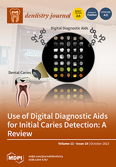
Cover Story (view full-size image): The detection and assessment of dental caries, especially at an early stage, are essential for the prevention and management of dental caries. The advances in digital diagnostic technologies have significantly facilitated the detection of dental caries. This review aims to provide an overview of digital diagnostic aids for the clinical detection of dental caries, particularly those at an initial stage. Currently available digital diagnostic aids for caries detection can be classified into four categories according to the initial source of energy, including radiation-based aids, light-based aids, ultrasound-based aids, and electric-based aids. In addition, many novel digital diagnostic aids for caries detection are still under development with promising results in laboratory settings. View this paper
  • Issues are regarded as officially published after their release is announced to the table of contents alert mailing list.
  • You may sign up for e-mail alerts to receive table of contents of newly released issues.
  • PDF is the official format for papers published in both, html and pdf forms. To view the papers in pdf format, click on the "PDF Full-text" link, and use the free Adobe Reader to open them.
Order results
Result details
Section
Select all
Export citation of selected articles as:
17 pages, 6128 KiB  
Review
Alveolar Ridge Preservation with Autologous Platelet-Rich Fibrin (PRF): Case Reports and the Rationale
by Cemal Ucer and Rabia S. Khan
Dent. J. 2023, 11(10), 244; https://doi.org/10.3390/dj11100244 - 23 Oct 2023
Viewed by 2930
Abstract
In dental implantology, alveolar ridge preservation (ARP) has emerged as a standard technique to address dimensional changes that affect alveolar ridge morphology following tooth loss. Various alternative graft materials, including xenografts, alloplasts, and allografts, have been effectively employed in fresh extraction sites for [...] Read more.
In dental implantology, alveolar ridge preservation (ARP) has emerged as a standard technique to address dimensional changes that affect alveolar ridge morphology following tooth loss. Various alternative graft materials, including xenografts, alloplasts, and allografts, have been effectively employed in fresh extraction sites for ARP. Current evidence suggests that these materials primarily serve as bio-scaffolds, which are slowly incorporated, thus necessitating a waiting period of at least 4–6 months before implant placement. Consequently, the ARP technique extends the overall duration of implant treatment by several months. Recently, the incorporation of a form of autologous platelet concentrate, known as platelet-rich fibrin (PRF), has been advocated in conjunction with ARP as a method of bioenhancement of soft- and hard-tissue healing and regeneration. PRF contains platelet-derived growth factors, hormones, and bioactive components like cytokines that have demonstrated the ability to stimulate angiogenesis and tissue regeneration throughout all phases of wound healing. Additionally, the concentration of leukocytes present in the PRF matrix plays a vital role in tissue healing and regeneration as part of the osteoimmune response. The reported advantages of incorporating autogenous PRF platelet concentrates during ARP encompass reduced healing time, improved angiogenesis and bone regeneration, socket sealing through the fibrin matrix, antibacterial properties, and decreased post-extraction pain and infection risk. Therefore, the objective of this paper is to review the existing evidence regarding the application of PRF in alveolar ridge preservation (ARP) following tooth extraction. Two clinical case studies are presented, wherein ARP was enhanced with PRF, followed by implant placement within a relatively short period of 8 weeks. These cases serve as further proof of concept for supporting the adjuvant use of PRF to enhance healing and accelerate implant placement after ARP. Full article
(This article belongs to the Special Issue Oral Implantology and Rehabilitation)
Show Figures

Figure 1

20 pages, 1274 KiB  
Review
Smartphone Applications in Dentistry: A Scoping Review
by Maurizio Pascadopoli, Paolo Zampetti, Maria Gloria Nardi, Matteo Pellegrini and Andrea Scribante
Dent. J. 2023, 11(10), 243; https://doi.org/10.3390/dj11100243 - 20 Oct 2023
Cited by 3 | Viewed by 2278
Abstract
This scoping review aims to investigate the latest literature concerning the use of smartphone applications (apps) in the prevention, management, and monitoring of oral diseases. Smartphone applications are software programs that are designed to run on smartphones. Nowadays, smartphones are regularly used by [...] Read more.
This scoping review aims to investigate the latest literature concerning the use of smartphone applications (apps) in the prevention, management, and monitoring of oral diseases. Smartphone applications are software programs that are designed to run on smartphones. Nowadays, smartphones are regularly used by people of all ages, and mobile health apps (MHAs) represent an important means of spreading information related to oral health, which is the state of the mouth and teeth, including the gums and other tissues. Several apps have been designed to promote prevention, diagnosis, and therapeutic adherence monitoring. This scoping review considered randomized clinical trials, cross-sectional studies, before–after (pre–post) studies with no control group, and observational studies. Once the inclusion and exclusion criteria had been defined, a preliminary confined search was performed on PubMed and Scopus; key terms from the collected articles were selected to design a search strategy, and then a search of all the included articles’ reference lists was run for further research. Studies were excluded if they did not fulfill the inclusion criteria. The preferred reporting items for scoping reviews (PRISMA-ScR) consensus was followed. The risk of bias was evaluated by providing a qualitative analysis of the clinical studies via the National Heart, Lung, and Blood Institute (NHLBI) Quality Assessment of Controlled Intervention Studies, Observational Cohort Studies, and Cross-Sectional Studies (NHLBI, NIH). A total of 21 studies were included in this review. As it is clear from the studies selected, the literature indicates that MHAs are effective in improving oral hygiene in adolescents and children and reducing the dental plaque index, including in patients undergoing orthodontic treatment. MHAs are also able to reduce the symptoms of patients affected by obstructive sleep apnea–hypopnea syndrome (OSAHS) and improve the swallowing-related quality of life of elderly patients. MHAs are furthermore recommended to decrease dental anxiety among patients, both during dental procedures and the post-operative period. MHAs are useful to spread knowledge about traumatic dental injuries among non-oral health professionals and to monitor dental erosion and awake bruxism. MHAs’ clinical outcomes might have been influenced by the demographic features of the subjects involved. Further studies considering a longer follow-up period and larger samples are needed. In conclusion, MHAs can be considered a useful tool to monitor oral disease and increase patients’ quality of life related to oral health. Full article
(This article belongs to the Special Issue Feature Review Papers in Dentistry)
Show Figures

Figure 1

12 pages, 3538 KiB  
Article
PRF Lysates Enhance the Proliferation and Migration of Oral Squamous Carcinoma Cell Lines
by Layla Panahipour, Rebecca Croci, Sara Guarnieri and Reinhard Gruber
Dent. J. 2023, 11(10), 242; https://doi.org/10.3390/dj11100242 - 19 Oct 2023
Viewed by 1674
Abstract
Platelet-rich fibrin (PRF) is an autologous fibrin-rich matrix where activated platelets and leucocytes accumulate. PRF has a wide spectrum of clinical indications with the overall aim of supporting tissue regeneration which in dentistry includes the healing of healthy oral mucosa with epithelial cells. [...] Read more.
Platelet-rich fibrin (PRF) is an autologous fibrin-rich matrix where activated platelets and leucocytes accumulate. PRF has a wide spectrum of clinical indications with the overall aim of supporting tissue regeneration which in dentistry includes the healing of healthy oral mucosa with epithelial cells. In oral squamous cell carcinoma lesions, however, epithelial cells undergo malignant transformation, indicated by their unrestricted proliferation and migration potential, which should not be further enhanced by a wound-healing formula. Yet, little is known about how oral squamous cell carcinomas respond to PRF lysates. The aim of the present study was, therefore, to test the capacity of PRF lysates to change the transcriptome of HSC2 oral squamous carcinoma cells and perform bioassays to support the findings. Based on the RNAseq analysis, PRF lysates caused an increase in the genes functionally linked to cell replication and migration. In support of this screening approach, PRF lysates enhanced the proliferation of HSC2 oral squamous carcinoma cells, as indicated by 3[H]-thymidine incorporation, cell counting, and the expression of proliferation-related genes. Moreover, PRF lysates sped up cell migration in a scratch assay requiring actin polymerization. Taken together, our data showing that PRF lysates are mitogenic and stimulate motility of oral squamous carcinoma cell lines could be an indication that treatment with PRF in cases of oral carcinoma should be carefully considered. Full article
(This article belongs to the Special Issue Regenerative Approaches in Dental Sciences)
Show Figures

Figure 1

12 pages, 748 KiB  
Systematic Review
Accuracy of Intraoral Scanner for Recording Completely Edentulous Arches—A Systematic Review
by Gunjan Srivastava, Subrat Kumar Padhiary, Neeta Mohanty, Pedro Molinero-Mourelle and Najla Chebib
Dent. J. 2023, 11(10), 241; https://doi.org/10.3390/dj11100241 - 18 Oct 2023
Cited by 3 | Viewed by 2430
Abstract
Scanning edentulous arches during complete denture fabrication is a crucial step; however, the quality of the resulting digital scan is still questionable. The purpose of this study is to systematically review studies (both clinical and in vitro) and determine whether intraoral scanners have [...] Read more.
Scanning edentulous arches during complete denture fabrication is a crucial step; however, the quality of the resulting digital scan is still questionable. The purpose of this study is to systematically review studies (both clinical and in vitro) and determine whether intraoral scanners have clinically acceptable accuracy when recording completely edentulous arches for the fabrication of removable complete dentures. An electronic search in medical databases like PubMed, Scopus, and Web of Science (WOS), using a combination of relevant keywords, retrieved 334 articles. After full-text evaluation, twelve articles fulfilled the inclusion criteria for this review (eight clinical studies and four in vitro studies). A quality analysis of the included studies was carried out using the QUADAS-2 tool. The accuracy values varied between different intraoral scanners. Different regions of the edentulous arches showed differences in trueness and precision values in both in vitro and clinical studies. Peripheral borders, the inner seal, and poorly traceable structures like the soft palate showed maximum discrepancies. The accuracy of intraoral scanners in recording clear anatomic landmarks like hard tissues with attached mucosa was comparable to conventional edentulous arch impressions. However, higher discrepancies were recorded when digitizing mobile and poorly traceable structures. Intraoral scanners can be used to digitize denture-bearing areas, but the interpretation of the peripheral border and the soft palate should be carefully carried out. Full article
Show Figures

Figure 1

15 pages, 763 KiB  
Article
Dental Anxiety and Its Association with Dietary Intake and Food Groups: A Cross-Sectional Study
by Lina Begdache, Eeshah Ahmed, Sana Malik and Muhammet Furkan Karakaya
Dent. J. 2023, 11(10), 240; https://doi.org/10.3390/dj11100240 - 17 Oct 2023
Viewed by 1674
Abstract
Although there is an established connection between diet and mental health, the relationship between diet and dental anxiety has not been examined yet. The purpose of this study was to fill this gap by assessing the association between diet quality, mental distress, and [...] Read more.
Although there is an established connection between diet and mental health, the relationship between diet and dental anxiety has not been examined yet. The purpose of this study was to fill this gap by assessing the association between diet quality, mental distress, and dental anxiety. The data was collected through an anonymous Google Forms survey. The survey consisted of a modified version of the validated Food–Mood Questionnaire and the Modified Dental Anxiety Scale with questions about demographics, dental health, and dental health anxieties. Data collection was performed over seven months, from April to October 2021. Data were analyzed using Pearson’s correlation coefficient in SPSS version 25.0 and STATA 17 for sample size calculation, data processing and regression analyses. A total of 506 responses were collected. Our data verified that diet quality modulates dental anxiety. Women exhibited a stronger link with dental anxiety than men (p < 0.01). Consumption of sugary foods was associated with different attributes of dental anxiety (p < 0.01). Low-quality energy-dense foods and dairy were associated with dental anxiety, whereas caffeine, meat, nuts, and green leafy vegetables produced a negative correlation. This cross-sectional study provides proof of concept that dietary patterns are potentially associated with dental anxiety. Full article
Show Figures

Figure 1

20 pages, 928 KiB  
Systematic Review
Efficacy/Safety of the Use of Glucocorticoids in Oral and Maxillofacial Surgery
by Heilyn Joanna Nils, Cristina Arce Recatala, Antonio Castano, David Ribas and Javier Flores-Fraile
Dent. J. 2023, 11(10), 239; https://doi.org/10.3390/dj11100239 - 17 Oct 2023
Viewed by 2179
Abstract
Introduction: Glucocorticoids, also known as corticosteroids or steroids, are drugs derived from cholesterol. They are synthesized by the adrenal cortex, along with other hormones, such as cortisol and aldosterone. Glucocorticoids are drugs recommended for patients undergoing surgery on the oral cavity, facial skeleton, [...] Read more.
Introduction: Glucocorticoids, also known as corticosteroids or steroids, are drugs derived from cholesterol. They are synthesized by the adrenal cortex, along with other hormones, such as cortisol and aldosterone. Glucocorticoids are drugs recommended for patients undergoing surgery on the oral cavity, facial skeleton, and related cervical structures due to their high efficacy against inflammatory and immune processes. However, these drugs are restricted due to their multiple and serious adverse effects. The objective of this study was to verify the efficacy of corticosteroids administered in major surgeries of the oral cavity, as well as of the cervical and facial structures, based on the characteristics of the patient so as to select the best therapeutic strategy. Methods: Articles in the databases of PubMed, Nature Portfolio, Medline, Cochrane Library, and Google Scholar were thoroughly examined. Results: A total of 54 articles were selected to address the proposed objectives. The results obtained show that it is effective and safe to use glucocorticoids as pre- or postsurgical therapy in oral and maxillofacial surgery to control the processes of inflammation, pain, lockjaw, and edema. However, when referring to the use of these drugs, one must proceed with caution and pay particular attention when handling them. The concentration of the glucocorticoids used must be individualized, as well as the selection of the route of administration. Various studies show that, although the oral route is the most used route, the most effective route is the intramuscular route due to its easy absorption. However, for patients who have recurrent inflammatory and vesiculobullous ulcerative lesions, the topical route should be chosen to mitigate side effects, considering that recurrent applications must be made to prevent the worsening of the lesion and to avoid having to use medications enterally. In patients with cervicofacial infections, antibiotics continue to be the main drugs used to manage the condition in conjunction with corticosteroids. It is important to know the possible interactions of glucocorticoids with other medicines or food: it has been described that the interaction between Ritonavir, an antiretroviral drug that inhibits human immunodeficiency virus (HIV) proteases, and prednisone causes an increase in the concentration of prednisone, leading to possible toxicity in normally safe doses and, in many cases, iatrogenic Cushing’s syndrome. It is also important to know the systemic or topical adverse effects of the chronic or high-dose use of glucocorticoids. Conclusions: It can be concluded that by making adequate use of glucocorticoid therapy in oral and maxillofacial surgery to manage clinical manifestations, it is possible to attenuate the morbidities of treatment and intervention. Full article
(This article belongs to the Section Oral and Maxillofacial Surgery)
Show Figures

Graphical abstract

13 pages, 307 KiB  
Article
Influence of Surgical Technique on Post-Operative Complications in the Extraction of the Lower Third Molar: A Retrospective Study
by Massimo Albanese, Alessandro Zangani, Federica Manfrin, Dario Bertossi, Rachele De Manzoni, Nicolò Tomizioli, Paolo Faccioni and Alessia Pardo
Dent. J. 2023, 11(10), 238; https://doi.org/10.3390/dj11100238 - 17 Oct 2023
Cited by 3 | Viewed by 1488
Abstract
The surgical extraction of the impacted third molar is frequently associated with several complications. The purpose of this study is to assess how two different surgical protocols affect post-operative complications during the extraction of the lower impacted third molars. In order to compare [...] Read more.
The surgical extraction of the impacted third molar is frequently associated with several complications. The purpose of this study is to assess how two different surgical protocols affect post-operative complications during the extraction of the lower impacted third molars. In order to compare and evaluate two different techniques (triangular flap vs. envelope flap), and the relative post-extraction complications, two groups of 150 patients each underwent to surgical impacted third molar extraction and 60 days of follow-up. The complication rate in the two groups was 14.00% in group A and 17.33% in group B. There was a strong association between smoking (OR: 2.8) and the use of oral contraceptives (OR: 1.75) with complications. The age- and sex-related incidence of complications in hard tissue healing has great variability in the literature; the analysis performed on our data did not show a statistically significant association between them. Even though related to a higher incidence of transient changes in sensitivity, it was found that the envelope flap saw a lower percentage of complications. There is still no clarity on which is the best protocol for the extraction of the lower impacted third molar, and the choice often depends on the surgeon’s experience. Full article
12 pages, 611 KiB  
Article
Descriptive Study of Oral Health in an Indigenous Child Population of Baka Pygmies in Cameroon
by Nicias Afoumpam Poni, David Ribas-Pérez, Javier Flores-Fraile, Paloma Villalva Hernández-Franch, Diego Rodríguez-Menacho and Antonio Castaño-Séiquer
Dent. J. 2023, 11(10), 237; https://doi.org/10.3390/dj11100237 - 12 Oct 2023
Viewed by 1449
Abstract
Indigenous populations around the world experience a disproportionate burden of oral diseases and health conditions. These inequalities are likely due to a complex web of socioeconomic, cultural, and health determinants. The Baka pygmies of southern Cameroon find themselves in this context of an [...] Read more.
Indigenous populations around the world experience a disproportionate burden of oral diseases and health conditions. These inequalities are likely due to a complex web of socioeconomic, cultural, and health determinants. The Baka pygmies of southern Cameroon find themselves in this context of an indigenous population with health inequities. The purpose of this study was to describe the oral health status, diet, hygiene habits, and access to health services of the Baka pygmy children, from which different care needs will emerge in order to develop health strategies. A descriptive cross-sectional study was conducted in 22 Baka pygmy camps randomly selected. The study population consisted of children aged 5–6 years and 11–12 years chosen by a consecutive sampling technique. The examination was performed using a data sheet based on World Health Organization (WHO) criteria and recommendations which consisted of an oral health questionnaire for children and an oral health assessment form for children. A total of 120 children participated in the study. Extraoral examination of the study population revealed the presence of noma (1%) in the age range of 5–6 years. A total of 2713 teeth were examined, and the DMFT/dft index of the sample was 0.71 with a predominant caries component. The periodontal status showed 87% bleeding on probing. Seven percent of the sample presented a need for immediate urgent treatment due to pain and/or infection. Eighty-seven percent of the sample reported never having been examined by a dentist. The examination and oral care they received was only from nongovernmental organizations (NGOs). The conclusion of this descriptive study is that the precarious oral health situation of pygmy children combined with the absence of care services in general for the Baka pygmies generate a situation of great vulnerability. Full article
Show Figures

Figure 1

15 pages, 677 KiB  
Review
Current Insights on Fiber Posts: A Narrative Review of Laboratory and Clinical Studies
by Dayana Campanelli de Morais, Sheila Butler and Maria Jacinta Moraes Coelho Santos
Dent. J. 2023, 11(10), 236; https://doi.org/10.3390/dj11100236 - 10 Oct 2023
Viewed by 2101
Abstract
Purpose: The aim of this study was to review the literature related to the clinical performance and laboratory findings regarding fiber posts, as well as the cementation technique employed with their use. Materials and Methods: A literature search was performed using an electronic [...] Read more.
Purpose: The aim of this study was to review the literature related to the clinical performance and laboratory findings regarding fiber posts, as well as the cementation technique employed with their use. Materials and Methods: A literature search was performed using an electronic database, PubMed/Medline, between 2010 and 2023. The terms used were “intra coronal post, fiber post, post cementation, and post length”. Titles and abstracts were initially screened, and a full-text assessment was conducted for those that fulfilled the inclusion criteria. The reference list of the collected papers was also screened for further relevant citations. Results: In this work, 135 potentially eligible studies were analyzed. Titles and abstracts of 90 studies followed the inclusion criteria and were selected for a full-text assessment, resulting in 50 studies selected. Moreover, additional studies from relevant citations were included, totaling 57 studies. Conclusion: According to the laboratory and clinical studies revised, the survival rate between fiber and prefabricated and cast metal posts was similar, and failures were mainly related to the loss of retention. The intra-canal post length of less than two-thirds of the root length presented successful results when ferrule was present. Furthermore, the ferrule increased the longevity of teeth restored with fiber posts. Additionally, the use of a surface treatment protocol for fiber posts and the adhesive cementation technique both contributed to the clinical success and longevity of the intra-canal post. Full article
(This article belongs to the Special Issue Feature Review Papers in Dentistry)
Show Figures

Figure 1

21 pages, 6091 KiB  
Technical Note
Mixed-Thickness Tunnel Access (MiTT) through a Linear Vertical Mucosal Incision for a Minimally Invasive Approach for Root Coverage Procedures in Anterior and Posterior Sites: Technical Description and Case Series with 1-Year Follow-Up
by Tiago Marques, Nuno Bernardo Malta dos Santos, Manuel Sousa, Juliana Campos Hasse Fernandes and Gustavo Vicentis Oliveira Fernandes
Dent. J. 2023, 11(10), 235; https://doi.org/10.3390/dj11100235 - 7 Oct 2023
Cited by 1 | Viewed by 1716
Abstract
Purpose: The goal of this article was to introduce a new root coverage (RC) technique, the mixed-thickness tunnel access (MiTT) technique, which approaches a full-split design and intends to augment soft tissues coronal to the gingival margin. It was shown step-by-step, and the [...] Read more.
Purpose: The goal of this article was to introduce a new root coverage (RC) technique, the mixed-thickness tunnel access (MiTT) technique, which approaches a full-split design and intends to augment soft tissues coronal to the gingival margin. It was shown step-by-step, and the results were presented in a case series. Methods: Healthy individuals (non-diabetics) and non-smokers with gingival recession (GR) type 1 or 2 (RT1 or RT2) were included. After evaluation, prophylaxis was performed 14 days before the surgical procedure. During the surgical appointment, one or two vertical incision(s) on the mucosa (around 1–2 mm apical to the MGJ), lateral to the papilla base, was/were performed after anesthesia. Initially, there was a partial incision to detach the mucosa of the muscles (split design). It was permitted (but not mandatory) to perform intrasulcular incisions. Through the vertical incision, internally, subperiosteal access from the MGJ toward the gingival margin (coronally) was performed to create a full-thickness tunnel. Then, communication from the vertical incision with the gingival sulcus and the papilla base occurred, keeping the papilla tip intact. A connective tissue graft was harvested and inserted through the linear incision or intrasulcularly. There were interrupted sutures. An adjunctive material may be applied (e.g., Endogain). The root coverage was measured using a periodontal probe and considered fully covered when the gingival margin was 1 mm coronal to the cementum–enamel junction (CEJ). Results: Nine healthy individuals (seven females and two males) aged 19 and 43 were enrolled. They were treated following the MiTT steps. Four cases had a single GR; two patients had two teeth involved; and three others had three or four GR. There were seven cases of RT1 and two RT2. All RT1 cases achieved 100% RC, while the mean RC obtained for RT2 was around 80%. Conclusion: The MiTT technique can be considered a more straightforward approach for minimally invasive surgical techniques, which is a feasible option to treat RC with a high success rate, predictability, and esthetic preservation. Therefore, there is a technical sensitivity to performing the full-split design procedure. Full article
Show Figures

Figure 1

18 pages, 1298 KiB  
Systematic Review
β-Tricalcium Phosphate as Alveolar Bone Grafting in Cleft Lip/Palate: A Systematic Review
by Alexander Patera Nugraha, Hui Yang, Junduo Chen, Kunhua Yang, Ploypim Kraisintu, Kyaw Zaww, Aobo Ma, Ruixian Wang, Nada Emad Alshafei Mohamed Alhadi, Juan Ramón Vanegas Sáenz and Guang Hong
Dent. J. 2023, 11(10), 234; https://doi.org/10.3390/dj11100234 - 7 Oct 2023
Viewed by 2238
Abstract
The aim of this systematic review is to describe and identify the prospects of β-Tricalcium Phosphate (β-TCP) as an alveolar bone grafting (ABG) material in cleft lip/palate (CL/P) or alveolar bone cleft defects. A systematic review protocol based on the Preferred Reporting Items [...] Read more.
The aim of this systematic review is to describe and identify the prospects of β-Tricalcium Phosphate (β-TCP) as an alveolar bone grafting (ABG) material in cleft lip/palate (CL/P) or alveolar bone cleft defects. A systematic review protocol based on the Preferred Reporting Items for Systematic Reviews and Meta-Analyses 2020 (PRISMA 2020) was drafted. The literature search was conducted using MEDLINE/PubMed, Web of Science/ISI Web of Knowledge, Scopus, and the Cochrane Library, with English as the inclusion criterion and no publication year limits. The keywords yielded a total of 5824 publications. After removing duplicates and non-English articles, there were 3196 suitable articles available for evaluation. Subsequently, 1315 studies remained after reviewing titles and abstracts. Furthermore, 85 full articles were assessed for eligibility. After reading the complete texts of those papers, 20 were eventually selected that matched the inclusion requirements. Thirteen out of the twenty studies included in this systematic review were deemed to have a low risk of bias; one had a high risk of bias; and six had a moderate risk of bias due to not reporting randomization. β-TCP, when used as an ABG material, is biocompatible, visible, practical, offers a less invasive procedure, and does not interfere with orthodontic treatment. Synthetic β-TCP for ABG can be an alternative to autologous bone grafts under certain terms and conditions. The efficacy of β-TCP for ABG in CL/P or alveolar bone cleft defects can be enhanced through a tissue engineering approach that combines β-TCP with growth factors, mesenchymal stem cells, or other graft materials, along with modifications to β-TCP’s physical properties. Full article
(This article belongs to the Collection Novel Ceramic Materials in Dentistry)
Show Figures

Figure 1

16 pages, 1876 KiB  
Article
Dental Student Perceptions of Distance Education over Time: A Mixed-Methods Study
by Susanna Y. Yeh, Nithya Puttige Ramesh, Kristie Kaczmarek-Stewart, Chiho Ahn, Alice Z. Li and Hiroe Ohyama
Dent. J. 2023, 11(10), 233; https://doi.org/10.3390/dj11100233 - 2 Oct 2023
Cited by 1 | Viewed by 1662
Abstract
Objectives: Amid the COVID-19 pandemic, the transition to distance learning raised pertinent questions regarding advantages and challenges compared to traditional in-person learning. This study aimed to investigate dental students’ perceptions of distance learning throughout the pandemic, examining its impact on their education. Methods: [...] Read more.
Objectives: Amid the COVID-19 pandemic, the transition to distance learning raised pertinent questions regarding advantages and challenges compared to traditional in-person learning. This study aimed to investigate dental students’ perceptions of distance learning throughout the pandemic, examining its impact on their education. Methods: This study employed a convergent mixed-methods design. Three online surveys were conducted in 2020, 2021, and 2022 to collect quantitative data. Additionally, qualitative semi-structured interviews were carried out in 2022. Interviews were recorded and transcribed; then, thematic analysis was performed following an inductive approach. Results: As perceived by the participants, distance learning entails advantages and challenges. Initially, they felt uncertainty and negativity about the new environment with distance learning. However, their perceptions shifted positively as they adapted, even after returning to hybrid and in-person modules. Furthermore, most participants felt that distance learning is better suited for didactic content. It was suggested that didactic courses contain approximately 25–50% of online methods. Conclusions: Distance learning has provided valuable opportunities to reinforce curricula and improve learning efficacy during the pandemic. Our findings suggest that a hybrid learning model that combines traditional and distance modules appears to be an effective approach for future dental education. Full article
(This article belongs to the Collection Feature Papers in Dental Education)
Show Figures

Figure 1

15 pages, 889 KiB  
Review
Use of Digital Diagnostic Aids for Initial Caries Detection: A Review
by Emma Kay Chan, Yuet Ying Wah, Walter Yu-Hang Lam, Chun-Hung Chu and Ollie Yiru Yu
Dent. J. 2023, 11(10), 232; https://doi.org/10.3390/dj11100232 - 28 Sep 2023
Cited by 1 | Viewed by 3661
Abstract
The advance in digital diagnostic technologies has significantly facilitated the detection of dental caries. Despite the increase in clinically available digital diagnostic aids for dental caries, there is yet to be a comprehensive summary of all available technology. This review aims to provide [...] Read more.
The advance in digital diagnostic technologies has significantly facilitated the detection of dental caries. Despite the increase in clinically available digital diagnostic aids for dental caries, there is yet to be a comprehensive summary of all available technology. This review aims to provide an overview of digital diagnostic aids for the clinical detection of dental caries, particularly those at an initial stage. Currently available digital diagnostic aids for caries detection can be classified into four categories according to the initial source of energy, including radiation-based aids, light-based aids, ultrasound-based aids, and electric-based aids. Radiation-based aids use ionizing radiation, normally X-ray, to produce images of dental structures. Radiation-based aids encompass digital bitewing radiography and cone beam computed tomography. Light-based aids employ light or laser to induce signals for the detection of the changes in the carious dental hard tissue. Common light-based aids include digital transillumination and light/laser-induced fluorescence. Ultrasound-based aids detect the signal of ultrasound waves to assess the acoustic impedance of the carious teeth. The ultrasound caries detector is an available ultrasound-based aid. Electric-based aids assess the changes in the electric current conductance or impedance of the teeth with caries. Available electric-based aids include electrical conductance measurement and alternating current impedance spectroscopy. Except for these clinically available digital diagnostic aids, many digital diagnostic aids for caries detection are still under development with promising results in laboratory settings. Full article
(This article belongs to the Special Issue Feature Papers in Digital Dentistry)
Show Figures

Figure 1

8 pages, 3057 KiB  
Case Report
Oral Secondary Syphilis in an HIV-Positive Transgender Patient: A Case Report and Review of the Literature
by Rodolfo Mauceri, Martina Coppini, Antonio Cascio, Marcello Trizzino, Valentina Crivello, Ada Maria Florena and Giuseppina Campisi
Dent. J. 2023, 11(10), 231; https://doi.org/10.3390/dj11100231 - 26 Sep 2023
Viewed by 2222
Abstract
Background: Syphilis is a worldwide sexually transmitted infection caused by Treponema pallidum. In most cases, the oral manifestations of syphilis infection are associated with cutaneous involvement. However, the present case report is noteworthy since the oral lesions are the sole clinical sign in [...] Read more.
Background: Syphilis is a worldwide sexually transmitted infection caused by Treponema pallidum. In most cases, the oral manifestations of syphilis infection are associated with cutaneous involvement. However, the present case report is noteworthy since the oral lesions are the sole clinical sign in an HIV-positive transgender patient. Case presentations: We reported an uncommon case of secondary syphilis in a 37-year-old seropositive transgender male, whose diagnostic suspect was based only on oral mucosal lesions. The patient was referred to the Oral Medicine Unit for the presence of multiple undiagnosed painful oral lesions. The intraoral examination revealed the presence of white and red plaques on the right and the left buccal mucosa and several painful lesions localized on the upper and lower labial mucosa. No cutaneous lesions were observed. Considering the sexual history of the patient and clinical findings, secondary syphilis infection was suspected. The serologic analysis was conducted, and the diagnosis of syphilis was confirmed. Moreover, to exclude the presence of oral epithelial dysplasia or malignant disease, an incisional biopsy was performed. Discussion: Compared to the literature data, oral lesions as lone signs of secondary syphilis infection are uncommon, especially in HIV-positive patients. Syphilis and HIV coinfection create a concerning situation as they interact synergistically, leading to an increased risk of transmission and faster disease progression. Conclusions: This case report emphasizes the importance of considering syphilis as a diagnostic possibility, even when oral lesions are the only clinical manifestations, especially in HIV-positive patients. Comprehensive evaluation, including a detailed sexual history and careful oral examination, is essential for accurate diagnosis and appropriate management in such cases. Full article
Show Figures

Figure 1

11 pages, 686 KiB  
Article
Supernumerary Tooth Patterns in Non-Syndromic White European Subjects
by Eva Henninger, Luca Friedli, Miltiadis A. Makrygiannakis, Vasileios F. Zymperdikas, Moschos A. Papadopoulos, Georgios Kanavakis and Nikolaos Gkantidis
Dent. J. 2023, 11(10), 230; https://doi.org/10.3390/dj11100230 - 25 Sep 2023
Cited by 1 | Viewed by 1893
Abstract
Supernumerary teeth form at an incidence of about 3% in the population, with differences among races and various clinical consequences. Information on detailed patterns, and especially on white subjects, is scarce in the literature. Therefore, we aimed to investigate the patterns of non-syndromic [...] Read more.
Supernumerary teeth form at an incidence of about 3% in the population, with differences among races and various clinical consequences. Information on detailed patterns, and especially on white subjects, is scarce in the literature. Therefore, we aimed to investigate the patterns of non-syndromic permanent supernumerary teeth in a white European population. A record review was performed in different orthodontic clinics and identified 207 eligible individuals with 258 supernumerary teeth. Approximately 80% of the subjects had one supernumerary tooth, while 15% had two. Supernumerary tooth formation was more often evident in males (male/female: 1.65). However, there was no sexual dimorphism in its severity. The following pattern sequences, with decreasing prevalence order, were observed in the maxilla: 21 > 11 > 12 > 18 > 28 and in the mandible: 34 > 44 > 35 > 45 > 42. Supernumerary teeth were most often unilaterally present, without sexual dimorphism. In the maxilla, they were more often anteriorly present, whereas in the mandible, an opposite tendency was observed. Supernumerary teeth were consistently more often observed in the maxilla than in the mandible; 74% were impacted, 80% had normal orientation (13% horizontal, 7% inverted), and 53% had normal size. The present thorough supernumerary tooth pattern assessment enables a better understanding of this condition with clinical, developmental, and evolutionary implications. Full article
Show Figures

Graphical abstract

12 pages, 3093 KiB  
Article
Salivary Duct Carcinoma with Rhabdoid Features of the Parotid Gland with No E-Cadherin Expression: A Report with Anti-HER2 Therapy and Review of the Literature
by Richa Jain, E. Ritter Sansoni, Jacqueline Angel, John P. Gleysteen, D. Neil Hayes and Adepitan A. Owosho
Dent. J. 2023, 11(10), 229; https://doi.org/10.3390/dj11100229 - 25 Sep 2023
Viewed by 1536
Abstract
Salivary duct carcinoma with rhabdoid features (SDC-RF) is a rare form of salivary gland neoplasm that was recently described. We report a case of SDC-RF of the parotid gland with loss of E-cadherin and decreased β-catenin expression in a 73-year-old male who presented [...] Read more.
Salivary duct carcinoma with rhabdoid features (SDC-RF) is a rare form of salivary gland neoplasm that was recently described. We report a case of SDC-RF of the parotid gland with loss of E-cadherin and decreased β-catenin expression in a 73-year-old male who presented with right facial/neck swelling and intermittent pain. Morphologically, the tumor presented with a discohesive infiltrate of isolated and cords of pleomorphic round cells containing moderate amount of eosinophilic to fine-vacuolated cytoplasm and hyperchromatic nuclei infiltrating through fibroadipose tissue and salivary parenchyma. Immunophenotypically, the tumor was positive for Cytokeratins Oscar and 7, GATA3, GCDFP, HER2, and an androgen receptor but negative for CK20, S100, p40, Melan A, CDX2, TTF1, ER, SATB2, DOG1, synaptophysin, and chromogranin. Due to its diffuse infiltrating pattern, involvement of the parapharyngeal space, supraclavicular fat pad, dermis, and skin without a defined surgical target, the tumor was deemed unresectable. Anti-HER2 therapy (Herceptin and Pertuzumab) was utilized. At the last follow-up, the patient is alive, with complete locoregional control and brain metastases. An electronic search was performed in the following registries for papers published up to June 2023: PubMed, Embase, and Web of Science. For the database searches, the keywords searched were “salivary gland”, “salivary duct carcinoma”, and “salivary duct carcinoma with rhabdoid features”. Our review of the literature identified 30 cases of SDC-RF that reveal there is a predilection for males (83%), parotid gland (72%), and patients older than the 6th decade of life (83%). Immunophenotypically, all SDC-RF cases except one were positive for AR and GCDFP (97%), 81% were positive for HER2, and loss or decreased expression of E-cadherin in 93% of cases. In conclusion, we described a rare case of SDF-RF of the parotid gland with no E-cadherin expression, decreased β-catenin expression, and its immunophenotypic profile. Full article
(This article belongs to the Special Issue Oral and Maxillofacial Surgery: Latest Advances and Prospects)
Show Figures

Figure 1

14 pages, 2664 KiB  
Article
Long-Term Efficacy of Chlorhexidine Gel in Single-Crown Implant Rehabilitation: A Five-Year Follow-Up Study
by Gianmaria D’Addazio, Eugenio Manciocchi, Giuseppe Tafuri, Ruggero Schiavone, Giovanna Murmura, Luan Mavriqi, Bruna Sinjari and Sergio Caputi
Dent. J. 2023, 11(10), 228; https://doi.org/10.3390/dj11100228 - 25 Sep 2023
Viewed by 1371
Abstract
Chlorhexidine digluconate (CHX) has shown the ability to significantly reduce inflammation and marginal bone loss (MBL) at the 1-year follow-up but limited data exist regarding its long-term efficacy in peri-implant stability. The objective was to compare the long-term effects (5 years of follow-up) [...] Read more.
Chlorhexidine digluconate (CHX) has shown the ability to significantly reduce inflammation and marginal bone loss (MBL) at the 1-year follow-up but limited data exist regarding its long-term efficacy in peri-implant stability. The objective was to compare the long-term effects (5 years of follow-up) of a placebo gel (16 patients in Group A) or a 0.20% CHX gel (15 patients in Group B) used at each previous surgical and prosthetic stage. Control visits were conducted in 2022, investigating the long-term effects by biological, radiological, and clinical evaluation. The data were statistically analyzed. The research achieved a 96.7% implant success rate over five years, but 41.9% of patients did not attend annual oral hygiene check-ups. The average MBL was 1.04 ± 0.39 mm, with no significant differences between the two groups. Notably, patients who attended regular periodontal check-ups experienced significantly less MBL than those who did not (p < 0.05). At five years, direct effects of CHX were absent, with both groups showing moderate bone loss. However, the results suggest that early disinfection could enhance both short- and long-term outcomes. In fact, patients with initial minor MBL due to use of CHX, preserve this advantage also after 5 years of follow-up. Additionally, the data underscore the importance of annual check-ups in early detection and management of biological complications. Full article
(This article belongs to the Special Issue Dentistry Journal: 10th Anniversary)
Show Figures

Graphical abstract

11 pages, 2547 KiB  
Article
Effect of Implant Macro-Design and Magnetodynamic Surgical Preparation on Primary Implant Stability: An In Vitro Investigation
by Alessandro Antonelli, Selene Barone, Ferdinando Attanasio, Marianna Salviati, Maria Giulia Cerra, Elena Calabria, Francesco Bennardo and Amerigo Giudice
Dent. J. 2023, 11(10), 227; https://doi.org/10.3390/dj11100227 - 24 Sep 2023
Cited by 7 | Viewed by 1692
Abstract
Background: Macro-geometry and surgical implant site preparation are two of the main factors influencing implant stability and potentially determining loading protocol. The purpose of this study was to assess the initial stability of various implant macro-designs using both magnetodynamic and traditional osteotomy techniques [...] Read more.
Background: Macro-geometry and surgical implant site preparation are two of the main factors influencing implant stability and potentially determining loading protocol. The purpose of this study was to assess the initial stability of various implant macro-designs using both magnetodynamic and traditional osteotomy techniques in low-density bone. The parameters examined included peak insertion torque (PIT), implant stability quotient (ISQ), and peak removal torque (PRT). Methods: Four groups of 34 implants each were identified in accordance with the surgery and implant shape: T5 group (Five implant and osteotomy using drills); M5 group (Five implant and magnetodynamic osteotomy using Magnetic Mallet); TT group (TiSmart implant and osteotomy with drills); and MT group (TiSmart implant and magnetodynamic osteotomy). Every implant was placed into a low-density bone animal model and scanned using CBCT. The PIT and PRT were digitally measured in Newton-centimeters (Ncm) using a torque gauge device. The ISQ was analyzed by conducting resonance frequency analysis. Results: The PIT values were 25.04 ± 4.4 Ncm for T5, 30.62 ± 3.81 Ncm for M5, 30 ± 3.74 Ncm for TT, and 32.05 ± 3.55 Ncm for MT. The average ISQ values were 68.11 ± 3.86 for T5, 71.41 ± 3.69 for M5, 70.88 ± 3.08 for TT, and 73 ± 3.5 for MT. The PRT values were 16.47 ± 4.56 Ncm for T5, 26.02 ± 4.03 Ncm for M5, 23.91 ± 3.28 Ncm for TT, and 26.93 ± 3.96 Ncm for MT. Based on our data analysis using a t-test with α = 0.05, significant differences in PIT were observed between TT and T5 (p < 0.0001), M5 and T5 (p < 0.0001), and MT and TT (p = 0.02). Significant differences in the ISQ were found between TT and T5 (p = 0.001), M5 and T5 (p < 0.001), and MT and TT (p = 0.01). The PRT also exhibited significant differences between TT and T5, M5 and T5, and MT and TT (p < 0.0001). Conclusion: Our data showed favorable primary implant stability (PS) values for both implant macro-geometries. Furthermore, the magnetodynamic preparation technique appears to be more effective in achieving higher PS values in low-density bone. Full article
(This article belongs to the Special Issue Oral Implantology and Rehabilitation)
Show Figures

Figure 1

13 pages, 22796 KiB  
Protocol
Five Steps for the Maintenance and Interception of Complications in Zygomatic Implants
by Consuela Sanavia, Edoardo Vallerga, Fanny Alessi, Tiziano Tealdo, Marco Bevilacqua, Christian Alberti, Maria Menini and Paolo Pesce
Dent. J. 2023, 11(10), 226; https://doi.org/10.3390/dj11100226 - 22 Sep 2023
Viewed by 1351
Abstract
Zygomatic implants are used for the rehabilitation of the upper jaw of patients with severe and moderate bone atrophy. Possible post-surgical complications include soft tissue dehiscence, sinusitis, and prosthodontic fractures, and maintaining an accurate control is crucial. Additionally, zygomatic implants have a unique [...] Read more.
Zygomatic implants are used for the rehabilitation of the upper jaw of patients with severe and moderate bone atrophy. Possible post-surgical complications include soft tissue dehiscence, sinusitis, and prosthodontic fractures, and maintaining an accurate control is crucial. Additionally, zygomatic implants have a unique peri-implant anatomy, making traditional periodontal parameters unsuitable. The present paper aims to provide guidelines for the maintenance and interception of complications in patients rehabilitated with these kinds of implants. The proposed protocol includes: 1. intra/extraoral and temporo-mandibular joint examination; 2. soft tissue and transmucosal path observation with magnifiers; 3. peri-implant health indices and digital stimulation of tissues; 4. examination of prosthodontic devices; and 5. photographic recording. These steps facilitate the comprehensive evaluation and monitoring of clinical conditions of zygomatic-supported rehabilitations, including dehiscence and occlusal wear during follow-up appointments. Full article
Show Figures

Figure 1

Previous Issue
Next Issue
Back to TopTop