Next Issue
Volume 9, January
Previous Issue
Volume 8, November


Bioengineering, Volume 8, Issue 12 (December 2021) – 36 articles

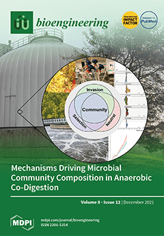
Cover Story (view full-size image): Anaerobic co-digestion uses waste to reduce the carbon footprint of wastewater treatment and relies on stable microbial communities. Here, food waste and canola lecithin were tested as co-substrates in lab-scale reactors, with amplicon sequencing employed to assess changes and stability of the communities in comparison to a life plant without co-digestion. With food waste, high levels of functional redundancy but few total changes were found. The second, more specialized co-substrate increased the selective pressure, and only there did we observe selection, diversity loss, and invasion into the otherwise stable resident community, elsewise mostly defined by neutral effects. View this paper
  • Issues are regarded as officially published after their release is announced to the table of contents alert mailing list.
  • You may sign up for e-mail alerts to receive table of contents of newly released issues.
  • PDF is the official format for papers published in both, html and pdf forms. To view the papers in pdf format, click on the "PDF Full-text" link, and use the free Adobe Reader to open them.
Order results
Result details
Section
Select all
Export citation of selected articles as:
14 pages, 1161 KiB  
Review
Current Therapeutics and Future Perspectives to Ocular Melanocytic Neoplasms in Dogs and Cats
by Tarcísio Guerra Guimarães, Karla Menezes Cardoso, Pedro Tralhão, Carlos Miguel Marto, Nuno Alexandre, Maria Filomena Botelho and Mafalda Laranjo
Bioengineering 2021, 8(12), 225; https://doi.org/10.3390/bioengineering8120225 - 20 Dec 2021
Cited by 6 | Viewed by 7502
Abstract
Neoplasms of melanocytic origin are diseases relevant to dogs and cats’ ophthalmic oncology due to their incidence, potential visual loss, and consequent decrease in life quality and expectancy. Despite its non-specific clinical presentation, melanocytic neoplasms can be histologically distinguished in melanocytomas, which present [...] Read more.
Neoplasms of melanocytic origin are diseases relevant to dogs and cats’ ophthalmic oncology due to their incidence, potential visual loss, and consequent decrease in life quality and expectancy. Despite its non-specific clinical presentation, melanocytic neoplasms can be histologically distinguished in melanocytomas, which present benign characteristics, and malignant melanomas. The diagnosis often occurs in advanced cases, limiting the therapeutic options. Surgery, cryotherapy, radiotherapy, photodynamic therapy (PDT), and laser are currently available therapeutic strategies. As no clinical guidelines are available, the treatment choice is primarily based on the clinician’s preference, proficiency, and the owner’s financial constraints. While surgery is curative in benign lesions, ocular melanomas present a variable response to treatments, besides the potential of tumour recurrences or metastatic disease. This review presents the currently available therapies for ocular melanocytic neoplasms in dogs and cats, describing the therapeutic, indications, and limitations. Additionally, new therapeutics being developed are presented and discussed, as they can improve the current treatment options. Full article
Show Figures

Figure 1

10 pages, 1439 KiB  
Article
Wool Keratin-Based Nanofibres—In Vitro Validation
by Diego Omar Sanchez Ramirez, Iriczalli Cruz-Maya, Claudia Vineis, Vincenzo Guarino, Cinzia Tonetti and Alessio Varesano
Bioengineering 2021, 8(12), 224; https://doi.org/10.3390/bioengineering8120224 - 18 Dec 2021
Cited by 11 | Viewed by 3000
Abstract
Protein-based nanofibres are commonly used in the biomedical field to support cell growth. For this study, the cell viability of wool keratin-based nanofibres was tested. Membranes were obtained by electrospinning using formic acid, hexafluoroisopropanol, and water as solvents. For aqueous solutions, polyethylene oxide [...] Read more.
Protein-based nanofibres are commonly used in the biomedical field to support cell growth. For this study, the cell viability of wool keratin-based nanofibres was tested. Membranes were obtained by electrospinning using formic acid, hexafluoroisopropanol, and water as solvents. For aqueous solutions, polyethylene oxide blended with keratin was employed, and their use to support in vitro cell interactions was also validated. Morphological characterization and secondary structure quantification were carried out by SEM and FTIR analyses. Although formic acid produced the best nanofibres from a morphological point of view, the results showed a better response to cell proliferation after 14 days in the case of fibres from hexafluoroisopropanol solution. Polyethylene oxide in keratin nanofibres was demonstrated, over time, to influence in vitro cell interactions, modifying membranes-wettability and reducing the contact between keratin chains and water molecules, respectively. Full article
Show Figures

Figure 1

12 pages, 3330 KiB  
Article
Human iPSC-Derived Vascular Smooth Muscle Cells in a Fibronectin Functionalized Collagen Hydrogel Augment Endothelial Cell Morphogenesis
by Kaiti Duan, Biraja C. Dash, Daniel C. Sasson, Sara Islam, Jackson Parker and Henry C. Hsia
Bioengineering 2021, 8(12), 223; https://doi.org/10.3390/bioengineering8120223 - 18 Dec 2021
Cited by 11 | Viewed by 4449
Abstract
Tissue-engineered constructs have immense potential as autologous grafts for wound healing. Despite the rapid advancement in fabrication technology, the major limitation is controlling angiogenesis within these constructs to form a vascular network. Here, we aimed to develop a 3D hydrogel that can regulate [...] Read more.
Tissue-engineered constructs have immense potential as autologous grafts for wound healing. Despite the rapid advancement in fabrication technology, the major limitation is controlling angiogenesis within these constructs to form a vascular network. Here, we aimed to develop a 3D hydrogel that can regulate angiogenesis. We tested the effect of fibronectin and vascular smooth muscle cells derived from human induced pluripotent stem cells (hiPSC-VSMC) on the morphogenesis of endothelial cells. The results demonstrate that fibronectin increases the number of EC networks. However, hiPSC-VSMC in the hydrogel further substantiated the number and size of EC networks by vascular endothelial growth factor and basic fibroblast growth factor secretion. A mechanistic study shows that blocking αvβ3 integrin signaling between hiPSC-VSMC and fibronectin impacts the EC network formation via reduced cell viability and proangiogenic growth factor secretion. Collectively, this study set forth initial design criteria in developing an improved pre-vascularized construct. Full article
Show Figures

Figure 1

12 pages, 3412 KiB  
Article
Assessing the Asynchrony Event Based on the Ventilation Mode for Mechanically Ventilated Patients in ICU
by Nur Sa’adah Muhamad Sauki, Nor Salwa Damanhuri, Nor Azlan Othman, Belinda Chong Chiew Meng, Yeong Shiong Chiew and Mohd Basri Mat Nor
Bioengineering 2021, 8(12), 222; https://doi.org/10.3390/bioengineering8120222 - 18 Dec 2021
Cited by 6 | Viewed by 2419
Abstract
Respiratory system modelling can assist clinicians in making clinical decisions during mechanical ventilation (MV) management in intensive care. However, there are some cases where the MV patients produce asynchronous breathing (asynchrony events) due to the spontaneous breathing (SB) effort even though they are [...] Read more.
Respiratory system modelling can assist clinicians in making clinical decisions during mechanical ventilation (MV) management in intensive care. However, there are some cases where the MV patients produce asynchronous breathing (asynchrony events) due to the spontaneous breathing (SB) effort even though they are fully sedated. Currently, most of the developed models are only suitable for fully sedated patients, which means they cannot be implemented for patients who produce asynchrony in their breathing. This leads to an incorrect measurement of the actual underlying mechanics in these patients. As a result, there is a need to develop a model that can detect asynchrony in real-time and at the bedside throughout the ventilated days. This paper demonstrates the asynchronous event detection of MV patients in the ICU of a hospital by applying a developed extended time-varying elastance model. Data from 10 mechanically ventilated respiratory failure patients admitted at the International Islamic University Malaysia (IIUM) Hospital were collected. The results showed that the model-based technique precisely detected asynchrony events (AEs) throughout the ventilation days. The patients showed an increase in AEs during the ventilation period within the same ventilation mode. SIMV mode produced much higher asynchrony compared to SPONT mode (p < 0.05). The link between AEs and the lung elastance (AUC Edrs) was also investigated. It was found that when the AEs increased, the AUC Edrs decreased and vice versa based on the results obtained in this research. The information of AEs and AUC Edrs provides the true underlying lung mechanics of the MV patients. Hence, this model-based method is capable of detecting the AEs in fully sedated MV patients and providing information that can potentially guide clinicians in selecting the optimal ventilation mode of MV, allowing for precise monitoring of respiratory mechanics in MV patients. Full article
Show Figures

Figure 1

21 pages, 2593 KiB  
Perspective
Back to the Cradle of Cytotherapy: Integrating a Century of Clinical Research and Biotechnology-Based Manufacturing for Modern Tissue-Specific Cellular Treatments in Switzerland
by Alexis Laurent, Philippe Abdel-Sayed, Corinne Scaletta, Philippe Laurent, Elénie Laurent, Murielle Michetti, Anthony de Buys Roessingh, Wassim Raffoul, Nathalie Hirt-Burri and Lee Ann Applegate
Bioengineering 2021, 8(12), 221; https://doi.org/10.3390/bioengineering8120221 - 17 Dec 2021
Cited by 3 | Viewed by 2576
Abstract
Empirically studied by Dr. Brown-Séquard in the late 1800s, cytotherapies were later democratized by Dr. Niehans during the twentieth century in Western Switzerland. Many local cultural landmarks around the Léman Riviera are reminiscent of the inception of such cell-based treatments. Despite the discreet [...] Read more.
Empirically studied by Dr. Brown-Séquard in the late 1800s, cytotherapies were later democratized by Dr. Niehans during the twentieth century in Western Switzerland. Many local cultural landmarks around the Léman Riviera are reminiscent of the inception of such cell-based treatments. Despite the discreet extravagance of the remaining heirs of “living cell therapy” and specific enforcements by Swiss health authorities, current interest in modern and scientifically sound cell-based regenerative medicine has never been stronger. Respective progress made in bioengineering and in biotechnology have enabled the clinical implementation of modern cell-based therapeutic treatments within updated medical and regulatory frameworks. Notably, the Swiss progenitor cell transplantation program has enabled the gathering of two decades of clinical experience in Lausanne for the therapeutic management of cutaneous and musculoskeletal affections, using homologous allogeneic cell-based approaches. While striking conceptual similarities exist between the respective works of the fathers of cytotherapy and of modern highly specialized clinicians, major and important iterative updates have been implemented, centered on product quality and risk-analysis-based patient safety insurance. This perspective article highlights some historical similarities and major evolutive differences, particularly regarding product safety and quality issues, characterizing the use of cell-based therapies in Switzerland over the past century. We outline the vast therapeutic potential to be harnessed for the benefit of overall patient health and the importance of specific scientific methodological aspects. Full article
Show Figures

Figure 1

20 pages, 4365 KiB  
Review
Is Culture Expansion Necessary in Autologous Mesenchymal Stromal Cell Therapy to Obtain Superior Results in the Management of Knee Osteoarthritis?—Meta-Analysis of Randomized Controlled Trials
by Sathish Muthu, Randhi Rama Kartheek, Naveen Jeyaraman, Ramya Lakshmi Rajendran, Manish Khanna, Madhan Jeyaraman, Rathinavelpandian Perunchezhian Packkyarathinam, Prakash Gangadaran and Byeong-Cheol Ahn
Bioengineering 2021, 8(12), 220; https://doi.org/10.3390/bioengineering8120220 - 16 Dec 2021
Cited by 9 | Viewed by 2998
Abstract
Study Design: Meta-analysis. Objectives: We aimed to analyze the impact of cultured expansion of autologous mesenchymal stromal cells (MSCs) in the management of osteoarthritis of the knee from randomized controlled trials (RCTs) available in the literature. Materials and Methods: We conducted independent and [...] Read more.
Study Design: Meta-analysis. Objectives: We aimed to analyze the impact of cultured expansion of autologous mesenchymal stromal cells (MSCs) in the management of osteoarthritis of the knee from randomized controlled trials (RCTs) available in the literature. Materials and Methods: We conducted independent and duplicate electronic database searches including PubMed, Embase, Web of Science, and Cochrane Library until August 2021 for RCTs analyzing the efficacy and safety of culture-expanded compared to non-cultured autologous MSCs in the management of knee osteoarthritis. The Visual Analog Score (VAS) for pain, Western Ontario McMaster University’s Osteoarthritis Index (WOMAC), Lysholm score, Knee Osteoarthritis Outcome Score (KOOS), and adverse events were the analyzed outcomes. Analysis was performed in R-platform using OpenMeta [Analyst] software. Results: Overall, 17 studies involving 767 patients were included for analysis. None of the studies made a direct comparison of the culture expanded and non-cultured MSCs, hence we pooled the results of all the included studies of non-cultured and cultured types of MSC sources and made a comparative analysis of the outcomes. At six months, culture expanded MSCs showed significantly better improvement (p < 0.001) in VAS outcome. Uncultured MSCs, on the other hand, demonstrated significant VAS improvement in the long term (12 months) in VAS (p < 0.001), WOMAC (p = 0.025), KOOS score (p = 0.016) where cultured-expanded MSCs failed to demonstrate a significant change. Culturing of MSCs did not significantly increase the complications noted (p = 0.485). On sub-group analysis, adipose-derived uncultured MSCs outperformed culture-expanded MSCs at both short term (six months) and long term (12 months) in functional outcome parameters such as WOMAC (p < 0.001, p = 0.025), Lysholm (p < 0.006), and KOOS (p < 0.003) scores, respectively, compared to their controls. Conclusions: We identified a void in literature evaluating the impact of culture expansion of MSCs for use in knee osteoarthritis. Our indirect analysis of literature showed that culture expansion of autologous MSCs is not a necessary factor to obtain superior results in the management of knee osteoarthritis. Moreover, while using uncultured autologous MSCs, we recommend MSCs of adipose origin to obtain superior functional outcomes. However, we urge future trials of sufficient quality to validate our findings to arrive at a consensus on the need for culture expansion of MSCs for use in cellular therapy of knee osteoarthritis. Full article
Show Figures

Figure 1

15 pages, 5274 KiB  
Article
Attenuating Effect of Vitamin E against Silver Nano Particles Toxicity in Submandibular Salivary Glands
by Mahmoud M. Bakr, Mahmoud M. Al-Ankily, Sara M. Shogaa and Mohamed Shamel
Bioengineering 2021, 8(12), 219; https://doi.org/10.3390/bioengineering8120219 - 16 Dec 2021
Cited by 5 | Viewed by 2868
Abstract
Silver nanoparticles (AgNPs) are extensively used in many industries due to their superior antimicrobial properties. However, it is evident from many studies that AgNPs has cytotoxic potential through its effect on excessive formation of reactive oxygen species (ROS). The aim of this study [...] Read more.
Silver nanoparticles (AgNPs) are extensively used in many industries due to their superior antimicrobial properties. However, it is evident from many studies that AgNPs has cytotoxic potential through its effect on excessive formation of reactive oxygen species (ROS). The aim of this study was to examine the toxic effect of AgNPs on the submandibular salivary glands and the attenuating effect of vitamin E, as a natural antioxidant, against this toxicity. Thirty Albino rats were divided into 3 groups (n = 10): control group, AgNPs group receiving 2 mg/kg daily for 28 days, and AgNPs and vitamin E group receiving AgNPs the same as the previous group in addition to vitamin E at a dose of 100 mg/kg. Microscopic, ultrastructural, and cytokeratin immune-reactivity examination of the glands were performed. The AgNPs group showed noticeable degeneration in all structures of the gland as evident in the histological and ultrastructural examination. The AgNPs and vitamin E group revealed an improvement of the glandular elements. A significant increase in cytokeratin immune expression was found after comparison of both groups (p = 0.01). This current study shows that vitamin E has powerful antioxidant properties, which can combat the cytotoxic effect caused by AgNPs. Further studies are deemed necessary to confirm this finding using other immunohistochemical markers, such as myosin and E-cadherin. Full article
Show Figures

Figure 1

18 pages, 2118 KiB  
Review
An Update on Nickel-Titanium Rotary Instruments in Endodontics: Mechanical Characteristics, Testing and Future Perspective—An Overview
by Alessio Zanza, Maurilio D’Angelo, Rodolfo Reda, Gianluca Gambarini, Luca Testarelli and Dario Di Nardo
Bioengineering 2021, 8(12), 218; https://doi.org/10.3390/bioengineering8120218 - 16 Dec 2021
Cited by 52 | Viewed by 7135
Abstract
Since the introduction of Nickel-Titanium alloy as the material of choice for the manufacturing of endodontic rotary instruments, the success rate of the root canal therapies has been significantly increased. This success mainly arises from the properties of the Nickel-Titanium alloy: the biocompatibility, [...] Read more.
Since the introduction of Nickel-Titanium alloy as the material of choice for the manufacturing of endodontic rotary instruments, the success rate of the root canal therapies has been significantly increased. This success mainly arises from the properties of the Nickel-Titanium alloy: the biocompatibility, the superelasticity and the shape memory effect. Those characteristics have led to a reduction in time of endodontic treatments, a simplification of instrumentation procedures and an increase of predictability and effectiveness of endodontic treatments. Nevertheless, the intracanal separation of Nickel-Titanium rotary instruments is still a major concern of endodontists, with a consequent possible reduction in the outcome rate. As thoroughly demonstrated, the two main causes of intracanal separation of endodontic instruments are the cyclic fatigue and the torsional loads. As results, in order to reduce the percentage of intracanal separation research and manufacturers have been focused on the parameters that directly or indirectly influence mechanical properties of endodontic rotary instruments. This review describes the current state of the art regarding the Nickel-Titanium alloy in endodontics, the mechanical behavior of endodontic rotary instruments and the relative stresses acting on them during intracanal instrumentation, highlighting the limitation of the current literature. Full article
Show Figures

Figure 1

7 pages, 347 KiB  
Article
Peracetic Acid: A Practical Alternative to Formalin for Disinfection of Extracted Human Teeth
by Riaz Ali, Justin Bartholomew, Randolph Coffey, Caroline Carrico, Todd Kitten and Parthasarathy Madurantakam
Bioengineering 2021, 8(12), 217; https://doi.org/10.3390/bioengineering8120217 - 15 Dec 2021
Cited by 2 | Viewed by 2432
Abstract
Extracted human teeth provide the closest approximation to teeth in situ and play important roles in dental education and materials research. Since extracted teeth are potentially infectious, the Centers for Disease Control recommend their sterilization by autoclaving or disinfection by formalin immersion to [...] Read more.
Extracted human teeth provide the closest approximation to teeth in situ and play important roles in dental education and materials research. Since extracted teeth are potentially infectious, the Centers for Disease Control recommend their sterilization by autoclaving or disinfection by formalin immersion to ensure safe handling. However, autoclaving is not recommended for teeth with amalgam fillings and formalin is hazardous. The goal of the present study was to investigate the potential of peracetic acid (PA) as an alternative method to achieve reliable disinfection of freshly extracted teeth. A total of 80 extracted teeth were collected for this study. Whole teeth were incubated in one of four solutions for defined periods of time: sterile water (2 weeks), formalin (2 weeks), PA 1000 ppm (2 weeks), and PA 2000 ppm (1 week). After sectioning, the crown and root fragments were transferred into separate tubes containing brain–heart infusion broth and incubated at 37 °C under anaerobic conditions for 72 h. Absence of broth turbidity was used to assess effectiveness of disinfection. No turbidity was observed in any of the formalin-treated or peracetic acid-treated samples, signifying complete disinfection. Our results indicate that PA can effectively disinfect extracted human teeth, providing a reliable alternative to formalin and autoclaving. Full article
Show Figures

Figure 1

13 pages, 3367 KiB  
Article
The Interventricular Septum Is Biomechanically Distinct from the Ventricular Free Walls
by Michael Nguyen-Truong, Wenqiang Liu, Courtney Doherty, Kristen LeBar, Kevin M. Labus, Christian M. Puttlitz, Jeremiah Easley, Eric Monnet, Adam Chicco and Zhijie Wang
Bioengineering 2021, 8(12), 216; https://doi.org/10.3390/bioengineering8120216 - 15 Dec 2021
Cited by 8 | Viewed by 4629
Abstract
The interventricular septum contributes to the pumping function of both ventricles. However, unlike the ventricular wall, its mechanical behavior remains largely unknown. To fill the knowledge gap, this study aims to characterize the biaxial and transmural variation of the mechanical properties of the [...] Read more.
The interventricular septum contributes to the pumping function of both ventricles. However, unlike the ventricular wall, its mechanical behavior remains largely unknown. To fill the knowledge gap, this study aims to characterize the biaxial and transmural variation of the mechanical properties of the septum and compare it to the free walls of the left and right ventricles (LV/RV). Fresh hearts were obtained from healthy, adult sheep. The septal wall was sliced along the mid-line into two septal sides and compared to the epicardial layers of the LV- and RV-free walls. Biaxial tensile mechanical tests and constitutive modeling were performed to obtain the passive mechanical properties of the LV- and RV-side of the septum and ventricular walls. We found that both sides of the septum were significantly softer than the respective ventricular walls, and that the septum presented significantly less collagen than the ventricular walls. At low strains, we observed the symmetric distribution of the fiber orientations and a similar anisotropic behavior between the LV-side and RV-side of the septum, with a stiffer material property in the longitudinal direction, rather than the circumferential direction. At high strains, both sides showed isotropic behavior. Both septal sides had similar intrinsic elasticity, as evidenced by experimental data and constitutive modeling. These new findings offer important knowledge of the biomechanics of the septum wall, which may deepen the understanding of heart physiology. Full article
Show Figures

Graphical abstract

26 pages, 1912 KiB  
Review
Control of Blood Coagulation by Hemocompatible Material Surfaces—A Review
by Janna Kuchinka, Christian Willems, Dmitry V. Telyshev and Thomas Groth
Bioengineering 2021, 8(12), 215; https://doi.org/10.3390/bioengineering8120215 - 15 Dec 2021
Cited by 44 | Viewed by 7151
Abstract
Hemocompatibility of biomaterials in contact with the blood of patients is a prerequisite for the short- and long-term applications of medical devices such as cardiovascular stents, artificial heart valves, ventricular assist devices, catheters, blood linings and extracorporeal devices such as artificial kidneys (hemodialysis), [...] Read more.
Hemocompatibility of biomaterials in contact with the blood of patients is a prerequisite for the short- and long-term applications of medical devices such as cardiovascular stents, artificial heart valves, ventricular assist devices, catheters, blood linings and extracorporeal devices such as artificial kidneys (hemodialysis), extracorporeal membrane oxygenation (ECMO) and cardiopulmonary bypass. Although lower blood compatibility of materials and devices can be handled with systemic anticoagulation, its side effects, such as an increased bleeding risk, make materials that have a better hemocompatibility highly desirable, particularly in long-term applications. This review provides a short overview on the basic mechanisms of blood coagulation including plasmatic coagulation and blood platelets, as well as the activation of the complement system. Furthermore, a survey on concepts for tailoring the blood response of biomaterials to improve the hemocompatibility of medical devices is given which covers different approaches that either inhibit interaction of material surfaces with blood components completely or control the response of the coagulation system, blood platelets and leukocytes. Full article
(This article belongs to the Special Issue The New Frontiers of Artificial Organs Engineering)
Show Figures

Figure 1

9 pages, 3050 KiB  
Technical Note
Computer-Guided Bone Biopsy: A Technical Note with the Description of a Clinical Case
by Federica Altieri, Giovanna Iezzi, Valeria Luzzi, Gianni Di Giorgio, Antonella Polimeni and Michele Cassetta
Bioengineering 2021, 8(12), 214; https://doi.org/10.3390/bioengineering8120214 - 15 Dec 2021
Cited by 3 | Viewed by 2272
Abstract
Aim: The aim of this technical note is to present a computer-aided design–computer-aided manufacturing (CAD–CAM) surgical guide to perform a computer-guided bone biopsy. Traditionally, to diagnose abnormal conditions affecting jawbone, a bone biopsy is performed with the use of a trephine bur. The [...] Read more.
Aim: The aim of this technical note is to present a computer-aided design–computer-aided manufacturing (CAD–CAM) surgical guide to perform a computer-guided bone biopsy. Traditionally, to diagnose abnormal conditions affecting jawbone, a bone biopsy is performed with the use of a trephine bur. The positioning of the bur, during the biopsy, is based on the skill of the surgeon; therefore, an inaccurate placement of a trephine bur may occur. The use of a guide, however, can minimize this risk and achieve a better result. Materials and Methods: To determine the site and the extension of bone sampling, the stereolithography file (STL) file of cone–beam computed tomography (CBCT) images is acquired using a specific planning software and superimposed with the STL file of a dental cast; a virtual surgical guide is designed, using the same software that allows a 3D (three-dimensional) view of the guide from different perspectives and planes. The number and site of guide tubes are determined on the basis of the width and the extension of the sampling; thanks to a 3D printer, the surgical guide is manufactured. Results: The use of a customized surgical guide realized with CAD–CAM technology allows a precise and minimally invasive approach, with an accurate three-dimensional localization of the biopsy site. Conclusions: The high precision, great predictability, time-effectiveness and versatility of the present guide should encourage the clinician to use this minimally invasive surgical approach, but controlled clinical trials should be conducted to evaluate the advantages as well as any possible complications. Full article
Show Figures

Figure 1

14 pages, 2107 KiB  
Article
How Localized Z-Disc Damage Affects Force Generation and Gene Expression in Cardiomyocytes
by Dominik Müller, Sören Donath, Emanuel Georg Brückner, Santoshi Biswanath Devadas, Fiene Daniel, Lara Gentemann, Robert Zweigerdt, Alexander Heisterkamp and Stefan Michael Klaus Kalies
Bioengineering 2021, 8(12), 213; https://doi.org/10.3390/bioengineering8120213 - 14 Dec 2021
Cited by 4 | Viewed by 2838
Abstract
The proper function of cardiomyocytes (CMs) is highly related to the Z-disc, which has a pivotal role in orchestrating the sarcomeric cytoskeletal function. To better understand Z-disc related cardiomyopathies, novel models of Z-disc damage have to be developed. Human pluripotent stem cell (hPSC)-derived [...] Read more.
The proper function of cardiomyocytes (CMs) is highly related to the Z-disc, which has a pivotal role in orchestrating the sarcomeric cytoskeletal function. To better understand Z-disc related cardiomyopathies, novel models of Z-disc damage have to be developed. Human pluripotent stem cell (hPSC)-derived CMs can serve as an in vitro model to better understand the sarcomeric cytoskeleton. A femtosecond laser system can be applied for localized and defined damage application within cells as single Z-discs can be removed. We have investigated the changes in force generation via traction force microscopy, and in gene expression after Z-disc manipulation in hPSC-derived CMs. We observed a significant weakening of force generation after removal of a Z-disc. However, no significant changes of the number of contractions after manipulation were detected. The stress related gene NF-kB was significantly upregulated. Additionally, α-actinin (ACTN2) and filamin-C (FLNc) were upregulated, pointing to remodeling of the Z-disc and the sarcomeric cytoskeleton. Ultimately, cardiac troponin I (TNNI3) and cardiac muscle troponin T (TNNT2) were significantly downregulated. Our results allow a better understanding of transcriptional coupling of Z-disc damage and the relation of damage to force generation and can therefore finally pave the way to novel therapies of sarcomeric disorders. Full article
Show Figures

Figure 1

14 pages, 2721 KiB  
Article
Pheno-SELEX: Engineering Anti-Metastatic Aptamers through Targeting the Invasive Phenotype Using Systemic Evolution of Ligands by Exponential Enrichment
by Greg Shelley, Jinlu Dai, Jill M. Keller and Evan T. Keller
Bioengineering 2021, 8(12), 212; https://doi.org/10.3390/bioengineering8120212 - 13 Dec 2021
Cited by 4 | Viewed by 2626
Abstract
Multiple methods (e.g., small molecules and antibodies) have been engineered to target specific proteins and signaling pathways in cancer. However, many mediators of the cancer phenotype are unknown and the ability to target these phenotypes would help mitigate cancer. Aptamers are small DNA [...] Read more.
Multiple methods (e.g., small molecules and antibodies) have been engineered to target specific proteins and signaling pathways in cancer. However, many mediators of the cancer phenotype are unknown and the ability to target these phenotypes would help mitigate cancer. Aptamers are small DNA or RNA molecules that are designed for therapeutic use. The design of aptamers to target cancers can be challenging. Accordingly, to engineer functionally anti-metastatic aptamers we used a modification of systemic evolution of ligands by exponential enrichment (SELEX) we call Pheno-SELEX to target a known phenotype of cancer metastasis, i.e., invasion. A highly invasive prostate cancer (PCa) cell line was established and used to identify aptamers that bound to it with high affinity as opposed to a less invasive variant to the cell line. The anti-invasive aptamer (AIA1) was found to inhibit in vitro invasion of the original highly invasive PCa cell line, as well as an additional PCa cell line and an osteosarcoma cell line. AIA1 also inhibited in vivo development of metastasis in both a PCa and osteosarcoma model of metastasis. These results indicate that Pheno-SELEX can be successfully used to identify aptamers without knowledge of underlying molecular targets. This study establishes a new paradigm for the identification of functional aptamers. Full article
(This article belongs to the Special Issue Targeted Therapy for Cancer)
Show Figures

Figure 1

23 pages, 3054 KiB  
Review
Alzheimer’s Disease: Current Perspectives and Advances in Physiological Modeling
by E. Josephine Boder and Ipsita A. Banerjee
Bioengineering 2021, 8(12), 211; https://doi.org/10.3390/bioengineering8120211 - 12 Dec 2021
Cited by 12 | Viewed by 4771
Abstract
Though Alzheimer’s disease (AD) is the most common cause of dementia, complete disease-modifying treatments are yet to be fully attained. Until recently, transgenic mice constituted most in vitro model systems of AD used for preclinical drug screening; however, these models have so far [...] Read more.
Though Alzheimer’s disease (AD) is the most common cause of dementia, complete disease-modifying treatments are yet to be fully attained. Until recently, transgenic mice constituted most in vitro model systems of AD used for preclinical drug screening; however, these models have so far failed to adequately replicate the disease’s pathophysiology. However, the generation of humanized APOE4 mouse models has led to key discoveries. Recent advances in stem cell differentiation techniques and the development of induced pluripotent stem cells (iPSCs) have facilitated the development of novel in vitro devices. These “microphysiological” systems—in vitro human cell culture systems designed to replicate in vivo physiology—employ varying levels of biomimicry and engineering control. Spheroid-based organoids, 3D cell culture systems, and microfluidic devices or a combination of these have the potential to replicate AD pathophysiology and pathogenesis in vitro and thus serve as both tools for testing therapeutics and models for experimental manipulation. Full article
(This article belongs to the Special Issue Material and Engineering-Based Approaches for Organoids)
Show Figures

Figure 1

33 pages, 3718 KiB  
Article
Tryptophan Production Maximization in a Fed-Batch Bioreactor with Modified E. coli Cells, by Optimizing Its Operating Policy Based on an Extended Structured Cell Kinetic Model
by Gheorghe Maria and Laura Renea
Bioengineering 2021, 8(12), 210; https://doi.org/10.3390/bioengineering8120210 - 10 Dec 2021
Cited by 5 | Viewed by 3327
Abstract
Hybrid kinetic models, linking structured cell metabolic processes to the dynamics of macroscopic variables of the bioreactor, are more and more used in engineering evaluations to derive more precise predictions of the process dynamics under variable operating conditions. Depending on the cell model [...] Read more.
Hybrid kinetic models, linking structured cell metabolic processes to the dynamics of macroscopic variables of the bioreactor, are more and more used in engineering evaluations to derive more precise predictions of the process dynamics under variable operating conditions. Depending on the cell model complexity, such a math tool can be used to evaluate the metabolic fluxes in relation to the bioreactor operating conditions, thus suggesting ways to genetically modify the microorganism for certain purposes. Even if development of such an extended dynamic model requires more experimental and computational efforts, its use is advantageous. The approached probative example refers to a model simulating the dynamics of nanoscale variables from several pathways of the central carbon metabolism (CCM) of Escherichia coli cells, linked to the macroscopic state variables of a fed-batch bioreactor (FBR) used for the tryptophan (TRP) production. The used E. coli strain was modified to replace the PTS system for glucose (GLC) uptake with a more efficient one. The study presents multiple elements of novelty: (i) the experimentally validated modular model itself, and (ii) its efficiency in computationally deriving an optimal operation policy of the FBR. Full article
(This article belongs to the Topic Bioreactors: Control, Optimization and Applications)
Show Figures

Figure 1

17 pages, 4063 KiB  
Article
Guided Self-Assembly of ES-Derived Lung Progenitors into Biomimetic Tube Structures That Impact Cell Differentiation
by John P. Soleas, Linwen Huang, Elisa D’Arcangelo, Maria Cristina Nostro, Thomas K. Waddell, Alison P. McGuigan and Golnaz Karoubi
Bioengineering 2021, 8(12), 209; https://doi.org/10.3390/bioengineering8120209 - 10 Dec 2021
Cited by 2 | Viewed by 2696
Abstract
Chemically directed differentiation of pluripotent stem cells (PSCs) into defined cell types is a potent strategy for creating regenerative tissue models and cell therapies. In vitro observations suggest that physical cues can augment directed differentiation. We recently demonstrated that confining human PSC-derived lung [...] Read more.
Chemically directed differentiation of pluripotent stem cells (PSCs) into defined cell types is a potent strategy for creating regenerative tissue models and cell therapies. In vitro observations suggest that physical cues can augment directed differentiation. We recently demonstrated that confining human PSC-derived lung progenitor cells in a tube with a diameter that mimics those observed during lung development results in the alteration of cell differentiation towards SOX2SOX9+ lung cells. Here we set out to assess the robustness of this geometric confinement effect with respect to different culture parameters in order to explore the corresponding changes in cell morphometry and determine the feasibility of using such an approach to enhance directed differentiation protocols. Culture of progenitor cells in polydimethylsiloxane (PDMS) tubes reliably induced self-organization into tube structures and was insensitive to a variety of extracellular matrix coatings. Cellular morphology and differentiation status were found to be sensitive to the diameter of tube cells that were cultured within but not to seeding density. These data suggest that geometric cues impose constraints on cells, homogenize cellular morphology, and influence fate status. Full article
Show Figures

Figure 1

27 pages, 4099 KiB  
Article
Metabolic and Transcriptional Changes across Osteogenic Differentiation of Mesenchymal Stromal Cells
by Thora Bjorg Sigmarsdottir, Sarah McGarrity, Adrián López García de Lomana, Aristotelis Kotronoulas, Snaevar Sigurdsson, James T. Yurkovich, Ottar Rolfsson and Olafur Eysteinn Sigurjonsson
Bioengineering 2021, 8(12), 208; https://doi.org/10.3390/bioengineering8120208 - 10 Dec 2021
Cited by 9 | Viewed by 3823
Abstract
Mesenchymal stromal cells (MSCs) are multipotent post-natal stem cells with applications in tissue engineering and regenerative medicine. MSCs can differentiate into osteoblasts, chondrocytes, or adipocytes, with functional differences in cells during osteogenesis accompanied by metabolic changes. The temporal dynamics of these metabolic shifts [...] Read more.
Mesenchymal stromal cells (MSCs) are multipotent post-natal stem cells with applications in tissue engineering and regenerative medicine. MSCs can differentiate into osteoblasts, chondrocytes, or adipocytes, with functional differences in cells during osteogenesis accompanied by metabolic changes. The temporal dynamics of these metabolic shifts have not yet been fully characterized and are suspected to be important for therapeutic applications such as osteogenesis optimization. Here, our goal was to characterize the metabolic shifts that occur during osteogenesis. We profiled five key extracellular metabolites longitudinally (glucose, lactate, glutamine, glutamate, and ammonia) from MSCs from four donors to classify osteogenic differentiation into three metabolic stages, defined by changes in the uptake and secretion rates of the metabolites in cell culture media. We used a combination of untargeted metabolomic analysis, targeted analysis of 13C-glucose labelled intracellular data, and RNA-sequencing data to reconstruct a gene regulatory network and further characterize cellular metabolism. The metabolic stages identified in this proof-of-concept study provide a framework for more detailed investigations aimed at identifying biomarkers of osteogenic differentiation and small molecule interventions to optimize MSC differentiation for clinical applications. Full article
Show Figures

Figure 1

18 pages, 1677 KiB  
Article
Developing a Novel Integrated Generalised Data Envelopment Analysis (DEA) to Evaluate Hospitals Providing Stroke Care Services
by Mirpouya Mirmozaffari, Elham Shadkam, Seyed Mohammad Khalili and Maziar Yazdani
Bioengineering 2021, 8(12), 207; https://doi.org/10.3390/bioengineering8120207 - 10 Dec 2021
Cited by 37 | Viewed by 3711
Abstract
Stroke is the biggest cause of adult disability and the third biggest cause of death in the US. Stroke is a medical emergency, and the treatment given in the early hours is important in shaping the patient’s long-term recovery and prognosis. Despite the [...] Read more.
Stroke is the biggest cause of adult disability and the third biggest cause of death in the US. Stroke is a medical emergency, and the treatment given in the early hours is important in shaping the patient’s long-term recovery and prognosis. Despite the fact that substantial attention has been dedicated to this complex and difficult issue in healthcare, novel strategies such as operation research-based approaches have hardly been used to deal with the difficult challenges associated with stroke. This study proposes a novel approach with data envelopment analysis (DEA) and multi-objective linear programming (MOLP) in hospitals that provide stroke care services to select the most efficient approach, which will be a new experiment in literature perception. DEA and MOLP are widely used for performance evaluation and efficiency measurement. Despite their similarities and common concepts, the two disciplines have evolved separately. The generalised DEA (GDEA) cannot incorporate the preferences of decision-makers (DMs) preferences and historical efficiency data. In contrast, MOLP can incorporate the DM’s preferences into the decision-making process. We transform the GDEA model into MOLP through the max-ordering approach to (i) solve the problem interactively; (ii) use the step method (STEM) and consider DM’s preferences; (iii) eliminate the need for predetermined preference information; and (iv) apply the most preferred solution (MPS) to identify the most efficient approach. A case study of hospitals that provide stroke care services is taken as an example to illustrate the potential application of the proposed approach method. Full article
Show Figures

Figure 1

14 pages, 4703 KiB  
Article
Second Generation of Tissue-Engineered Ligament Substitutes for Torn ACL Replacement: Adaptations for Clinical Applications
by Franck Simon, Jadson Moreira-Pereira, Jean Lamontagne, Rejean Cloutier, Francine Goulet and Stéphane Chabaud
Bioengineering 2021, 8(12), 206; https://doi.org/10.3390/bioengineering8120206 - 10 Dec 2021
Cited by 1 | Viewed by 2926
Abstract
The anterior cruciate ligament (ACL) of the knee joint is one of the strongest ligaments of the body and is often the target of traumatic injuries. Unfortunately, its healing potential is limited, and the surgical options for its replacement are frequently associated with [...] Read more.
The anterior cruciate ligament (ACL) of the knee joint is one of the strongest ligaments of the body and is often the target of traumatic injuries. Unfortunately, its healing potential is limited, and the surgical options for its replacement are frequently associated with clinical issues. A bioengineered ACL (bACL) was developed using a collagen matrix, seeded with autologous cells and successfully grafted and integrated into goat knee joints. We hypothesize that, in order to reduce the cost and simplify the model, an acellular bACL can be used as a substitute for a torn ACL, and bone plugs can be replaced by endobuttons to fix the bACL in situ. First, acellular bACLs were successfully grafted in the goat model with 18% recovery of ultimate tensile strength 6 months after implantation (94 N/mm2 vs. 520). Second, a bACL with endobuttons was produced and tested in an exvivo bovine knee model. The natural collagen scaffold of the bACL contributes to supporting host cell migration, growth and differentiation in situ post-implantation. Bone plugs were replaced by endobuttons to design a second generation of bACLs that offer more versatility as biocompatible grafts for torn ACL replacement in humans. A robust collagen bACL will allow solving therapeutic issues currently encountered by orthopedic surgeons such as donor-site morbidity, graft failure and post-traumatic osteoarthritis. Full article
Show Figures

Figure 1

27 pages, 15216 KiB  
Review
Implantable Drug Delivery Systems and Foreign Body Reaction: Traversing the Current Clinical Landscape
by Alexey Fayzullin, Alesia Bakulina, Karen Mikaelyan, Anatoly Shekhter and Anna Guller
Bioengineering 2021, 8(12), 205; https://doi.org/10.3390/bioengineering8120205 - 9 Dec 2021
Cited by 28 | Viewed by 16114
Abstract
Precise delivery of therapeutics to the target structures is essential for treatment efficiency and safety. Drug administration via conventional routes requires overcoming multiple transport barriers to achieve and maintain the local drug concentration and commonly results in unwanted off-target effects. Patients’ compliance with [...] Read more.
Precise delivery of therapeutics to the target structures is essential for treatment efficiency and safety. Drug administration via conventional routes requires overcoming multiple transport barriers to achieve and maintain the local drug concentration and commonly results in unwanted off-target effects. Patients’ compliance with the treatment schedule remains another challenge. Implantable drug delivery systems (IDDSs) provide a way to solve these problems. IDDSs are bioengineering devices surgically placed inside the patient’s tissues to avoid first-pass metabolism and reduce the systemic toxicity of the drug by eluting the therapeutic payload in the vicinity of the target tissues. IDDSs present an impressive example of successful translation of the research and engineering findings to the patient’s bedside. It is envisaged that the IDDS technologies will grow exponentially in the coming years. However, to pave the way for this progress, it is essential to learn lessons from the past and present of IDDSs clinical applications. The efficiency and safety of the drug-eluting implants depend on the interactions between the device and the hosting tissues. In this review, we address this need and analyze the clinical landscape of the FDA-approved IDDSs applications in the context of the foreign body reaction, a key aspect of implant–tissue integration. Full article
Show Figures

Figure 1

24 pages, 3734 KiB  
Article
Conductive Bioimprint Using Soft Lithography Technique Based on PEDOT:PSS for Biosensing
by Nor Azila Abd. Wahid, Azadeh Hashemi, John J. Evans and Maan M. Alkaisi
Bioengineering 2021, 8(12), 204; https://doi.org/10.3390/bioengineering8120204 - 9 Dec 2021
Cited by 5 | Viewed by 3363
Abstract
Culture platform surface topography plays an important role in the regulation of biological cell behaviour. Understanding the mechanisms behind the roles of surface topography in cell response are central to many developments in a Lab on a Chip, medical implants and biosensors. In [...] Read more.
Culture platform surface topography plays an important role in the regulation of biological cell behaviour. Understanding the mechanisms behind the roles of surface topography in cell response are central to many developments in a Lab on a Chip, medical implants and biosensors. In this work, we report on a novel development of a biocompatible conductive hydrogel (CH) made of poly (3,4-ethylenedioxythiophene):polystyrene sulfonate (PEDOT:PSS) and gelatin with bioimprinted surface features. The bioimprinted CH offers high conductivity, biocompatibility and high replication fidelity suitable for cell culture applications. The bioimprinted conductive hydrogel is developed to investigate biological cells’ response to their morphological footprint and study their growth, adhesion, cell–cell interactions and proliferation as a function of conductivity. Moreover, optimization of the conductive hydrogel mixture plays an important role in achieving high imprinting resolution and conductivity. The reason behind choosing a conducive hydrogel with high resolution surface bioimprints is to improve cell monitoring while mimicking cells’ natural physical environment. Bioimprints which are a 3D replication of cellular morphology have previously been shown to promote cell attachment, proliferation, differentiation and even cell response to drugs. The conductive substrate, on the other hand, enables cell impedance to be measured and monitored, which is indicative of cell viability and spread. Two dimensional profiles of the cross section of a single cell taken via Atomic Force Microscopy (AFM) from the fixed cell on glass, and its replicas on polydimethylsiloxane (PDMS) and conductive hydrogel (CH) show unprecedented replication of cellular features with an average replication fidelity of more than 90%. Furthermore, crosslinking CH films demonstrated a significant increase in electrical conductivity from 10−6 S/cm to 1 S/cm. Conductive bioimprints can provide a suitable platform for biosensing applications and potentially for monitoring implant-tissue reactions in medical devices. Full article
(This article belongs to the Special Issue Multifunctional Scaffolds for Musculoskeletal Regeneration)
Show Figures

Figure 1

7 pages, 911 KiB  
Technical Note
A Wireless Wearable Doppler Ultrasound Detects Changing Stroke Volume: Proof-of-Principle Comparison with Trans-Esophageal Echocardiography during Coronary Bypass Surgery
by Jon-Émile Stuart Kenny, Geoffrey Clarke, Matt Myers, Mai Elfarnawany, Andrew M. Eibl, Joseph K. Eibl, Bhanu Nalla and Rony Atoui
Bioengineering 2021, 8(12), 203; https://doi.org/10.3390/bioengineering8120203 - 8 Dec 2021
Cited by 9 | Viewed by 3622
Abstract
Background: A novel, wireless, ultrasound biosensor that adheres to the neck and measures real-time Doppler of the carotid artery may be a useful functional hemodynamic monitor. A unique experimental set-up during elective coronary artery bypass surgery is described as a means to compare [...] Read more.
Background: A novel, wireless, ultrasound biosensor that adheres to the neck and measures real-time Doppler of the carotid artery may be a useful functional hemodynamic monitor. A unique experimental set-up during elective coronary artery bypass surgery is described as a means to compare the wearable Doppler to trans-esophageal echocardiography (TEE). Methods: A total of two representative patients were studied at baseline and during Trendelenburg position. Carotid Doppler spectra from the wearable ultrasound and TEE were synchronously captured. Areas under the receiver operator curve (AUROC) were performed to assess the accuracy of changing common carotid artery velocity time integral (ccVTI) at detecting a clinically significant change in stroke volume (SV). Results: Synchronously measuring and comparing Doppler spectra from the wearable ultrasound and TEE is feasible during Trendelenburg positioning. In two representative cardiac surgical patients, the ccVTI accurately detected a clinically significant SV with AUROCs of 0.89, 0.91, and 0.95 when single-beat, 3-consecutive beat and 10-consecutive beat averages were assessed, respectively. Conclusion: In this proof-of-principle research communication, a wearable Doppler ultrasound system is successfully compared to TEE. Preliminary data suggests that the diagnostic accuracy of carotid Doppler ultrasonography at detecting clinically significant SV is enhanced by averaging more cardiac cycles. Full article
Show Figures

Figure 1

29 pages, 754 KiB  
Review
The Effect of Diabetes Mellitus on IGF Axis and Stem Cell Mediated Regeneration of the Periodontium
by Nancy M. S. Hussein, Josie L. Meade, Hemant Pandit, Elena Jones and Reem El-Gendy
Bioengineering 2021, 8(12), 202; https://doi.org/10.3390/bioengineering8120202 - 3 Dec 2021
Cited by 2 | Viewed by 3985
Abstract
Periodontitis and diabetes mellitus (DM) are two of the most common and challenging health problems worldwide and they affect each other mutually and adversely. Current periodontal therapies have unpredictable outcome in diabetic patients. Periodontal tissue engineering is a challenging but promising approach that [...] Read more.
Periodontitis and diabetes mellitus (DM) are two of the most common and challenging health problems worldwide and they affect each other mutually and adversely. Current periodontal therapies have unpredictable outcome in diabetic patients. Periodontal tissue engineering is a challenging but promising approach that aims at restoring periodontal tissues using one or all of the following: stem cells, signalling molecules and scaffolds. Mesenchymal stem cells (MSCs) and insulin-like growth factor (IGF) represent ideal examples of stem cells and signalling molecules. This review outlines the most recent updates in characterizing MSCs isolated from diabetics to fully understand why diabetics are more prone to periodontitis that theoretically reflect the impaired regenerative capabilities of their native stem cells. This characterisation is of utmost importance to enhance autologous stem cells based tissue regeneration in diabetic patients using both MSCs and members of IGF axis. Full article
Show Figures

Figure 1

10 pages, 1770 KiB  
Article
Prediction of Protein Sites and Physicochemical Properties Related to Functional Specificity
by Florencio Pazos
Bioengineering 2021, 8(12), 201; https://doi.org/10.3390/bioengineering8120201 - 3 Dec 2021
Cited by 1 | Viewed by 2384
Abstract
Specificity Determining Positions (SDPs) are protein sites responsible for functional specificity within a family of homologous proteins. These positions are extracted from a family’s multiple sequence alignment and complement the fully conserved positions as predictors of functional sites. SDP analysis is now routinely [...] Read more.
Specificity Determining Positions (SDPs) are protein sites responsible for functional specificity within a family of homologous proteins. These positions are extracted from a family’s multiple sequence alignment and complement the fully conserved positions as predictors of functional sites. SDP analysis is now routinely used for locating these specificity-related sites in families of proteins of biomedical or biotechnological interest with the aim of mutating them to switch specificities or design new ones. There are many different approaches for detecting these positions in multiple sequence alignments. Nevertheless, existing methods report the potential SDP positions but they do not provide any clue on the physicochemical basis behind the functional specificity, which has to be inferred a-posteriori by manually inspecting these positions in the alignment. In this work, a new methodology is presented that, concomitantly with the detection of the SDPs, automatically provides information on the amino-acid physicochemical properties more related to the change in specificity. This new method is applied to two different multiple sequence alignments of homologous of the well-studied RasH protein representing different cases of functional specificity and the results discussed in detail. Full article
Show Figures

Figure 1

27 pages, 1780 KiB  
Review
Tissue Engineering Strategies for Treating Avascular Necrosis of the Femoral Head
by Sumit Murab, Teresa Hawk, Alexander Snyder, Sydney Herold, Meghana Totapally and Patrick W. Whitlock
Bioengineering 2021, 8(12), 200; https://doi.org/10.3390/bioengineering8120200 - 2 Dec 2021
Cited by 13 | Viewed by 11431
Abstract
Avascular necrosis (AVN) of the femoral head commonly leads to symptomatic osteoarthritis of the hip. In older patients, hip replacement is a viable option that restores the hip biomechanics and improves pain but in pediatric, adolescent, and young adult patients hip replacements impose [...] Read more.
Avascular necrosis (AVN) of the femoral head commonly leads to symptomatic osteoarthritis of the hip. In older patients, hip replacement is a viable option that restores the hip biomechanics and improves pain but in pediatric, adolescent, and young adult patients hip replacements impose significant activity limitations and the need for multiple revision surgeries with increasing risk of complication. Early detection of AVN requires a high level of suspicion as diagnostic techniques such as X-rays are not sensitive in the early stages of the disease. There are multiple etiologies that can lead to this disease. In the pediatric and adolescent population, trauma is a commonly recognized cause of AVN. The understanding of the pathophysiology of the disease is limited, adding to the challenge of devising a clinically effective treatment strategy. Surgical techniques to prevent progression of the disease and avoid total hip replacement include core decompression, vascular grafts, and use of bone-marrow derived stem cells with or without adjuncts, such as bisphosphonates and bone morphogenetic protein (BMP), all of which are partially effective only in the very early stages of the disease. Further, these strategies often only improve pain and range of motion in the short-term in some patients and do not predictably prevent progression of the disease. Tissue engineering strategies with the combined use of biomaterials, stem cells and growth factors offer a potential strategy to avoid metallic implants and surgery. Structural, bioactive biomaterial platforms could help in stabilizing the femoral head while inducing osteogenic differentiation to regenerate bone and provide angiogenic cues to concomitantly recover vasculature in the femoral head. Moreover, injectable systems that can be delivered using a minimal invasive procedure and provide mechanical support the collapsing femoral head could potentially alleviate the need for surgical interventions in the future. The present review describes the limitations of existing surgical methods and the recent advances in tissue engineering that are leading in the direction of a clinically effective, translational solution for AVN in future. Full article
(This article belongs to the Special Issue Multifunctional Scaffolds for Musculoskeletal Regeneration)
Show Figures

Figure 1

14 pages, 3596 KiB  
Article
Biocompatible Customized 3D Bone Scaffolds Treated with CRFP, an Osteogenic Peptide
by Vamiq M. Mustahsan, Amith Anugu, David E. Komatsu, Imin Kao and Srinivas Pentyala
Bioengineering 2021, 8(12), 199; https://doi.org/10.3390/bioengineering8120199 - 30 Nov 2021
Cited by 6 | Viewed by 3281
Abstract
Background: Currently used synthetic bone graft substitutes (BGS) are either too weak to bear the principal load or if metallic, they can support loading, but can lead to stress shielding and are unable to integrate fully. In this study, we developed biocompatible, 3D [...] Read more.
Background: Currently used synthetic bone graft substitutes (BGS) are either too weak to bear the principal load or if metallic, they can support loading, but can lead to stress shielding and are unable to integrate fully. In this study, we developed biocompatible, 3D printed scaffolds derived from µCT images of the bone that can overcome these issues and support the growth of osteoblasts. Methods: Cylindrical scaffolds were fabricated with acrylonitrile butadiene styrene (ABS) and Stratasys® MED 610 (MED610) materials. The 3D-printed scaffolds were seeded with Mus musculus calvaria cells (MC3T3). After the cells attained confluence, osteogenesis was induced with and without the addition of calcitonin receptor fragment peptide (CRFP) and the bone matrix production was analyzed. Mechanical compression testing was carried out to measure compressive strength, stiffness, and elastic modulus. Results: For the ABS scaffolds, there was a 9.8% increase in compressive strength (p < 0.05) in the scaffolds with no pre-coating and the treatment with CRFP, compared to non-treated scaffolds. Similarly, MED610 scaffolds treated with CRFP showed an 11.9% (polylysine pre-coating) and a 20% (no pre-coating) increase (p < 0.01) in compressive strength compared to non-treated scaffolds. Conclusions: MED610 scaffolds are excellent BGS as they support osteoblast growth and show enhanced bone growth with enhanced compressive strength when augmented with CRFP. Full article
(This article belongs to the Special Issue Multifunctional Scaffolds for Musculoskeletal Regeneration)
Show Figures

Figure 1

12 pages, 2627 KiB  
Article
Synergistic Effect of Magnetite and Bioelectrochemical Systems on Anaerobic Digestion
by Nhlanganiso Ivan Madondo, Emmanuel Kweinor Tetteh, Sudesh Rathilal and Babatunde Femi Bakare
Bioengineering 2021, 8(12), 198; https://doi.org/10.3390/bioengineering8120198 - 30 Nov 2021
Cited by 20 | Viewed by 3671
Abstract
Conventionally, the anaerobic digestion of industrial effluent to biogas constitutes less than 65% methane, which warrants its potential methanation to mitigate carbon dioxide and other anthropogenic gas emissions. The performance of the anaerobic digestion process can be enhanced by improving biochemical activities. The [...] Read more.
Conventionally, the anaerobic digestion of industrial effluent to biogas constitutes less than 65% methane, which warrants its potential methanation to mitigate carbon dioxide and other anthropogenic gas emissions. The performance of the anaerobic digestion process can be enhanced by improving biochemical activities. The aim of this study was to examine the synergistic effect of the magnetite and bioelectrochemical systems (BES) on anaerobic digestion by comparing four digesters, namely a microbial fuel cell (MFC), microbial electrolysis cell (MEC), MEC with 1 g of magnetite nanoparticles (MECM), and a control digester with only sewage sludge (500 mL) and inoculum (300 mL). The MFC digester was equipped with zinc and copper electrodes including a 100 Ω resistor, whereas the MEC was supplied with 0.4 V on the electrodes. The MECM digester performed better as it improved microbial activity, increased the content of methane (by 43% compared to 41% of the control), and reduced contaminants (carbon oxygen demand, phosphates, colour, turbidity, total suspended solids, and total organic carbon) by more than 81.9%. Current density (jmax = 25.0 mA/m2) and electrical conductivity (275 µS/cm) were also high. The prospects of combining magnetite and bioelectrochemical systems seem very promising as they showed a great possibility for use in bioelectrochemical methane generation and wastewater treatment. Full article
Show Figures

Figure 1

18 pages, 1378 KiB  
Article
Mechanisms Driving Microbial Community Composition in Anaerobic Co-Digestion of Waste-Activated Sewage Sludge
by Jan Torsten Jeske and Claudia Gallert
Bioengineering 2021, 8(12), 197; https://doi.org/10.3390/bioengineering8120197 - 30 Nov 2021
Cited by 4 | Viewed by 3276
Abstract
Anaerobic co-digestion (Co-AD) is used to increase the effectiveness of anaerobic digestion (AD) using local “wastes”, adding economic and environmental benefits. Since system stability is of existential importance for the operation of wastewater treatment plants, thorough testing of potential co-substrates and their effects [...] Read more.
Anaerobic co-digestion (Co-AD) is used to increase the effectiveness of anaerobic digestion (AD) using local “wastes”, adding economic and environmental benefits. Since system stability is of existential importance for the operation of wastewater treatment plants, thorough testing of potential co-substrates and their effects on the respective community and system performance is crucial for understanding and utilizing Co-AD to its best capacity. Food waste (FW) and canola lecithin (CL) were tested in mesophilic, lab-scale, semi-continuous reactors over a duration of 120 days with stepwise increased substrate addition. Key performance indicators (biogas, total/volatile solids, fatty acids) were monitored and combined with 16S-rRNA amplicon sequencing to assess the impact of co-substrate addition on reactor performance and microbial community composition (MCC). Additionally, the latter was then compared with natural shifts occurring in the wastewater treatment plant (WWTP, source) at the same time. An almost linear increase in biogas production with both co-substrates at an approximate 1:1 ratio with the organic loading rate (OLR) was observed. The MCCs in both experiments were mostly stable, but also prone to drift over time. The FW experiment MCC more closely resembled the original WWTP community and the observed shifts indicated high levels of functional redundancy. Exclusive to the CL co-substrate, a clear selection for a few operational taxonomic units (OTUs) was observed. There was little evidence for a persistent invasion and establishment of microorganisms from typical primary substrates into the stable resident community of the reactors, which is in line with earlier findings that suggested that the inoculum and history mostly define the MCC. However, external factors may still tip the scales in favor of a few r-strategists (e.g., Prolixibacter) in an environment that otherwise favors K-strategists, which may in fact also be recruited from the primary substrate (Trichococcus). In our study, specialization and diversity loss were also observed in response to the addition of the highly specialized CL, which in turn, may have adverse effects on the system’s stability and reduced resilience and recovery. Full article
(This article belongs to the Topic Bioreactors: Control, Optimization and Applications)
Show Figures

Figure 1

11 pages, 3743 KiB  
Article
Versatile Covalent Postsynthetic Modification of Metal Organic Frameworks via Thermal Condensation for Fluoride Sensing in Waters
by Eugenio Hernan Otal, Manuela Leticia Kim, Yoshiyuki Hattori, Yu Kitazawa, Juan Paulo Hinestroza and Mutsumi Kimura
Bioengineering 2021, 8(12), 196; https://doi.org/10.3390/bioengineering8120196 - 29 Nov 2021
Cited by 3 | Viewed by 2848
Abstract
Having access to safe drinking water is one of the 17 sustainable development goals defined by the United Nations (UN). However, many settlements around the globe have limited access to drinkable water due to non-anthropogenic pollution of the water sources. One of those [...] Read more.
Having access to safe drinking water is one of the 17 sustainable development goals defined by the United Nations (UN). However, many settlements around the globe have limited access to drinkable water due to non-anthropogenic pollution of the water sources. One of those pollutants is fluoride, which can induce major health problems. In this manuscript, we report on a post synthetic functionalization of metal organic frameworks for the sensing of fluoride in water. The proposed thermal condensation methodology allows for a high yield of functionalization using few steps, reducing reagent costs and generating minimal by-products. We identified a Rhodamine B functionalized Al-BDC-NH2 metal organic framework as one particularly suitable for fluoride detection in water. Full article
(This article belongs to the Special Issue Biosensors in Biomedical Applications)
Show Figures

Figure 1

Previous Issue
Next Issue
Back to TopTop