Article

Pathogenicity, Host Resistance, and Genetic Diversity of Fusarium Species under Controlled Conditions from Soybean in Canada

1
Department of Agricultural, Food and Nutritional Science, University of Alberta, Edmonton, AB T6G 2P5, Canada
2
Centre de Recherche en Innovation des Végétaux, Université Laval, Québec, QC G1V 0A6, Canada
3
Harrow Research and Development Centre, Agriculture and Agri-Food Canada, Harrow, ON N0R 1G0, Canada
4
Brandon Research and Development Centre, Agriculture and Agri-Food Canada, Brandon, MB R7C 5Y3, Canada
*
Author to whom correspondence should be addressed.
J. Fungi 2024, 10(5), 303; https://doi.org/10.3390/jof10050303
Submission received: 25 January 2024 / Revised: 26 March 2024 / Accepted: 17 April 2024 / Published: 23 April 2024
(This article belongs to the Special Issue Fusarium spp.: A Trans-Kingdom Fungus)

Abstract

:
Fusarium spp. are commonly associated with the root rot complex of soybean (Glycine max). Previous surveys identified six common Fusarium species from Manitoba, including F. oxysporum, F. redolens, F. graminearum, F. solani, F. avenaceum, and F. acuminatum. This study aimed to determine their pathogenicity, assess host resistance, and evaluate the genetic diversity of Fusarium spp. isolated from Canada. The pathogenicity of these species was tested on two soybean cultivars, ‘Akras’ (moderately resistant) and ‘B150Y1′ (susceptible), under greenhouse conditions. The aggressiveness of the fungal isolates varied, with root rot severities ranging from 1.5 to 3.3 on a 0–4 scale. Subsequently, the six species were used to screen a panel of 20 Canadian soybean cultivars for resistance in a greenhouse. Cluster and principal component analyses were conducted based on the same traits used in the pathogenicity study. Two cultivars, ‘P15T46R2′ and ‘B150Y1′, were consistently found to be tolerant to F. oxysporum, F. redolens, F. graminearum, and F. solani. To investigate the incidence and prevalence of Fusarium spp. in Canada, fungi were isolated from 106 soybean fields surveyed across Manitoba, Saskatchewan, Ontario, and Quebec. Eighty-three Fusarium isolates were evaluated based on morphology and with multiple PCR primers, and phylogenetic analyses indicated their diversity across the major soybean production regions of Canada. Overall, this study contributes valuable insights into host resistance and the pathogenicity and genetic diversity of Fusarium spp. in Canadian soybean fields.

1. Introduction

Soybean (Glycine max L.) is one of the most important legume crops worldwide, known as one of the few ‘complete protein’ sources with all nine human-essential amino acids. Cultivation is mainly centered in North and South America, comprising 80% of total world production [1,2,3]. In Canada, soybean production has increased over the past decades, and it has become a crucial crop for human and livestock consumption as well as global exportation. Starting in Ontario and expanding to the Prairie provinces, soybean ranks behind only wheat, canola, and barley in acreage [4,5]. In 2022, 1.6 million ha of soybeans were grown in eastern Canada (mainly in Ontario and Quebec), yielding 5.1 million tonnes, with an additional 0.5 million ha (1.4 million tonnes) grown in western Canada (primarily Manitoba and Saskatchewan). The value of the crop in Canada was estimated at more than USD $33 billion [5,6].
Despite this rapid expansion, several biotic constraints continue to be a major limitation to soybean production. To date, over 300 diseases have been reported in soybean, causing yield losses averaging 11% [7]. A soybean survey conducted in the USA and Ontario found 23 specific diseases from 2015 to 2019, resulting in yield losses of 6–11% [8]. Addressing these biotic constraints is essential for enhancing soybean production, ensuring food security, and supporting farmers worldwide.
Fusarium spp. are significant soybean pathogens, causing root rot, wilt, sudden death syndrome (SDS), seed decay, and seedling blight [9,10,11]. In some cases, soybean diseases are caused by a single Fusarium species, for instance, Fusarium wilt caused by F. oxysporum Schlecht [12,13,14]. Others involve multiple species, constituting a pathogen ‘complex’. The soybean root rot complex, widespread in North America in recent years, results from infection by multiple Fusarium spp. [9,10,15,16,17]. These species include F. solani, F. oxysporum, F. acuminatum, F. avenaceum, F. cerealis, F. culmorum, F. equiseti, F. graminearum, F. poae, F. proliferatum, F. pseudograminearum, F. redolens, F. sporotrichioides, F. fujikuroi, F. incarnatum-equiseti, F. tricinctum, F. semitectum, F. armeniacum, F. commune, and F. verticillioides [9,10,15,16,18,19,20,21,22,23]. While F. solani and F. oxysporum were initially considered the dominant species in the soybean root rot complex, the composition of Fusarium spp. and their prevalence varied across different geographic regions [9,24,25]. Zhao et al. [19] reported that F. proliferatum is the most virulent species in soybean in Hubei, China. In contrast, in Sichuan province, the most aggressive species were F. oxysporum, F. equiseti, and F. graminearum [26]. At the reproductive stage of the crop, F. acuminatum, F. graminearum, and F. solani were more prevalent than other Fusarium spp. in Iowa [10]. Zhang et al. [27] reported eight Fusarium spp. associated with soybean root rot in Ontario, with the most aggressive species identified as F. graminearum. Moreover, F. oxysporum and F. acuminatum were the dominant species in surveys of soybean in Manitoba and Alberta, respectively [9,14].
Given the host-specificity and genetic diversity found within Fusarium spp., these can be further subdivided into formae speciales and/or races [14]. For example, within the Fusarium solani species complex (FSSC), F. solani is classified into 12 formae speciales and two races, distinguished by their host specificity and phylogenetic differences [28,29,30]. Four other members of the FSSC, F. virguliforme, F. tucumaniae, F. brasiliense, and F. cuneirostrum, serve as the primary causal agents of SDS [28]. However, there are instances where host-specific formae speciales have been observed to infect different hosts, underscoring the uncertainty and complexity involved in the classification of these special forms [31]. Fusarium spp. can also be categorized according to sexual compatibility, which grouped the members of the FSSC into seven distinct biological species (mating populations I–VII) [32]. Taking the Fusarium oxysporum species complex (FOSC) as another example, its formae speciales and races have been documented as pathogenic on over 150 hosts, with F. oxysporum f. sp. vasinfectum race 2 and F. oxysporum f. sp. tracheiphilium race 1 causing vascular wilt in soybean [33]. In western Canada, Hafez et al. [9] summarized four common species complexes: the Fusarium tricinctum species complex (FTSC), the Fusarium incarnatum-equiseti species complex (FIESC), the Fusarium sambucinum species complex (FSAMSC), and the FOSC, which included 10 of 12 Fusarium spp. identified in soybean and cereal crops in Manitoba. The remaining Fusarium spp., F. solani and F. redolens, belong to the FSSC and Fusarium redolens species complex (FRSC), respectively [34].
Generally, Fusarium spp. are classified based on various morphological characters, including colony features and pigmentation in different media, as well as the appearance, size, and presence of the three spore types: microconidia, macroconidia, and chlamydospores [35]. In addition to spores, mycelium and mycelial fragments may also cause infections [36,37,38,39]. The morphology, ecology, physiology, and even genetic traits of Fusarium spp. often exhibit variations across different studies [14,35,40,41]. Currently, the most common and effective approach for identifying Fusarium spp. in soybean relies on the DNA Sanger sequencing of PCR amplification products obtained with primers for specific genes or genomic regions. This method offers rapid, precise, sensitive, and convenient results [9,42]. The internal transcribed spacer (ITS) region of nuclear-encoded ribosomal DNA (ITS rDNA) is commonly used to generate primers to identify plant pathogens [43,44]. Both the ITS1/ITS4 and ITS5/ITS4 primer sets are popular for the identification of Fusarium spp. in various crops [14,19,45,46,47]. Additionally, numerous other primers targeting genes within the Fusarium genus have been widely applied in DNA sequence analysis. These alternate targets include the translation elongation factor 1-alpha (TEF1α) genes, mating type locus genes, phosphate permease gene, and beta-tubulin gene, as well as the largest and second largest subunit nucleotide sequences of RNA polymerase II, RPB1, and RPB2, respectively [9,48,49]. Furthermore, the most up to date Fusarium database, FUSARIUM-ID v.3.0, primarily identifies Fusarium spp. based on TEF1α sequences, complemented with some other loci, including RPB1, RPB2, and ITS [34]. At present, molecular methods with multiple primers, combined with the evaluation of morphological characters, are necessary to differentiate Fusarium spp. and analyze their phylogenetic relationships [19,26,50,51,52].
Fusarium spp. have a wide host range, infecting various crops such as cereals, soybean, other legume crops, canola, and corn [53,54,55,56]. Notably, F. graminearum, known for causing severe Fusarium head blight (FHB) in cereal crops, also exhibits high aggressiveness towards soybean [9,57,58]. Cross-pathogenicity among different crops can limit the efficacy of crop rotation in controlling Fusarium diseases [9,54]. As such, seed treatments are widely applied in North America to enhance seedling emergence and provide protection against soilborne pathogens on soybean [59,60,61]. Studies of fungicides and biocontrol agents as soybean seed treatments have also reported potential efficacy against single species of Fusarium under controlled conditions. The effectiveness of these treatments, however, remains limited in field trials conducted in natural environments [25,62,63].
The most promising strategy to manage soybean diseases caused by Fusarium spp. lies in the selection and breeding of resistant cultivars. However, globally available commercial cultivars with complete resistance are yet to be developed [27,64,65]. Incomplete resistance, controlled by polygenetic loci [66,67,68,69], is influenced by the interaction between the cultivar and the environment [70,71,72]. Cultivar screening and the detection of resistance to SDS caused by F. virguliforme in soybean have been reported in the United States [58]. Compared with complete resistance controlled by dominant genes, partial resistance is usually not race or species specific [73]. Considering the involvement of multiple Fusarium spp. in the development of Fusarium root rot (FRR) in soybean, broad-spectrum resistance (BSR) is a desirable trait for minimizing the risk of this disease. Tolerance or incomplete resistance to virulent isolates of Fusarium have also been identified based on reduced disease severity and other agronomic traits [27,62,74]. However, broad-spectrum resistance to Fusarium spp. has not been evaluated in Manitoba or other regions of Canada.
To improve the management of Fusarium diseases in Canadian soybean cultivation, it is essential to understand the species diversity, distribution, and pathogenicity of Fusarium spp., as well as to assess soybean cultivar resistance in the major production areas. This study aims to achieve three primary objectives: (1) to evaluate the pathogenicity of six common Fusarium spp. on soybean under controlled conditions; (2) to assess resistance/tolerance to these six common Fusarium spp. in a selection of 20 soybean cultivars; and (3) to investigate the genetic diversity and distribution of Fusarium spp. across the major soybean production regions of Canada.

2. Materials and Methods

2.1. Fungal Material and Inoculum Preparation

Twelve isolates representing the six most common Fusarium species (two isolates per species) in Manitoba as reported by Kim et al. [75], consisting of F. oxysporum, F. redolens, F. graminearum, F. solani, F. avenaceum, and F. acuminatum, were obtained from the culture collection of the Agriculture and Agri-Food Canada, Brandon Research and Development Centre, Brandon, Manitoba (Table 1). Monosporic purification and grain inoculum preparation for each isolate were accomplished according to a procedure modified from Chang et al. [15]. Briefly, each of the 12 isolates was subcultured from single-spores on PDA for 13–15 days at room temperature, and cultured on Difco™ potato-dextrose agar (PDA) (Becton, Dickinson and Company, Sparks, MD, USA) for one week at 25 °C in the dark. Eight kg (approximately 10 L) of wheat grain was soaked in distilled water overnight, and the soaked grain was transferred to an autoclave bag (each bag containing 600 mL of soaked grains) and autoclaved at 121 °C for 90 min. After cooling, the grain was inoculated with 10 pieces of 7-day-old mycelial plugs (5 mm × 5 mm) excised from the PDA colonies of each fungus and incubated at 25 °C in the dark for 2 weeks. The infested wheat grain was dried and ground into a powder (particle sizes between 0.25 and 1 mm). A final volume of 3 L dry grain inoculum was obtained for each isolate.

2.2. Pathogenicity Test

Two soybean cultivars, ‘Akras’ and ‘B150Y1′, were selected to evaluate the pathogenicity of the 12 fungal isolates. To avoid the potential interactions of other microorganisms with Fusarium spp., the potting mix (Sun Gro Horticulture Canada Ltd., Seba Beach, AB, USA) was autoclaved twice at 121 °C for 120 min. Plastic cups (473 mL vol.) were filled with sterilized soil mix, and a layer of grain inoculum powder (10 mL) was applied to each cup and covered with 1 cm of soil mix. Seven seeds of each soybean cultivar were sown into each cup and covered with an additional 1 cm of soil mix. The cups were then placed in a greenhouse maintained at 25 °C with natural light supplemented by artificial lighting (light/dark cycle of 18 h/6 h). Non-inoculated controls of each soybean cultivar received sterilized wheat grain powder that had not been inoculated with any of the Fusarium spp. The pathogenicity test was arranged in a randomized complete block design (RCBD) with five replicates and repeated twice.

2.3. Evaluation of Resistance

The more virulent isolate of each Fusarium spp. was selected based on the results of the pathogenicity test and applied to screen a germplasm collection of 20 soybean commercial cultivars, including ‘HS11Ry07’, ‘S15B4’, ‘P15T46R2’, ‘B150Y1’, ‘Williams’, ‘Akras’, ‘TH32004R2Y’, ‘AC Proteus’, ‘AAC Edward’, ‘AAC Springfield’, ‘OAC Prudence’, ‘Misty’, ‘NSC Reston’, ‘OAC Ayton’, ‘Bloomfield’, ‘AC Harmony’, ‘OAC Petrel’, ‘Mandor’, ‘OT15-02′, and ‘NSC Dauphin’. Seeds were provided by the University of Alberta, and Agriculture and Agri-Food Canada (Harrow Research and Development Centre, Morden Research and Development Centre, and Brandon Research and Development Centre). The same seeding and inoculation methods were used as described above for the pathogenicity test. To ensure adequate inoculum pressure, 15 mL of grain inoculum was applied to each cup. All 20 cultivars were inoculated with each of the six Fusarium spp. Non-inoculated controls, prepared as above, were also included for each cultivar. The experiment was arranged in an RCBD with five replicates and repeated twice.

2.4. Greenhouse Data Collection

Seedling emergence, plant height, root rot severity (RRS), and dry shoot and root weights were recorded in the pathogenicity testing and evaluation of host resistance. Emergence counts were conducted on the 7th, 14th, and 21st day after seeding. The plant height from stem to the top leaf was measured on the 14th day in centimeters. Symptoms on the soybean seedlings varied in response to the six Fusarium spp.; for instance, F. avenaceum caused reddish-brown discoloration on the roots, F. solani caused brown discoloration, while F. oxysporum attacked the root vascular system and caused a general tracheal mycosis. To enable comparisons, a 0–4 scale of RRS was applied as previously described in other studies [15,47,76], with minor modifications. Briefly, the extent of lesions and discoloration of the total root system was considered, together with the adverse effects of the damaged root system on overall plant health. Ratings consisted of 0 = no symptoms; 1 = limited visible lesions or discoloration on roots, with aboveground growth appearing normal; 2 = extended lesions or discoloration on roots, with slightly reduced aboveground growth; 3 = severe lesions or discoloration on roots, with severely reduced aboveground growth; and 4 = dead plant. This disease scale was applied to rate symptoms caused by each of the six Fusarium spp. evaluated (Figure 1). The 21-day-old seedlings from each replicate were collected after disease rating; their shoots and roots were separated, dried at 35 °C for 48 h, and weighed.

2.5. Fungal Isolation from Field Samples

Soybean root samples were collected from 106 fields in Manitoba (55 fields), Saskatchewan (18 fields), Ontario (30 fields), and Quebec (3 fields) during the 2022 growing season. Fifteen roots with symptoms of root rot were chosen from each field for pathogen isolation. Two symptomatic root pieces (3–5 mm long) were excised from the root tip and crown of each selected root, surface-sterilized in 1% NaOCl for 60 s, and then rinsed three times with sterilized water. The surface-sterilized root pieces were transferred to PDA containing 0.2 mg/mL streptomycin and incubated for 7 days at 24 °C. The cultures were examined and any colonies of Fusarium were transferred to water agar and incubated for 5 days. Afterward, a single hyphal tip was cut and transferred to PDA in Petri dishes for purification. The purified cultures were grouped and sub-grouped based on their morphological characteristics, including color, mycelium type, and released pigment on both sides of the Petri dishes [35].

2.6. DNA Extraction, PCR Amplification, and Sanger Sequencing

The purified cultures were grouped based on their morphology on PDA and one isolate was randomly selected from each group for molecular identification. A total of 336 isolates from 89 fields and six reference isolates of Fusarium spp. were used to extract genomic DNA using a modified CTAB method following O’Donnell et al. [77]. Approximately 100 mg of the mycelium was scraped from the surface of a 10-day-old colony grown on PDA and transferred to a 1.5 mL microcentrifuge tube. The samples were flash-frozen in liquid nitrogen and mechanically homogenized using a TissueLyser II (Qiagen, Hilden, Germany) at 25 Hz with 3 mm beads for 1 min. Seven hundred µL of CTAB buffer (Teknova, Hollister, CA, USA) was added to each 1.5 mL microcentrifuge tube and incubated at 65 °C for 30 min. A 500 µL aliquot of phenol:chloroform:isoamylalcohol (25:24:1) (Invitrogen, Waltham, MA, USA) was added to each extraction tube and centrifuged at 1107× g (3000 rpm) for 30 min. The aqueous phase was transferred to a new microcentrifuge tube and mixed with an equal volume of isopropanol, incubated at −20 °C for 1 h, and then centrifuged for 30 min at 1107× g (3000 rpm). The supernatant was decanted, and the DNA pellet was washed twice with 70% ethanol and allowed to dry overnight at room temperature. The DNA pellet was dissolved in 40 µL TE buffer (10 mM Tris-HCl, 1 mM EDTA), the DNA concentration was estimated using a Nanodrop ND2000 (Thermo Fisher Scientific, Wilmington, CA, USA), and diluted to a final concentration of 50 ng DNA/µL in distilled water.
The primer sets ITS4/5 (5′GGAAGTAAAAGTCGTAACAAGG 3′/5′TCCTCCGCTTATTGATATGC 3′; ITS region) [44] and T12 (5′AACATGCGTGAGATTGTAAGT 3′/5′TAGTGACCCTTGGCCCAGTTG 3′; beta-tubulin gene) [46,77] were used for PCR amplification and sequencing of each DNA sample. A subset of DNA samples identified as Fusarium spp. by ITS4/5 and T12 was further selected for species distinction with the primer set EF1/2 (5′-ATGGGTAAGGARGACAAGAC-3′/5′-GGARGTACCAGTSATCATG-3′; TEF1α gene) [78]. Amplifications were conducted in a 50 µL reaction volume, which included 1 ng/μL DNA template (5 µL), 10× Go Taq buffer (10 µL), MgCl2 buffer (5 µL), sterile distilled water (26.75 µL), 2 mM dNTPs (1 µL), 1 μL each of forward and reverse primers (2.5 μM), and 0.25 μL of DNA Taq polymerase (Titanium Taq, Promega, Madison, CA, USA). The PCR conditions were set as follows: initial denaturation at 95 °C for 120 s; followed by 40 cycles at 95 °C for 45 s, annealing for 45 s, and extension at 72 °C for 1 min; and a final extension at 72 °C for 10 min followed by cooling at 4 °C until recovery of the samples. The annealing temperatures were 55 °C and 52 °C for the ITS4/5 and T12 beta-tubulin genes, respectively. A 2 µL aliquot from each PCR was examined for the presence/absence of amplicons by electrophoresis in 2% agarose. The amplicons in the remaining 48 µL volume were purified using a QIAquick PCR Purification Kit (Qiagen, Hilden, Germany) and sent for Sanger sequencing at the Molecular Biology Services Unit (MBSU) of the University of Alberta in Edmonton, AB. The sequences obtained were used in BLAST searches of the National Center for Biotechnology Information (NCBI) databases (https://blast.ncbi.nlm.nih.gov/Blast.cgi; accessed on 1 January 2024). The BLAST comparison was conducted in standard databases (nr etc.) with the organism of nucleotide collection (nr/nt) using highly similar sequences (megablast). The BLAST results were sorted by percent identity, of which the first result was used to identify the species of the DNA sample.

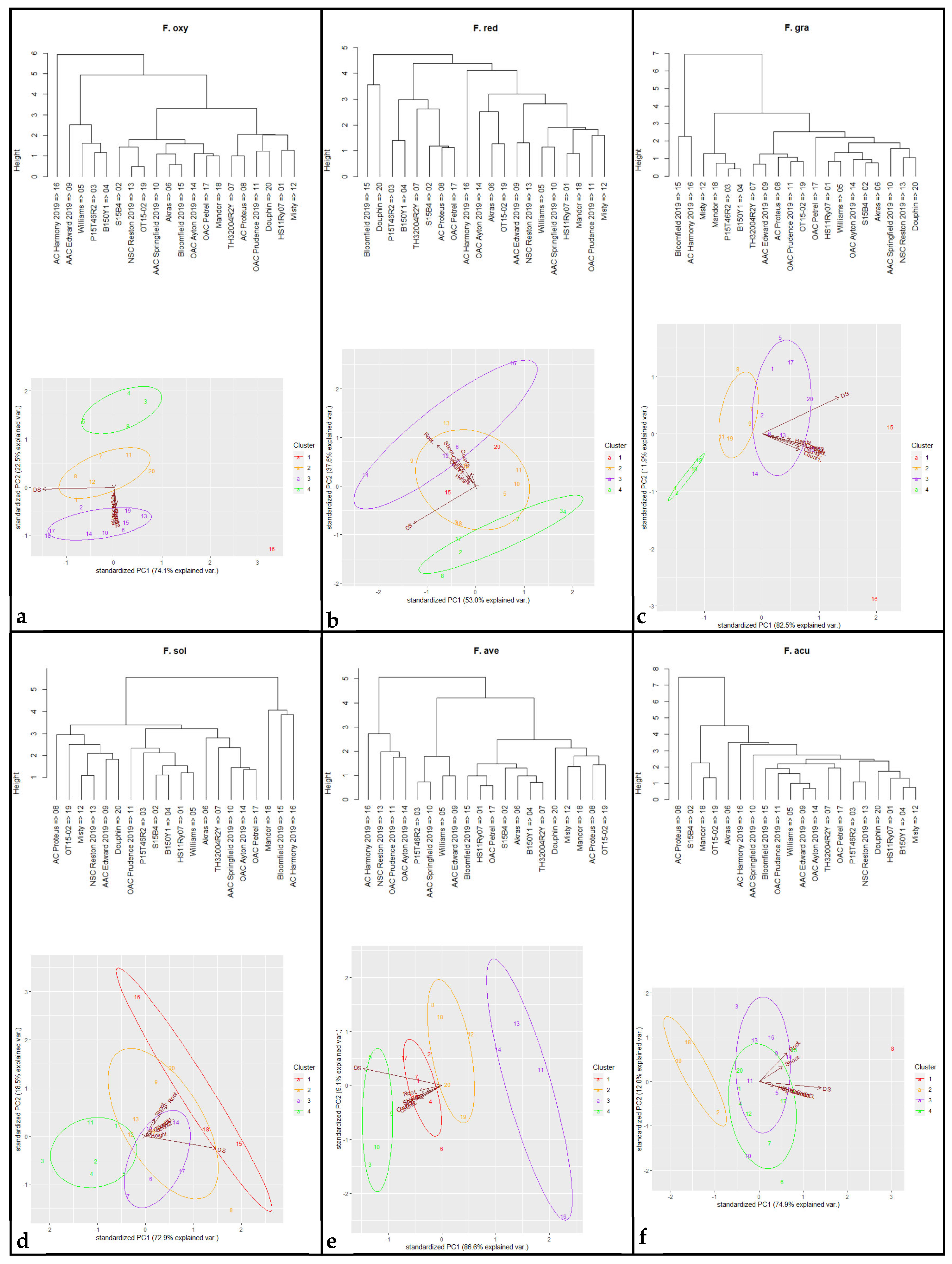
2.7. Phylogenetic Analysis

All 227 sequences amplified by ITS4/5 and T12, as well as 89 sequences generated by EF1/2 from the isolates obtained from the soybean root samples collected in 2022 and the six reference isolates, were edited using Bio-Edit software v. 7.2.5 [79], with manual adjustment. The six reference sequences that best matched F. oxysporum, F. redolens, F. graminearum, F. solani, F. avenaceum, and F. acuminatum were downloaded from GenBank. All the sequences identified as Fusarium spp. and the six reference sequences were included in a phylogenetic analysis by the maximum likelihood (ML) method using the Jukes Cantor model with default parameters in the CLC main Workbench v.23.0.3 (QIAGEN, Aarhus, Denmark). Bootstrap values (BV) (%) were calculated with 1000 replicates, and a phylogenetic tree was constructed with BV > 70. The phylogenetic tree was finalized by iTOL version 6.8.1 (https://itol.embl.de/; accessed on 12 January 2024).

2.8. Data Analysis

Analysis of variance (ANOVA) was conducted for all traits analyzed in the pathogenicity test and cultivar resistance evaluation trials in the greenhouse using R v. 4.2.0 [80]. As the experiments were arranged in a RCBD with five replicates, the experimental unit was defined as each plastic cup. The seven seedlings in each cup (whether germinated or non-germinated) were considered as pseudo-replicates. Thus, the germinated seedlings in each cup on the 7th, 14th, and 21st day after seeding were counted as “count1”, “count2”, “count3”. The plant height and RRS of each experimental unit were estimated as “Height” and “RRS”, respectively, based on the mean of the seven pseudo-replicates in each cup, of which non-germinated/non-emerged seeds or seedlings were regarded as dead (height = 0 cm; RRS = 4). The RRS of non-germinated/non-emerged seeds or seedlings for the non-inoculated controls was rated as 0, since the germination failure was not caused by pathogen infection. The total dry shoot and root weights (g) in each cup were denoted as “shoot” and “root”, respectively. The estimated mean of all traits for the two greenhouse experiments was calculated based on the five replicated experimental units. A least significant difference (LSD) at p < 0.05 was applied to compare the estimated means of all traits among different treatments using the R package “agricolae” version 1.3-5. Correlation coefficients among all the traits collected in the pathogenicity test were calculated with the R package “PerformanceAnalytics” version 2.0.4. The reduction percentage was calculated for cluster analysis using R v. 4.2.0 [80]. To demonstrate the levels of high tolerance, moderate tolerance, moderate susceptibility, and high susceptibility, the cluster group number was set as four. A principal components analysis (PCA) was carried out, estimating the reduction percentage of the three germination counts, height, shoot weight, and root weight, as well as the increase in RRS (DS) in R using the package “ggbiplot” version 0.55. For the Fusarium spp. isolation, the incidence of each species was calculated using the formula: Incidence % = (n/N) × 100, where n is the number of fields where the species was detected in each province, and N is the total field number in each province.

3. Results

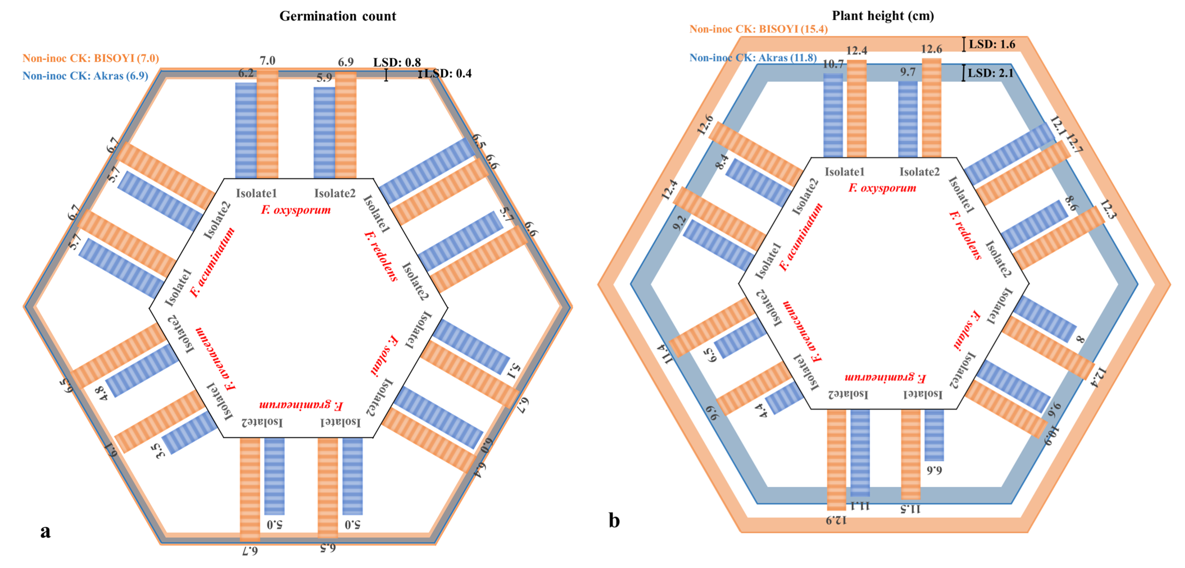
3.1. Pathogenicity Test

Symptoms caused by the six Fusarium spp. varied and included rotting, girdling, and the development of brown sunken lesions (Figure 1). An analysis of variance indicated significant interactions between Fusarium spp. and the soybean cultivars for all the traits, while the repeat effect of two greenhouse experiments was not significant (Table 1). Consequently, data from the two repeated experiments were combined for all traits. Mean and LSD estimates for the six Fusarium spp. and non-inoculated control were calculated using ‘Akras’ and ‘B150Y1’ for all traits. Correlation analysis indicated highly significant correlation coefficients among all the tested traits (Supplementary Figure S1). RRS, as a direct reflection of pathogen infection, was negatively correlated with the other traits, including the three emergence counts and their average, plant height, and dry shoot and root weights. The reduction in these growth parameters was considered together with RRS when evaluating the aggressiveness of the Fusarium species in this study.
Reductions in the emergence of the cultivar ‘B150Y1′ were not significant for any of the six Fusarium species or isolates tested except for F. avenaceum isolate 1. In contrast, significant reductions in emergence were observed for ‘Akras’ following inoculation with each of the isolates (Figure 2a). In general, ‘B150Y1′ had a higher emergence rate than ‘Akras’ under both inoculated and non-inoculated conditions. Differences between the two isolates of each species were significant only for F. redolens, F. solani, and F. avenaceum on the cultivar ‘Akras’. The greatest reduction in emergence (50%) was observed with F. avenaceum isolate 1.
A significant reduction in plant height was observed for ‘B150Y1′ inoculated with each of the 12 isolates, as well as for ‘Akras’ inoculated with F. redolens isolate 2, F. graminearum isolate 1, and both isolates of each of F. solani, F. avenaceum, and F. acuminatum (Figure 2b). Variance within each Fusarium species reached a significant level for F. redolens, F. graminearum, and F. avenaceum on ‘Akras’, while isolate effects within species were not significant for ‘B150Y1′. In addition, ‘B150Y1′ had relatively higher plant height than ‘Akras’. The lowest plant height was obtained for ‘Akras’ inoculated with F. avenaceum isolate 1, followed by the same host inoculated with F. avenaceum isolate 2 and F. graminearum isolate 1.
All isolates of the six Fusarium spp. caused significant increases in RRS relative to the non-inoculated controls for both cultivars (Figure 2c). In the case of F. oxysporum, RRS caused by isolate 2 was significantly greater than RRS caused by isolate 1. Similarly, F. redolens isolate 2 caused significantly more severe disease on ‘Akras’ than isolate 1, while disease severities on ‘B150Y1′ were similar for both isolates. On ‘Akras’, F. solani isolate 1 caused the highest RRS, but no significant differences were observed between the two host cultivars. Fusarium graminearum caused greater RRS on ‘Akras’ than on ‘B150Y1′, with both isolates of this species inducing similar levels of disease. Isolate 1 of F. avenaceum caused higher RRS than isolate 2 on both cultivars, while F. acuminatum isolate 2 was more virulent than isolate 1 on these hosts.
Reductions in shoot dry weight were significant following inoculation with each of the isolates and ranged from 24.0% for ‘B150Y1′ in response to inoculation with F. graminearum isolate 2 to 70.8% for ‘Akras’ in response to F. avenaceum isolate 1 (Figure 2d). Similarly, shoot weight reductions were significant for all the isolates and both soybean cultivars. In general, the root dry weight of ‘B150Y1′ was greater than for ‘Akras’. Reductions in root dry weight following the inoculation of both cultivars were mostly significant (Figure 2e), with the only exceptions found for F. avenaceum isolate 1 on ‘B150Y1′, F. solani isolate 2 on ‘Akras’, and F. acuminatum isolate 1 and isolate 2 on ‘Akras’. Distinct differences between the root weight reductions caused by the two isolates of each species were detected only in F. redolens, F. solani, and F. avenaceum.
In the pathogenicity study, the aggressiveness of each isolate was consistent across all measured traits including RRS and reductions in emergence, plant height, and dry shoot and root weights. The most aggressive isolate of each species, identified as F. oxysporum isolate 2, F. redolens isolate 2, F. solani isolate 1, F. graminearum isolate 2, F. acuminatum isolate 2, and F. avenaceum isolate 1 based on these parameters, was selected to evaluate cultivar resistance.

3.2. Cultivar Resistance Evaluation

Both greenhouse experiments yielded corresponding results for all measured variables, with a non-significant repeat effect (p > 0.05) (Table 2). Cultivar effect, Fusarium spp. effect, and their interaction were all found to be significant for all the traits (p < 0.05) (Table 2). As such, the reaction of each of the 20 soybean cultivars was evaluated in response to each of the six Fusarium spp. Notably, significant differences among the soybean cultivars were observed for all traits in the non-inoculated control, with ranges of 3.9–7.0 for the three germination counts (Count1, Count2, Count3), 5.4 cm–17.1 cm for plant height, 0.9 g–1.5 g for dry shoot weight, and 0.2g–0.4 g for dry root weight. As expected, however, the non-inoculated controls of all 20 cultivars remained completely healthy, with a score of 0 for RRS (Supplementary Table S1). Consequently, the disease reactions for the six Fusarium spp. were evaluated using percentage reduction for Count1, Count2, Count3, Height, Shoot, and Root, as well as RRS increase.
Fusarium avenaceum caused the greatest average reduction in emergence (94.1%), while the maximum average height reduction was observed for F. oxysporum (99.5%). For shoot and root loss, F. oxysporum had the most pronounced effects, with reductions of 97.6% and 98.7%, respectively. Regarding RRS, inoculation with F. oxysporum and F. avenaceum caused the greatest severity scores for RRS (3.9). In contrast, F. solani had the lowest impact on all the traits compared with the other five Fusarium spp.
Principal component analysis with four clusters revealed a positive correlation among variables and cultivar reactions following inoculation with F. graminearum, F. avenaceum, F. acuminatum, F. oxysporum, F. redolens, and F. solani (Figure 3). Notably, four cultivars, ‘B150Y1′, ‘P15T46R2′, ‘Misty’, and ‘Mandor’, clustered in the high-tolerance group against F. graminearum. Against F. avenaceum, the most tolerant cultivars were ‘OAC Prudence’, ‘NSC Reston’, ‘OAC Ayton’, and ‘AC Harmony’. The cultivars ‘S15B4′, ‘OT15-2′, and ‘Mandor’ displayed the greatest tolerance to F. acuminatum, and ‘P15T46R2′, ‘B150Y1′, ‘Williams’, and ‘AAC Edward’ were most tolerant to F. oxysporum. Additionally, the cultivars most tolerant to F. redolens were ‘S15B4′, ‘P15T46R2′, ‘B150Y1′, ‘TH32004R2Y’, ‘AC Proteus’, and ‘OAC Petrel’. The cultivars, ‘HS11Ry07′, ‘S15B4′, ‘P15T46R2′, ‘B150Y1′, ‘Williams’, and ‘OAC Prudence’ showed the greatest tolerance to F. solani. Overall, the soybean cultivars ‘P15T46R2′ and ‘B150Y1′ displayed suppression against F. graminearum, F. oxysporum, F. redolens, and F. solani.

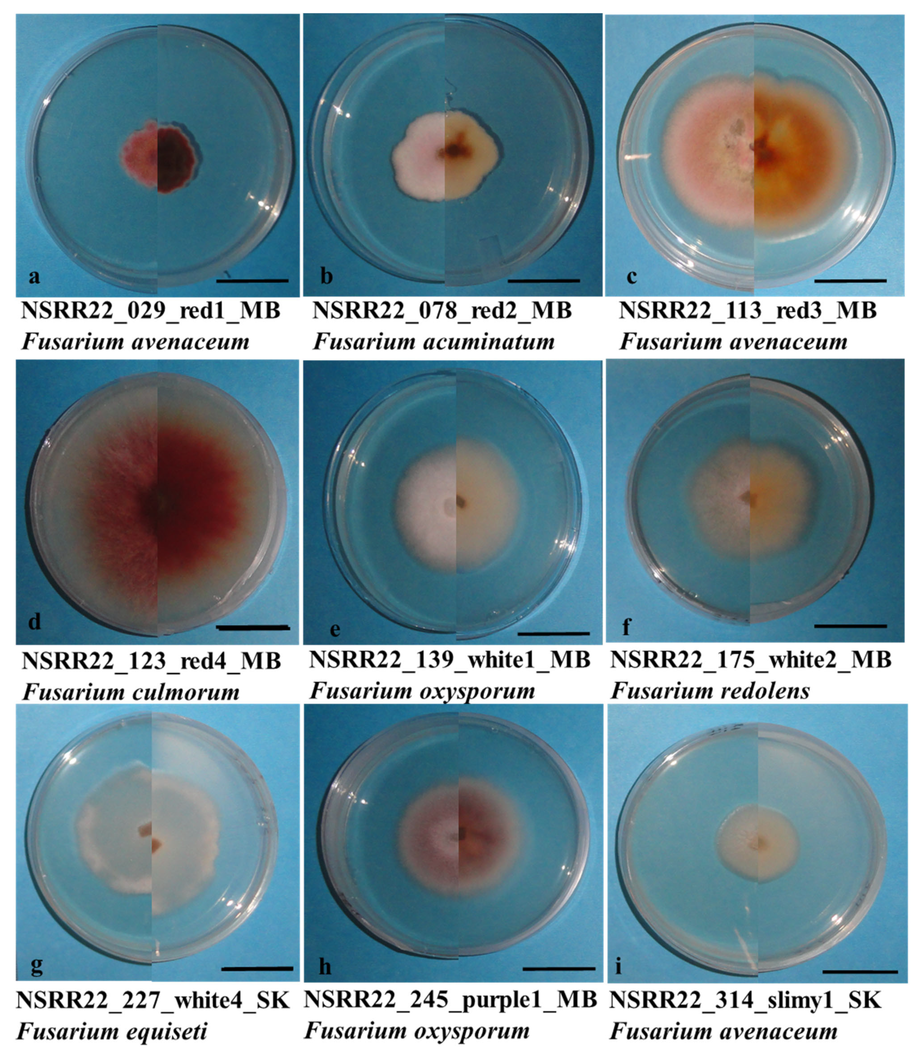
3.3. Fusarium spp. Identification

A total of 983 purified isolates were obtained from symptomatic root samples and separated into four morphological groups (“Red”, “White”, “Purple”, and “Other”) and nine subgroups (“Red1”, “Red2”, “Red3”, “Red4”, “White1”, “White2”, “White4”, “Purple1”, and “Slimy1”) (Figure 4). After filtering and grouping based on colony morphology, 336 isolates were selected for molecular identification. The primer sets ITS4/5 (ITS region), T12 (beta-tubulin gene), and EF1/2 (TEF1α gene) produced single bands of ~500 bp [47], ~580 bp [46], and ~690 bp [78], respectively, from the six reference isolates (Figure 5) and isolates collected in this study. Following the removal of non-Fusarium species and isolates with poor sequence quality, 221 isolates were confirmed as Fusarium spp., primarily based on the ITS4/5-amplified gene sequences complemented with the T12-amplified sequences.
Within the 221 Fusarium spp. isolates detected by ITS4/5 and T12, 33 (14.9%), 47 (21.2%), and 141 (63.8%) isolates had coincident identification, species conflict, and single primer identification, respectively. The inconsistency of sequence identification between the ITS region and the beta-tubulin gene was mainly observed in the species within the FTSC as well as those belonging to FEISC. Thus, 83 isolates and 6 reference isolates were further selected for confirmation by EF1/2 as a subgroup, consisting of 6 coincident isolates, 29 isolates with conflict, 48 isolates with a single result, as well as 6 reference Fusairum spp. (Supplementary Table S2). Based on the sequence blast of the TEF1α gene, 19 of 80 isolates exhibited coincidence with results based on the ITS region. For the comparison with 35 sequences of the beta-tubulin gene, 21 sequences were identified as the same Fusarium species by the TEF1α gene. In the identification conflicts between F. avenaceum and F. acuminatum with the primer sets ITS4/5 and T12, the species identifications obtained with EF1/2 were consistent with 17 of 20 sequences amplified by T12. Among conflicting isolates belonging to the FIESC, 13 of 14 were identified by the TEF1α gene as F. avenaceum while only one was F. equiseti. None of these matched the species identifications based on the ITS region or the beta-tubulin gene.
Overall, the main species identified with the EF1/2 primer set were F. avenaceum (39 isolates), F. acuminatum (25 isolates), F. oxysporum (10 isolates), and F. redolens (3 isolates). On the other hand, only one isolate each of F. culmorum, F. equiseti, F. flocciferum, F. sporotrichioides, F. solani, and F. falciforme were confirmed with these primers. Isolates identified as F. avenaceum in the current study had the greatest diversity in morphology, and were observed to occur in all the colony morphology subgroups. Fusarium avenaceum and F. acuminatum both occurred in the morphology subgroups “Red1” and “Red2”. Morphological variation was also detected in F. oxysporum. In contrast, F. redolens was only detected in the “White2” group. Isolates of F. culmorum, F. flocciferum, and F. sporotrichioides, all belonging to the FSSC, were found in the “Red4” morphology subgroup. All the major species were detected in both western and eastern Canada apart from F. redolens, which was identified only in Manitoba and Saskatchewan. While the current study identified other Fusarium spp. in the FSSC, the reference species F. graminearum was not identified in this soybean survey.

3.4. Phylogenetic Analysis

The ML phylogenetic analysis based on the TEF1α-amplified sequences clearly segregated all the identified Fusarium spp. into seven clades with bootstrap values ranging from 89 to 100%, except for NSRR22_062_red1_SK_F. flocciferum, which was grouped with isolates identified as F. avenaceum (Figure 6). The phylogenetic trees were rooted using sequences of all F. avenaceum and the single F. flocciferum, with the reference sequence of F. avenaceum in Clade1. Clade2 included all the isolates identified as F. acuminatum and its reference sequence, with a bootstrap value of 89%. Clade3 included one F. solani, one F. falciforme, and the F. solani reference sequence in the FSSC, with a bootstrap value of 99% [81]. NSRR22_230_white4_SK_F. equiseti was the only isolate in the FIESC and was classified as Clade4, distinguished from Clade5 with a 99% supporting value. The latter (Clade5) consisted of Fusarium spp. in the FSAMSC, including F. culmorum, F. sporotrichiodes, and the reference sequence of F. graminearum with a bootstrap value of 100. The three F. redolens isolates, along with the reference sequence, were strongly linked together as Clade6 (bootstrap value = 100%). Similarly, all 10 isolates of F. oxysporum and its reference sequence were clustered as Clade7 with a bootstrap value of 92%.
The phylogenetic tree generated by ITS4/5-amplified sequences generally included seven groups (Supplementary Figure S2). However, species mixture was observed in groups 1, 3, and 6, corresponding to FTSC, FIESC, and FOSC. Meanwhile, the bootstrap values of nodes for grouping were relatively low. Considering the high failure rate of sequence BLAST and inconsistencies in species identification by ITS4/5, T12, and EF1/2 of isolates in morphology subgroup “White4”, only sequences identified as a species of FTSC by amplification of the beta-tubulin gene were analyzed for their phylogenetic relationships (Supplementary Figure S3), of which, F. avenaceum was significantly distinguished from F. acuminatum.

4. Discussion

Root rot in soybean is a global concern involving numerous soilborne pathogens, with Fusarium spp. found to be predominant in disease surveys conducted in eastern [74] and western Canada [9,14,16]. However, studies investigating the pathogenicity of Fusarium spp. in Canada, especially in the western Prairie region (Alberta, Saskatchewan, and Manitoba), are limited.
While several Fusarium spp., including F. oxysporum and F. solani [28,33], consist of distinct formae speciales or physiological races, these species are also reported to be pathogenic in multiple crops [28,57,82]. The F. graminearum species complex, which is the primary cause of Fusarium head blight in cereals such as wheat, barley, and other small grains [83], exhibits a wide host range that also extends to soybean, rice, and maize [58,84]. The pathogenicity and aggressiveness of the FGSC complex on cereals are well described and associated with the production of the mycotoxins nivalenol (NIV) and deoxynivalenol (DON) [85,86]. While F. graminearum is capable of infecting soybeans, there have not been any specific formae speciales reported for this crop. Assessing fungal aggressiveness based on a single parameter is likely insufficient, particularly for Fusarium species that are pathogenic but not specialized for soybeans. Thus, the evaluation in this study, not only of RRS but also of emergence, plant height, and dry shoot and root weights, provided a more comprehensive description of the aggressiveness of Fusarium spp. on soybean. Likewise, by examining multiple traits and incorporating principal components analysis, a more comprehensive assessment of the tolerance of various soybean cultivars to Fusarium root rot was achieved.
In Canada, Abdelmagid et al. [16] evaluated the pathogenicity of five F. sporotrichioides isolates from Manitoba, which caused up to 70% RRS and significant reductions in root and shoot lengths in soybean. The cross-pathogenicity of five Fusarium spp., including isolates of F. cerealis, F. culmorum, F. graminearum, and F. sporotrichioides collected in Manitoba, was also examined on soybean and wheat; the resulting RRS on soybean ranged from 1.89 to 3.33 on a 0–4 scale [9]. In Alberta, F. proliferatum was reported as the most aggressive species on soybean based on greenhouse trials, while other tested Fusarium spp. caused mild to moderate levels of disease [14]. In this study, the pathogenicity of six Fusarium species was compared on soybean, using isolates previously collected from Manitoba. These species included F. oxysporum, F. graminearum, F. solani, and F. acuminatum, which are common in North America, as well as F. avenaceum, which is also prevalent in Canada, and F. redolens, which was previously reported in the country [9,14]. The severity of root rot caused by these six species varied, with F. oxysporum, F. avenaceum, and F. graminearum causing the most severe root rot on the soybean cultivar ‘Akras’. Despite this variability, the root rot severities caused by the six Fusarium spp. tested in this study were generally greater than in previous reports [14,62,63], suggesting increased aggressiveness in these species. This trend of increasing virulence should be emphasized to farmers when implementing control measures for Fusarium root rot in soybean.
Several studies have detected horizontal resistance in soybean controlled by polygenetic loci against Fusarium spp. Acharya et al. [67] identified one major and one minor QTL on soybean chromosomes 8 and 6 controlling partial resistance to F. graminearum. Quantitative resistance was also detected against SDS on all 20 soybean chromosomes [66,68]. In this study, 20 commercial soybean cultivars were screened for resistance to virulent isolates of F. oxysporum, F. graminearum, F. solani, F. acuminatum, F. avenaceum, and F. redolens selected from the earlier pathogenicity test. The host reactions indicated varying degrees of tolerance. Fusarium oxysporum and F. avenaceum were identified as the most virulent species, while F. solani caused the lowest RRS, consistent with the findings of the pathogenicity testing. Complete resistance (RRS ≤ 1) was not observed in any of the 20 soybean cultivars evaluated. Moderate resistance was only observed against F. redolens and F. solani (1 ≤ RRS ≤ 2) (Supplementary Table S1). In this context, the level of tolerance was taken into consideration, evaluating all the traits investigated in this study. The six Fusarium spp. tested also had varying effects on germination counts, plant height, dry shoot weight, and dry root weight. In another study, 57 commercial soybean cultivars were evaluated against F. oxysporum, F. graminearum, F. avenaceum, and F. tricinctum, with resistance against all four species identified in the soybean cultivar ‘Maple Amber’, based on RRS rather than emergence, plant height, or dry root weight [74]. However, in that study, RRS ranged from 0.5 to 2.7 on a 0–4 scale, and reductions in emergence, plant height, and dry root weight were generally <50%.
Nyandoro et al. [62] evaluated the resistance of 12 soybean cultivars against F. avenaceum in greenhouse trials and found high RRS among cultivars (ranging from 2.6 to 3.4) and considerable reductions in emergence (26.7 to 75.5%). Given the variable performance of soybean cultivars across different trials, PCA was conducted in the current study to evaluate tolerance to Fusarium spp., taking into account RRS, germination counts, plant height, dry shoot weight, and dry root weight. Positive correlations were observed among RRS, reduction of emergence, decline in plant height, and weight loss of the shoot and root, illustrating the adverse effects of the six Fusarium spp. on the entire soybean plant. As the cultivars were clustered into four groups based on multiple parameters, the direct identification of high tolerance to infection was determined by selecting cultivars located in the circle farthest from the parameter arrowhead. Some tolerant cultivars were identified against each Fusarium spp., with broad-spectrum resistance detected in ‘P15T46R2′ and ‘B150Y1′. The cultivars were tolerant/partially resistant to F. graminearum, F. oxysporum, F. redolens, and F. solani. Similarly, in recent studies, broad-spectrum resistance to multiple races of Phytophthora sojae was identified in soybean genotypes carrying the RpsX, Rps11, Rps12, and Rps13 resistance genes, suggesting a promising approach for control of this pathogen [87,88,89]. Broad-spectrum resistance was also evaluated for the management of soybean mosaic disease, providing information helpful for reducing viral infections and mitigating their impact on soybean [90,91]. Nevertheless, broad-spectrum resistance to Fusarium species and/or races in soybean still appears limited. Further study of ‘P15T46R2′ and ‘B150Y1′ may contribute to improved knowledge of the genetic control of host reactions to infection.
Numerous studies have highlighted the variability of Fusarium spp. associated with the soybean root rot complex across different environments, locations, and years [11,92,93,94]. In the current study, 10 different Fusarium spp. were identified from a large collection of symptomatic roots using a combination of morphological and molecular methods (Supplementary Table S2). All six species included in the pathogenicity test and cultivar evaluation trials were also recovered in the fungal isolation study, with F. avenaceum, F. oxysporum, F. acuminatum, and F. redolens as the major groups. The current study also isolated one culture of F. equiseti, commonly reported in northern California and South Dakota in the USA [95,96], as well as in Ontario, Alberta, and Manitoba [9,14,27]. However, species such as F. poae, F. graminearum, F. solani, F. sporotrichioides, F. tricinctum, F. torulosum, F. commune, and F. proliferatum, which were previously reported as predominant in Canada [9,14,27], were only sparsely identified or absent in this study. Notably, isolates of F. redolens displayed location-specific characteristics, consistent with previous reports [9,14]. The absence of F. graminearum in the current soybean survey suggests that it may not play a significant role as a major pathogen causing root rot disease in soybean, although it showed significant levels of pathogenicity in the pathogenicity test. Factors such as different methodologies, variations in field locations, soybean cultivars, or environmental conditions may have contributed to discrepancies.
Colony morphology played a crucial role in the primary grouping of isolates from the same field, allowing for the selection of 336 isolates from the initial 983 for molecular identification. All isolates identified as F. redolens with the primer sets ITS4/5 or EF1/2 were found in the morphology subgroup “White2”. Likewise, all isolates identified as F. acuminatum with EF1/2 and T12 were found in morphology subgroups “Red1” and “Red2”, characterized by small colonies containing short and dense mycelium with a light to dark reddish color. Fusarium oxysporum had three morphology types, including “White1”, “White4”, and “Purple1”, while the only F. equiseti isolate occurred in “White4”. The similarity in morphology between F. oxysporum and F. equiseti has also been reported in tomato in northeast India [97]. Within the “Red” group, however, the four subgroups (“Red1”, “Red2”, “Red3”, and “Red4”) failed to distinguish whether the isolates belonged to FTSC or FSAMSC. Additionally, Fusarium avenaceum showed the largest variability in morphology, and occurred with other Fusarium spp. in all nine morphology subgroups. Consequently, no morphologically unique species were found in the current study. Non-negligible differences in species identification by morphology and molecular phylogenetics have been reported in other recent studies, illustrating the need to integrate both morphological characteristics and molecular technologies in the identification of Fusarium spp. [98,99].
The sequencing of the DNA fragments amplified by PCR based on the ITS region and beta-tubulin gene identified 225 and 79 isolates belonging to Fusarium spp., respectively. Inconsistencies were frequently observed among species identified within the same Fusarium species complex, leading to the need for further confirmation by the sequencing of the TEF1α gene. For example, while five isolates were classified as either F. incarnatum or F. equiseti with the primer set ITS4/5, they were recognized as F. flagelliforme (also a member of FIESC) with the T12 primer set (beta-tubulin gene). Notably, F. flagelliforme was not detected using ITS4/5 in this study. Based on an analysis of the TEF1α gene, however, only one of these isolates was confirmed as F. equiseti, while the rest were identified as F. avenaceum. Chang et al. [26] demonstrated that variable identification of isolates across species complexes or among species within the Fusarium genus is common in studies with multiple primers, including those targeting the beta-tubulin and ITS regions. For instance, ITS sequences have been reported as unsuitable for distinguishing F. equiseti or F. incarnatum [45]. Similarly, the beta-tubulin sequence has been applied to identify species in the FIESC and F. chlamydosporum species complexes (FCSC) [98], but failed to distinguish F. armeniacum, F. acuminatum, F. sporotrichioides, and F. langsethiae [100]. While the TEF1α gene has been widely used to define species and reveal phylogenetic relationships within the genus Fusarium in the past, multiple primers have been more frequently employed in recent studies [9,19,51,101,102].
Phylogenetic analysis using the TEF1α sequence successfully distinguished the 10 identified Fusarium spp., except for F. flocciferum, which was grouped with 25 F. avenaceum isolates. Moreover, the phylogenetic tree generated based on the TEF1α gene sequences clearly showed the genetic distances within and among the species complexes, including the FTSC, FSSC, FIESC, FSAMSC, and FOSC. The classification among species or species complexes was strongly supported, with bootstrap values ranging from 89% to 100%. On the other hand, phylogenetic analysis based on the ITS region could generally distinguish complexes, such as FTSC, FOSC, FIESC, FSAMSC, and FSSC. However, only F. incarnatum-equiseti, F. redolens, and F. solani had strong bootstrap (≥70) support. Beta-tubulin sequences clearly distinguished F. avenaceum and F. acuminatum in the FTSC. O’Donnell et al. [102] concluded that the beta-tubulin gene is not universally informative within Fusarium and is suitable only for distinguishing Fusarium spp., forming part of the F. solani and F. incarnatum-equiseti species complexes. This study was the first to use the T12 primer set, specific for the beta-tubulin gene, to separate Fusarium spp. in FTSC. Hafez et al. [9] also generated a phylogenetic tree that clearly distinguished Fusarium spp. of the FTSC, FSAMSC, FOSC, FIESC, F. redolens, and F. solani collected from Carman and Melita, Manitoba. In a study investigating Fusarium isolates from central and southern Alberta, overlapping species were found in phylogenetic trees based on both the ITS and TEF1α sequences [14]. Fusarium oxysporum was reported to have extensive genetic variation [10] and morphological variability [103]. A whole genome sequence study demonstrated the large diversity in FOSC in Australia among the identified clades [94]. Overall, the TEF1α genes clearly distinguished Fusarium species and species complexes in the phylogenetic tree, demonstrating the accuracy of the BLAST sequence alignment. Phylogenetic analysis based solely on ITS4/5 was insufficient to classify the different Fusarium species complexes or a single species, while beta-tubulin clearly distinguished Fusarium spp. in the FTSC.

5. Conclusions

The evaluation of the pathogenicity of six Fusarium spp. on soybean indicated that F. avenaceum and F. oxysporum were the most strongly virulent, while F. graminearum, F. acuminatum, and F. redolens also caused significant levels of disease. In contrast, F. solani was weakly virulent, causing mild symptoms of Fusarium root rot in soybean. An assessment of the reaction of a suite of 20 soybean cultivars to inoculation with each of the Fusarium spp., based on RRS, emergence, plant height, and dry shoot and root weight, indicated that while no hosts were completely resistant, some cultivars showed partial resistance or tolerance to the disease. The soybean cultivars ‘P15T46R2′ and ‘B150Y1′ were consistently tolerant to F. graminearum, F. oxysporum, F. redolens, and F. solani, making them promising candidates for farmers seeking to minimize the risk of Fusarium root rot. Furthermore, this study provided valuable insights into the distribution and composition of Fusarium spp. in the major soybean production areas of Canada, updating and complementing existing information on the Fusarium root rot complex in soybean cultivation. These findings may help to guide the development of effective measures to mitigate the risk of Fusarium root rot of soybean.

Supplementary Materials

The following supporting information can be downloaded at: https://www.mdpi.com/article/10.3390/jof10050303/s1, Figure S1: Correlation analysis of all traits in the pathogenictiy test under controlled conditions; Figure S2: phylogenetic tree of 221 isolates identified as Fusarium spp. by ITS sequence; Figure S3: phylogenetic tree identified as Fusarium spp. by T12 sequence; Table S1: Estimated mean and grouping letter determined by least significant difference (LSD) method of all traits including three emergence counts (Count1, Count2 and Count3), plant height (Height), Root rot disease severity (DS), dry shoot weight (Shoot) and dry root weight (Root) in response to inoculation with each of six Fusarium spp.; Table S2: Full list of fungal isolates and associated details.

Author Contributions

Conceptualization, S.E.S., Y.-M.K. and S.-F.H.; methodology, R.F.-A. and L.W.; validation, Y.-M.K. and S.E.S.; formal analysis, L.W. and S.-H.O.; writing—original draft preparation, L.W.; writing—review and editing, S.E.S., O.W., R.R.B. and Y.-M.K.; visualization, L.W. and S.-H.O.; supervision, S.E.S. and Y.-M.K.; project administration, S.-F.H., Y.-M.K. and S.E.S.; funding acquisition, S.-F.H., Y.-M.K. and S.E.S. All authors have read and agreed to the published version of the manuscript.

Funding

This research was funded by the Agriculture and Agri-Food Canada AgriScience Program through the Canadian Agricultural Partnership, with industry support from the Canadian Field Crop Research Alliance (CFCRA) whose members include: Atlantic Grains Council; Producteurs de grains du Québec; Grain Farmers of Ontario; Manitoba Crop Alliance; Manitoba Pulse & Soybean Growers; Saskatchewan Pulse Growers; Prairie Oat Growers Association; SeCan; and FP Genetics. Project Number: ASC-09.

Institutional Review Board Statement

Not applicable.

Informed Consent Statement

Not applicable.

Data Availability Statement

The analysis of variance, least significant difference analysis of estimated mean in the pathogenicity test, cultivar evaluation in greenhouse trials, and sequence identification in genetic diversity evaluation are available in the main manuscript or as Supplementary Materials.

Acknowledgments

The authors acknowledge the technical support of Ashley Wragg, Vanessa Tremblay, Waldo Penner, Dennis Stoesz, Shelby Zatylny, and Tom Henderson. The authors thank Manitoba Agriculture, Manitoba Pulse and Soybean Growers, Saskatchewan Ministry of Agriculture, and Saskatchewan Pulse Growers for contributing soybean samples. The in-kind support of the University of Alberta and Agriculture and Agri-Food Canada is also gratefully acknowledged.

Conflicts of Interest

The authors declare no conflict of interest.

References

  1. Michelfelder, A.J. Soy: A Complete Source of Protein. Am. Fam. Physician 2009, 79, 43–47. [Google Scholar]
  2. Voora, V.; Larrea, C.; Bermúdez, S. Global Market Report: Soybeans. 2020. Available online: https://www.jstor.org/stable/resrep26554 (accessed on 4 July 2023).
  3. Shahbandeh, M. Production of Soybeans in Leading Countries Worldwide. 2012–2023. Available online: https://www.statista.com/statistics/263926/soybean-production-in-selected-countries-since-1980/ (accessed on 25 April 2023).
  4. Government of Canada; Statistics Canada. Estimated Areas, Yield, Production, Average Farm Price and Total Farm Value of Principal Field Crops, in Metric and Imperial Units. Available online: https://www150.statcan.gc.ca/t1/tbl1/en/tv.action?pid=3210035901 (accessed on 17 April 2023).
  5. Barthet, V.J.; Puvirajah, A. Quality of Canadian Oilseed-Type Soybeans 2022. 2022. Available online: https://grainscanada.gc.ca/en/grain-research/export-quality/oilseeds/soybean-oil/2022/pdf/quality-report-soybean-oilseed-type-2022.pdf (accessed on 4 July 2023).
  6. Agriculture and Agri-Food Canada. Canada: Outlook for Principal Field Crops, 2022-11-18. Available online: https://publications.gc.ca/collections/collection_2022/aac-aafc/A77-1-2022-11-18-eng.pdf (accessed on 25 April 2023).
  7. Hartman, G.L.; Sinclair, J.B.; Rupe, J.C. Compendium of Soybean Diseases, 4th ed.; APS Press, The American Phytopathological Society: St. Paul, MN, USA, 1999; ISBN 0-89054-238-4. [Google Scholar]
  8. Bradley, C.A.; Allen, T.W.; Sisson, A.J.; Bergstrom, G.C.; Bissonnette, K.M.; Bond, J.; Byamukama, E.; Chilvers, M.I.; Collins, A.A.; Damicone, J.P.; et al. Soybean Yield Loss Estimates Due to Diseases in the United States and Ontario, Canada, from 2015 to 2019. Plant Health Prog. 2021, 22, 483–495. [Google Scholar] [CrossRef]
  9. Hafez, M.; Abdelmagid, A.; Aboukhaddour, R.; Adam, L.R.; Daayf, F. Fusarium Root Rot Complex in Soybean: Molecular Characterization, Trichothecene Formation, and Cross-Pathogenicity. Phytopathology 2021, 111, 2287–2302. [Google Scholar] [CrossRef]
  10. Díaz Arias, M.M.; Munkvold, G.P.; Ellis, M.L.; Leandro, L.F.S. Distribution and Frequency of Fusarium Species Associated with Soybean Roots in Iowa. Plant Dis. 2013, 97, 1557–1562. [Google Scholar] [CrossRef]
  11. Díaz Arias, M.M.; Leandro, L.; Munkvold, G. Frequency of Isolation, Aggressiveness, and Impact on Yield of Fusarium Root Rot Species in Soybean in Iowa. Phytopathology 2012, 102, S4.30. [Google Scholar]
  12. Michielse, C.B.; Rep, M. Pathogen Profile Update: Fusarium oxysporum. Mol. Plant Pathol. 2009, 10, 311–324. [Google Scholar] [CrossRef]
  13. Armstrong, G.M.; Armstrong, J.K. Biological races of the Fusarium causing wilt of Cowpea and Soybeans. Phytopathology 1950, 40, 181–193. [Google Scholar]
  14. Zhou, Q.; Li, N.; Chang, K.-F.; Hwang, S.-F.; Strelkov, S.E.; Conner, R.L.; McLaren, D.L.; Fu, H.; Harding, M.W.; Turnbull, G.D. Genetic Diversity and Aggressiveness of Fusarium Species Isolated from Soybean in Alberta, Canada. Crop Prot. 2018, 105, 49–58. [Google Scholar] [CrossRef]
  15. Chang, K.F.; Hwang, S.F.; Conner, R.L.; Ahmed, H.U.; Zhou, Q.; Turnbull, G.D.; Strelkov, S.E.; McLaren, D.L.; Gossen, B.D. First Report of Fusarium proliferatum Causing Root Rot in Soybean (Glycine max L.) in Canada. Crop Prot. 2015, 67, 52–58. [Google Scholar] [CrossRef]
  16. Abdelmagid, A.; Hafez, M.; Soliman, A.; Adam, L.R.; Daayf, F. First Report of Fusarium sporotrichioides Causing Root Rot of Soybean in Canada and Detection of the Pathogen in Host Tissues by PCR. Can. J. Plant Pathol. 2021, 43, 527–536. [Google Scholar] [CrossRef]
  17. Yang, X.B.; Feng, F. Ranges and Diversity of Soybean Fungal Diseases in North America. Phytopathology 2001, 91, 769–775. [Google Scholar] [CrossRef]
  18. Pioli, R.N.; Mozzoni, L.; Morandi, E.N. First Report of Pathogenic Association Between Fusarium graminearum and Soybean. Plant Dis. 2004, 88, 220. [Google Scholar] [CrossRef]
  19. Zhao, L.; Wei, X.; Zheng, T.; Gou, Y.-N.; Wang, J.; Deng, J.-X.; Li, M. Evaluation of Pathogenic Fusarium Spp. Associated with Soybean Seed (Glycine max L.) in Hubei Province, China. Plant Dis. 2022, 106, 3178–3186. [Google Scholar] [CrossRef]
  20. Abdelmagid, A.; Hafez, M.; Lawley, Y.; Adam, L.R.; Daayf, F. First Report of Fusarium cerealis Causing Root Rot on Soybean. Plant Dis. 2018, 102, 2638. [Google Scholar] [CrossRef]
  21. Hartman, G.L. Compendium of Soybean Diseases and Pests, 5th ed.; APS Press, The American Phytopathological Society: St. Paul, MN, USA, 2015; ISBN 978-0-89054-473-0. [Google Scholar]
  22. Ellis, M.L.; Díaz Arias, M.M.; Leandro, L.F.; Munkvold, G.P. First Report of Fusarium armeniacum Causing Seed Rot and Root Rot on Soybean (Glycine max) in the United States. Plant Dis. 2012, 96, 1693. [Google Scholar] [CrossRef]
  23. Ellis, M.L.; Díaz Arias, M.M.; Cruz Jimenez, D.R.; Munkvold, G.P.; Leandro, L.F. First Report of Fusarium commune Causing Damping-off, Seed Rot, and Seedling Root Rot on Soybean (Glycine max) in the United States. Plant Dis. 2013, 97, 284. [Google Scholar] [CrossRef]
  24. Killebrew, J.F. Greenhouse and Field Evaluation of Fusarium solani Pathogenicity to Soybean Seedlings. Plant Dis. 1988, 72, 1067. [Google Scholar] [CrossRef]
  25. Cruz Jimenez, D.R.; Ellis, M.L.; Munkvold, G.P.; Leandro, L.F.S. Isolate–Cultivar Interactions, In Vitro Growth, and Fungicide Sensitivity of Fusarium oxysporum Isolates Causing Seedling Disease on Soybean. Plant Dis. 2018, 102, 1928–1937. [Google Scholar] [CrossRef]
  26. Chang, X.; Dai, H.; Wang, D.; Zhou, H.; He, W.; Fu, Y.; Ibrahim, F.; Zhou, Y.; Gong, G.; Shang, J.; et al. Identification of Fusarium Species Associated with Soybean Root Rot in Sichuan Province, China. Eur. J. Plant Pathol. 2018, 151, 563–577. [Google Scholar] [CrossRef]
  27. Zhang, J.X.; Xue, A.G.; Cober, E.R.; Morrison, M.J.; Zhang, H.J.; Zhang, S.Z.; Gregorich, E. Prevalence, Pathogenicity and Cultivar Resistance of Fusarium and Rhizoctonia Species Causing Soybean Root Rot. Can. J. Plant Sci. 2013, 93, 221–236. [Google Scholar] [CrossRef]
  28. Coleman, J.J. The Fusarium solani Species Complex: Ubiquitous Pathogens of Agricultural Importance. Mol. Plant Pathol. 2016, 17, 146–158. [Google Scholar] [CrossRef]
  29. Aoki, T.; O’Donnell, K.; Homma, Y.; Lattanzi, A.R. Sudden-Death Syndrome of Soybean Is Caused by Two Morphologically and Phylogenetically Distinct Species within the Fusarium solani Species Complex: F. virguliforme in North America and F. tucumaniae in South America. Mycologia 2003, 95, 660–684. [Google Scholar] [CrossRef]
  30. Šišić, A.; Baćanović-Šišić, J.; Al-Hatmi, A.M.S.; Karlovsky, P.; Ahmed, S.A.; Maier, W.; de Hoog, G.S.; Finckh, M.R. The ‘Forma Specialis’ Issue in Fusarium: A Case Study in Fusarium solani f. sp. pisi. Sci. Rep. 2018, 8, 1252. [Google Scholar] [CrossRef]
  31. O’Donnell, K. Molecular Phylogeny of the Nectria haematococca-Fusarium Solani Species Complex. Mycologia 2000, 92, 919–938. [Google Scholar] [CrossRef]
  32. Matuo, T. Use of Morphology and Mating Populations in the Identification of Formae Speciales in Fusarium solani. Phytopathology 1973, 63, 562. [Google Scholar] [CrossRef]
  33. Ellis, M.L.; Cruz Jimenez, D.R.; Leandro, L.F.; Munkvold, G.P. Genotypic and Phenotypic Characterization of Fungi in the Fusarium oxysporum Species Complex from Soybean Roots. Phytopathology 2014, 104, 1329–1339. [Google Scholar] [CrossRef]
  34. Torres-Cruz, T.J.; Whitaker, B.K.; Proctor, R.H.; Broders, K.; Laraba, I.; Kim, H.-S.; Brown, D.W.; O’Donnell, K.; Estrada-Rodríguez, T.L.; Lee, Y.-H.; et al. FUSARIUM-ID v.3.0: An Updated, Downloadable Resource for Fusarium Species Identification. Plant Dis. 2022, 106, 1610–1616. [Google Scholar] [CrossRef]
  35. Leslie, J.F.; Summerell, B.A. The Fusarium Laboratory Manual; John Wiley & Sons: Hoboken, NJ, USA, 2008; ISBN 978-0-470-27646-4. [Google Scholar]
  36. Toth, B.; Gyorgy, A.; Varga, M.; Mesterhazy, A. The Influence of the Dilution Rate on the Aggressiveness of Inocula and the Expression of Resistance against Fusarium Head Blight in Wheat. Plants 2020, 9, 943. [Google Scholar] [CrossRef]
  37. Takegami, S.; Sasai, K. Studies on Resistance of Wheat Varieties to Scab (Gibberella zeae (Schw.) Petch.): X. The Improved Method of Inoculation by Either Conidiospores or Hyphae of the Scab Cultured on Potato Agar Medium. Jpn. J. Crop Sci. 1970, 39, 1–6. [Google Scholar] [CrossRef]
  38. Grausgruber, H.; Lemmens, M.; Buerstmayr, H.; Ruckenbauer, P. Evaluation of Inoculation Methods for Testing Fusarium Head Blight Resistance of Winter Wheat on Single Plant Basis. Bodenkult. Wien Munch. 1995, 46, 39–49. [Google Scholar]
  39. Stack, R.W. A Comparison of the Inoculum Potential of Ascospores and Conidia of Gibberella zeae. Can. J. Plant Pathol. 1989, 11, 137–142. [Google Scholar] [CrossRef]
  40. Vujanovic, V.; Hamel, C.; Yergeau, E.; St-Arnaud, M. Biodiversity and Biogeography of Fusarium Species from Northeastern North American Asparagus Fields Based on Microbiological and Molecular Approaches. Microb. Ecol. 2006, 51, 242–255. [Google Scholar] [CrossRef]
  41. Feng, J.; Hwang, R.; Chang, K.F.; Hwang, S.F.; Strelkov, S.E.; Gossen, B.D.; Conner, R.L.; Turnbull, G.D. Genetic Variation in Fusarium avenaceum Causing Root Rot on Field Pea. Plant Pathol. 2010, 59, 845–852. [Google Scholar] [CrossRef]
  42. van Diepeningen, A.D.; Brankovics, B.; Iltes, J.; van der Lee, T.A.J.; Waalwijk, C. Diagnosis of Fusarium Infections: Approaches to Identification by the Clinical Mycology Laboratory. Curr. Fungal Infect. Rep. 2015, 9, 135–143. [Google Scholar] [CrossRef]
  43. Porter, T.M.; Brian Golding, G. Are Similarity- or Phylogeny-Based Methods More Appropriate for Classifying Internal Transcribed Spacer (ITS) Metagenomic Amplicons? New Phytol. 2011, 192, 775–782. [Google Scholar] [CrossRef] [PubMed]
  44. White, T.; Bruns, T.; Lee, S.; Taylor, J.; Innis, M.; Gelfand, D.; Sninsky, J. Amplification and Direct Sequencing of Fungal Ribosomal RNA Genes for Phylogenetics. In PCR Protocols: A Guide to Methods and Applications; Academic Press: London, UK, 1990; Volume 31, pp. 315–322. [Google Scholar]
  45. Wang, M.; Chen, Q.; Diao, Y.; Duan, W.; Cai, L. Fusarium incarnatum-equiseti Complex from China. Persoonia 2019, 43, 70–89. [Google Scholar] [CrossRef]
  46. Kalman, B.; Abraham, D.; Graph, S.; Perl-Treves, R.; Meller Harel, Y.; Degani, O. Isolation and Identification of Fusarium spp., the Causal Agents of Onion (Allium cepa) Basal Rot in Northeastern Israel. Biology 2020, 9, 69. [Google Scholar] [CrossRef] [PubMed]
  47. Chen, Y.; Zhou, Q.; Strelkov, S.E.; Hwang, S.-F. Genetic Diversity and Aggressiveness of Fusarium spp. Isolated from Canola in Alberta, Canada. Plant Dis. 2014, 98, 727–738. [Google Scholar] [CrossRef]
  48. Kashyap, P.L.; Rai, S.; Kumar, S.; Srivastava, A.K.; Anandaraj, M.; Sharma, A.K. Mating Type Genes and Genetic Markers to Decipher Intraspecific Variability among Fusarium udum Isolates from Pigeonpea: Diversity Analysis of Fusarium udum Isolates. J. Basic Microbiol. 2015, 55, 846–856. [Google Scholar] [CrossRef]
  49. O’Donnell, K.; Rooney, A.P.; Proctor, R.H.; Brown, D.W.; McCormick, S.P.; Ward, T.J.; Frandsen, R.J.N.; Lysøe, E.; Rehner, S.A.; Aoki, T.; et al. Phylogenetic Analyses of RPB1 and RPB2 Support a Middle Cretaceous Origin for a Clade Comprising All Agriculturally and Medically Important Fusaria. Fungal Genet. Biol. 2013, 52, 20–31. [Google Scholar] [CrossRef]
  50. Dash, A.; Gurdaswani, V.; D’Souza, J.; Ghag, S. Functional Characterization of an Inducible Bidirectional Promoter from Fusarium oxysporum f. sp. cubense. Sci. Rep. 2020, 10, 2323. [Google Scholar] [CrossRef]
  51. Khuna, S.; Kumla, J.; Thitla, T.; Nuangmek, W.; Lumyong, S.; Suwannarach, N. Morphology, Molecular Identification, and Pathogenicity of Two Novel Fusarium Species Associated with Postharvest Fruit Rot of Cucurbits in Northern Thailand. J. Fungi 2022, 8, 1135. [Google Scholar] [CrossRef]
  52. Spanic, V.; Lemmens, M.; Drezner, G. Morphological and Molecular Identification of Fusarium Species Associated with Head Blight on Wheat in East Croatia. Eur. J. Plant Pathol. 2010, 128, 511–516. [Google Scholar] [CrossRef]
  53. Parikh, L.; Kodati, S.; Eskelson, M.J.; Adesemoye, A.O. Identification and Pathogenicity of Fusarium spp. in Row Crops in Nebraska. Crop Prot. 2018, 108, 120–127. [Google Scholar] [CrossRef]
  54. Moparthi, S.; Burrows, M.; Mgbechi-Ezeri, J.; Agindotan, B. Fusarium spp. Associated with Root Rot of Pulse Crops and Their Cross-Pathogenicity to Cereal Crops in Montana. Plant Dis. 2021, 105, 548–557. [Google Scholar] [CrossRef]
  55. Shabeer, S.; Tahira, R.; Jamal, A. Fusarium spp. Mycotoxin Production, Diseases and Their Management: An Overview. Pak. J. Agric. Res. 2021, 34, 278. [Google Scholar] [CrossRef]
  56. Safarieskandari, S.; Chatterton, S.; Hall, L.M. Pathogenicity and Host Range of Fusarium Species Associated with Pea Root Rot in Alberta, Canada. Can. J. Plant Pathol. 2021, 43, 162–171. [Google Scholar] [CrossRef]
  57. Ellis, M.L.; Munkvold, G.P. Trichothecene Genotype of Fusarium graminearum Isolates from Soybean (Glycine max) Seedling and Root Diseases in the United States. Plant Dis. 2014, 98, 1012. [Google Scholar] [CrossRef]
  58. Cruz, D.R.; Leandro, L.F.S.; Mayfield, D.A.; Meng, Y.; Munkvold, G.P. Effects of Soil Conditions on Root Rot of Soybean Caused by Fusarium graminearum. Phytopathology 2020, 110, 1693–1703. [Google Scholar] [CrossRef] [PubMed]
  59. Gaspar, A.P.; Marburger, D.A.; Mourtzinis, S.; Conley, S.P. Soybean Seed Yield Response to Multiple Seed Treatment Components across Diverse Environments. Agron. J. 2014, 106, 1955–1962. [Google Scholar] [CrossRef]
  60. Esker, P.D.; Conley, S.P. Probability of Yield Response and Breaking Even for Soybean Seed Treatments. Crop Sci. 2012, 52, 351–359. [Google Scholar] [CrossRef]
  61. McKenzie-Gopsill, A.; Beaton, A.; Foster, A.J. Investigating the Effects of Seed Treatments on the Economically Optimal Seeding Rate of Conventional Soybean in Atlantic Canada. Can. J. Plant Sci. 2022, 103, 201–213. [Google Scholar] [CrossRef]
  62. Nyandoro, R.; Chang, K.F.; Hwang, S.F.; Ahmed, H.U.; Turnbull, G.D.; Strelkov, S.E. Management of Root Rot of Soybean in Alberta with Fungicide Seed Treatments and Genetic Resistance. Can. J. Plant Sci. 2019, 99, 499–509. [Google Scholar] [CrossRef]
  63. Zhang, J.X.; Xue, A.G.; Tambong, J.T. Evaluation of Seed and Soil Treatments with Novel Bacillus Subtilis Strains for Control of Soybean Root Rot Caused by Fusarium oxysporum and F. graminearum. Plant Dis. 2009, 93, 1317–1323. [Google Scholar] [CrossRef]
  64. Wang, J.; Jacobs, J.L.; Roth, M.G.; Chilvers, M.I. Temporal Dynamics of Fusarium virguliforme Colonization of Soybean Roots. Plant Dis. 2019, 103, 19–27. [Google Scholar] [CrossRef]
  65. Herman, T.K.; Bowen, R.; Mahan, A.L.; Hartman, G.L. Evaluation of Soybean Germplasm for Resistance to Fusarium virguliforme, the Major Pathogen Causing Sudden Death Syndrome of Soybean in the United States. Crop Sci. 2023, 63, 1344–1353. [Google Scholar] [CrossRef]
  66. Kazi, S.; Shultz, J.; Afzal, J.; Johnson, J.; Njiti, V.N.; Lightfoot, D.A. Separate Loci Underlie Resistance to Root Infection and Leaf Scorch during Soybean Sudden Death Syndrome. Theor. Appl. Genet. 2008, 116, 967–977. [Google Scholar] [CrossRef]
  67. Acharya, B.; Lee, S.; Rouf Mian, M.A.; Jun, T.-H.; McHale, L.K.; Michel, A.P.; Dorrance, A.E. Identification and Mapping of Quantitative Trait Loci (QTL) Conferring Resistance to Fusarium graminearum from Soybean PI 567301B. Theor. Appl. Genet. 2015, 128, 827–838. [Google Scholar] [CrossRef]
  68. Swaminathan, S.; Abeysekara, N.S.; Liu, M.; Cianzio, S.R.; Bhattacharyya, M.K. Quantitative Trait Loci Underlying Host Responses of Soybean to Fusarium virguliforme Toxins That Cause Foliar Sudden Death Syndrome. Theor. Appl. Genet. 2016, 129, 495–506. [Google Scholar] [CrossRef]
  69. Almeida-Silva, F.; Venancio, T.M. Integration of Genome-Wide Association Studies and Gene Coexpression Networks Unveils Promising Soybean Resistance Genes against Five Common Fungal Pathogens. Sci. Rep. 2021, 11, 24453. [Google Scholar] [CrossRef] [PubMed]
  70. Weinig, C.; Schmitt, J. Environmental Effects on the Expression of Quantitative Trait Loci and Implications for Phenotypic Evolution. BioScience 2004, 54, 627–635. [Google Scholar] [CrossRef]
  71. Li, S.; Wang, J.; Zhang, L. Inclusive Composite Interval Mapping of QTL by Environment Interactions in Biparental Populations. PLoS ONE 2015, 10, e0132414. [Google Scholar] [CrossRef] [PubMed]
  72. Luckew, A.S.; Cianzio, S.R.; Leandro, L.F. Screening Method for Distinguishing Soybean Resistance to Fusarium virguliforme in Resistant × Resistant Crosses. Crop Sci. 2012, 52, 2215–2223. [Google Scholar] [CrossRef]
  73. Li, W.; Deng, Y.; Ning, Y.; He, Z.; Wang, G.-L. Exploiting Broad-Spectrum Disease Resistance in Crops: From Molecular Dissection to Breeding. Annu. Rev. Plant Biol. 2020, 71, 575–603. [Google Scholar] [CrossRef] [PubMed]
  74. Zhang, J.X.; Xue, A.G.; Zhang, H.J.; Nagasawa, A.E.; Tambong, J.T. Response of Soybean Cultivars to Root Rot Caused by Fusarium Species. Can. J. Plant Sci. 2010, 90, 767–776. [Google Scholar] [CrossRef]
  75. Kim, Y.M.; McLaren, D.; Conner, R.; Henriquez, M.A.; Abdelmagid, A.; Wu, L.; Hwang, S.-F.; Strelkov, S.; Chatterton, S.; Wally, O. The Fusarium Root Rot Complex of Soybean, Dry Bean and Field Pea in Manitoba, Canada. In Proceedings of the 12th International Congress of Plant Pathology, Lyon, France, 20–25 August 2023; pp. 1124–1125. [Google Scholar]
  76. Esmaeili Taheri, A.; Hamel, C.; Gan, Y.; Vujanovic, V. First Report of Fusarium redolens from Saskatchewan and Its Comparative Pathogenicity. Can. J. Plant Pathol. 2011, 33, 559–564. [Google Scholar] [CrossRef]
  77. O’Donnell, K.; Cigelnik, E. Two Divergent Intragenomic rDNA ITS2 Types within a Monophyletic Lineage of the Fungus Fusarium Are Nonorthologous. Mol. Phylogenetics Evol. 1997, 7, 103–116. [Google Scholar] [CrossRef] [PubMed]
  78. O’Donnell, K.; Kistler, H.C.; Cigelnik, E.; Ploetz, R.C. Multiple Evolutionary Origins of the Fungus Causing Panama Disease of Banana: Concordant Evidence from Nuclear and Mitochondrial Gene Genealogies. Proc. Natl. Acad. Sci. USA 1998, 95, 2044–2049. [Google Scholar] [CrossRef] [PubMed]
  79. Hall, T. BioEdit: A User-Friendly Biological Sequence Alignment Editor and Analysis Program for Windows 95/98/NT. Nucleic Acids Symp. Ser. 1999, 41, 95–98. [Google Scholar] [CrossRef]
  80. R Core Team. R: A Language and Environment for Statistical Computing; R Foundation for Statistical Computing: Vienna, Austria, 2022. [Google Scholar]
  81. Hoh, D.Z.; Lee, H.-H.; Wada, N.; Liu, W.-A.; Lu, M.R.; Lai, C.-K.; Ke, H.-M.; Sun, P.-F.; Tang, S.-L.; Chung, W.-H.; et al. Comparative Genomic and Transcriptomic Analyses of Trans-Kingdom Pathogen Fusarium solani Species Complex Reveal Degrees of Compartmentalization. BMC Biol. 2022, 20, 236. [Google Scholar] [CrossRef]
  82. Kolander, T.M.; Bienapfl, J.C.; Kurle, J.E.; Malvick, D.K. Symptomatic and Asymptomatic Host Range of Fusarium virguliforme, the Causal Agent of Soybean Sudden Death Syndrome. Plant Dis. 2012, 96, 1148–1153. [Google Scholar] [CrossRef] [PubMed]
  83. Chittem, K.; Khan, M.F.R.; Goswami, R.S. Efficacy of Precipitated Calcium Carbonate in Managing Fusarium Root Rot of Field Pea. Phytoparasitica 2016, 44, 295–303. [Google Scholar] [CrossRef]
  84. Goswami, R.S.; Kistler, H.C. Heading for Disaster: Fusarium graminearum on Cereal Crops. Mol. Plant Pathol. 2004, 5, 515–525. [Google Scholar] [CrossRef] [PubMed]
  85. Mueller, B.; Groves, C.L.; Smith, D.L. Chemotype and Aggressiveness Evaluation of Fusarium graminearum and Fusarium culmorum Isolates from Wheat Fields in Wisconsin. Plant Dis. 2021, 105, 3686–3693. [Google Scholar] [CrossRef] [PubMed]
  86. Laraba, I.; Ward, T.J.; Cuperlovic-Culf, M.; Azimi, H.; Xi, P.; McCormick, S.P.; Hay, W.T.; Hao, G.; Vaughan, M.M. Insights into the Aggressiveness of the Emerging North American Population 3 (NA3) of Fusarium graminearum. Plant Dis. 2023, 107, 2687–2700. [Google Scholar] [CrossRef] [PubMed]
  87. Wang, W.; Chen, L.; Fengler, K.; Bolar, J.; Llaca, V.; Wang, X.; Clark, C.B.; Fleury, T.J.; Myrvold, J.; Oneal, D.; et al. A Giant NLR Gene Confers Broad-Spectrum Resistance to Phytophthora sojae in Soybean. Nat. Commun. 2021, 12, 6263. [Google Scholar] [CrossRef] [PubMed]
  88. Sahoo, D.K.; Das, A.; Huang, X.; Cianzio, S.; Bhattacharyya, M.K. Tightly Linked Rps12 and Rps13 Genes Provide Broad-Spectrum Phytophthora Resistance in Soybean. Sci. Rep. 2021, 11, 16907. [Google Scholar] [CrossRef] [PubMed]
  89. Zhong, C.; Li, Y.; Sun, S.; Duan, C.; Zhu, Z. Genetic Mapping and Molecular Characterization of a Broad-Spectrum Phytophthora sojae Resistance Gene in Chinese Soybean. Int. J. Mol. Sci. 2019, 20, 1809. [Google Scholar] [CrossRef]
  90. Wang, D.; Chen, S.; Huang, Z.; Lin, J. Identification and Mapping of Genetic Locus Conferring Resistance to Multiple Plant Viruses in Soybean. Theor. Appl. Genet. 2022, 135, 3293–3305. [Google Scholar] [CrossRef]
  91. Yan, T.; Zhou, Z.; Wang, R.; Bao, D.; Li, S.; Li, A.; Yu, R.; Wuriyanghan, H. A Cluster of Atypical Resistance Genes in Soybean Confers Broad-Spectrum Antiviral Activity. Plant Physiol. 2021, 188, 1277–1293. [Google Scholar] [CrossRef]
  92. Pfordt, A.; Ramos Romero, L.; Schiwek, S.; Karlovsky, P.; von Tiedemann, A. Impact of Environmental Conditions and Agronomic Practices on the Prevalence of Fusarium Species Associated with Ear- and Stalk Rot in Maize. Pathogens 2020, 9, 236. [Google Scholar] [CrossRef] [PubMed]
  93. Pramunadipta, S.; Widiastuti, A.; Wibowo, A.; Suga, H.; Priyatmojo, A. Identification and Pathogenicity of Fusarium spp. Associated with the Sheath Rot Disease of Rice (Oryza sativa) in Indonesia. J. Plant Pathol. 2022, 104, 251–267. [Google Scholar] [CrossRef]
  94. Achari, S.R.; Kaur, J.; Dinh, Q.; Mann, R.; Sawbridge, T.; Summerell, B.A.; Edwards, J. Phylogenetic Relationship between Australian Fusarium oxysporum Isolates and Resolving the Species Complex Using the Multispecies Coalescent Model. BMC Genom. 2020, 21, 248. [Google Scholar] [CrossRef]
  95. Escamilla, D.; Rosso, M.L.; Zhang, B. Identification of Fungi Associated with Soybeans and Effective Seed Disinfection Treatments. Food Sci. Nutr. 2019, 7, 3194–3205. [Google Scholar] [CrossRef] [PubMed]
  96. Okello, P.N.; Petrović, K.; Kontz, B.; Mathew, F.M. Eight Species of Fusarium Cause Root Rot of Corn (Zea mays) in South Dakota. Plant Health Prog. 2019, 20, 38–43. [Google Scholar] [CrossRef]
  97. Singha, I.M.; Kakoty, Y.; Unni, B.G.; Das, J.; Kalita, M.C. Identification and Characterization of Fusarium spp. Using ITS and RAPD Causing Fusarium Wilt of Tomato Isolated from Assam, North East India. J. Genet. Eng. Biotechnol. 2016, 14, 99–105. [Google Scholar] [CrossRef]
  98. O’Donnell, K.; Sutton, D.A.; Rinaldi, M.G.; Gueidan, C.; Crous, P.W.; Geiser, D.M. Novel Multilocus Sequence Typing Scheme Reveals High Genetic Diversity of Human Pathogenic Members of the Fusarium incarnatum-F. equiseti and F. chlamydosporum Species Complexes within the United States. J. Clin. Microbiol. 2009, 47, 3851–3861. [Google Scholar] [CrossRef] [PubMed]
  99. James, J.E.; Santhanam, J.; Zakaria, L.; Mamat Rusli, N.; Abu Bakar, M.; Suetrong, S.; Sakayaroj, J.; Abdul Razak, M.F.; Lamping, E.; Cannon, R.D. Morphology, Phenotype, and Molecular Identification of Clinical and Environmental Fusarium solani Species Complex Isolates from Malaysia. J. Fungi 2022, 8, 845. [Google Scholar] [CrossRef] [PubMed]
  100. Nosratabadi, M.; Kachuei, R.; Rezaie, S.; Harchegani, A.B. Beta-Tubulin Gene in the Differentiation of Fusarium Species by PCR-RFLP Analysis. Infez. Med. 2018, 26, 52–60. [Google Scholar]
  101. O’Donnell, K.; Gräfenhan, T.; Laraba, I.; Busman, M.; Proctor, R.H.; Kim, H.-S.; Wiederhold, N.P.; Geiser, D.M.; Seifert, K.A. Fusarium abutilonis and F. guadeloupense, Two Novel Species in the Fusarium buharicum Clade Supported by Multilocus Molecular Phylogenetic Analyses. Mycologia 2022, 114, 682–696. [Google Scholar] [CrossRef]
  102. O’Donnell, K.; Whitaker, B.K.; Laraba, I.; Proctor, R.H.; Brown, D.W.; Broders, K.; Kim, H.-S.; McCormick, S.P.; Busman, M.; Aoki, T.; et al. DNA Sequence-Based Identification of Fusarium: A Work in Progress. Plant Dis. 2022, 106, 1597–1609. [Google Scholar] [CrossRef] [PubMed]
  103. Tian, Y.H.; Hou, Y.Y.; Peng, C.Y.; Wang, Y.Y.; He, B.L.; Gao, K.X. Genetic diversity and phylogenetic analysis of Fusarium oxysporum strains isolated from the Cucurbitaceae hosts revealed by SRAPs. Ying Yong Sheng Tai Xue Bao 2017, 28, 947–956. [Google Scholar] [CrossRef] [PubMed]
Figure 1. Root rot severity following inoculation of soybean with (a) Fusarium oxysporum, (b) Fusarium redolens, (c) Fusarium graminearum, (d) Fusarium solani, (e) Fusarium avenaceum, and (f) Fusarium acuminatum, assessed on a 0–4 scale following the methods outlined by Chang et al. [15] and Zhou et al. [14]. A brown discoloration was observed in soybean plants inoculated with all Fusarium spp. except for F. graminearum and F. avenaceum, which caused a dark reddish discoloration. Lesions between the root and stem were visible on plants inoculated with F. oxysporum and F. redolens, while F. oxysporum, F. redolens, and F. acuminatum caused a girdling at the base of the stem.
Jof 10 00303 g001
Figure 2. Effect of inoculation with each of the 12 Fusarium isolates on (a) germination count, (b) plant height, (c) root rot severity, and (d) dry shoot weight and (e) dry root weight of two soybean cultivars, ‘Akras’ and ‘B150Y1′. The orange and blue lines represent the estimated mean of the non-inoculated controls for ‘Akras’ and ‘B150Y1′, respectively. The bars indicate the values in response to the different fungal isolates. The least significant differences for ‘Akras’ and ‘B150Y1′ are also presented in the graphs, and indicate a significant effect among the 12 isolates as well as the six Fusarium species.
Jof 10 00303 g002aJof 10 00303 g002b
Figure 3. Cluster and principal component analyses of 20 soybean cultivars evaluated against six Fusarium spp., including (a) Fusarium oxysporum, (b) Fusarium redolens, (c) Fusarium graminearum, (d) Fusarium solani, (e) Fusarium avenaceum, and (f) Fusarium acuminatum. The evaluated traits included three emergence count (Count1, Count2 and Count3), plant height (Height), root rot disease severity (DS), dry shoot weight (Shoot) and dry root weight (Root). The hosts were divided into four groups (denoted by circles; high tolerance, moderate tolerance, moderate susceptibility, and high susceptibility) based on cluster analysis.
Jof 10 00303 g003
Figure 4. Colony growth on potato dextrose agar of the Fusarium isolates (a) NSRR22_029, (b) NSRR22_078, (c) NSRR22_113, (d) NSRR22_123, (e) NSRR22_139, (f) NSRR22_175, (g) NSRR22_230, (h) NSRR22_245, and (i) NSRR22_314, representing the colony morphology subgroups “Red1”, “Red2”, “Red3”, “Red4”, “White1”, “White2”, “White4”, “Purple1”, and “Slimy1”, respectively. The bar in each panel = 1 cm.
Jof 10 00303 g004
Figure 5. Molecular identification of reference isolates of Fusarium oxysporum (F. oxy), Fusarium redolens (F. red), Fusarium graminearum (F. gra), Fusarium solani (F. sol), Fusarium avenaceum (F. ave), and Fusarium acuminatum (F. acu). Genomic DNA from each isolate was amplified with each of three primer sets: (a) ITS4/5 (targeting the ITS region), (b) T12 (targeting the beta-tubulin gene), and (c) EF1/2 (targeting the TEF1α gene), and the products were resolved by electrophoresis on 2% agarose, where a band (500 bp–700 bp) is visible for each isolate. A DNA ladder is included on the left of each panel.
Jof 10 00303 g005
Figure 6. Phylogenetic tree of Fusarium isolates identified based on their TEF1α sequences. The isolates clustered into seven clades, corresponding to six species complexes: the Fusarium tricinctum species complex (FTSC), the Fusarium solani species complex (FSSC), the Fusarium incarnatum-equiseti species complex (FIESC), the Fusarium sambucinum species complex (FSAMSC), the Fusarium redolens species complex (FRSC), and the Fusarium oxysporum species complex (FOSC). Reference sequences of the six Fusarium spp. previously obtained from Manitoba are in bold text. Provinces of origin are color-coded, with Manitoba (MB) in red, Saskatchewan (SK) in green, Ontario (ON) in blue, and Quebec (QC) in purple. Bootstrap values, indicated by circles of varying sizes on the branches, are based on 1000 replicates.
Jof 10 00303 g006
Table 1. ANOVA table for three germination counts (Count1, Count2, and Count3) recorded at 7, 14, and 21 days after seeding, plant height (Height), root rot severity (RRS), dry shoot weight (Shoot), and dry root weight (Root) of two soybean cultivars, ‘Akras’ and ‘B150Y1′, inoculated under greenhouse conditions with each of 12 fungal isolates representing Fusarium oxysporum, Fusarium redolens, Fusarium graminearum, Fusarium solani, Fusarium avenaceum, and Fusarium acuminatum.
Source of VarianceDfMean Square
Count1Count2Count3CountAveHeightRRSShootRoot
F.spp 11210.4 *,24.1 *4.7 *5.5 *53.9 *11 *4.3 *0.2 *
CV 11128.8 *70.1 *57.3 *82.7 *696.9 *10.7 *175.9 *9.5 *
Repeat 110.21.40.30.21.40.00.10.1
F.spp:CV123.8 *2.6 *2.0 *2.6 *11.7 *1.0 *0.9 *0.1 *
F.spp:Repeat121.50.81.00.92.80.20.30.0
CV:Repeat11.70.90.40.94.50.00.50.1
F.spp:CV:Repeat120.80.91.00.83.50.20.20.0
Residuals2081.00.60.60.44.70.10.20.0
1 “F.spp”, “CV”, and “Repeat” refer to the variances from 12 Fusarium isolates and non-inoculated controls, two soybean cultivars, and two repeated greenhouse experiments, respectively. 2 A bold mean squares denoted with an asterisk (*) indicates that the treatment effect was significant (p < 0.001).
Table 2. ANOVA table for the evaluation of 20 soybean cultivars in response to Fusarium oxysporum, Fusarium redolens, Fusarium graminearum, Fusarium solani, Fusarium avenaceum, and Fusarium acuminatum in greenhouse studies. The estimated traits include three germination counts (Count1, Count2, and Count3) taken at 7, 14, and 21 days after seeding, plant height (Height), root rot severity (RRS), dry shoot weight (Shoot), and dry root weight (Root).
Source of VarianceDfMean Square
Count1Count2Count3HeightRRSShoot Root
F.spp 16643.2 *,2657.1 *709 *2946.8 *227.1 *28.9 *2.12 *
CV 11928.0 *26.8 *25.8 *56.8 *2.0 *2.2 *0.18 *
Repeat 110.10.10.00.60.30.00.00
CV:F.spp1143.1 *2.8 *3.1 *15.8 *0.5 *0.2 *0.02 *
CV:Repeat190.60.60.61.30.20.00.00
F.spp:Repeat61.01.71.30.60.50.00.00
CV:F.spp:Repeat1140.80.70.70.90.20.00.00
Residuals8391.61.61.52.40.30.10.01
1 “F.spp”, “CV”, and “Repeat” refer to the variances from six Fusarium species and non-inoculated controls, 20 soybean cultivars, and two repeated greenhouse experiments, respectively. 2 Bold mean squares denoted with an asterisk (*) indicate that the treatment effect was significant (p < 0.001).
Disclaimer/Publisher’s Note: The statements, opinions and data contained in all publications are solely those of the individual author(s) and contributor(s) and not of MDPI and/or the editor(s). MDPI and/or the editor(s) disclaim responsibility for any injury to people or property resulting from any ideas, methods, instructions or products referred to in the content.

