Deep Learning techniques based on Neural Networks represent an important area in the Digital Pathology field. These machine learning methods, such as Convolutional Neural Networks (CNN), are becoming central in VS analysis since they give promising results in difficult tasks like region detection and classification [
2]. CNN allows a relatively simple configuration thanks to its unsupervised feature generation, the availability of larger datasets to train the models, and the possibility to import pre-trained models to partially set-up the neural networks. The major drawback of these techniques is the huge amount of computational power necessary to correctly train the networks in a reasonable time. Madabushi et al. [
3] show machine learning methods applied to different Digital Pathology challenges.
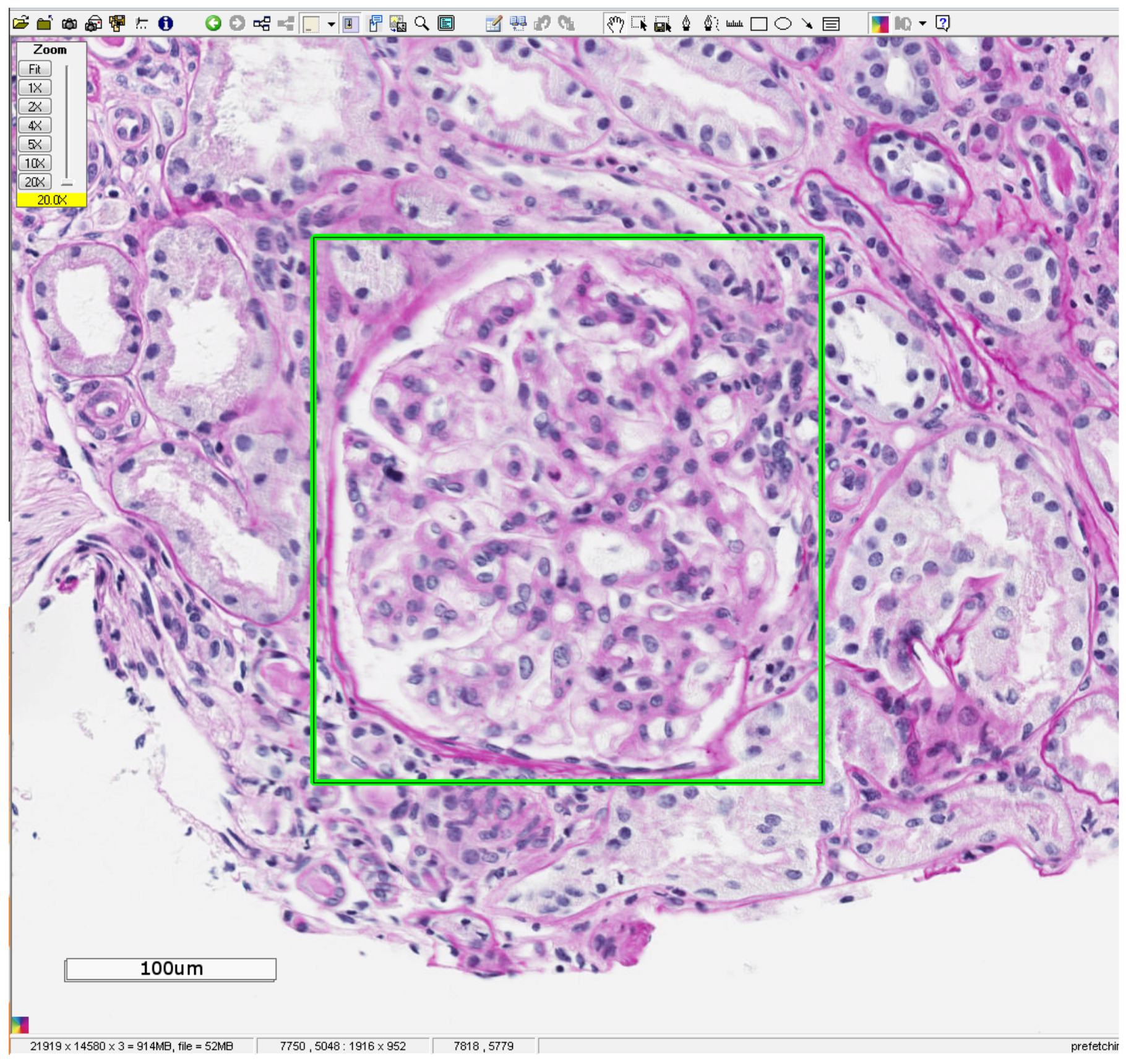
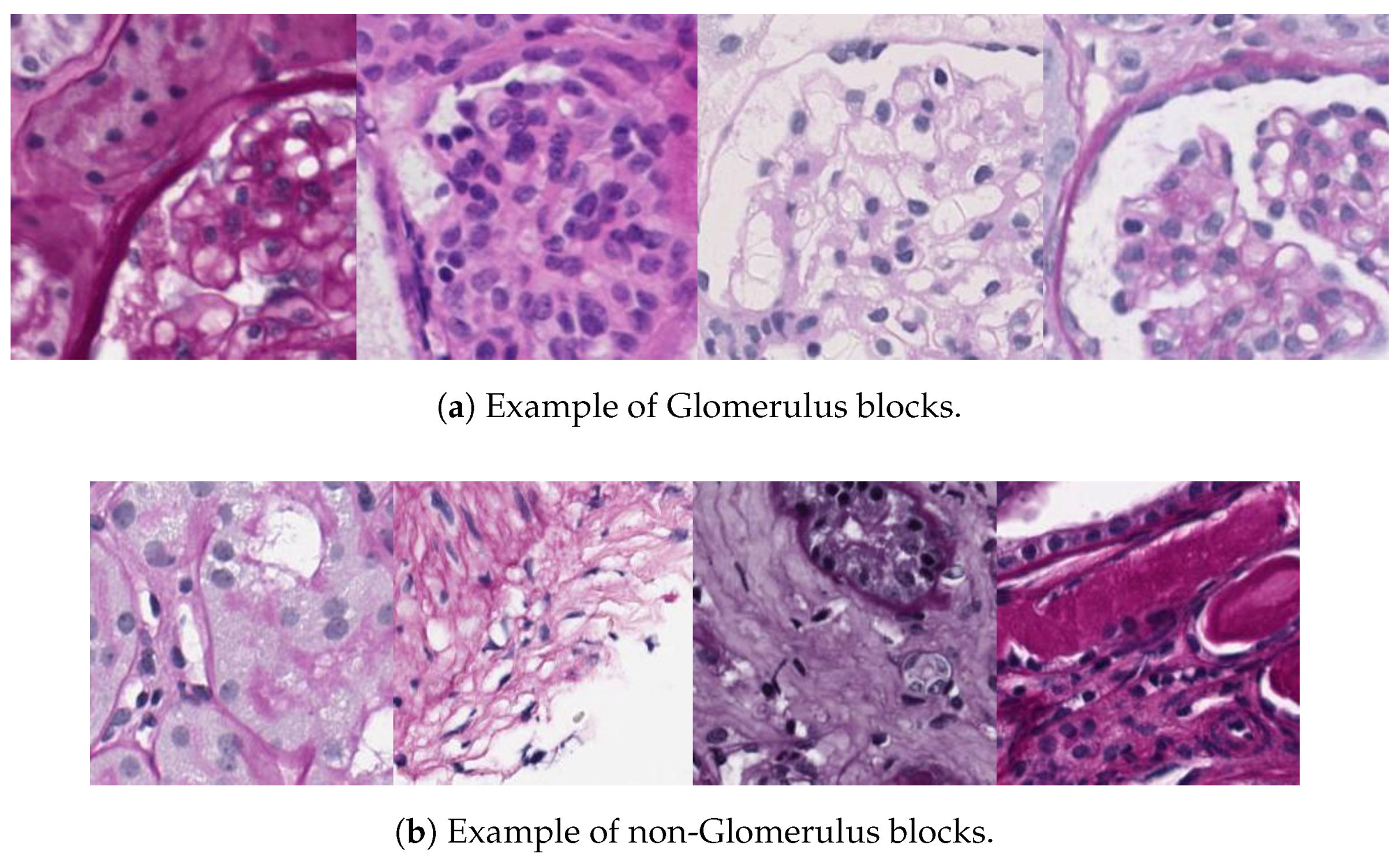
In this paper, we will focus on Glomerulus classification based on deep learning in the context of digital pathology applied to kidney tissue slides. Glomerulus classification is central in nephropathology field to get a correct medical diagnosis. These networks of capillaries serve as the first stage in the filtering process of the blood that retains higher molecular weight proteins in blood circulation in the formation of urine. Therefore, any pathological change associated to them inside a tissue section, like the number of Glomeruli or their sizes, is essential to detect diseases from the patients.
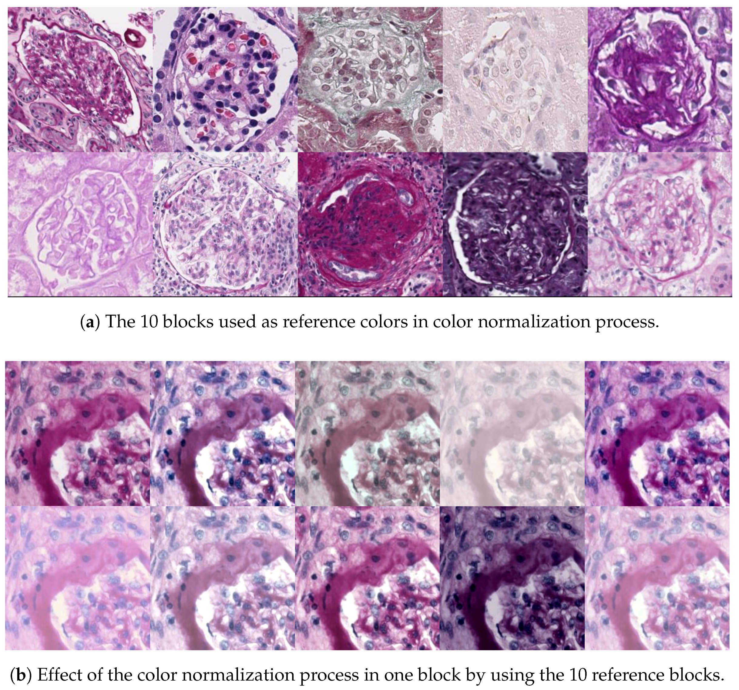
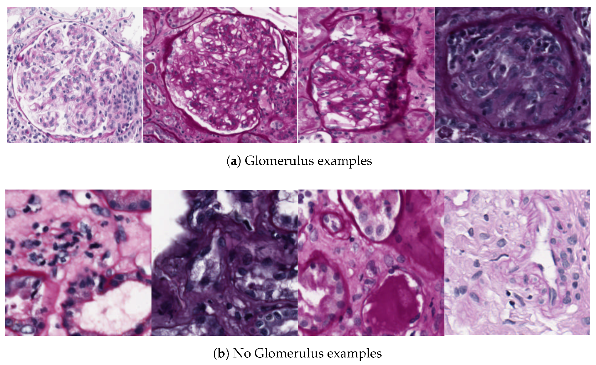
Glomeruli structures visualized in a VS present high variability in terms of size, shape and color. The reasons of this high variability in samples are multiple: the relative position of the Glomerulus inside the renal section, the heterogeneity in immunohistochemistry staining, or the presence of internal biological processes.
All this variability related to the Glomerulus appearance, and in general to VS images, makes it difficult to automate the classification using a digital pathology framework. Therefore, any proposal devoted to this task must be robust to these aspects.
Previous Work
Many authors have been working in digital pathology developing techniques to automate quantitative analysis tasks. Methods devoted to classify and detect Glomeruli in the nephropathology field have become of main importance in diseases diagnosis and research. The classification methods of the literature can be divided into two groups: handcrafted features approaches and unsupervised features approaches.
Handcrafted features: These classification methods try to find specific measurable attributes in the image that can be utilized to correctly identify the structures. In [
6], the authors propose to use a genetic algorithm for edge patching by using a Canny edge detector to get discontinuous edges of Glomerulus and its final detection and classification. The main problem of this technique is that borders detected by means of Canny edge detector are not robust and is prone to errors under color intensity variations. In [
7,
8], the authors use Rectangular-Histogram of Gradients (R-HOG [
9]) as feature vector to classify Glomeruli. The main problem of these techniques is the fact that R-HOG has rigid block division that results into considerable number of false positives. In order to improve the R-HOG framework, Kato et al. [
10] suggest the Segmental HOG (S-HOG) as potential candidate descriptor for Glomerulus detection, where the block division is not rigid, and uses nine discretized oriented gradients and Support Vector Machine (SVM [
11]) as a supervised learning classifier. In [
12], an analysis of renal microscopic images is applied by using a detection of borders improved with the Convex Hull algorithm ([
13]). This method detects and classifies Glomeruli for the measurement of both diameter and Bowman’s Space thickness. In [
14], the authors propose an approach for Glomeruli detection based on the ellipse shape of Glomeruli to obtain candidate regions. These regions will be classified in a final step with a trained SVM classifier. The method uses a color normalization step to deal with variations that appear in the stain process. Brandon Ginley et al. [
15] present a method to identify glomerular boundaries based on Gabor filtering, Gaussian blurring, statistical F-testing and distance transform. In [
16], the authors propose a computer-assisted topological analysis to detect tubules and interstitium, glomeruli, and peritubular capillaries in kidney samples. The method is based on double immunostaining of both capillaries and an immune cell population, so that the decomposition of colors allows identification of the nuclei of all cells present in the biopsy specimen, of the cytoplasm of stained immune cells, of capillary walls, and the lumens of capillaries and Glomeruli. Coupling decomposed colors allow precise identification of the regions of interest, that is, the stained immune cells, capillaries, and Glomeruli. Simon et al. [
17], designed a method for the segmentation of glomeruli using an adaptation of the local binary patterns (LBP) image feature vector to train a support vector machine (SVM) model. Finally, as an example of Glomeruli classification in 3D dataset, Zhang et al. [
18] propose a Glomerulus detection and classification using 3D Magnetic Resonance Imaging (MRI) by means of Hessian based Difference of Gaussians (HDoG) detector.
Unsupervised features: These techniques avoid the necessity to define handcrafted features to perform the classification. Deep Learning methods such as CNNs belong to this group. They learn the best features for the problem, so that the scientists are only responsible of providing large representative datasets, as well as choosing the network architecture. Kothari et al. [
19] perform a thorough review of how the application of this kind of techniques is currently the most suitable one, pointing at the challenges and future opportunities that are involved.
Regarding the application of these techniques to specific problems, some of the most interesting works that have been published focus on different medical areas: Wang et al. [
20] use a cascaded approach for mitosis detection in breast cancer that combines a CNN model and handcrafted features (morphology, color and texture features). In [
21], the authors deal with mitotic figure classification combining manually designed nuclear features with learned features extracted by CNN. Xu et al. [
22] explore the use of very deep CNNs to automatically classify diabetic retinopathy by configuring a network with 18 layers. Havaei et al. [
23] apply CNN to automatic brain tumor segmentation in MRI images that exploits both local features as well as more global contextual features simultaneously, in a CNN cascade configuration.
Table 1 shows a summary of the different experiments performed in the publications mentioned above, specifying the kind of problem, network architecture (and any particularity if necessary to be mentioned), the dataset used and the obtained results. The revision of every publication stated before is out of the scope of this article and therefore we focus on those works related to CNN.
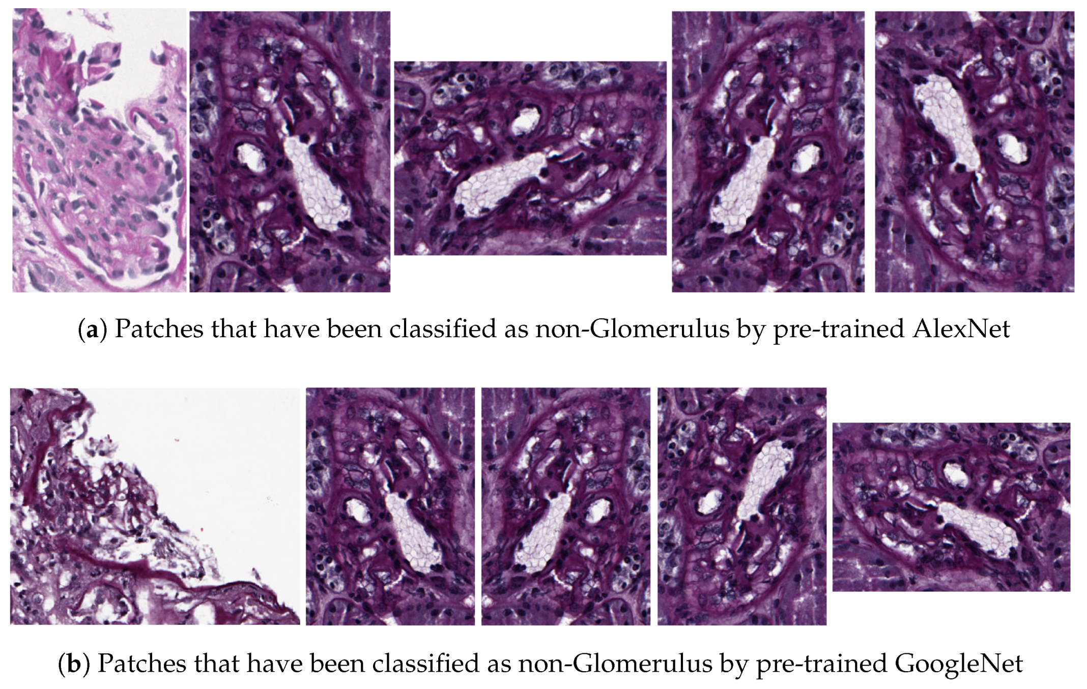
Some recent publications focus on unsupervised Glomerulus detection by means of Deep Learning. In [
24], the authors evaluate the detection of glomerular structures in WSIs stained with multiple histochemical staining using HOG feature classifier and CNN with a specific model. They compare the performance of both methods for each stain, and the combination of the detection in consecutive sections stained with different histochemical staining. Their results showed that HOG detector perform between 10% and 20% worse than CNN detector. Besides, their method using CNN detector with PAS samples performed on average 66.38% F1-score. Combining consecutive sections, after WSI registration, the final score reaches 78%. The main drawback of this proposal is the low F1-score using only the PAS WSI and the complexity of combining consecutive sections. The training method, which does not take into account the differences between stains, and the CNN analysis carried out to accept a region as a Glomerulus are partially the cause of these inferior results. More recently, Gadermayr et al. [
25] deal with Glomerulus detection and segmentation using CNN cascades where first, a network is used to detect regions of interest, and next, a U-Net network is applied to achieve the Glomerulus segmentation. The resulting detection rate is 91%, which is higher than previous proposals, but no color normalization is applied to the data, which led us to consider miss-detection under different conditions like PAS samples belonging to different laboratories.
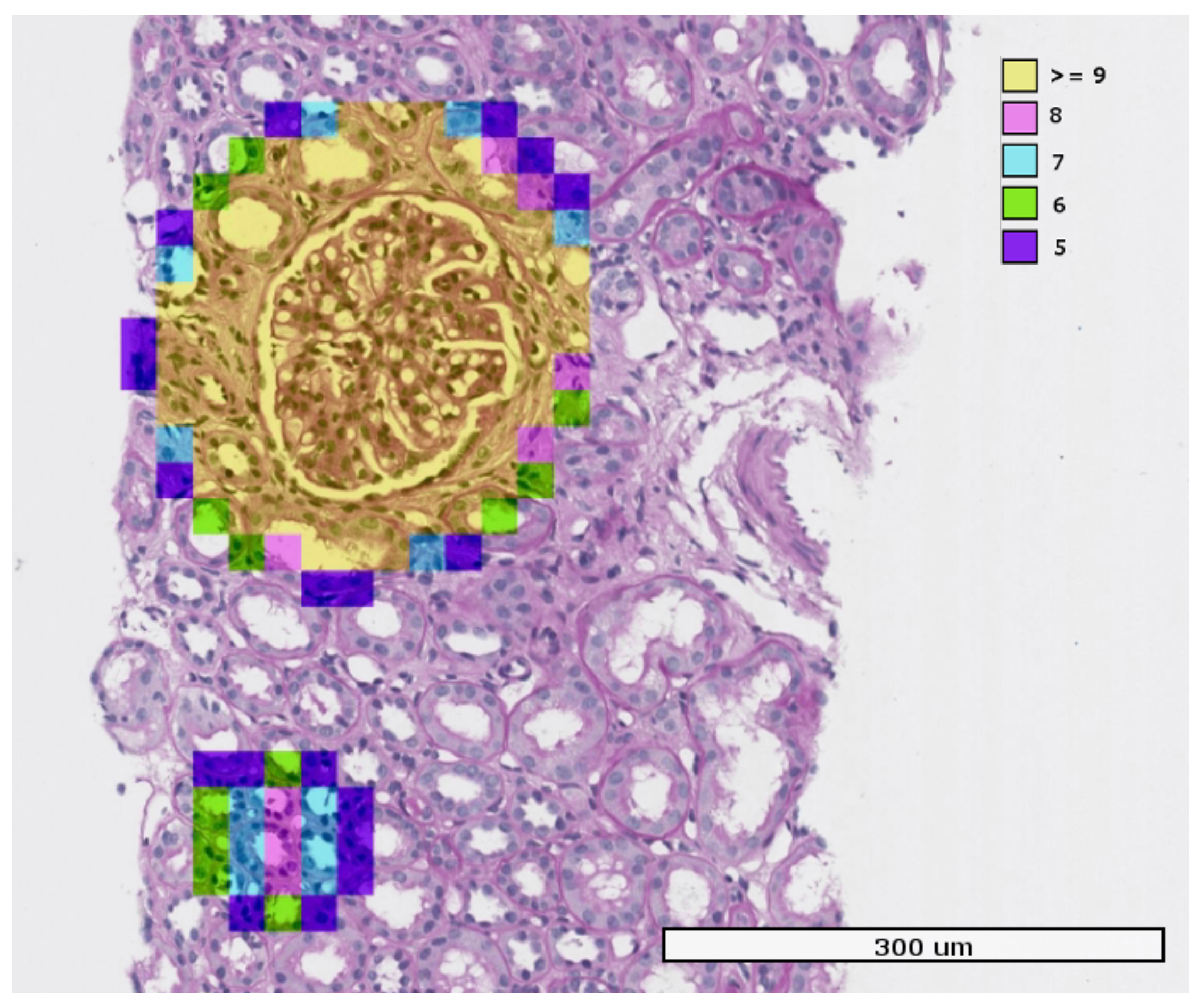
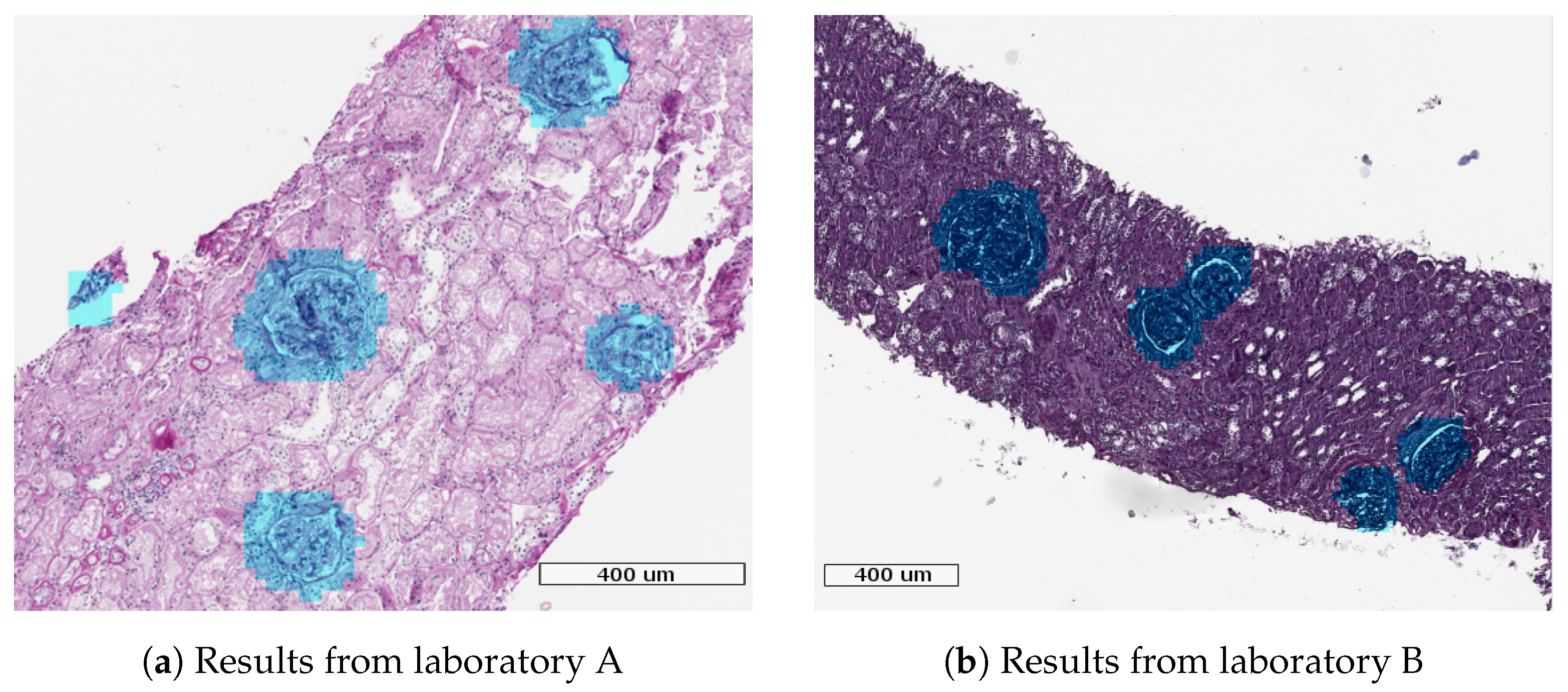
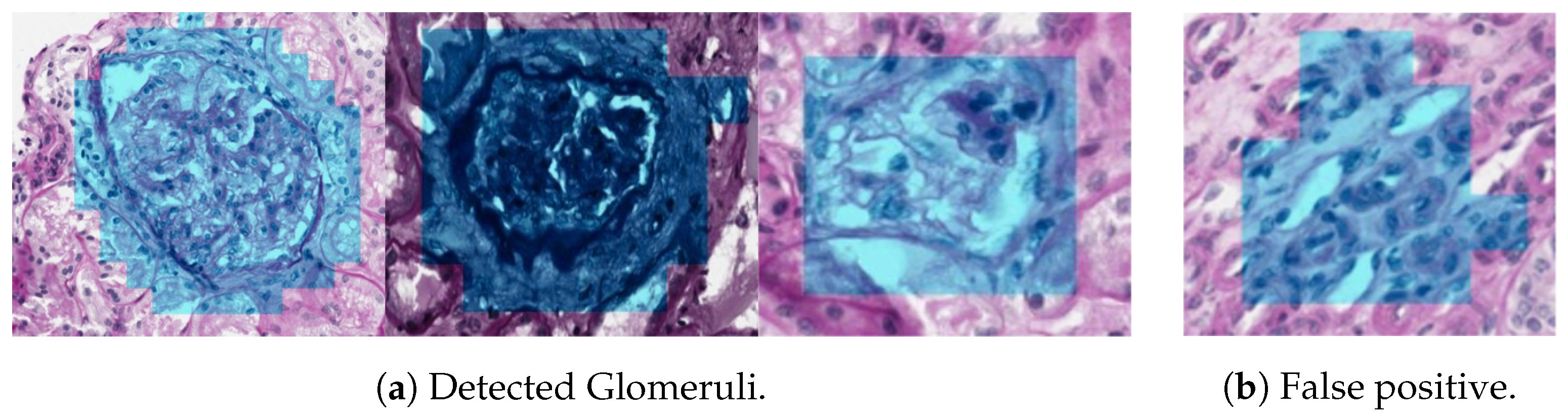
In this paper we propose two methods devoted to: Glomerulus classification and Glomerulus detection. In the Glomerulus classification proposal (Method I), we study the viability of the CNN to achieve a correct Glomerulus classification into Glomerulus/non-Glomerulus classes. In the Glomerulus detection method (Method II), we use the CNN configuration that gave the best results and performance for Glomerulus classification (Method I) to achieve the Glomeruli detection in a WSI. In this proposal, we present a method suitable for PAS samples belonging to different laboratories, thanks to color normalization process used in the data augmentation step. Our method uses a sliding window with overlapping to perform the detection, and utilizes a majority vote decision for each pixel to reduce false positive detections. With this proposal, we achieve a detection score of 93.7%, which improves upon the results obtained in reference methods.
The remainder of the paper is as follows. The Materials and Methods are explained in
Section 2, where Glomerulus classification and Glomerulus detection methods are described in detail. Results for Glomerulus classification and Glomerulus detection are shown in
Section 3. Finally,
Section 4 is devoted to the Discussion.


 ,
, 


