Article

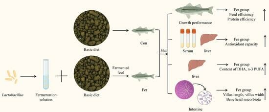
Effects of Co-Fermented Feed Using Lactobacillus acidophilus, Limosilactobacillus reuteri and Lactiplantibacillus plantarum on Growth, Antioxidant Capacity, Fatty Acids and Gut Microbiota of Largemouth Bass (Micropterus salmoides)

1
College of Marine Sciences, South China Agricultural University, Guangzhou 510642, China
2
Guangdong Laboratory for Linnan Modern Agriculture, Guangzhou 510642, China
3
State Key Laboratory of Applied Microbiology Southern China, Guangdong Provincial Key Laboratory of Microbial Culture Collection and Application, Guangdong Institute of Microbiology, Guangdong Academy of Sciences, Guangzhou 510070, China
*
Authors to whom correspondence should be addressed.
Fishes 2023, 8(9), 433; https://doi.org/10.3390/fishes8090433
Submission received: 25 July 2023 / Revised: 20 August 2023 / Accepted: 21 August 2023 / Published: 23 August 2023

Abstract

:
The effects of diets fermented with compound probiotics, namely Lactobacillus acidophilus, Limosilactobacillus reuteri and Lactiplantibacillus plantarum, on the growth performance, physiological and biochemical indexes, fatty acid composition and intestinal health of juvenile largemouth bass (Micropterus salmoides) were investigated. Three hundred healthy juvenile M. salmoides (5.29 ± 0.02 g) were selected and randomly divided into two groups with triplicates for each. The basic diet was set as the control group (CON), and fermentation of the basic diet with a mixed bacterial solution (1.8 × 10⁹ cfu/mL, L. acidophilus:L. reuteri:L. plantarum = 1:1:1) was set as the fermentation group (FER). Fish were hand fed to satiation for 56 days and two-thirds of the culture water was renewed every 3 days. The results showed that feed intake of fish in the FER group was significantly lowered, thereby increasing feed efficiency (FE) and protein efficiency (PER) (p < 0.05). Serum alanine aminotransferase (ALT) activity was significantly decreased, and catalase (CAT) activity was significantly increased in the FER group (p < 0.05). The liver superoxide dismutase activity (SOD) was significantly enhanced, and intestinal trypsin was significantly increased in the FER group (p < 0.05). Being fed with the fermentation diet significantly increased the content of n-3 polyunsaturated fatty acids (PUFA), docosahexaenoic acid (DHA) and the n-3/n-6 PUFAs ratios in the liver (p < 0.05). Intestinal histology showed that villus height and width of the intestine and the number of goblet cells were significantly increased in the FER group (p < 0.05). Those fed with fermentation diets had limited diversity of gut microbiota. Compared to the CON group, the relative abundance of Aeromonas decreased significantly (p < 0.05), while the relative abundance of Fusobacteria, Cetobacteria and Lactobacillusis in FER increased greatly in the gut microbiota of the FER group. In conclusion, fermented feed with the three probiotics effectively improved the feed utilization and antioxidant capacity, promoted digestion and absorption of dietary protein, improved the ability of synthesize DHA and n-3 PUFAs in the liver and reduced the abundance of pathogenic bacteria in the gut. Therefore, the present research provided a new way of co-fermented feed with three probiotics for the aquaculture of M. salmoides.
Key Contribution: Compound probiotic fermented feed improves growth performance and promotes the intestinal health of fish.

Graphical Abstract

1. Introduction

Largemouth bass, Micropterus salmoides, originally from the United States, is a freshwater carnivorous fish characterized by rapid growth, high adaptability and a short reproductive cycle [1,2]. As M. salmoides is popular among consumers, it is gradually occupying a higher position in China’s aquaculture market. However, the development of M. salmoides was more and more limited by deterioration of the water quality that resulted from intensive aquaculture, low feed utilization, more and more expensive fish feed and antibiotic abuse [3]. Current research on M. salmoides mainly focuses on macronutrient requirements and alternative protein and lipid sources [4]. There was a great need for research on the development of unconventional feeds that can decrease food waste to improve feed utilization and decrease the use of the antibiotics to promote the healthy aquaculture of the fish.
Probiotics show promise in aquaculture for disease prevention and supervision [5]. Studies on M. salmoides have shown that probiotics, such as Lactobacillus casei and Bacillus subtilis, can improve lipid metabolism, antioxidant and immune capabilities, intestinal digestion and absorption capabilities, and reduce the abundance of pathogenic bacteria in the intestine [6,7]. Feed ingredients can be decomposed into microbial bacterial proteins, small bioactive peptides and amino acids by probiotic fermentation [8]. Fermentation destroys anti-nutrients and increases nutrient levels in feed ingredients, which improves feed utilization in fish. Furthermore, fermented feed can promote the balance of intestinal flora and the organism’s intestinal health [9]. Existing studies frequently combine different probiotics in order to sum up the fermentation advantages of multiple strains and fully utilize the probiotic effects of the strains [10,11]. Lactobacillus acidophilus, Limosilactobacillus reuteri and Lactiplantibacillus plantarum are common lactic acid bacteria that can maintain intestinal microbial balance and inhibit pathogenicity [12,13,14]. L. acidophilus improves intestinal morphology and boosts serum antioxidant enzyme activity in the organism, whereas L. reuteri and L. plantarum can increase the diversity of intestinal microbiota [15,16,17,18,19]. Although the effects of the three microbial species were widely used and researched in aquaculture, the effects of the three species applied in combination were not evaluated.
Therefore, in this study, a multistrain probiotic (L. acidophilus, L. reuteri and L. plantarum) was selected for fermentation in the basal diet to investigate its role in the growth, antioxidants, digestion, fatty acid composition and intestinal flora composition of M. salmoides.

2. Materials and Methods

2.1. Diet Preparation

The feed formula of the basic diet is presented in Table 1. The raw materials were ground and mixed with water according to the formula, extruded into strips and pelletized, then dried and stored in a 4 °C refrigerator [20]. Fermented feed was prepared based on a basic diet.
The probiotics used for the fermentation of the feed were provided by Guangdong Microbial Culture Collection Center (GDMCC) and prepared by Microbiological Analysis and Testing Center of Guangdong Institute of Microbiology (Guangzhou, China). Compound probiotics solution was the 1:1:1 mixture of L. reuteri (GDMCC 1.614), L. plantarum (GDMCC 1.191), L. acidophilus (GDMCC 1.208) with 1.8 × 10⁹ cfu/mL for each bacterium. Briefly, 10 mL probiotics mixture was diluted in 400 mL sterile water in, which was sprayed evenly into 1 kg basic diets and fermented in a sealed bag with the air discharging at 37 ± 5 °C for 3 days. The fermented diets smelled of an obvious sour flavor, and were then fed to the fish within 3 days. The proximate composition of the fermented diets was evaluated after drying to constant weight, which is presented in Table 1. The fatty acid composition of the feed is shown in the Appendix A Table A1.

2.2. Feeding Trial

Juvenile M. salmoides were purchased from Huaxuan Aquatic Co., Ltd. (Guangzhou, China). The fish were acclimated in the aquaculture system and fed with basic diets for 2 weeks. The aquaculture system was the same one used by Wang et al. [21]. After acclimatization, 300 healthy fish with similar physical size (5.29 ± 0.02 g) were selected and randomly divided into 2 groups with triplicates of 50 fish per tank. The group fed with the basic diets was set as control (CON) and the experimental group was fed with the fermented feed (FER). All fish were fed twice (08:00 a.m. and 17:00 p.m.) daily to satiation and the daily feed intake was recorded. During the 56-day experimental period, culture water was continuously aerated and two-thirds was renewed every 3 days to ensure the water quality.

2.3. Sample Collection and Procession

At the end of the feeding test, all the fish were fasted for 24 h. The total final weight and number of fish in each tank was recorded, and the individual weight and body length were measured. Three fish from each tank were randomly selected and frozen at −20 °C for the determination of whole body proximate composition. Eight randomly selected fish from each tank were anesthetized with 2-phenoxyethanol (Sigma-Aldrich, St. Louis, MO, USA). Blood samples from the fish were obtained from the tail vein with a 1.5 mL sterile syringe. Serum was prepared for the determination of serum biochemical indicators according to the methods described in Liao et al. [20]. After blood collection, the fish liver and visceral and intraperitoneal fat were dissected to determine fish morphometric parameters. Livers and intestines were sampled for the determination of liver biochemical indicators and digestive enzyme activities. The intestines collected for histological analysis were preserved with 4% paraformaldehyde (PFA, Guangzhou Yunran Biotechnology Co., Ltd., Guangzhou, China). The dorsal muscle and liver were collected for the determination of proximate compositions and fatty acid compositions. Gut contents were squeezed out and instantly frozen in liquid nitrogen for subsequent 16S rRNA sequence determination.

2.4. Growth Assessment

Final body weight (FBW), weight gain rate (WGR), specific growth rate (SGR), feed efficiency (FE), food intake (FI), survival rate (SR), protein efficiency (PER), condition factor (CF), hepatosomatic index (HSI), viscerasomatic index (VSI) and intraperitoneal fat ratio (IPF) are calculated through the following equations [20]:
FBW (g) = Wt/Nt
WGR (%) = 100 × (Wt − W0)/W0
SGR (%) = 100 × [(Wt − W0)/t]
FE = Wf/(Wt − W0 + Wd)
FI (%/day) = Wf × 100 × 2/[56 × (W0 + Wt + Wd)]
SR (%) = (Nt/N0) × 100
PER (%) = 100 × (Wt − W0)/(Wf × CP)
CF (g/cm3) = 100 × (Ws/Ls3)
HSI (%) = 100 × (W1/Ws)
VSI (%) = 100 × (Vl/Ws)
IPF (%) = 100 × (Fl/Ws)
where Wt is the total final fish weight; W0 is the initial total fish weight; Wd is the total dead fish weight; t is the number of breeding days; Wf is the total feeding dry weight; N0 and Nt are the initial and final fish tail numbers, respectively; CP is crude protein content in feed; and Ws, Ls, W1, V1 and F1 are the fish weight (g), body length (cm), liver weight (g), visceral weight (g) and abdominal fat weight (g), respectively.

2.5. Proximate Composition and Fatty Acid Profiles Analysis

Proximate composition of moisture, crude protein (CP), crude lipid (CL), ash and fillet fatty acid profiles in the diets, whole fish and muscles were determined according to the methods described by Liao et al. [20] and Perez-Velazquez et al. [22], respectively.

2.6. Biochemical Indices and Enzymatic Activities Analysis

Serum biochemical parameters including alanine aminotransferase (ALT), aspartate aminotransferase (AST), alkaline phosphatase (AKP), total bile acid (TBA), triglyceride (TG), total cholesterol (TC), high-density lipoprotein cholesterol (HDL), low-density lipoprotein cholesterol (LDL), albumin (ALB) were determined by the Guangzhou Kingmed Center for Clinical Laboratory (Guangzhou, China). All the measurements were performed by the same operator to ensure the accuracy of the test data.
Antioxidant indicators, namely superoxide dismutase (SOD), catalase (CAT), malondialdehyde (MDA), total antioxidant capacity (T-AOC) in serum and liver, lipase (LPS), amylase (AMS) and trypsin (TPS) in intestine were measured through diagnostic reagent kits according to the instructions (Nanjing Jiancheng Bioengineering Institute, Nanjing, China).

2.7. Liver and Intestine Histological Analysis

After fixation with 4% formaldehyde, liver and intestine tissues were sent to Wuhan Servicebio Technology CO., LTD (Wuhan, Hubei Province, China) for dehydration, paraffin embedding, sectioning and staining. The morphological structures of the intestinal wall and liver of M. salmoides were observed with microscope (E200MV, Nikon Optical Instruments Co., Ltd., Nanjing, China). The length of the villi and the thickness of the muscular layer of the intestinal tissues were measured, and the diameters of the hepatocytes and nuclei of the liver tissues were measured. Twenty values were randomly measured in each section, and the average value was taken as the measurement result.

2.8. Intestinal Microbiota Analysis

The intestinal contents were transported in liquid nitrogen to Shanghai Meiji Biomedical Technology Co., Ltd. for DNA extraction and PCR amplification. Then, the 16S rRNA gene V3-V4 was amplified with universal primers 338F and 806R. The amplified region was about 450 bp in length, and the library construction and Illumina Miseq sequencing were conducted at Shanghai Meiji Biomedical Technology Co. Before bioinformatics analysis, the raw data were quality-controlled by Trimmomatic software (V0.32), and then sequence splicing was performed by FLASH software (1.2.11). Bioinformatics analysis was performed using the bioconfidence cloud platform of Shanghai Meiji Biomedical Technology Co. Sequences were clustered at 97% similarity using UPARSE software (7.1) for OTU. The sequences were annotated for species classification using RDP classifier (V2.2), compared to the reference database of Silva_138 16S rRNA database, and the composition of microbial taxonomic levels were counted [23].

2.9. Statistical Analysis

All the data presented as means ± SE. The differences in values between 2 groups were analyzed through t-test using the SPSS software (25.0). A statistical significance level of p = 0.05 was employed. * indicates p < 0.05 and ** indicates p < 0.01. The α-diversity of gut microbial communities was assessed using Mothur software (V 1.30.2); the β-diversity of microbial communities was assessed by non-metric multidimensional scaling analysis (NMDS).

3. Results

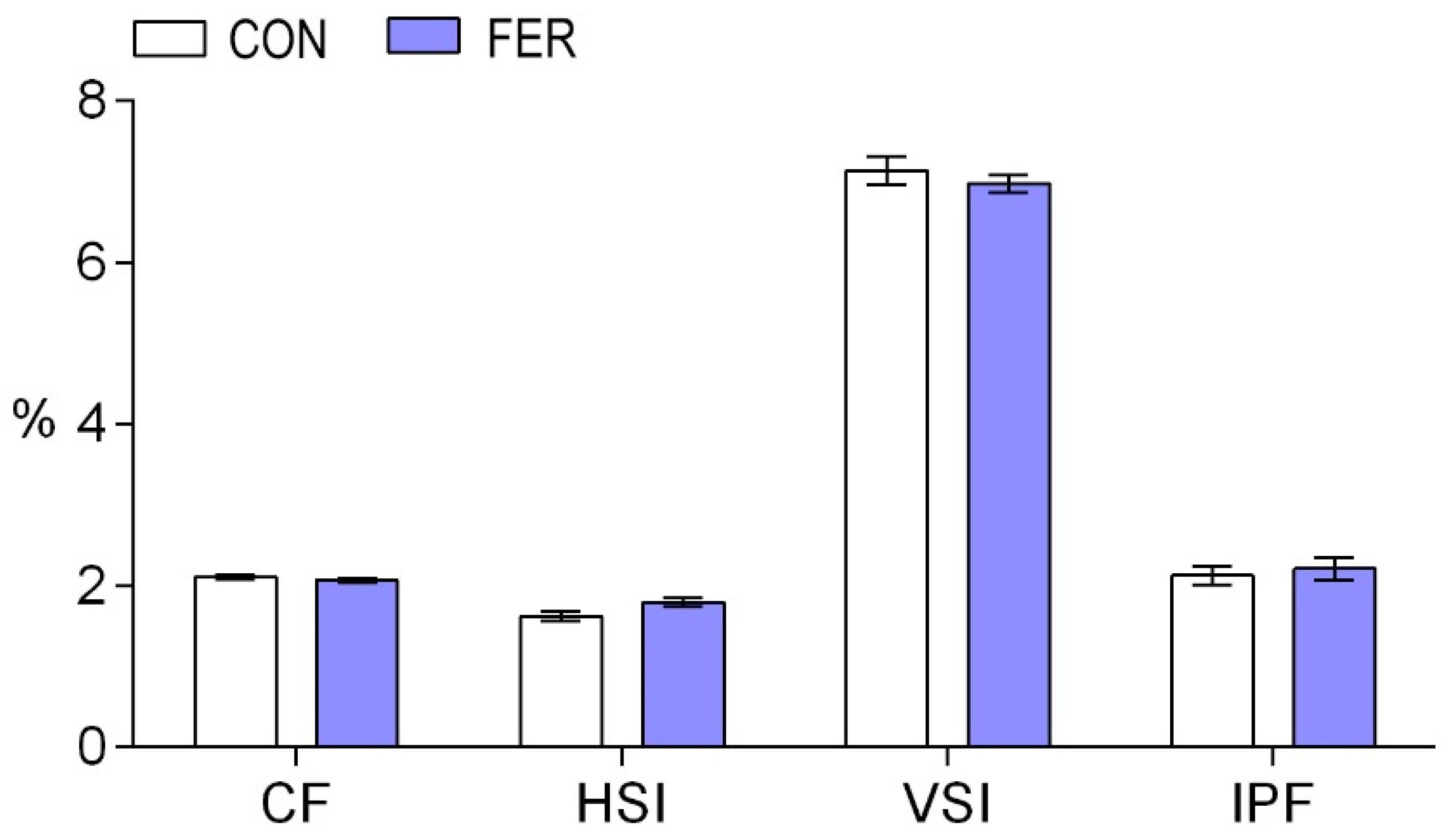
3.1. Growth Performance

There is no significant difference in SR between CON and FER (p > 0.05) (Table 2). Compared with CON, the FI of fish was significantly decreased (p < 0.05), but the average FBW, WGR and SGR in FER were increased, though it did not reach significant differences (p > 0.05). Compared with CON, 33% higher FE and 21% higher PER were found in FER (p < 0.05). There were no significant differences in whole-body and muscle-proximate compositions of HIS, VSI, CF and IPF between the two groups (p > 0.05) (Figure 1).

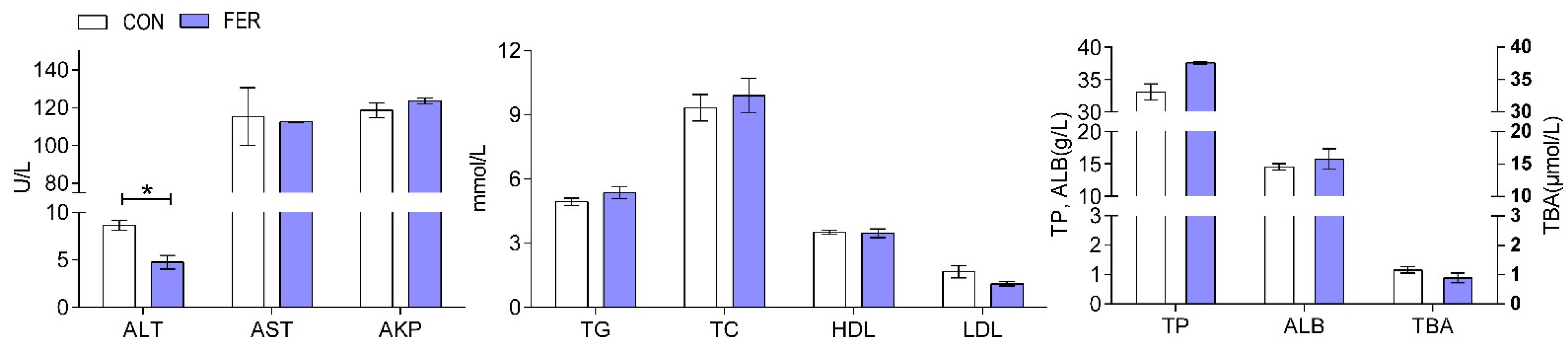
3.2. Biochemical Indices and Enzymatic Activities

The results of the serum biochemical parameters are shown in Figure 2. Compared with CON, the activity of ALT in FER was significantly lowered (p < 0.05). There are no significant differences between CON and FER in other indicators, namely AST, AKP, TG, TC, HDL, LDL, ALB, TP and TBA (p > 0.05).
The results of the serum and liver antioxidant indexes are shown in Figure 3. The serum CAT and liver SOD activities was significantly enhanced in the FER group (p < 0.05), and there were no significant differences in liver MDA, CAT and T-AOC activities between the two groups (p > 0.05).
The intestinal digestive enzyme activities were shown in Figure 4. Compared with the CON group, the activity of TPS in the intestine was significantly higher in the FER group (p < 0.05), while the activities of AMS and LPS showed no significant difference (p > 0.05).

3.3. Fatty Acids

3.3.1. Muscle Fatty Acids

A total of 16 fatty acids were detected in muscle (Table 3), of which saturated fatty acids (SFA) accounted for 28.8–30.0%, monounsaturated fatty acids (MUFA) accounted for 22.6–23.1% and polyunsaturated fatty acids (PUFA) accounted for 46.9–48.3%. The content of C20:3n-3 in FER was significantly lower than that in CON (p < 0.05), and the other fatty acids did not reach significant levels (p > 0.05).

3.3.2. Liver Fatty Acid Compositions

The liver fatty acid compositions are shown in Table 4. A total of 17 fatty acids were detected in the liver, with SFA accounting for 26.7–27.0%, MUFA accounting for 23.5–27.0%, and PUFA accounting for 37.1–41.2%. The contents of C16, C18:2n-6 (linoleic acid), C18:3n-6 and n-6 PUFAs in the liver of the FER group were significantly lower than those in the CON group, and the contents of C20:3n-3, C22:6n-3 (docosahexaenoic acid, DHA), n-3 PUFAs and n-3/n-6 PUFAs were significantly higher than those in CON (p < 0.05).

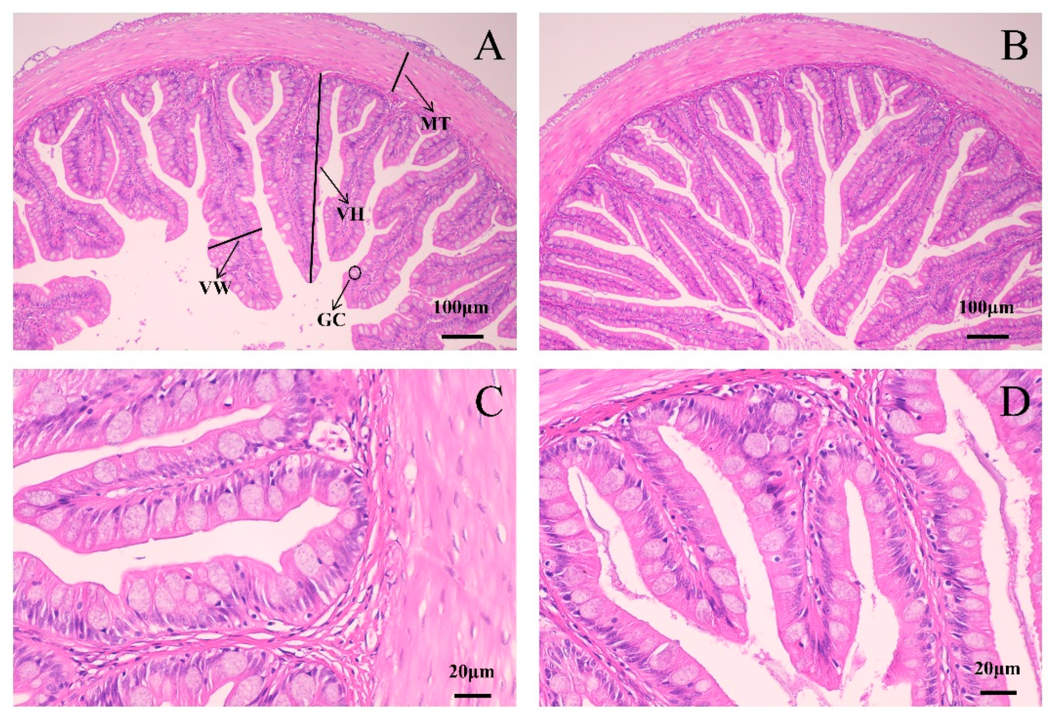
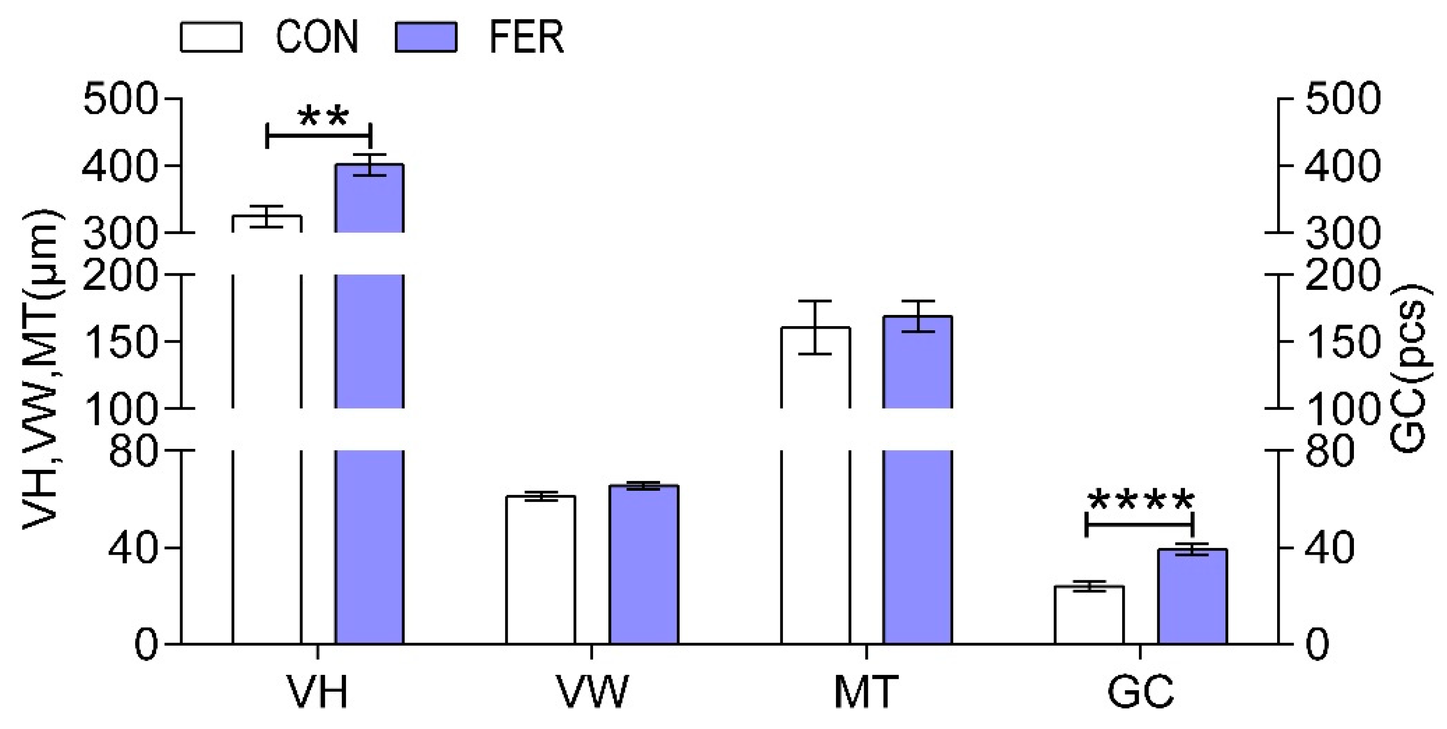
3.4. Histomorphology of the Intestine

The intestinal morphology and morphologically relevant parameters are shown in Figure 5 and Figure 6. Fish in FER had significantly higher intestinal villus height, villus width and number of goblet cells than those in CON (p < 0.05), while there was no significant difference in myofibrillar thickness between the two groups (p > 0.05).

3.5. Intestinal Flora Analysis

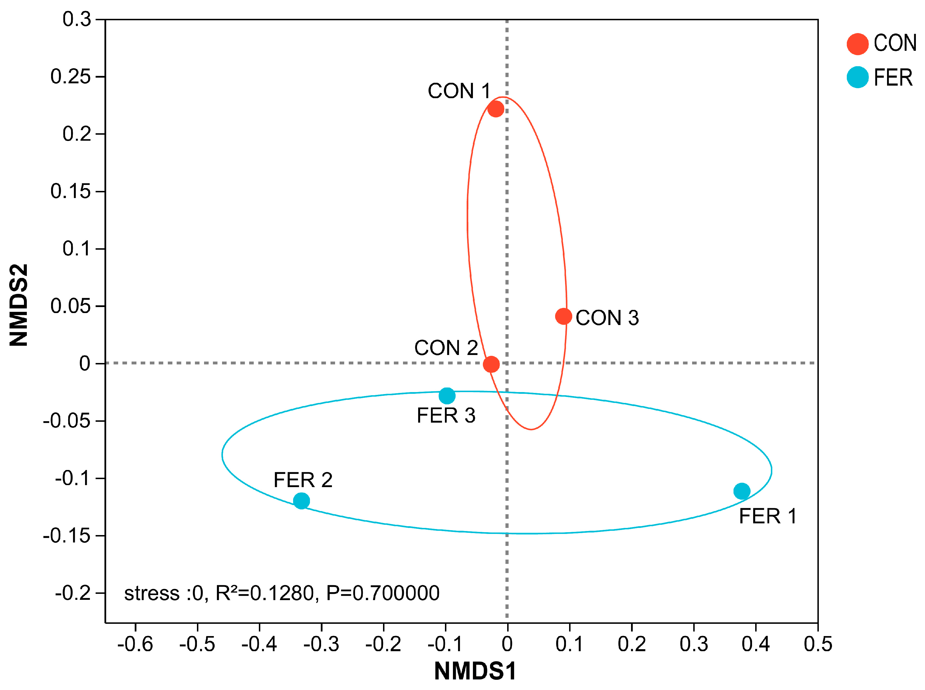
3.5.1. Sequencing Data and Diversity Analysis

A total of 231,674 sequences were obtained from Illumina sequencing, with an average of 38,612 sequences per sample and an average sequence length of 417 bp; 330 OTUs were clustered at 97% similarity, belonging to 20 phyla, 38 orders, 92 families, 140 families and 223 genera. The sequence number and the α-diversity index of the intestinal microbiota in two groups are shown in Table 5. There were no significant differences in any of the four indices (p > 0.05), although the FER group had relatively higher species richness (as shown by Ace and Chao1 index) and lower species evenness (as shown by Shannon and Simpson index) compared with CON. The β-diversity of gut microorganisms (Figure 7) showed an overall trend of better clustering, but the intergroup difference was not significant (p > 0.05).

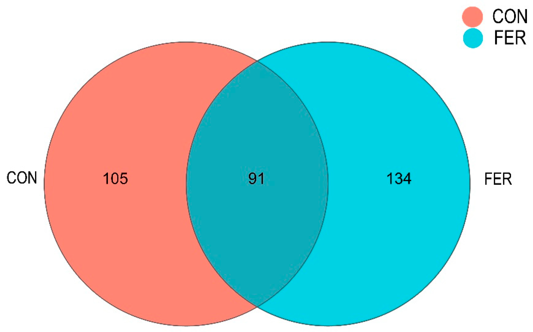
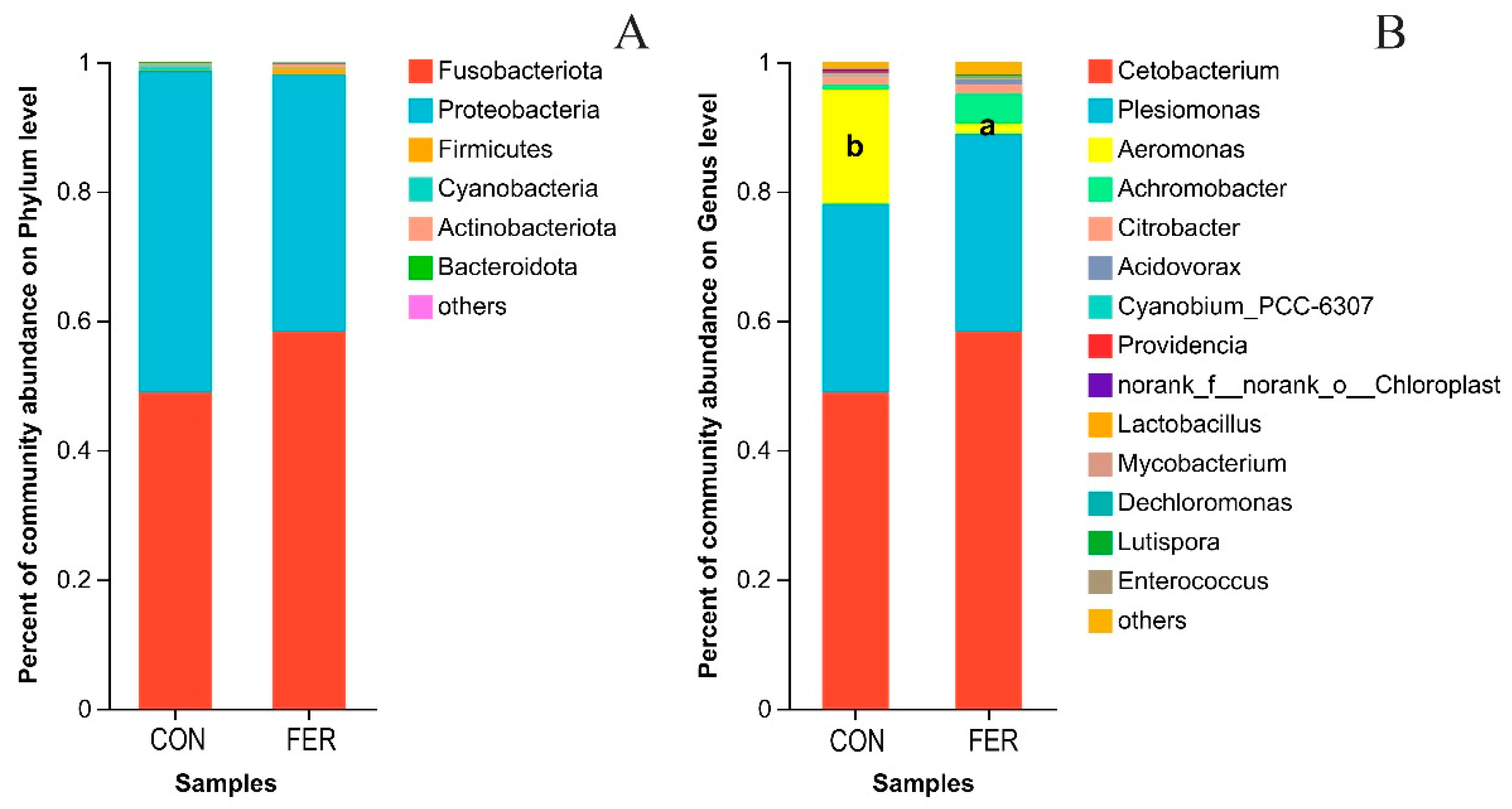
3.5.2. Intestinal Microbial Community Composition

Venn diagrams show that two groups shared 91 OTUs (Figure 8). More intestinal microflora OTUs were determined in FER than in CON.
Fusobacteria and Proteobacteria were the most dominant phyla (Figure 9A). The relative abundance of Fusobacteria in CON and FER was 48.85% and 58.24%, respectively, while the relative abundance of Proteobacteria in CON and FER was 49.77% and 39.9%, respectively. At the genus level, Cetobacterium, Plesiomonas, Aeromonas, Achromobacter, Citrobacter and Acidovorax, were the dominant genus (Figure 9B). The most dominant genera in CON were Cetobacterium (48.85%), Plesiomonas (29.21%) and Aeromonas (17.76%), while the most dominant genera in FER were Cetobacterium (58.24%), Plesiomonas (30.73%) and Achromobacter (4.66%). The relative abundance of Aeromonas in the FER group decreased significantly compared with that in CON (p < 0.05). It was notable that the relative abundance of Lactobacillus in FER was 0.21%, while it was decreased to 0.01% in CON.

4. Discussion

FBW and WGR in FER were comparable or even better than those of CON during the 56-day feeding experiment, which was consistent with research on Procambarus clarkii [24]. Previous research also found that L. plantarum and L. acidophilus compound-fermented feed improved the WGR and reduced the feed conversion rate of laying hen chicks [25]. However, it had been reported that fermented feed can reduce appetite [26]. The feeding rate of M. salmoides in FER in this experiment was significantly lower than that of CON, which was presumably related to the sour smell and changes in the feed pellet shapes of the fermented feed, although at a lower feeding rate, the growth of fish in the FER group was comparable or better than the CON, suggesting a better feed utilization which was reflected by higher FE and PER in the FER group. It was probably due to increased bioavailability of the diet nutrients during the fermentation process, such as the degradation of diet protein into small peptides, thus improving the quality of the feed [27]. Other studies also suggested dietary supplementation with probiotics such as L. plantarum and L. acidophilus significantly increase feed utilization [28,29]. It can be concluded that fermentation with the three probiotics was beneficial to feed utilization, thus decreasing food dilapidation.
Serum ALT and AST were generally used to evaluate liver health status and nutritional metabolism of the organisms [30]. In general, serum ALT and AST were decreased when ALT and AST enzymes formed new amino acids in the liver. And only when liver damage occurs, such as from anti-oxidative stress, would the ALT and AST enter the bloodstream in large quantities [31,32]. A decrease in ALT activity in the FER group indicate feed with fermentation diets promoted liver health. It was similar to the results based on Macrobrachium nipponense that serum ALT and AST activities were significantly lowered after being fed with Lactobacillus plantarum fermented feed [33]. M. salmoides fed with fermented feed could alleviate liver damage and improve the liver health.
During growth and development, the organisms will generate reactive oxygen species (ROS) under stress conditions, which are prone to lipid peroxidation and hence cause damage [34]. T-AOC can accurately reflect the antioxidant capacity of both enzymatic and non-enzymatic defense systems, while SOD and CAT represent antioxidant enzyme systems, which are the first lines of defense against free radical damage and reflect the antioxidant capability of the organisms [25,35]. MDA is a byproduct of lipid peroxidation, which represents lipid oxidation and tissue oxidative damage [36]. In the present study, M. salmoides fed with fermented feed significantly increased the antioxidant capability, suggesting that Lactobacillus can efficiently eliminate excess free radicals and improve the antioxidant capacity of M. salmoides. Similarly, Pichiah et al. and Yadav et al. reported that L. plantarum fermented feed increased serum T-AOC and SOD activities of Litopenaeus vannamei, while L. plantarum fermented feed decreased Procambarus clarkii’s serum MDA content [24,37]. In addition, it was presumable that the fermented diet produced a large number of metabolites containing probiotic enzymatic components and immune factors, making the nutrients of the feed more balanced and stimulating the epidemic system of the organism to improve the antioxidant capacity of the fish.
Digestive enzymes, such as lipase and amylase, play important roles in intestinal digestion and absorptive capacity [38]. Trypsin is involved in the hydrolytic absorption of dietary proteins [39]. Sufficiently high digestive enzyme activities along with adequate food supply contributed to rapid growth of the fish [40]. In this study, intestine TPS activity of fish in the FER group was significantly higher than that of CON (p < 0.05), suggesting higher digestion and absorption of dietary protein after fermentation. Pichiah et al. and Yadav et al. obtained similar results that fermented feeds containing L. plantarum increased the lipase, amylase and trypsin activity of P. clarkii and L. vannamei, which contributed to improve the digestive capability of fish [24,37]. The fish in the FER group had better digestive function, which may be related to the fact that fermented feeds had higher contents of protein, and the protein was more digestible. It was also reflected by the significant increase in FE and PER in FER.
The fatty acid composition of muscle depends on the fish species [41]. In this study, there was no great difference in the fatty acid composition of the muscles of M. salmoides. Long-chain polyunsaturated fatty acids (LC-PUFAs), including DHA and EPA, are not only essential nutrients to fish but also to human beings [42,43,44]. EPA and DHA are important parts of n-3 PUFAs, while their contents in fish were generally related to the diets [43,45]. The n-3-to-n-6 PUFA ratio was also critical for decreasing inflammation and improving human mental health [46,47]. Carbonium (C20:3n-3) is one of the intermediates of the DHA synthesis pathway [41]. In the present experiment, both carbonium and DHA were significantly increased, which contributed to increase the content of n-3 PUFAs. Similar results were also found in the tilapia fed diets with added L. plantarum and Pediococcus acidilactici, with significantly decreased n-3-to-n-6 PUFA ratios in the liver and muscle [48].
The intestine is not only the main place of digestion and nutrient absorption but also the innate barrier to prevent bacteria and other pathogens from entering the fish [49]. Studies have shown that 21% Lactobacillus fermented soybean meal can increase the intestinal villi height in Oreochromis niloticus [50]. Bacillus subtilis fermentation products showed excellent protection in Danio rerio fed high-fat diets, with structural integrity of the intestinal mucosa layer, muscle layer, and plasma membrane layer, and an elevated number of goblet cells on the villi [51]. In this experiment, the intestinal structure of M. salmoides were intact without inflammation, and the height and width of intestinal villi and the number of goblet cells in FER were significantly higher than those in CON. The increase in intestinal villus height and width indicated an increase in absorption surface area, which could lead to better nutrient absorption, which was reflected by increased PER and FE.
Probiotics were vital in maintaining gut health through modulation of the microbial community [52]. It was generally believed that the greater the diversity of the intestinal microbiota, the more comprehensive physiological functions of the microbiota [53]. Li et al. found that fermented feed increased the diversity of gut microbiota and the relative abundance of probiotics in Macrobrachium nipponense [33]. Meanwhile, Zhang et al. found that appropriate concentration of probiotic fermentation could improve the species richness and uniformity of Litopenaeus vannamei [37]. In the present experiment, species richness and uniformity of gut microbiota did not show significant differences between the two groups. Meanwhile, NMDS analysis showed a good clustering tendency, which indicated that fermentation had some effects on the gut microbiota of M. salmoides.
The gut microbiota composition in the experiment showed that Fusobacteria, Proteobacteria and Firmicutes were the predominant phyla, which was similar to the results reported by other studies on the composition of the dominant microbiota of M. salmoides [23,53,54,55]. It suggested that compound fermentation of the feed with probiotics did not change the dominant phylum of gut microbiota. Proteobacteria are common and predominant in the aquatic environment and in the intestines of aquatic animals, which can lead to metabolic disorders in the host and is closely associated with intestinal inflammation [53,55,56]. Cetobacterium, belonging to Fusobacteria, was a B12-vitamin-producing bacteria [55,57,58], which increased the abundance in the FER group. Plesiomonas and Aeromonas species were generally identified as potential opportunistic pathogens [59,60]. Compared to CON, the abundance of Aeromonas was greatly decreased in FER. Lactobacillus have an antibacterial effect and protect against infectious diseases in fish [55,61]. In this experiment, the relative abundance of Lactobacillus in Con was only 0.01%, while it reached 0.21% in FER. Based on the above analysis, it could be concluded that fermented feeds were beneficial for reducing the proportion of pathogenic bacteria, increasing the proportion of probiotics in the gut, and finally improving the intestinal health of M. salmoides.

5. Conclusions

The effects of fermented diets with compound probiotics, namely L. acidophilus, L. reuteri and L. plantarum, on the growth performance, physiological and biochemical indexes, fatty acid composition and intestinal health of M. salmoides were investigated. Results showed that fermentation diets with compound probiotics L. acidophilus, L. reuteri and L. plantarum effectively improved the feed utilization, increased antioxidant capacity, enhanced digestion and absorption of dietary protein, improved the ability of the liver to synthesize DHA and n-3 PUFAs and reduced the proportion of pathogenic bacteria in the gut. The present research provided a new way to use co-fermented feed with three probiotics for the aquaculture of M. salmoides.

Author Contributions

Z.Y.: Conceptualization, research performance, methodology, formal analysis, data curation, writing—original draft; P.L.: Visualization, investigation, validation, writing—original draft; H.T.: Conceptualization, methodology, formal analysis, writing, review; G.X.: Funding acquisition, conceptualization; Q.K., Y.D. and W.Z.: Software, validation, sampling. All authors have read and agreed to the published version of the manuscript.

Funding

This research was funded by Guangzhou Science and Technology Program key projects: 2023B03J1359, the Key Realm R&D Program of Guangdong Province: No. 2020B0202080005 and CARS: CARS-45-50.

Institutional Review Board Statement

The animal subjects used in the study were fish; all experiments were performed according to the Experimental Animal Management Law of China and approved by Animal Welfare of South China Agricultural University (protocol code 2022g024, 24 July 2022).

Data Availability Statement

Research data are available upon request to the authors.

Conflicts of Interest

The authors declare no conflict of interest.

Appendix A

Table A1. Fatty acid composition of the feed (dry matter basis).
%CONFER
C:145.74 ± 0.125.62 ± 0.34
C:150.45 ± 0.020.67 ± 0.10
C:1617.2 ± 0.4617.16 ± 1.06
C:16-16.17 ± 0.135.97 ± 0.23
C:170.67 ± 0.000.65 ± 0.05
C:17-11.03 ± 0.240.95 ± 0.17
C:183.99 ± 0.153.87 ± 0.25
C18:1n-914.6 ± 0.3214.3 ± 0.95
C18:2n-617.4 ± 0.0316.0 ± 0.82
C18:3n-30.79 ± 0.080.85 ± 0.06
C18:3n-62.55 ± 0.032.44 ± 0.03
C20:3n-31.49 ± 0.061.37 ± 0.04
C20:5n-3(EPA)9.60 ± 0.269.07 ± 0.40
C20:3n-61.89 ± 0.101.77 ± 0.10
C22:6n-3(DHA)10.4 ± 0.1610.7 ± 0.58
C24:1n-90.67 ± 0.090.39 ± 0.08
C24:6n-30.97 ± 0.171.02 ± 0.05
SFA28.1 ± 0.5128.0 ± 1.60
MUFA22.5 ± 0.1521.9 ± 1.43
PUFA45.1 ± 0.1143.2 ± 2.08
n-3 PUFA23.3 ± 0.0523.0 ± 1.13
n-6 PUFA21.8 ± 0.1620.2 ± 0.95
n-3/n-6 PUFA1.07 ± 0.011.14 ± 0.00
EPA (eicosapentaenoic acid), DHA (docosahexaenoic acid), SFA (saturated fatty acids), MUFA (monounsaturated fatty acids) and PUFA (polyunsaturated fatty acids). The results are expressed as mean ± standard error (n = 3). No letter in the same row indicates no significant difference (p > 0.05).

References

  1. Romano, N.; Egnew, N.; Quintero, H.; Kelly, A.; Sinha, A.K. The Effects of Water Hardness on the Growth, Metabolic Indicators and Stress Resistance of Largemouth Bass Micropterus salmoides. Aquaculture 2020, 527, 735469. [Google Scholar] [CrossRef]
  2. Aguilar, G.L.; Sakmar, J.; Nicholls, A.; Litvak, M.K.; Hess, H.N.; Bruce, T.J.; Montague, H.R.; Kelly, A.M.; Roy, L.A.; Bernal, M.A.; et al. Effects of Temperature and Subspecies during Critical Early Life History Stages of Largemouth Bass (Micropterus salmoides). Aquaculture 2023, 570, 739350. [Google Scholar] [CrossRef]
  3. Guo, H.Y.; Chen, J.; Yuan, X.M.; Zhang, J.; Wang, J.Y.; Yao, J.Y.; Ge, H.X. The Combined Effect of a Novel Formula of Herbal Extracts on Bacterial Infection and Immune Response in Micropterus salmoides. Front. Microbiol. 2023, 14, 1185234. [Google Scholar] [CrossRef] [PubMed]
  4. Subhadra, B.; Lochmann, R.; Rawles, S.; Chen, R.G. Effect of Fish-Meal Replacement with Poultry by-Product Meal on the Growth, Tissue Composition and Hematological Parameters of Largemouth Bass (Micropterus salmoides) Fed Diets Containing Different Lipids. Aquaculture 2006, 260, 221–231. [Google Scholar] [CrossRef]
  5. Loh, J.Y.; Chan, H.K.; Yam, H.C.; In, L.L.A.; Lim, C.S.Y. An Overview of the Immunomodulatory Effects Exerted by Probiotics and Prebiotics in Grouper Fish. Aquacult. Int. 2020, 28, 729–750. [Google Scholar] [CrossRef]
  6. Wang, J.S.; Zhu, Z.Y.; Tian, S.H.; Fu, H.Y.; Leng, X.J.; Chen, L.M. Dietary Lactobacillus casei K17 Improves Lipid Metabolism, Antioxidant Response, and Fillet Quality of Micropterus salmoides. Animals 2021, 11, 2564. [Google Scholar] [CrossRef]
  7. Du, R.Y.; Zhang, H.Q.; Chen, J.X.; Zhu, J.; He, J.Y.; Luo, L.; Lin, S.M.; Chen, Y.J. Effects of Dietary Bacillus subtilis DSM 32315 Supplementation on the Growth, Immunity and Intestinal Morphology, Microbiota and Inflammatory Response of Juvenile Largemouth Bass Micropterus salmoides. Aquac. Nutr. 2021, 27, 2119–2131. [Google Scholar] [CrossRef]
  8. Sun, H.X.; Chen, D.; Cai, H.Y.; Chang, W.; Wang, Z.D.; Liu, G.H.; Deng, X.J.; Chen, Z.M. Effects of Fermenting the Plant Fraction of a Complete Feed on the Growth Performance, Nutrient Utilization, Antioxidant Functions, Meat Quality, and Intestinal Microbiota of Broilers. Animals 2022, 12, 2870. [Google Scholar] [CrossRef]
  9. Liu, Y.L.; Feng, J.; Wang, Y.M.; Lv, J.; Li, J.H.; Guo, L.J.; Min, Y.N. Fermented Corn–Soybean Meal Mixed Feed Modulates Intestinal Morphology, Barrier Functions and Cecal Microbiota in Laying Hens. Animals 2021, 11, 3059. [Google Scholar] [CrossRef]
  10. Kuebutornye, F.K.A.; Abarike, E.D.; Lu, Y.S. A Review on the Application of Bacillus as Probiotics in Aquaculture. Fish Shellfish Immunol. 2019, 87, 820–828. [Google Scholar] [CrossRef]
  11. Vazirzadeh, A.; Roosta, H.; Masoumi, H.; Farhadi, A.; Jeffs, A. Long-Term Effects of Three Probiotics, Singular or Combined, on Serum Innate Immune Parameters and Expressions of Cytokine Genes in Rainbow Trout during Grow-Out. Fish Shellfish Immunol. 2020, 98, 748–757. [Google Scholar] [CrossRef]
  12. Kos, B.; Šušković, J.; Vuković, S.A.; Šimpraga, M.; Frece, J.; Matošić, S. Adhesion and Aggregation Ability of Probiotic Strain Lactobacillus acidophilus M92. J. Appl. Microbiol. 2003, 94, 981–987. [Google Scholar] [CrossRef] [PubMed]
  13. Jiang, P.P.; Yang, W.T.; Jin, Y.B.; Huang, H.B.; Shi, C.W.; Jiang, Y.L.; Wang, J.Z.; Kang, Y.H.; Wang, C.F.; Yang, G.L. Lactobacillus reuteri Protects Mice against Salmonella typhimurium Challenge by Activating Macrophages to Produce Nitric Oxide. Microb. Pathog. 2019, 137, 103754. [Google Scholar] [CrossRef] [PubMed]
  14. Wang, J.Y.; Yao, L.; Su, J.; Fan, R.R.; Zheng, J.Q.; Han, Y.Z. Effects of Lactobacillus plantarum and Its Fermentation Products on Growth Performance, Immune Function, Intestinal PH, and Cecal Microorganisms of Lingnan Yellow Chicken. Poult. Sci. 2023, 102, 102610. [Google Scholar] [CrossRef] [PubMed]
  15. Hassaan, M.S.; El-Sayed, A.M.I.; Mohammady, E.Y.; Zaki, M.A.A.; Elkhyat, M.M.; Jarmołowicz, S.; El-Haroun, E.R. Eubiotic Effect of a Dietary Potassium Diformate (KDF) and Probiotic (Lactobacillus acidophilus) on Growth, Hemato-Biochemical Indices, Antioxidant Status and Intestinal Functional Topography of Cultured Nile Tilapia Oreochromis niloticus Fed Diet Free Fishmeal. Aquaculture 2021, 533, 736147. [Google Scholar] [CrossRef]
  16. Ljubobratovic, U.; Kosanovic, D.; Vukotic, G.; Molnar, Z.; Stanisavljevic, N.; Ristovic, T.; Peter, G.; Lukic, J.; Jeney, G. Supplementation of Lactobacilli Improves Growth, Regulates Microbiota Composition and Suppresses Skeletal Anomalies in Juvenile Pike-Perch (Sander lucioperca) Reared in Recirculating Aquaculture System (RAS): A Pilot Study. Res. Vet. Sci. 2017, 115, 451–462. [Google Scholar] [CrossRef]
  17. Sagada, G.; Gray, N.; Wang, L.; Xu, B.Y.; Zheng, L.; Zhong, Z.W.; Ullah, S.; Tegomo, A.F.; Shao, Q.J. Effect of Dietary Inactivated Lactobacillus plantarum on Growth Performance, Antioxidative Capacity, and Intestinal Integrity of Black Sea Bream (Acanthopagrus schlegelii) Fingerlings. Aquaculture 2021, 535, 736370. [Google Scholar] [CrossRef]
  18. Andani, H.R.R.; Tukmechi, A.; Meshkini, S.; Sheikhzadeh, N. Antagonistic Activity of Two Potential Probiotic Bacteria from Fish Intestines and Investigation of Their Effects on Growth Performance and Immune Response in Rainbow Trout (Oncorhynchus mykiss). J. Appl. Ichthyol. 2012, 28, 728–734. [Google Scholar] [CrossRef]
  19. Abdel-Latif, H.M.R.; Chaklader, M.R.; Shukry, M.; Ahmed, H.A.; Khallaf, M.A. A Multispecies Probiotic Modulates Growth, Digestive Enzymes, Immunity, Hepatic Antioxidant Activity, and Disease Resistance of Pangasianodon Hypophthalmus Fingerlings. Aquaculture 2023, 563, 738948. [Google Scholar] [CrossRef]
  20. Liao, H.P.; Liu, P.Q.; Deng, Y.Y.; Zhang, W.Q.; Pan, C.G.; Jia, Y.M.; Long, F.P.; Tang, H.J. Feeding Effects of Low-Level Fish Meal Replacement by Algal Meals of Schizochytrium Limacinum and Nannochloropsis Salina on Largemouth Bass (Micropterus salmoides). Aquaculture 2022, 557, 738311. [Google Scholar] [CrossRef]
  21. Wang, S.D.; Zou, Z.H.; Wang, Q.J.; Zhou, A.G.; Wang, G.Q.; Xu, G.H.; Zou, J.X. Replacing Starch with Resistant Starch (Laminaria japonica) Improves Water Quality, Nitrogen and Phosphorus Budget and Microbial Community in Hybrid Snakehead (Channa maculata ♀ × Channa argus ♂). Water Environ. Res. 2023, 95, e10836. [Google Scholar] [CrossRef] [PubMed]
  22. Perez-Velazquez, M.; Gatlin, D.M.; González-Félix, M.L.; García-Ortega, A.; Cruz, C.R.D.; Juárez-Gómez, M.L.; Chen, K.Q. Effect of Fishmeal and Fish Oil Replacement by Algal Meals on Biological Performance and Fatty Acid Profile of Hybrid Striped Bass (Morone crhysops ♀ × M. saxatilis ♂). Aquaculture 2019, 507, 83–90. [Google Scholar] [CrossRef]
  23. Chen, S.W.; Jiang, X.L.; Liu, N.; Ren, M.C.; Wang, Z.J.; Li, M.K.; Chen, N.S.; Li, S.L. Effects of Dietary Berberine Hydrochloride Inclusion on Growth, Antioxidant Capacity, Glucose Metabolism and Intestinal Microbiome of Largemouth Bass (Micropterus salmoides). Aquaculture 2022, 552, 738023. [Google Scholar] [CrossRef]
  24. Yao, H.H.; Cha, X.R.; Yun, H.W.; Luo, K.; Fang, L.; Chen, Y.; Gao, W.H.; Tian, J.; Liu, Y.S. Effects of Fermented Feed on Growth Performance, Muscle Quality, Antioxidant Capacity and Intestinal Microflora of Red Swamp Crayfish (Procambarus clarkii). J. Fish. China 2022, 1–15. [Google Scholar]
  25. Zhu, F.H.; Zhang, B.B.; Li, J.; Zhu, L.Q. Effects of Fermented Feed on Growth Performance, Immune Response, and Antioxidant Capacity in Laying Hen Chicks and the Underlying Molecular Mechanism Involving Nuclear Factor-ΚB. Poult. Sci. 2020, 99, 2573–2580. [Google Scholar] [CrossRef] [PubMed]
  26. Pichiah, P.B.T.; Cho, S.H.; Han, S.K.; Cha, Y.S. Fermented Barley Supplementation Modulates the Expression of Hypothalamic Genes and Reduces Energy Intake and Weight Gain in Rats. J. Med. Food 2016, 19, 418–426. [Google Scholar] [CrossRef]
  27. Zhang, Y.K.; Ishikawa, M.; Koshio, S.; Yokoyama, S.; Dossou, S.; Wang, W.L.; Zhang, X.X.; Ronick, S.S.; Mzengereza, K.; Zhu, K.H.; et al. Optimization of Soybean Meal Fermentation for Aqua-Feed with Bacillus Subtilis Natto Using the Response Surface Methodology. Fermentation 2021, 7, 306. [Google Scholar] [CrossRef]
  28. Buglione-Neto, C.; Mouriño, J.L.; Vieira, F.D.N.; Silva, B.C.D.; Jatobá, A.; Seiffert, W.; Fracalossi, D.M.; Andreatta, E. Métodos para determinação da digestibilidade aparente de dietas para camarão marinho suplementadas com probiótico. Pesq. Agropec. Bras. 2013, 48, 1021–1027. [Google Scholar] [CrossRef]
  29. Al-Dohail, M.A.; Hashim, R.; Aliyu-Paiko, M. Effects of the Probiotic, Lactobacillus Acidophilus, on the Growth Performance, Haematology Parameters and Immunoglobulin Concentration in African Catfish (Clarias gariepinus, Burchell 1822) Fingerling. Aquac. Res. 2009, 40, 1642–1652. [Google Scholar] [CrossRef]
  30. Anderson, F.H.; Zeng, L.; Rock, N.R.; Yoshida, E.M. An Assessment of the Clinical Utility of Serum ALT and AST in Chronic Hepatitis C. Hepatol. Res. 2000, 18, 63–71. [Google Scholar] [CrossRef]
  31. Fawole, F.J.; Sahu, N.P.; Shamna, N.; Phulia, V.; Emikpe, B.O.; Adeoye, A.A.; Aderolu, A.Z.; Popoola, O.M. Effects of Detoxified Jatropha Curcas Protein Isolate on Growth Performance, Nutrient Digestibility and Physio-Metabolic Response of Labeo Rohita Fingerlings. Aquac. Nutr. 2018, 24, 1223–1233. [Google Scholar] [CrossRef]
  32. Pan, W.J.; Miao, L.H.; Lin, Y.; Huang, X.; Ge, X.P.; Moosa, S.L.; Liu, B.; Ren, M.C.; Zhou, Q.L.; Liang, H.L.; et al. Regulation Mechanism of Oxidative Stress Induced by High Glucose through PI3K/Akt/Nrf2 Pathway in Juvenile Blunt Snout Bream (Megalobrama amblycephala). Fish Shellfish Immunol. 2017, 70, 66–75. [Google Scholar] [CrossRef] [PubMed]
  33. Li, Z.Z.; Liu, B.; Li, Z.H.; Sun, C.X.; Zhou, Q.L.; Zheng, X.C.; Sun, M.; Gao, L. Effect of Fermented Feed on Growth Performance, Antioxidant Capacity and Intestinal Microorganisms of Macrobrachium nipponense. Acta Hydrobiol. Sin. 2023, 47, 1435–1445. [Google Scholar] [CrossRef]
  34. Karbownik, M.; Lewiński, A. Melatonin Reduces Fenton Reaction-Induced Lipid Peroxidation in Porcine Thyroid Tissue. J. Cell. Biochem. 2003, 90, 806–811. [Google Scholar] [CrossRef]
  35. Wang, J.H.; Jia, R.; Liu, Y.J.; Du, J.L.; Cao, L.P.; Yin, G.J. Effects of Peptides from Swine Blood on Antioxidant Capacity and Lmmune Function of Jian Carp (Cyprinus carpio Var. Jian). Chin. J. Anim. Nutr. 2014, 26, 3689–3697. [Google Scholar] [CrossRef]
  36. Memudu, A.E.; Dongo, G.A. A Study to Demonstrate the Potential of Anabolic Androgen Steroid to Activate Oxidative Tissue Damage, Nephrotoxicity and Decline Endogenous Antioxidant System in Renal Tissue of Adult Wistar Rats. Toxicol. Rep. 2023, 10, 320–326. [Google Scholar] [CrossRef] [PubMed]
  37. Zhang, M.Z.; Pan, L.Q.; Fan, D.P.; He, J.J.; Su, C.; Gao, S.; Zhang, M.Y. Study of Fermented Feed by Mixed Strains and Their Effects on the Survival, Growth, Digestive Enzyme Activity and Intestinal Flora of Penaeus vannamei. Aquaculture 2021, 530, 735703. [Google Scholar] [CrossRef]
  38. Lin, Y.; Xiao, Q.Z. Dietary Glutamine Supplementation Improves Structure and Function of Intestine of Juvenile Jian Carp (Cyprinus carpio Var. Jian). Aquaculture 2006, 256, 389–394. [Google Scholar] [CrossRef]
  39. Jiang, W.; Zhang, Y.M.; Yuan, M.Y.; Liu, Y.; Deng, J.M.; Tan, B.P. Effects of Different Types of Non-Starch Polysaccharides on Growth, Digestive Enzyme Activity, Intestinal Barrier Function and Antioxidant Activity of Tilapia (Oreochromis niloticus). Aquac. Rep. 2022, 25, 101198. [Google Scholar] [CrossRef]
  40. Solovyev, M.M.; Kashinskaya, E.N.; Izvekova, G.I.; Gisbert, E.; Glupov, V.V. Feeding Habits and Ontogenic Changes in Digestive Enzyme Patterns in Five Freshwater Teleosts: Fry Feeding Habits of Five Siberian Teleosts. J. Fish Biol. 2014, 85, 1395–1412. [Google Scholar] [CrossRef]
  41. Xu, H.G.; Turchini, G.M.; Francis, D.S.; Liang, M.Q.; Mock, T.S.; Rombenso, A.; Ai, Q.H. Are Fish What They Eat? A Fatty Acid’s Perspective. Prog. Lipid Res. 2020, 80, 101064. [Google Scholar] [CrossRef]
  42. Eryalçın, K.M.; Roo, J.; Saleh, R.; Atalah, E.; Benítez, T.; Betancor, M.; Hernandez-Cruz, M.D.C.; Izquierdo, M. Fish Oil Replacement by Different Microalgal Products in Microdiets for Early Weaning of Gilthead Sea Bream (Sparus aurata, L.). Aquac. Res. 2013, 44, 819–828. [Google Scholar] [CrossRef]
  43. Yadav, A.K.; Rossi, W.; Habte-Tsion, H.-M.; Kumar, V. Impacts of Dietary Eicosapentaenoic Acid (EPA) and Docosahexaenoic Acid (DHA) Level and Ratio on the Growth, Fatty Acids Composition and Hepatic-Antioxidant Status of Largemouth Bass (Micropterus salmoides). Aquaculture 2020, 529, 735683. [Google Scholar] [CrossRef]
  44. Osmond, A.T.Y.; Arts, M.T.; Hall, J.R.; Rise, M.L.; Bazinet, R.P.; Armenta, R.E.; Colombo, S.M. Schizochytrium sp. (T18) Oil as a Fish Oil Replacement in Diets for Juvenile Rainbow Trout (Oncorhynchus mykiss): Effects on Growth Performance, Tissue Fatty Acid Content, and Lipid-Related Transcript Expression. Animals 2021, 11, 1185. [Google Scholar] [CrossRef]
  45. Gong, Y.Y.; Sørensen, S.L.; Dahle, D.; Nadanasabesan, N.; Dias, J.; Valente, L.M.P.; Sørensen, M.; Kiron, V. Approaches to Improve Utilization of Nannochloropsis Oceanica in Plant-Based Feeds for Atlantic Salmon. Aquaculture 2020, 522, 735122. [Google Scholar] [CrossRef]
  46. Young, K.L. Omega-6 (n-6) and Omega-3 (n-3) Fatty Acids in Tilapia and Human Health: A Review. Int. J. Food Sci. Nutr. 2009, 60, 203–211. [Google Scholar] [CrossRef] [PubMed]
  47. Artemis, P. Simopoulos Evolutionary Aspects of Diet: The Omega-6/Omega-3 Ratio and the Brain. Mol. Neurobiol. 2011, 44, 203–215. [Google Scholar] [CrossRef]
  48. Liao, H.P. Evaluation of the Application Effect of Two Lactic Acid Bacteria in Nile Tilapia (Oreochromis niloticus) Feed. Master’s Thesis, South China Agricultural University, Guangzhou, China, 2021. [Google Scholar]
  49. Zhou, Y.L.; He, G.L.; Jin, T.; Chen, Y.J.; Dai, F.Y.; Luo, L.; Lin, S.M. High Dietary Starch Impairs Intestinal Health and Microbiota of Largemouth Bass, Micropterus salmoides. Aquaculture 2021, 534, 736261. [Google Scholar] [CrossRef]
  50. Picoli, F.; Marques, S.D.O.; Oliveira, A.D.D.; Nunes, C.G.; Serafini, S.; Klein, B.; Oliveira, N.S.D.; Santos, N.N.O.D.; Zampar, A.; Lopes, D.L.D.A.; et al. Mixed Culture Microorganisms Fermented Soybean Meal Improves Productive Performance and Intestinal Health of Nile Tilapia (Oreochromis niloticus) Juveniles Fed Plant-Based Diets in a Biofloc System. Aquac. Res. 2022, 53, 4233–4245. [Google Scholar] [CrossRef]
  51. Wang, A.R.; Meng, L.; Hao, Q.; Xia, R.; Zhang, Q.S.; Ran, C.; Yang, Y.; Li, D.J.; Liu, W.S.; Zhang, Z.; et al. Effect of Supplementation of Solid-State Fermentation Product of Bacillus Subtilis HGcc-1 to High-Fat Diet on Growth, Hepatic Lipid Metabolism, Epidermal Mucus, Gut and Liver Health and Gut Microbiota of Zebrafish. Aquaculture 2022, 560, 738542. [Google Scholar] [CrossRef]
  52. Nayak, S.K. Role of Gastrointestinal Microbiota in Fish. Aquac. Res. 2010, 41, 1553–1573. [Google Scholar] [CrossRef]
  53. Liu, Y.; Huang, H.J.; Fan, J.T.; Zhou, H.; Zhang, Y.M.; Cao, Y.X.; Jiang, W.; Zhang, W.; Deng, J.M.; Tan, B.P. Effects of Dietary Non-Starch Polysaccharides Level on the Growth, Intestinal Flora and Intestinal Health of Juvenile Largemouth Bass Micropterus salmoides. Aquaculture 2022, 557, 738343. [Google Scholar] [CrossRef]
  54. Xie, X.Z.; Wang, J.; Guan, Y.; Xing, S.; Liang, X.F.; Xue, M.; Wang, J.J.; Chang, Y.; Leclercq, E. Cottonseed Protein Concentrate as Fishmeal Alternative for Largemouth Bass (Micropterus salmoides) Supplemented a Yeast-Based Paraprobiotic: Effects on Growth Performance, Gut Health and Microbiome. Aquaculture 2022, 551, 737898. [Google Scholar] [CrossRef]
  55. Zhou, M.; Liang, R.S.; Mo, J.F.; Yang, S.; Na, G.; Wu, Z.H.; Babu, V.S.; Li, J.; Huang, Y.M.; Lin, L. Effects of Brewer’s Yeast Hydrolysate on the Growth Performance and the Intestinal Bacterial Diversity of Largemouth Bass (Micropterus salmoides). Aquaculture 2018, 484, 139–144. [Google Scholar] [CrossRef]
  56. Zhu, L.X.; Baker, S.S.; Gill, C.; Liu, W.S.; Alkhouri, R.; Baker, R.D.; Gill, S.R. Characterization of Gut Microbiomes in Nonalcoholic Steatohepatitis (NASH) Patients: A Connection between Endogenous Alcohol and NASH. Hepatology 2013, 57, 601–609. [Google Scholar] [CrossRef] [PubMed]
  57. Tsuchiya, C.; Sakata, T.; Sugita, H. Novel Ecological Niche of Cetobacterium Somerae, an Anaerobic Bacterium in the Intestinal Tracts of Freshwater Fish. Lett. Appl. Microbiol. 2007, 46, 282. [Google Scholar] [CrossRef] [PubMed]
  58. Castañeda-Monsalve, V.A.; Junca, H.; García-Bonilla, E.; Montoya-Campuzano, O.I.; Moreno-Herrera, C.X. Characterization of the Gastrointestinal Bacterial Microbiome of Farmed Juvenile and Adult White Cachama (Piaractus brachypomus). Aquaculture 2019, 512, 734325. [Google Scholar] [CrossRef]
  59. Behera, B.K.; Bera, A.K.; Paria, P.; Das, A.; Parida, P.K.; Kumari, S.; Bhowmick, S.; Das, B.K. Identification and Pathogenicity of Plesiomonas Shigelloides in Silver Carp. Aquaculture 2018, 493, 314–318. [Google Scholar] [CrossRef]
  60. Wang, E.L.; Yuan, Z.H.; Wang, K.Y.; Gao, D.Y.; Liu, Z.J.; Liles, M.R. Consumption of Florfenicol-Medicated Feed Alters the Composition of the Channel Catfish Intestinal Microbiota Including Enriching the Relative Abundance of Opportunistic Pathogens. Aquaculture 2019, 501, 111–118. [Google Scholar] [CrossRef]
  61. Liu, W.S.; Wang, W.W.; Ran, C.; He, S.X.; Yang, Y.L.; Zhou, Z.G. Effects of Dietary ScFOS and Lactobacilli on Survival, Growth, and Disease Resistance of Hybrid Tilapia. Aquaculture 2017, 470, 50–55. [Google Scholar] [CrossRef]
Figure 1. Effects of co-fermented feed on morphological indicators of M. salmoides after 56 days. CF (condition factor); HSI (hepatosomatic index); VSI (viscerasomatic index); IPF (intraperitoneal fat ratio). No * means no significant difference between the CON and the FER groups (p > 0.05).
Fishes 08 00433 g001
Figure 2. Effects of co-fermented feed on serum biochemical indicators of M. salmoides after 56 days. ALT (alanine aminotransferase); AST (aspartate aminotransferase); AKP (alkaline phosphatase); TG (triglyceride); TC (total cholesterol); HDL (high-density lipoprotein cholesterol); LDL (low-density lipoprotein cholesterol); TP (total protein); ALB (albumin); TBA (total bile acid). No * means no significant difference between CON and the FER groups (p > 0.05). Marked * indicates the significant difference between the CON and the FER groups (p < 0.05), and different quantities indicate the degree of variation.
Fishes 08 00433 g002
Figure 3. Effects of co-fermented feed on serum and liver antioxidant indexes of M. salmoides after 56 days. A (serum antioxidant index), B (liver antioxidant index). SOD (superoxide dismutase); CAT (catalase); MDA (malondialdehyde); T-AOC (total antioxidant capacity); No * means no significant difference between the CON and the FER groups (p > 0.05). Marked ** indicates the significant difference between the CON and the FER groups (p < 0.01).
Fishes 08 00433 g003
Figure 4. Effects of co-fermented feed on digestive indexes of M. salmoides after 56 days. AMS (amylase), LPS (lipase) and TPS (trypsin). No * means the no significant difference between CON and the FER groups (p > 0.05). Marked ** indicates the significant difference between the CON and the FER groups (p < 0.01).
Fishes 08 00433 g004
Figure 5. Effects of co-fermented feed on histomorphology of the intestine of M. salmoides after 56 days. Hematoxylin–eosin staining. (AD) CON (10×), FER (10×), CON (40×), FER (40×), respectively. VH (villi height); VW (villi width); MT (thickness of the muscular layer); GC (goblet cell).
Fishes 08 00433 g005
Figure 6. Effects of co-fermented feed on intestinal structure parameters of M. salmoides after 56 days. VH (villi height); VW (villi width); MT (thickness of the muscular layer); GC (goblet cell). No * means no significant difference between the CON and the FER groups (p > 0.05). Marked ** indicates the significant difference between the CON and the FER groups (p < 0.01), marked **** indicates the significant difference between the CON and the FER groups (p < 0.0001).
Fishes 08 00433 g006
Figure 7. Effects of co-fermented feed on NMDS analysis of intestinal flora of M. salmoides after 56 days.
Fishes 08 00433 g007
Figure 8. Effects of co-fermented feed on Venn diagram of intestinal flora OTUs of M. salmoides after 56 days.
Fishes 08 00433 g008
Figure 9. Effects of co-fermented feed on relative abundance of the phylum (A) and genus (B) of M. salmoides after 56 days. Species with relative abundance less than 0.1% were considered the same as the others. Different letters in column of the same color indicate significant difference in relative abundance between CON and DER (p < 0.05).
Fishes 08 00433 g009
Table 1. Feed formulation and proximate composition.
Feed IngredientsBasic Diets (%)
Fish meal50.0
Soybean meal13.0
Soybean isolate protein10.0
Fish oil7.00
High-gluten flour15.0
Monocalcium phosphate1.00
Choline chloride0.20
Zeolite powder1.80
Multi-dimensional multi-mineral premixes 12.00
Total100
Nutritional compositionBasic diets (CON)Fermented feed (FER)
Moisture/%3.80 ± 0.563.78 ± 0.34
Crude Protein/%51.5 ± 0.13 a54.5 ± 0.52 b
Crude Lipid/%10.9 ± 1.749.58 ± 0.67
Crude Ash/%13.5 ± 0.5712.6 ± 0.51
Feed nutrient composition data are the average of three repeated measurements. Values labeled with different letters indicate significant differences (p < 0.05). 1 Multi-dimensional multi-mineral premixes: consists of a mixture of vitamins and mineral compounds. Vitamins (per kg diet): vitamin A: ≥350,000 IU; vitamin D: 100,000–250,000 IU; vitamin K: ≥900 mg; vitamin E: ≥3500 mg; vitamin B1: ≥700 mg; vitamin B2: ≥800 mg; vitamin B6: ≥600 mg; vitamin B12: ≥2.5 mg; vitamin C: ≥6000 mg; nicotinamide: ≥5000 mg; folic acid: ≥400 mg; pantothenic acid: ≥2000 mg; biotin: ≥10 mg; inositol: ≥3000 mg. Minerals (per kg diet): Mg: 3.0–20 g; Mn: 1.0–7.5 g; Fe: 3.5–20 g; Zn: 1.8–10 g; Cu: 0.8–2.0 g; I: 90–160 mg; Co: 120–200 mg; Se: 30–50 mg. The raw materials were provided by Guangdong Bi De Bio-Tech Co., Ltd. (Guangzhou, China), and machines procured from Jinan Dingrun (Bright) Machinery Co., Ltd. (Jinan, China).
Table 2. Effects of co-fermented feed on growth performance and body composition (whole fish and muscle) of M. salmoides for 56 days.
CONFER
Growth performance
IBW, g5.25 ± 0.045.34 ± 0.03
FBW, g52.2 ± 1.1256.0 ± 0.49
WGR, %892.4 ± 39.6949.5 ± 10.5
SGR, %3.95 ± 0.174.04 ± 0.08
FI, %/day2.82 ± 0.02 b2.14 ± 0.06 a
FE0.99 ± 0.03 a1.33 ± 0.04 b
SR, %92.7 ± 6.3692.0 ± 4.62
PER, %1.92 ± 0.06 a2.46 ± 0.12 b
Whole-body composition (%)
Moisture/%69.7 ± 1.0070.0 ± 0.45
Crude protein/%17.8 ± 0.1618.0 ± 0.20
Crude lipid/%3.64 ± 0.293.64 ± 0.11
Crude ash/%6.81 ± 0.206.82 ± 0.11
Muscle composition (%)
Moisture/%77.6 ± 0.3877.2 ± 0.34
Crude protein/%20.3 ± 0.0920.3 ± 0.22
Crude lipid/%1.33 ± 0.081.39 ± 0.19
Crude ash/%1.14 ± 0.071.15 ± 0.02
The results are expressed as mean ± standard error (n = 3). The same letter or no letter in the same row indicates that the difference is not significant (p > 0.05), and different letters in the superscript indicate that the difference is significant (p < 0.05). IBW (initial body weigh), FBW (final body weight), WGR (weight-gain rate), SGR (specific-growth rate), FE (feed efficiency), FI (food intake), SR (survival rate) and PER (protein efficiency).
Table 3. Effects of co-fermented feed on muscle fatty acids of M. salmoides for 56 days.
%CONFER
C:143.21 ± 0.123.39 ± 0.25
C:150.32 ± 0.000.34 ± 0.02
C:1620.4 ± 0.2520.7 ± 0.27
C:16-15.31 ± 0.145.51 ± 0.31
C:170.62 ± 0.050.47 ± 0.03
C:17-1//
C:184.71 ± 0.094.28 ± 0.24
C18:1n-9 (Oleic acid)16.9 ± 0.2517.9 ± 0.39
C18:2n-6 (linoleic acid)13.5 ± 0.2413.6 ± 0.28
C18:3n-3 (linolenic acid)0.82 ± 0.030.79 ± 0.01
C18:3n-61.23 ± 0.071.38 ± 0.10
C20:3n-31.65 ± 0.12 b1.23 ± 0.08 a
C20:5n-3 (EPA)5.17 ± 0.124.72 ± 0.17
C20:3n-60.45 ± 0.040.43 ± 0.00
C22:6n-3 (DHA)23.8 ± 0.3123.3 ± 1.11
C24:6n-32.57 ± 0.052.51 ± 0.07
C24:1n-90.57 ± 0.120.61 ± 0.03
SFA28.8 ± 0.1928.7 ± 0.29
MUFA22.6 ± 0.2723.6 ± 0.68
PUFA48.3 ± 0.2246.9 ± 1.03
n-3PUFAs33.5 ± 0.4131.8 ± 1.12
n-6PUFAs14.9 ± 0.2615.2 ± 0.3
n-3/n-6 PUFAs2.25 ± 0.072.11 ± 0.1
EPA (eicosapentaenoic acid), DHA (docosahexaenoic acid), SFA (saturated fatty acids), MUFA (monounsaturated fatty acids) and PUFA (polyunsaturated fatty acids). The results are expressed as mean ± standard error (n = 6). No superscript letter in the same row indicates no significant difference between the two groups (p > 0.05), and different letters in the superscript indicate significant difference (p < 0.05).
Table 4. Effects of co-fermented feed on liver fatty acids composition of M. salmoides for 56 days.
%CONFER
C:143.07 ± 0.242.94 ± 0.33
C:150.31 ± 0.020.24 ± 0.04
C:1618.7 ± 0.35 b17.4 ± 0.19 a
C:16-16.00 ± 0.285.56 ± 0.32
C:170.58 ± 0.040.54 ± 0.04
C:17-10.47 ± 0.140.34 ± 0.05
C:185.6 ± 0.435.68 ± 0.16
C18:1n-9 (Oleic acid)20.2 ± 1.0719.8 ± 1.43
C18:2n-6 (linoleic acid)9.25 ± 0.56 b7.71 ± 0.20 a
C18:3n-3 (linolenic acid)1.07 ± 0.051.10 ± 0.06
C18:3n-61.07 ± 0.2 b0.63 ± 0.06 a
C20:3n-31.53 ± 0.19 a2.60 ± 0.15 b
C20:5n-3 (EPA)2.67 ± 0.142.60 ± 0.18
C20:3n-60.55 ± 0.020.69 ± 0.07
C22:6n-3 (DHA)21.4 ± 1.36 a27.1 ± 1.38 b
C24:6n-32.08 ± 0.101.85 ± 0.10
C24:1n-90.52 ± 0.040.58 ± 0.02
SFA26.7 ± 1.3726.7 ± 0.51
MUFA27.0 ± 1.1226.2 ± 1.69
PUFA37.1 ± 1.340.7 ± 1.46
n-3 PUFAs29.1 ± 1.53 a35.2 ± 1.65 b
n-6 PUFAs11.0 ± 0.37 b9.04 ± 0.23 a
n-3/n-6 PUFAs2.49 ± 0.15 a3.76 ± 0.14 b
EPA (eicosapentaenoic acid), DHA (docosahexaenoic acid), SFA (saturated fatty acids), MUFA (monounsaturated fatty acids) and PUFA (polyunsaturated fatty acids). The results are expressed as mean ± standard error (n = 6). No superscript letter in the same row indicates no significant difference between the two groups (p > 0.05), and different letters in the superscript indicate significant difference (p < 0.05).
Table 5. Effects of co-fermented feed on sequence count and colony diversity analysis of M. salmoides for 56 days.
DietReadCoverage (%)Ace IndexChao1 IndexShannon IndexSimpson Index
CON38,657 ± 37990.999 ± 0.00110.17 ± 40.17107.14 ± 40.181.17 ± 0.150.39 ± 0.04
FER38,567 ± 52830.999 ± 0.00123.82 ± 23.73125.11 ± 27.20.98 ± 0.090.56 ± 0.06
The results are expressed as mean ± standard error (n = 3). No letter in the same line indicates that there is no significant difference (p > 0.05).
Disclaimer/Publisher’s Note: The statements, opinions and data contained in all publications are solely those of the individual author(s) and contributor(s) and not of MDPI and/or the editor(s). MDPI and/or the editor(s) disclaim responsibility for any injury to people or property resulting from any ideas, methods, instructions or products referred to in the content.

