
Sol–Gel Synthesis of Dy Co-Doped ZnO:V Nanoparticles for Optoelectronic Applications

1
Department of Physics, College of Science, Princess Nourah Bint Abdulrahman University, Riyadh 11564, Saudi Arabia
2
Department of Physics, College of Sciences, Imam Mohammad Ibn Saud University (IMSIU), Riyadh 11623, Saudi Arabia
3
Laboratory of Physics of Materials and Nanomaterials Applied at Environment (LaPhyMNE), Faculty of Sciences of Gabes, Gabes University, Gabès 6072, Tunisia
*
Author to whom correspondence should be addressed.
Condens. Matter 2021, 6(3), 35; https://doi.org/10.3390/condmat6030035
Submission received: 21 August 2021
/
Revised: 12 September 2021
/
Accepted: 16 September 2021
/
Published: 18 September 2021
Abstract
:In this paper, Dy co-doped ZnO:V1% was prepared using the sol–gel process. We studied the impact of doping on the physical properties of the synthesized nanoparticles. In our synthetic approach, under an esterification reaction the release of water was carried out slowly, and this step was followed by drying beyond the critical point of ethanol then by calcination in air at 500 °C for 2 h. The structural and morphological studies show the presence of wurtzite structure with an average crystallite size of about 30 nm. In addition, no secondary phase was detected, which shows that the doping elements reacted with the matrix. The reflectance measurements show that by increasing the doping concentration the energy of the band gap energy decreases. Photoluminescence (PL) indicates the presence of two emission bands situated at around 481 nm and 577 nm linked to doping with Dy.
1. Introduction
Due to their uses in different application areas such as optoelectronics, photonics, and storage devices [1,2,3,4], semiconductor nanomaterials have attracted remarkable attention in recent years. ZnO, possessing a large band gap (3.37 eV) and important exciton binding energy (60 meV), has attracted great attention as a promising material for different applications such as in optoelectronics and electroluminescence [5,6,7,8]. Furthermore, its large band gap energy made it an important candidate as a host lattice for the incorporation of trivalent lanthanide ions due to its exceptional optical properties [9,10]. The narrow and intense emission lines of the trivalent ions originating from the 4f-4f transitions made them good luminescence centers [11,12]. The Dy3+ ion is one of the lanthanide elements that produces the emission in the visible range by activating different inorganic lattices [13,14]. However, due to the parity forbidden nature of the 4f-4f transitions of these ions, it has been shown that direct excitation for Dy3+ ions is generally inefficient, unlike the host sensitized [13]. To synthesize ZnO doped with lanthanide elements, several methods have been used. Among the different methods, the sol–gel method offers certain advantages, in particular an almost uniform size and good dispersion of the dopant.
It is known that the use of ZnO in new magneto-optical applications is difficult because of their diamagnetic and paramagnetic behaviors at room temperature. Room temperature ferromagnetism (RTFM) in ZnO has been reported to result from vacancy mediation [15,16,17]. Several works have been carried out to improve the RTFM in ZnO by substituting the Zn atom with dopants of transition metals and rare earths. The doping of ZnO to transition metals provides an RTFM linked to the d-d exchange coupling between the non-localized 3d electron and the exterior of the transition metal [18,19,20,21]. Whereas the stronger magnetization, relative to transition metals, in the case of doping with rare earth ions is related to the interaction of indirect 4f electron exchange via 5d or 6s conduction electrons [22,23,24,25]. Recently, co-doping with two transition metals has enhanced magnetization as was the case of ZnO co-doped Ni, Co, Cu, and Fe [26,27,28]. In recent years, different teams have explored the co-doping of ZnO with a transition metal and rare earth element [29,30,31,32]. In this work, we study the vanadium doping jointly with dysprosium of ZnO nanoparticles prepared by sol–gel. Therefore, we report the role of these doping elements on the different physical properties of ZnO nanoparticles.
2. Materials and Methods
2.1. Preparation Details
For the synthesis of Zn0.99-xV0.01DyxO nanoparticles, we purchased from Sigma-Aldrich zinc acetate dihydrate (Zn(CH3COO)2·2H2O), ammonium metavanadate (NH4VO3), dysprosium nitrate hexahydrate Dy(NO3)3·6H2O, and methanol (CH3OH). All our samples were prepared by simple sol–gel route mixing the Zn(CH3COO)2·2H2O as precursor, NH4VO3 and Dy(NO3)3·6H2O as dopants in methanol. After 15 min of magnetic stirring, the solution was placed in an autoclave and dried under supercritical conditions of ethyl alcohol (EtOH) [33,34].
2.2. Characterization Techniques
X-ray diffractograms and transmission electron microscopy images of our samples were performed using a diffractometer Bruker D8 with Cu-Kα radiation (λ = 1.5406 Å) and a transmission electron microscope JEM-200CX, respectively. Preparation of samples for TEM is as described above in EL GHOUL et al. [30,34]. A SPECS using a PHIBOS100 energy analyzer and Al-Kα radiation (1486.61 eV) was used for the XPS analysis. A Renishaw inVia confocal Raman microscope with 785 nm excitation has been used for the Raman measurement. The reflectance measurement was released by a Shimadzu UV-3101 PC spectrophotometer coupled with an integrating sphere. The values of band gap energies were estimated by using the first derivative reflectance method (dR/dλ) [35].
3. Results and Discussion
3.1. Structural and Morphological Analysis
Figure 1 shows typical XRD spectra of the undoped ZnO and Zn0.99-xV0.01DyxO (x = 0.00, 0.04, and 0.08) samples. The diffractograms confirm the presence of polycrystalline with hexagonal (wurtzite) structure (ICDD file No. 36-1451) with lattice parameters a and c comparable to the undoped ZnO (Table 1) [36]. No diffraction peak linked to the doping elements appeared, this indicates that the doping is totally successful in the ZnO lattice. We notice, after doping, a small shift toward a larger angle, widening of the peaks, and a decrease in the intensity of the peaks compared to ZnO (inset Figure 1). This is probably linked to the fact that the ionic radii of the present elements are not close {(Zn = 0.74 Å), (V = 0.54 Å), and (Dy = 1.03 Å)}. Likewise, it can also be due to the introduction of a stress or a defect in the structure of crystal which decreases the crystallinity of the nanoparticles [37].
We used the Williamson–Hall formula to obtain some structural parameters like size and strain of synthesized samples using the full width at half height (FWHM) of peak (002) [30]. This result is in good agreement with the displacement of the peaks to higher angles while observing a decrease in the size of the crystallites and strain (Table 1).
The DRX results were confirmed by TEM micrographs of doped samples shown in Figure 2. These images reveal the presence of a spherical shape with crystallite sizes in the range 25–40 nm.
3.2. Elemental Analysis
Figure 3 shows the EDX analysis of the sample. All the elements supposed to be present have been identified, confirming the good synthesis of our samples. The substitution of doping ions was also confirmed with acceptable amounts. The elemental composition indicated in the table inserted in Figure 3 shows a slight excess of V and Dy, probably due to the uncertainty of the EDX analysis.
To know more about the characteristics of the constituent atoms, we used the XPS technique by determining the binding energies of each element. The XPS spectrum of Zn0.91V0.01Dy0.08O sample illustrated in Figure 4 shows the presence of peaks of the species zinc, carbon, oxygen, vanadium, and dysprosium. The peak positions of Zn2p1/2 and Zn 2p3/2 orbitals located at 1044.20, 1022.21 eV indicate that the Zn is in the oxidation state of Zn2+.
The dysprosium element is represented by the 4d core level located at 156.09 eV. The appearance of this peak is a signal indicating that the Dy ion is with oxidation state Dy+3 [38]. The peak located at 517.8 eV corresponds to the V 2p signal and confirms that it is probably present in the V5+ state [39]. From this, we can conclude that Dy and V are successfully doped into ZnO.
The asymmetric enlarged O1s peak, shown in Figure 5, reveal the presence of two fitting Gaussians peaks located at 531.9 eV and 530.1 eV. These peaks can be attributed to the Zn–OH bonding and O2 ions in the Zn–O bonding of the wurtzite structure of ZnO, respectively.
3.3. Raman Analysis
The chemical structure of the Zn0.91V0.01Dy0.08O nanoparticles was approved by the Raman analysis and illustrated in Figure 6. It is known that the active modes A1, E1 can move into longitudinal (LO) and transverse (TO) optical modes. The E1 (LO) is related to lattice defects, while B1 modes are considered as inactive Raman and infrared modes [40]. On the other hand, the vibrations of the oxygen and zinc elements in the lattice induce the presence of the sub-modes of E2 high and E2 low [41].
This spectrum shows the presence of different acoustic and optical modes which are known in the Wurtzite lattice [40]. The peaks around 660 cm−1, 430 cm−1, and 370 cm−1 are assigned to the two-phonon processes A1 (LO) + E2 (low), vibration mode E2 (high) and transverse optical phonon mode E1 (TO), respectively [41,42,43]. We observe the appearance of some peaks corresponding to the optical phonon mode of ZnO as B1(low) silent and A1(TO) modes, in the range 220–350 cm–1. The existence of these types of modes is probably related to the effects of different defects such as Vo and Zni. On the right side of the spectrum, the appearance of certain peaks could be a sign of the presence of V doping impurity phases or the optical-acoustic combinations [34,42].
3.4. Optical Properties
The reflectance spectra illustrated in Figure 7 approves characteristics of nano-ZnO, showing a low reflectance in the UV spectral range and high reflectance in the visible domain. The absorption edge, shown in inset, reveals a small shift leaving a modification in the band gap energy.
The plot of the first derivative of the reflectance (dR/dλ) as a function of λ, as shown in Figure 8, present a shift toward high wavelengths, that is to say a decrease in band gap value after doping. We suggest that the band gap drop can be attributed to the effect of doping elements in the host lattice [44].
3.5. Photoluminescence (PL)
Figure 9 shows the PL spectra of Zn0.99-xV0.01DyxO (x = 0.00, 0.04, and 0.08) nanoparticles excited with 330 nm at room temperature. The PL studies show that the appearance of a band-edge emission occurs at 380 nm for the Zn0.99V0.01O which shifts to the red after Dy doping, confirming the decrease in the bandgap. In addition, the PL spectra consist of two others bands centered at around 481 nm and 577 nm which are attributed to the transitions 4F9/2→6H15/2 and 4F9/2→6H13/2, respectively [45]. No significant change in the position or shape of the bands was manifested with the increase in the concentration of Dy3+. This amounts to the protection of the 4f electrons by the outer 5s and 5p electrons.
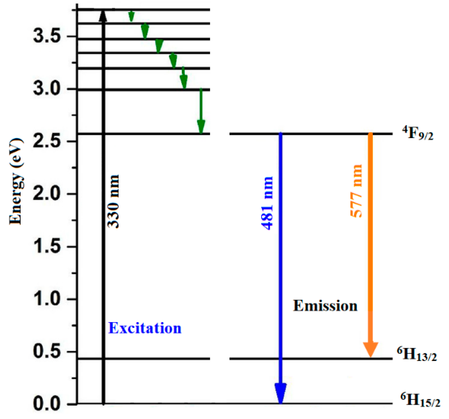
The two transitions 4F9/2→6H15/2 and 4F9/2→4H13/2 are ascribed to a magnetic and forced electric dipole transitions, respectively. The crystal field strength of host matrix does not strongly affect the transition to the 6H15/2 level, while that the one toward 4H13/2 level is hypersensitive to the surroundings. We have noticed that the spectral intensity of the 4F9/2→4H13/2 transition is higher than the 4F9/2→6H15/2 transition. This is due to the localization of Dy3+ ions on sites of low symmetry without centers of inversion. A large interaction between the host matrix and the RE ion is induced by asymmetry when the intensity of the hypersensitive transition is high [46]. For ZnO nanoparticles, the quantum efficiency of the visible emission can reach around 20% after UV excitation [47]. We see that the quantum efficiency increases slightly with the addition of Dy, this may be related to the decrease in particle size [47], confirming the result of XRD. Figure 10 shows the energy level diagram of Dy3+-doped ZnO:V. As the excitation energy (330 nm) is greater than the energy of 4F9/2 level (475 nm), the excess energy is lost through non-radiative channels. This produces the radiative emission of the populated level 4F9/2.
4. Conclusions
Zn0.99-xV0.01DyxO nanopowders were prepared by sol–gel route. XRD and TEM analysis show the presence of polycrystalline wurtzite structure and average crystallite size around 30 nm. The absorbance shows a red shift after doping, indicating the decrease of the band gap due to creation of defects in the band gap. The photoluminescence study shows the existence of two emission peaks centered at around 481 nm and 577 nm linked to the effect of co-doping by Dy and confirming the absorption results. We suggest that these emissions peaks are related to the transitions 4F9/2→6H15/2 and 4F9/2→6H13/2, respectively. These results confirm the good synthesis of our samples and offer major advantage for theirs use in the optoelectronic domain.
Author Contributions
Conceptualization, F.F.A.-H. and J.M.E.G.; methodology, F.F.A.-H. and J.M.E.G.; software, F.F.A.-H. and J.M.E.G.; validation, F.F.A.-H. and J.M.E.G.; formal analysis, F.F.A.-H. and J.M.E.G. All authors have read and agreed to the published version of the manuscript.
Funding
This research received no external funding.
Data Availability Statement
Data presented are original and not inappropriately selected, manipulated, enhanced, or fabricated. This includes (1) exclusion of data points to enhance significance of conclusions, (2) fabrication of data, (3) selection of results that support a particular conclusion at the expense of contradictory data, (4) deliberate selection of analysis tools or methods to support a particular conclusion (including p-hacking). We strongly recommend preregistration of methods and analysis.
Acknowledgments
This research was funded by the Deanship of Scientific Research at Princess Nourah bint Abdulrahman University through the Fast-track Research Funding program.
Conflicts of Interest
The authors declare no conflict of interest.
References
- Ellmer, K.; Klein, A. ZnO and Its Applications; Springer Series in Materials Science; Springer: Berlin/Heidelberg, Germany, 2008; Volume 104, p. 1. [Google Scholar]
- Calnan, S.; Tiwari, A.N. High mobility transparent conducting oxides for thin film solar cells. Thin Solid Film. 2010, 518, 1839. [Google Scholar] [CrossRef]
- El Ghoul, J. Synthesis of vanadium doped ZnO nanoparticles by sol–gel method and its characterization. J. Mater. Sci.-Mater. Electron. 2016, 27, 2159. [Google Scholar] [CrossRef]
- Janotti, A.; van de Walle, C.G. Fundamentals of zinc oxide as a semiconductor. Rep. Prog. Phys. 2009, 72, 126501. [Google Scholar] [CrossRef] [Green Version]
- El Ghoul, J.; Bouguila, N.; Gómez-Lopera, S.A.; El Mir, L. Structural and optical properties of nanoparticles (V, Al) co-doped ZnO synthesized by sol–gel processes. Superlattices Microstruct. 2013, 64, 451. [Google Scholar] [CrossRef]
- Mehrabian, M.; Azimirad, R.; Mirabbaszadeh, K.; Afarideh, H.; Davoudian, M. UV detecting properties of hydrothermal synthesized ZnO nanorods. Phys. E 2011, 43, 1141. [Google Scholar] [CrossRef]
- Willander, M.; Nur, O.; Sadaf, J.R.; Qadir, M.I.; Zaman, S.; Zainelabdin, A.; Bano, N.; Hussain, I. Luminescence from Zinc Oxide Nanostructures and Polymers and Their Hybrid Devices. Materials 2010, 3, 2643. [Google Scholar] [CrossRef]
- Willander, M.; Nur, O.; Zhao, Q.X.; Yang, L.L.; Lorenz, M.; Cao, B.Q.; Perez, J.Z.; Zekalla, C.; Zimmermann, G.; Grundmann, M.; et al. Zinc oxide nanorod based photonic devices: Recent progress in growth, light emitting diodes and lasers. Nanotechnology 2009, 20, 332001. [Google Scholar] [CrossRef]
- Ishizumi, A.; Kanemitsu, Y. Structural and luminescence properties of Eu-doped ZnO nanorods fabricated by a microemulsion method. Appl. Phys. Lett. 2005, 86, 253106. [Google Scholar] [CrossRef] [Green Version]
- Liu, Y.; Luo, W.; Li, R.; Chen, X. Spectroscopic evidence of the multiple-site structure of Eu3+ ions incorporated in ZnO nanocrystals. Opt. Lett. 2007, 32, 566. [Google Scholar] [CrossRef]
- Armelao, L.; Bottaro, G.; Pascolini, M.; Sessolo, M.; Tondello, A. Structure−Luminescence correlations in europium-doped sol−gel ZnO nanopowders. J. Phys. Chem. C 2008, 112, 4049. [Google Scholar] [CrossRef]
- Taguchi, S.; Ishizumi, A.; Tayagaki, T.; Kanemitsu, Y. Mn–Mn couplings in Mn-doped CdS nanocrystals studied by magnetic circular dichroism spectroscopy. Appl. Phys. Lett. 2009, 94, 173101. [Google Scholar] [CrossRef] [Green Version]
- Liu, Y.; Li, R.; Luo, W.; Zhu, H.; Chen, X. Optical spectroscopy of Sm3+ and Dy3+ doped ZnO nanocrystals. Spectrosc. Lett. 2010, 43, 343. [Google Scholar] [CrossRef]
- Wu, G.S.; Zhuang, Y.L.; Lin, Z.Q.; Yuan, X.Y.; Xie, T.; Zhang, L.D. Synthesis and photoluminescence of Dy-doped ZnO nanowires. Phys. E 2006, 31, 5. [Google Scholar] [CrossRef]
- Liu, E.Z.; Liu, Y.; He, J.Z. Ferromagnetism induced by defect complex in Co-doped ZnO. Appl. Phys. Lett. 2008, 93, 132506. [Google Scholar] [CrossRef]
- Kim, D.; Yang, J.; Hong, J. Ferromagnetism induced by Zn vacancy defect and lattice distortion in ZnO. J. Appl. Phys. 2009, 106, 013908. [Google Scholar] [CrossRef]
- Chen, B.; Yu, Q.X.; Gao, Q.Q.; Liao, Y.; Wang, G.Z. Structural reconstruction and defects transition in mediating room temperature ferromagnetism in Co-doped ZnO film. Appl. Phys. Lett. 2013, 102, 132405. [Google Scholar] [CrossRef]
- Kaushik, A.; Dalela, B.; Rathore, R.; Vats, V.S.; Choudhary, B.L.; Alvi, P.A.; Kumar, S.; Dalela, S. Influence of Co doping on the structural, optical and magnetic properties of ZnO nanocrystals. J. Alloys Compd. 2013, 578, 328. [Google Scholar] [CrossRef]
- Wibowo, J.A.; Djaja, N.F.; Saleh, R. Cu-and Ni-doping effect on structure and magnetic properties of Fe-doped ZnO nanoparticles. Adv. Mater. Phys. Chem. 2013, 3, 48. [Google Scholar] [CrossRef] [Green Version]
- Kumar, S.; Chen, C.L.; Dong, C.L.; Ho, Y.K.; Lee, J.F.; Chan, T.S.; Thangavel, R.; Chen, T.K.; Mok, B.H.; Rao, S.M.; et al. Structural, optical, and magnetic characterization of Co and N co-doped ZnO nanopowders. J. Mater. Sci. 2013, 48, 2618. [Google Scholar] [CrossRef]
- Kaur, P.; Pandey, S.K.; Kumar, S.; Negi, N.S.; Chen, C.L.; Rao, S.M.; Wu, M.K. Tuning ferromagnetism in zinc oxide nanoparticles by chromium doping. Appl. Nano 2015, 5, 975. [Google Scholar] [CrossRef] [Green Version]
- Murmu, P.P.; Kennedy, J.; Ruck, B.J.; Williams, G.V.M.; Markwit, A.; Rubanov, S.; Suvorova, A.A. Effect of annealing on the structural, electrical and magnetic properties of Gd-implanted ZnO thin films. J. Mater. Sci. 2012, 47, 1119. [Google Scholar] [CrossRef]
- Kumar, S.; Kaur, P.; Chen, C.L.; Thangavel, R.; Dong, C.L.; Ho, Y.K.; Lee, J.F.; Chan, T.S.; Chen, T.K.; Mok, B.H.; et al. optical and magnetic characterization of Ru doped ZnO nanorods. J. Alloys Compd. 2014, 588, 705. [Google Scholar] [CrossRef]
- Kumar, S.; Thangavel, R. Gd doping effect on structural, electrical and magnetic properties of ZnO thin films synthesized by sol-gel spin coating technique. Elecon. Mater. Lett. 2017, 13, 129. [Google Scholar] [CrossRef]
- Kaur, P.; Kumar, S.; Chen, C.L.; Hsu, Y.Y.; Chan, T.S.; Dong, C.L.; Srivastava, C.; Singh, A.; Rao, S.M. Investigations on structural, magnetic and electronic structure of Gd-doped ZnO nanostructures synthesized using sol–gel technique. Appl. Phys. A 2016, 122, 1. [Google Scholar] [CrossRef]
- Wu, Z.F.; Cheng, K.; Zhang, F.; Guan, R.F.; Wuc, X.M.; Zhuge, L.J. Effect of Al co-doping on the electrical and magnetic properties of Cu-doped ZnO nanorods. J. Alloys Compd. 2014, 615, 521. [Google Scholar] [CrossRef]
- Chattopadhyay, S.; Nath, T.K.; Behan, A.J.; Neal, J.R.; Score, D.; Feng, Q.; Fox, A.M.; Gehring, G.A. Enhancement of room temperature ferromagnetism of Fe-doped ZnO epitaxial thin films with Al co-doping. J. Magn. Magn. Mater. 2011, 323, 1033. [Google Scholar] [CrossRef]
- Liu, L.; Yu, P.Y.; Ma, Z.; Mao, S.S. Enhancement of room temperature ferromagnetism of Fe-doped ZnO epitaxial thin films with Al co-doping. Phys. Rev. Lett. 2008, 100, 127203. [Google Scholar] [CrossRef] [Green Version]
- Xu, Q.; Schmidt, H.; Hochmuth, H.; Lorenz, M.; Setzer, A.; Esquinazi, P.; Meinecke, C.; Grundmann, M. Room temperature ferromagnetism in Nd-and Mn-codoped ZnO films. J. Phys. D Appl. Phys. 2008, 41, 105012. [Google Scholar] [CrossRef]
- El Ghoul, J.; Al-Harbi, F.F. Synthesis, Structural and Optical Properties of Er and V Codoping ZnO Nanoparticles. J. Inorg. Organomet. Polym. Mater. 2021, 31, 272. [Google Scholar] [CrossRef]
- Assadi, M.H.N.; Zhang, Y.B.; Photongkam, P.; Li, S. Intrinsic ambient ferromagnetism in ZnO: Co induced by Eu codoping. J. Appl. Phys. 2011, 109, 013909. [Google Scholar] [CrossRef]
- Huang, H.; Ou, Y.; Xu, S.; Fang, G.; Li, M.; Zhao, X.Z. Properties of Dy-doped ZnO nanocrystalline thin films prepared by pulsed laser deposition. Appl. Surf. Sci. 2008, 254, 2013. [Google Scholar] [CrossRef]
- Omri, K.; el Ghoul, J.; Lemine, O.M.; Bououdina, M.; Zhang, B.; El Mir, L. Magnetic and optical properties of manganese doped ZnO nanoparticles synthesized by sol-gel technique. Superlattices Microstruct. 2013, 60, 139. [Google Scholar] [CrossRef]
- El Ghoul, J.; Al-Harbi, F.F. Synthesis, structural, optical and magnetic properties of Gd co-doped ZnO: V nanoparticles. Solid State Commun. 2020, 314, 113916. [Google Scholar] [CrossRef]
- Mourad, S.; El Ghoul, J.; Omri, K.; Khirouni, K. Indium doping effect on properties of ZnO nanoparticles synthesized by sol–gel method. Chin. Phys. B 2019, 28, 047701. [Google Scholar] [CrossRef]
- Powder Diffraction File. Joint Committee for Powder Diffraction Studies (JCPDS) File No. 36-1451.
- Thangeeswari, T.; Murugasen, P.; Velmuruga, J. Influence of Co and Dy doping on the optical and magnetic properties of ZnO nanoparticles for DMS application. J. Supercond. Nov. Magn. 2015, 28, 2505. [Google Scholar] [CrossRef]
- Zhan, F.; Yang, Y.; Li, W.; Li, J.; Liu, W.; Li, Y.; Chen, Q. Preparation of DyVO4/WO3 heterojunction plate array films with enhanced photoelectrochemical activity. RSC Adv. 2016, 6, 10393. [Google Scholar] [CrossRef]
- Zhang, L.; Chen, D.R.; Jiao, X.L. Monoclinic structured BiVO4 nanosheets: Hydrothermal preparation, formation mechanism, and coloristic and photocatalytic properties. J. Phys. Chem. B 2006, 110, 2668. [Google Scholar] [CrossRef] [PubMed]
- Jayachandraiah, C.; Kumar, K.S.; Krishnaiah, G.; Rao, N.M. Influence of Dy dopant on structural and photoluminescence of Dy-doped ZnO nanoparticles. J. Alloys Compd. 2015, 623, 248. [Google Scholar] [CrossRef]
- Russo, V.; Ghidelli, M.; Gondoni, P.; Casari, C.S.; Bassi, A.L. Multi-wavelength Raman scattering of nanostructured Al-doped zinc oxide. J. Appl. Phys. 2014, 115, 073508. [Google Scholar] [CrossRef] [Green Version]
- Lung, C.; Toma, M.; Pop, M.; Marconi, D.; Pop, A. Characterization of the structural and optical properties of ZnO thin films doped with Ga, Al and (Al+ Ga). J. Alloys Compd. 2017, 725, 1238. [Google Scholar] [CrossRef]
- Batista, C.; Teixeira, V.; Carneiro, J.O. Structural and morphological characterization of magnetron sputtered nanocrystalline vanadium oxide films for thermochromic smart surfaces. J. Nano Res. 2008, 2, 21. [Google Scholar] [CrossRef] [Green Version]
- Thaslin, S.; Fathima, N.; Anandhan, A.; Ganesan, A.R.K.P.; Karthikeyan, M.; Marimuthu, T. Surface texture and luminous analysis of Sol-Gel spin coated Dy-doped ZnO thin films. IRJET 2017, 4, 89. [Google Scholar]
- Kumar, J.S.; Pavani, K.; Babu, A.M.; Giri, N.K.; Rai, S.B.; Moorthy, L.R. Fluorescence characteristics of Dy3+ ions in calcium fluoroborate glasses. J. Lumin. 2010, 130, 1916. [Google Scholar] [CrossRef]
- Gruber, J.B.; Zandi, B.; Valiev, U.V.; Rakhimov, S.A. Energy levels of Dy3+(4f9) in orthoaluminate crystals. J. Appl. Phys. 2003, 94, 1030. [Google Scholar] [CrossRef]
- Van Dijken, A.; Makkinje, J.; Meijerink, A. The influence of particle size on the luminescence quantum efficiency of nanocrystalline ZnO particles. J. Lumin. 2001, 92, 323. [Google Scholar] [CrossRef]
Figure 1.
XRD patterns of undoped and Zn0.99-xV0.01DyxO (x = 0.00, 0.04, and 0.08) nanoparticles.
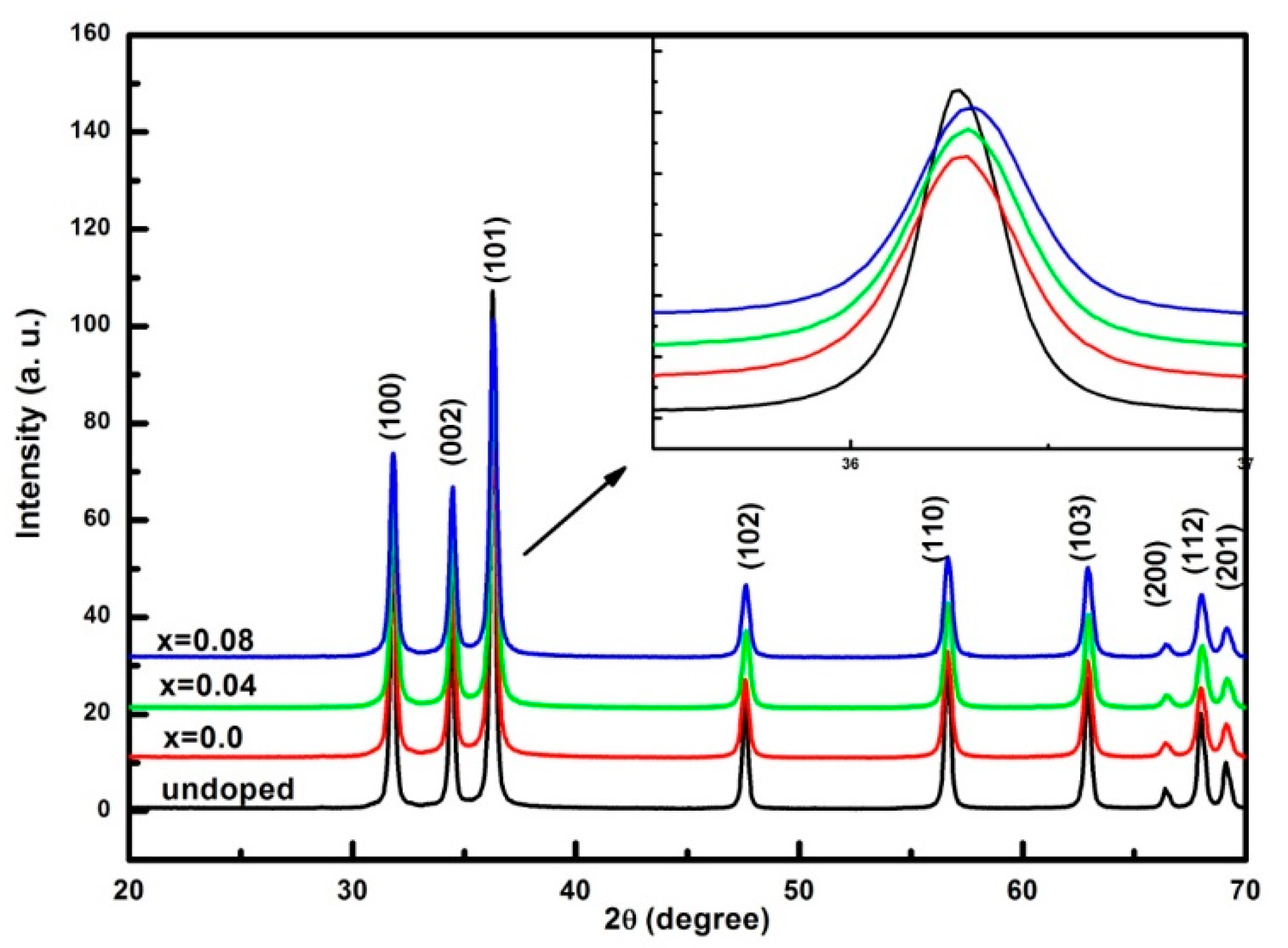
Figure 2.
TEM images of (a) Zn0.99V0.01O, (b) Zn0.95V0.01Dy0.04O, and (c) Zn0.91V0.01Dy0.08O nanoparticles.
Figure 2.
TEM images of (a) Zn0.99V0.01O, (b) Zn0.95V0.01Dy0.04O, and (c) Zn0.91V0.01Dy0.08O nanoparticles.
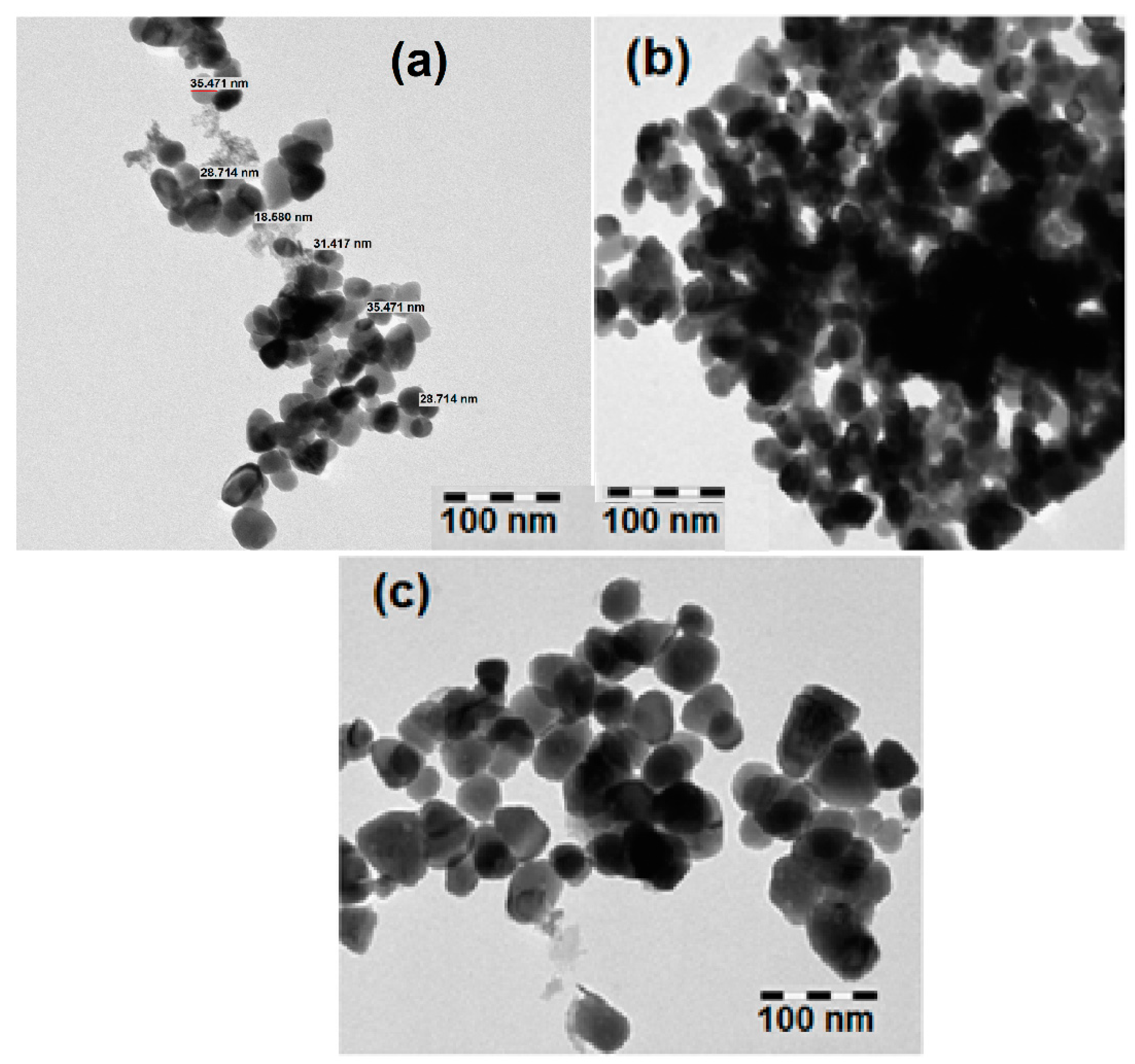
Figure 3.
EDX spectrum of Zn0.91V0.01Dy0.08O nanoparticles.
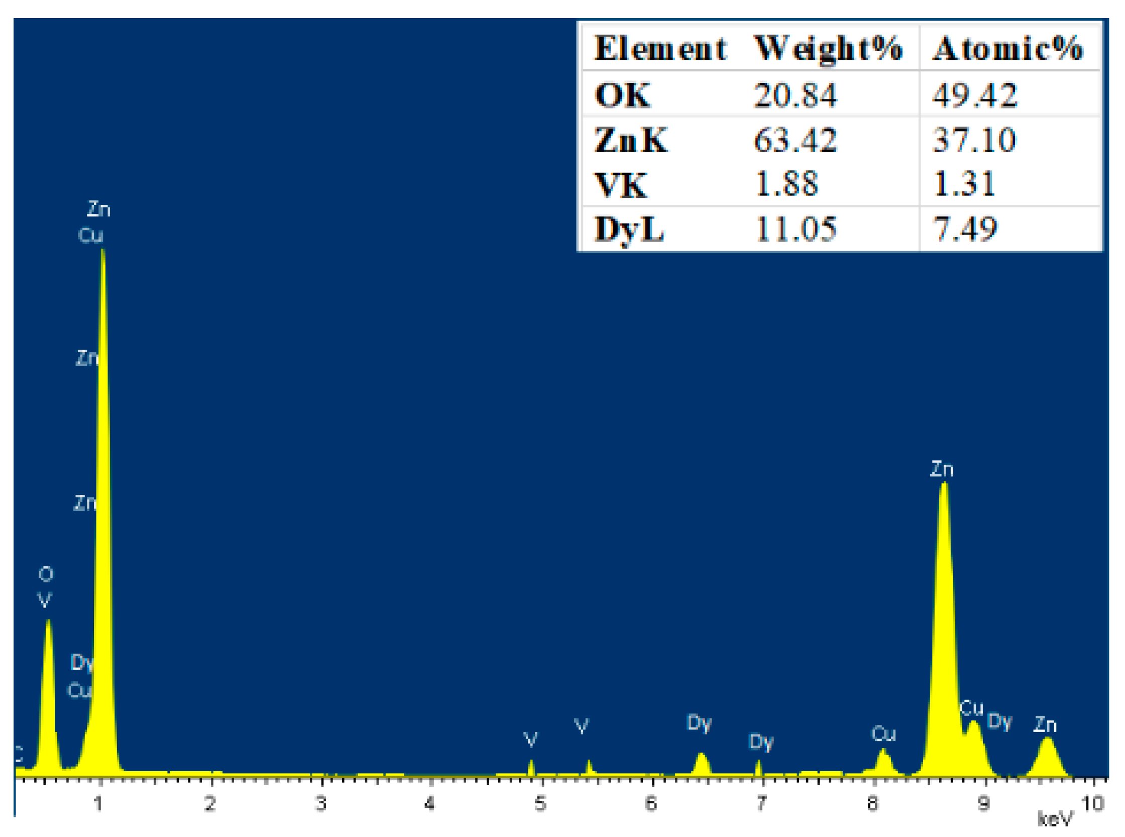
Figure 4.
XPS spectrum of Zn0.91V0.01Dy0.08O nanoparticles.
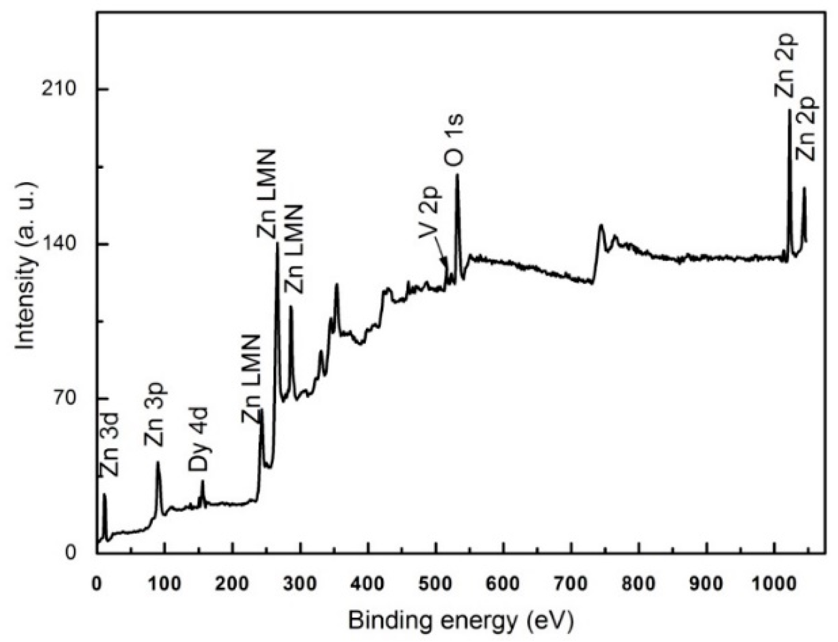
Figure 5.
XPS spectra of O 1s of Zn0.95V0.01Dy0.04O nanoparticles.
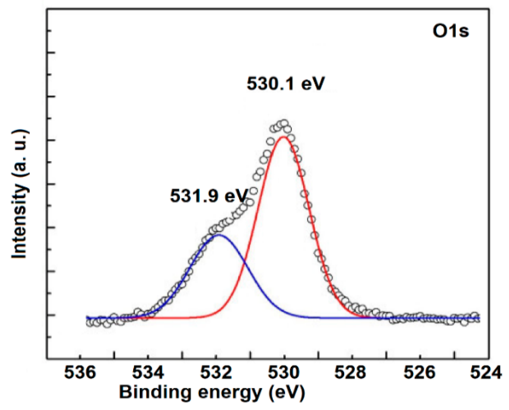
Figure 6.
Raman spectrum of Zn0.95V0.01Dy0.04O nanoparticles.
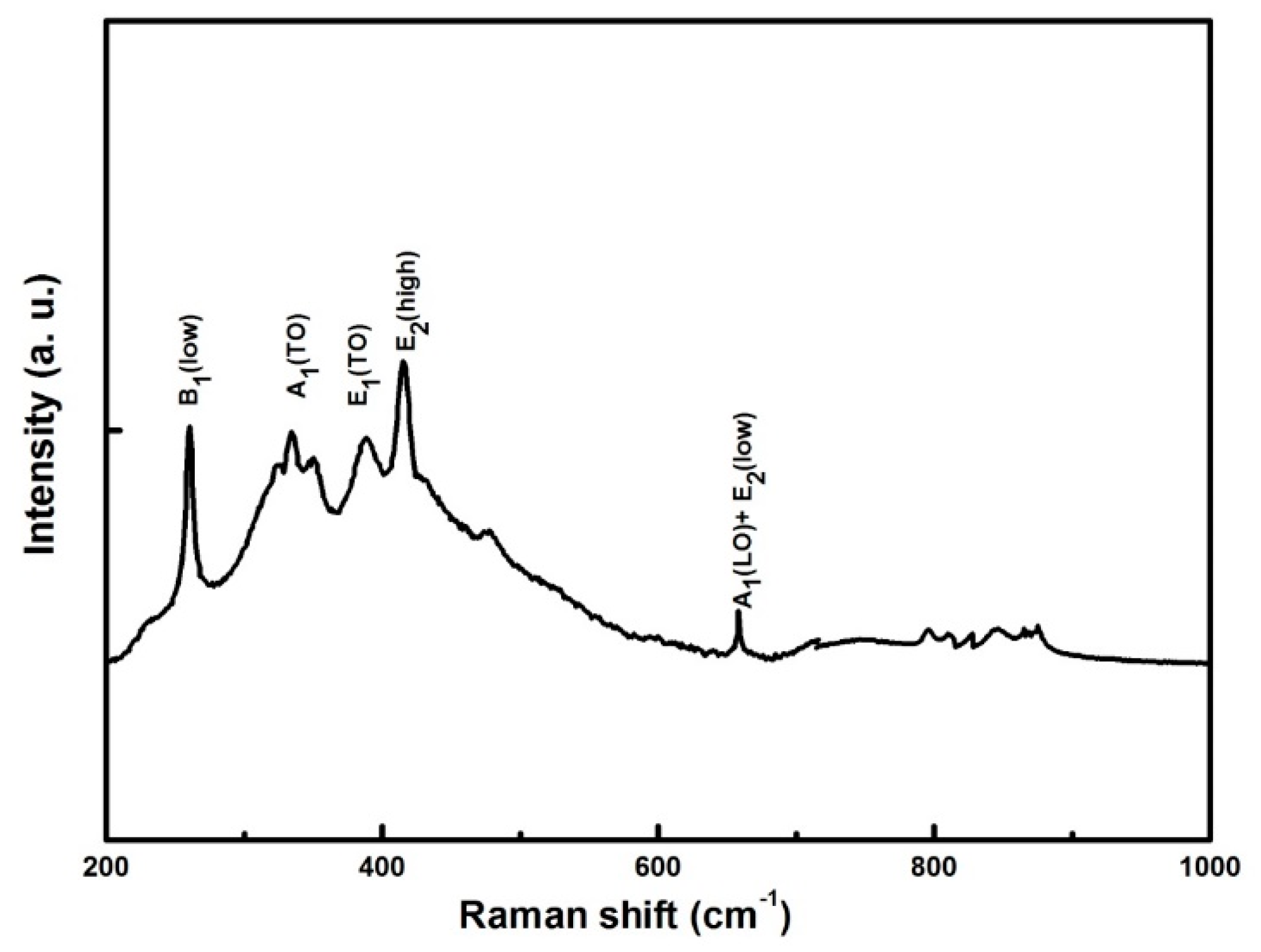
Figure 7.
Reflectance spectra of undoped and Zn0.99-xV0.01DyxO (x = 0.00, 0.04, and 0.08) nanoparticles. The inset shows enlarged portion of the absorption.
Figure 7.
Reflectance spectra of undoped and Zn0.99-xV0.01DyxO (x = 0.00, 0.04, and 0.08) nanoparticles. The inset shows enlarged portion of the absorption.
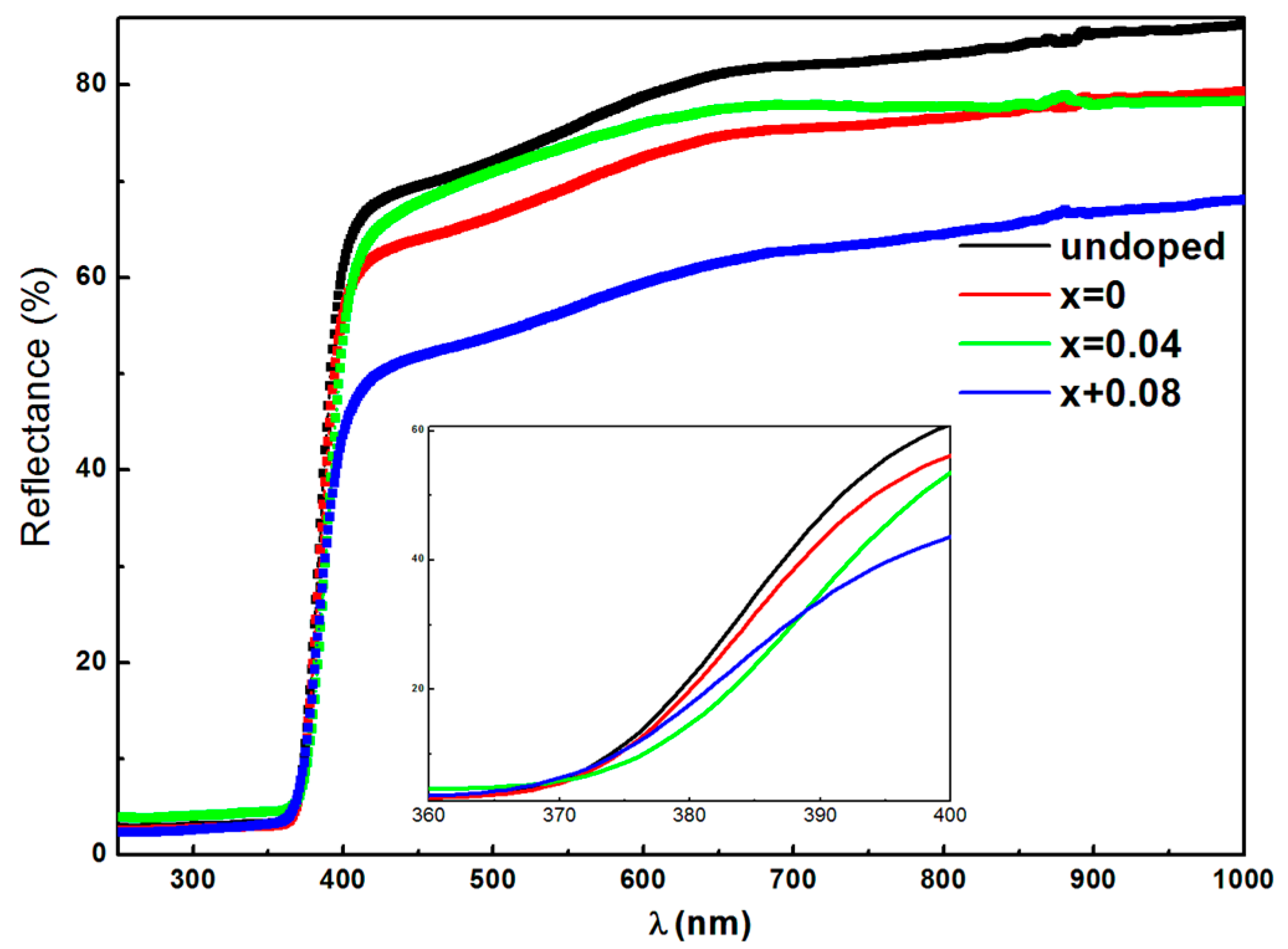
Figure 8.
First derivative of the reflectance (dR/dλ) vs. λ of undoped and Zn0.99-xV0.01DyxO (x = 0.00, 0.04, and 0.08) nanoparticles.
Figure 8.
First derivative of the reflectance (dR/dλ) vs. λ of undoped and Zn0.99-xV0.01DyxO (x = 0.00, 0.04, and 0.08) nanoparticles.
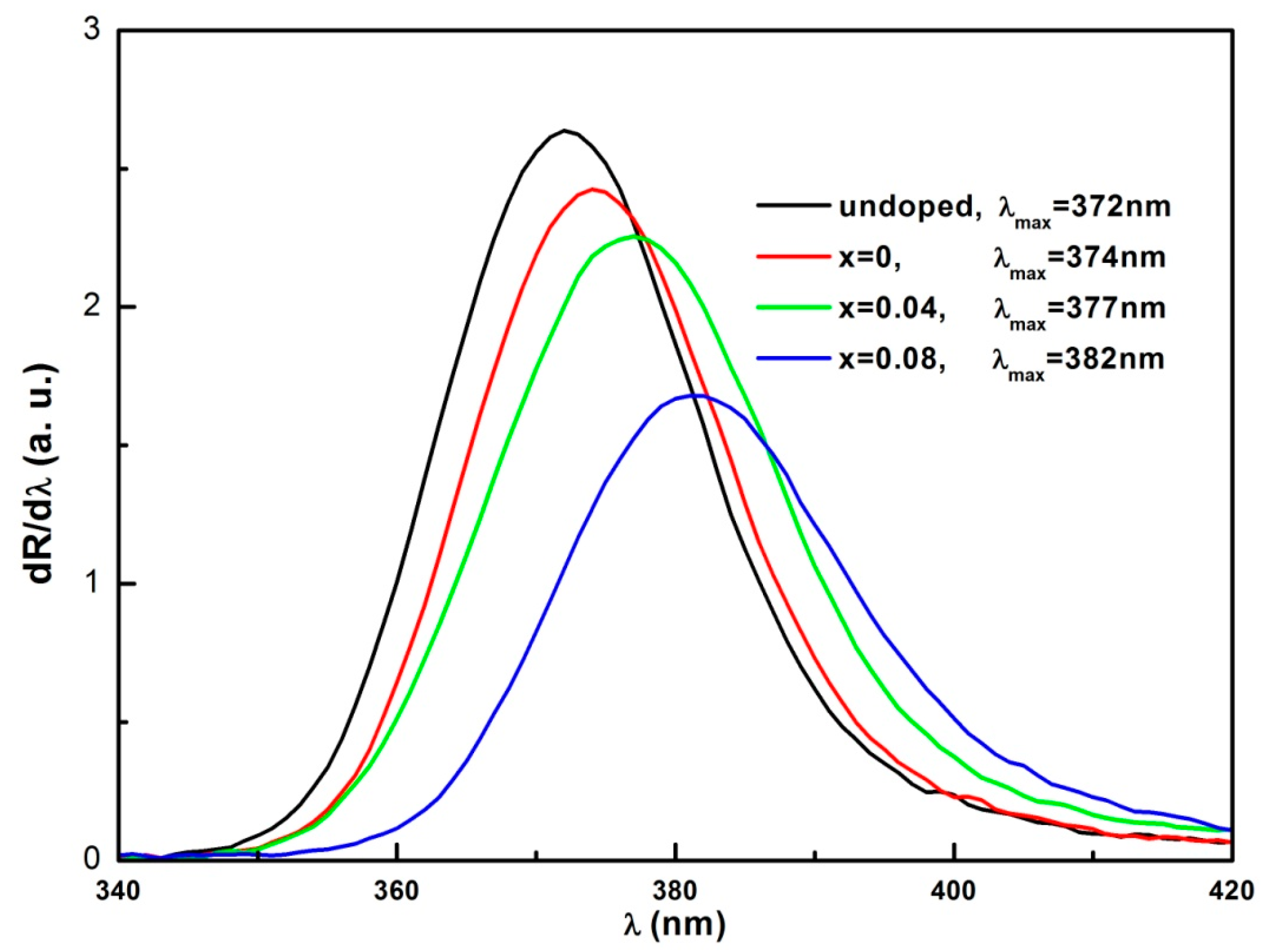
Figure 9.
PL spectra of Zn0.99-xV0.01DyxO (x = 0.00, 0.04, and 0.08) nanoparticles. The picture of Zn0.91V0.01Dy0.08O nanoparticles without and with excitation are shown in the upper left.
Figure 9.
PL spectra of Zn0.99-xV0.01DyxO (x = 0.00, 0.04, and 0.08) nanoparticles. The picture of Zn0.91V0.01Dy0.08O nanoparticles without and with excitation are shown in the upper left.
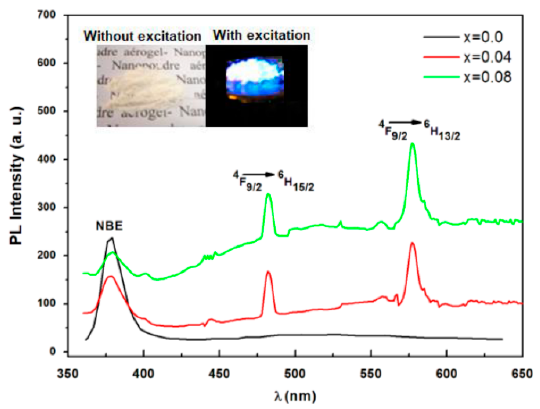
Figure 10.
Energy level diagram of Dy3+-doped ZnO:V.

Table 1.
Variation of different physical parameters for all samples.
| Sample | a (Å) | C (Å) | Crystallite Size (nm) | Strain (ε) × 10−4 | Bandgap (eV) |
|---|---|---|---|---|---|
| Undoped ZnO | 3.2498 | 5.2063 | 34.71 | 1.59 | 3.333 |
| x = 0.0 | 3.2497 | 5.2063 | 32.84 | 1.48 | 3.315 |
| x = 0.04 | 3.2494 | 5.2064 | 29.73 | 1.39 | 3.287 |
| x = 0.08 | 3.2495 | 5.2065 | 27.38 | 1.31 | 3.246 |
Publisher’s Note: MDPI stays neutral with regard to jurisdictional claims in published maps and institutional affiliations. |
© 2021 by the authors. Licensee MDPI, Basel, Switzerland. This article is an open access article distributed under the terms and conditions of the Creative Commons Attribution (CC BY) license (https://creativecommons.org/licenses/by/4.0/).
Share and Cite
MDPI and ACS Style
Al-Harbi, F.F.; El Ghoul, J.M. Sol–Gel Synthesis of Dy Co-Doped ZnO:V Nanoparticles for Optoelectronic Applications. Condens. Matter 2021, 6, 35. https://doi.org/10.3390/condmat6030035
AMA Style
Al-Harbi FF, El Ghoul JM. Sol–Gel Synthesis of Dy Co-Doped ZnO:V Nanoparticles for Optoelectronic Applications. Condensed Matter. 2021; 6(3):35. https://doi.org/10.3390/condmat6030035
Chicago/Turabian StyleAl-Harbi, Fatemah F., and Jaber Mohamed El Ghoul. 2021. "Sol–Gel Synthesis of Dy Co-Doped ZnO:V Nanoparticles for Optoelectronic Applications" Condensed Matter 6, no. 3: 35. https://doi.org/10.3390/condmat6030035