Article

Magnetically Recoverable and Reusable Antimicrobial Nanocomposite Based on Activated Carbon, Magnetite Nanoparticles, and Silver Nanoparticles for Water Disinfection

1
Math & Science Department, United States Merchant Marine Academy, Kings Point, NY 11024, USA
2
Neuroscience Department, Johns Hopkins University, Baltimore, MD 21218, USA
3
Instrumentation Division, Brookhaven National Laboratory, Upton, NY 11973, USA
*
Author to whom correspondence should be addressed.
Inventions 2017, 2(2), 10; https://doi.org/10.3390/inventions2020010
Submission received: 25 April 2017 / Revised: 28 May 2017 / Accepted: 30 May 2017 / Published: 6 June 2017
(This article belongs to the Special Issue Nanomaterials and Nanocomposites for Environmental Applications)

Abstract

:
Recent advancements in nanotechnology have led to the development of innovative, low-cost and highly efficient water disinfection technologies that may replace or enhance the conventional methods. In this study, we introduce a novel procedure for preparing a bifunctional activated carbon nanocomposite in which nanoscale-sized magnetic magnetite and antimicrobial silver nanoparticles are incorporated (MACAg). The antimicrobial efficacy of the nanocomposite was tested against Escherichia coli (E. coli). MACAg (0.5 g, 0.04% Ag) was found to remove and kill 106–107 CFU (colony-forming units) in 30 min via a shaking test and the removing and killing rate of the nanocomposites increased with increasing silver content and decreased with increasing CFU. The inhibition zone tests revealed, among the relevant components, only Ag nanoparticles and Ag+ ions showed antimicrobial activities. The MACAg was easily recoverable from treated water due to its magnetic properties and was able to remove and kill 106 CFU after multiple-repeated use. The MACAg nanocomposite also demonstrated its feasibility and applicability for treating a surface water containing 105 CFU. Combining low cost due to easy synthesis, recoverability, and reusability with high antimicrobial efficiency, MACAg may provide a promising water disinfection technology that will find wide applications.

1. Introduction

Microbial contamination of drinking water poses a serious health concern, especially in developing countries [1]. Marine invasive microorganisms spread across the globe by ships through their ballast water tanks can have devastating effects on ecosystems [2,3]. Disinfecting water, drinking or ballast, by removing, deactivating, or killing harmful microorganisms is therefore of great importance. Current water disinfection methods include chemical disinfection, filtration, and ultraviolet (UV) disinfection. Chemical disinfection using chlorine is the most popular method. However recent research has revealed that, while effective at disinfection, chemical disinfectants such as chlorine, chloramines, and ozone react with various constituents in natural water, forming disinfection by-products (DBPs) [4]. Many of these DBPs are carcinogens [5]. Although filtration and UV-disinfection methods are effective in eliminating microbial contamination, their operational costs are very high and some procedures can be quite time-consuming [6]. Innovative, low-cost, and highly efficient water disinfection methods are therefore urgently needed.
Recently, nanotechnology has opened an alternative method of water disinfection. Unlike the chemical disinfectants, the commonly known antimicrobial nanomaterials are relatively inert in water and are not expected to form harmful DBPs. They can, therefore, potentially replace or enhance the conventional disinfection methods. Among these nanomaterials, silver-based nanoparticles (AgNPs) have been most intensively studied due to their strong antimicrobial activity toward many different bacteria [7,8], fungi [9], algae [10], and viruses [11] and their relatively low toxicity to humans (AgNPs concentrations of 2–4 ppm effectively inhibit bacterial growth but are not toxic for human healthy cells) [12]. Since ancient times, silver has been proven as a well-known agent for water purifying and has long been used in the treatment of infections, burns and chronic wounds [13]. However, two challenges may limit the use of AgNPs for water disinfection: (1) their aggregation in water; and (2) their removal from treated water. Nanoparticles are known to have a strong tendency to form aggregates in water due to van der Waals forces and high surface energy. The antimicrobial activity of silver nanoparticles is a result of nanometer size, with the smaller particles having higher activity on the basis of equivalent silver mass content [10,14]. The aggregation of silver nanoparticles may reduce or diminish the particles’ ability to disinfect. Recently, it has been reported that high concentrations of AgNPs (>26 ppm) may harm mammalian cells [10]. Because of this, removing silver nanoparticles from water after disinfection is necessary. Moreover, the removed silver nanoparticles can also be reused for the next cycle of water disinfection, reducing the material cost and avoiding any adverse environmental effects which would be involved in their disposal [7].
Nanocomposites are a special class of nanomaterials. They are multiphase solid materials with at least one of the phases being nano-scaled. Nanocomposites represent an exciting class of advanced materials due to their synergistic or hybrid properties derived from their components. They offer unique properties that cannot be achieved through the use of conventional materials [15,16]. Nanocomposites are prepared by intimately binding one or more inorganic materials to an inorganic or organic host material. In recent years, incorporation of AgNPs into different filter or absorbent materials of both organic and inorganic host matrixes has been considered promising for water disinfection and treatment [17,18,19,20,21,22,23]. The host matrix provides high dispersion and prevents aggregation of embedded AgNPs. The most hopeful hosting material is activated carbon (AC), since it is the most widely used material in water treatment throughout the world [24]. AC is renowned for being an effective absorbent for the removal of a wide variety of organic and inorganic matter [15,24,25,26,27,28], due to its extended surface area, large porosity, total pore volume, and complex porous structure. Coupled with the presence of a wide spectrum of functional groups on its surface, AC provides high adsorption capacity, high degree of surface reactivity, and strong affinity for even low concentrations of pollutants. In 2016, Karthik et al. [22] prepared an antimicrobial nanocomposite by loading AgNPs (25–85 nm) onto AC. The nanocomposite was found to inhibit the growth of Escherichia coli (E. coli) and Bacillus subtilis (B. subtilis) and microbes in beverage, brewery, dairy, and dye industrial effluents. Eltugral et al. [23] investigated the effect of water soluble ligands on the preparation of AgNP (7–20 nm)-supported AC and found that AgNPs prepared by NaBH4 had the most dense and homogenous distribution on the AC support and seemed to remain the tightest on the AC surface after rinsing with water. However, nano-silver-support AC may have issues regarding separation and regeneration. AC-based nanocomposites are usually applied in columns or filter device. Due to their fine sizes, the carbon nanocomposite particles may block the water flow or pass through the filters. In large-scale water treatment processes, this and related problems may result in slow processes and high operational cost. Additionally, the reuse of these materials requires additional time-consuming procedures, often leading to material being discarded after use, a process that creates a secondary pollution. To overcome these disadvantages, the AgNP-supported AC may be combined with a magnetic carrier to develop a magnetic antimicrobial nanocomposite. Magnetic separation is attractive since it is instant and magnetically controllable. It allows the antimicrobial agents to be recovered easily and reused for the next cycle of water disinfection, conserving materials and reducing environmental impacts.
In this manuscript, we describe a facile process that prepares a magnetic antimicrobial AC nanocomposite in which nanoscale-sized magnetite and silver particles are incorporated (MACAg). At the U.S. Merchant Marine Academy, this procedure is modified and has been successfully implemented in the Chemistry for Marine Engineer course where 165 midshipmen have participated in preparing MACAg and testing its antimicrobial activity [29]. To our knowledge, such a nanocomposite has not been reported by other research groups. To prepare this bifunctional nanocomposite, magnetite nanoparticles (MNPs) are first immobilized on porous AC using the procedure reported previously to impart the magnetic properties to AC [15]. This intermediate product, magnetite-activated carbon nanocomposite, is referred to as MAC. This is followed by integrating AgNPs onto MAC, where AgNPs are formed via reducing silver ions from its salt solution by the borohydride (BH4) ions coated on MAC. This adds biocidal properties to MAC. The approach we take here aims to achieve the synergistic and/or hybrid properties of the nanocomposite derived from its components:
  • The superior antimicrobial properties of AgNPs for water disinfection;
  • The magnetic properties of MNPs for easy and fast removal of AgNPs from treated water and for making antimicrobial agent recoverable and reusable;
  • The superior adsorption ability of AC for organic and inorganic species;
  • And the immobilization of well dispersed nanoparticles on the AC and/or the MAC surfaces that prevents the aggregation of magnetite and silver nanoparticles in water, helping retain the superior properties of these nanoparticles.
The targeted antimicrobial properties of MACAg nanocomposite are proved effective against Escherichia coli (E. coli), a well-studied prokaryotic indicator organism, in water. The easily prepared MACAg is rapidly and readily separated from treated water due to its magnetic properties and is found to be able to remove microbes for repeated cycles with no observed decline in performance. The nanocomposite is shown to be stable and the AgNPs remain undetached after use. The nanocomposites are also shown to be feasible and applicable to disinfect surface water containing a wide range of microorganisms. The MACAg antimicrobial agent developed in this work is therefore a green and highly efficient product, demonstrating the concerted properties of its components, overcoming the challenges the AgNPs alone may face, and providing a promising new technology for water disinfection.

2. Materials and Methods

2.1. Materials

Concentrated hydrochloric acid (HCl), iron(II) tetrahydrate (FeCl2·4H2O), iron(III) hexahydrate (FeCl3·6H2O), concentrated ammonia (NH3), concentrated nitric acid (HNO3), activated carbon (AC) powder (20 μm), sodium borohydride (NaBH4), silver nitrate (AgNO3) were purchased from VWR International (Radnor, PA, USA). Silver nanoparticles (20–40 nm) were purchased from Aesar (Haverhill, MA, USA) and Neodymium magnets from Applied Magnets (Plano, TX, USA). Escherichia coli (E. coli) K-12 strain, Lysogeny Broth (LB) and LB nutrient agar were obtained from a Microbes and Health Kit purchased from Bio-Rad (Hercules, CA, USA). FD&C Red 40 (Allura Red, disodium 6-hydroxy-5-[(2-methoxy-5-methyl-4-sulfophenyl)azo]-2-napthalenesulfonate, C18H14N2Na2O8S2) was manufactured by Badia (Doral, FL, USA). Methylene blue (3,7-bis(dimenthylamino)-phenothiazub-5-ium chloride, ‎C16H18ClN3S) was from the Hubbard Scientific’s Microbiology Culture Kit purchased from Sigma-Aldrich (St. Louis, MO, USA).

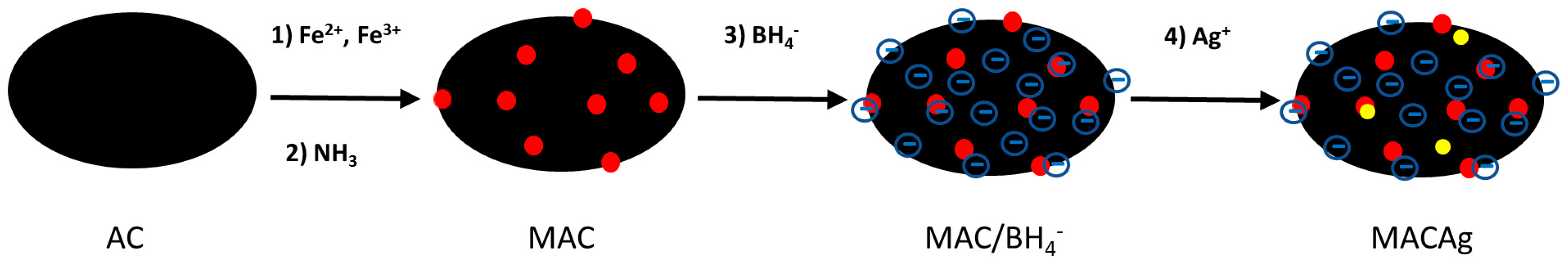
2.2. Synthesis of Magnetite-Activiated Carbon-Silver (MACAg) Nanocomposite

AgNPs, MNPs and MAC were prepared by adapting or modifying the procedures described in literature [15,30,31]. To prepare MACAg, 4.0 mL of 1.0 M FeCl3 and 1.0 mL of 2.0 M FeCl2 solution were mixed thoroughly with 0.10 g of activated carbon powder in a beaker for 3 min. The mixture was then titrated with 50 mL of 1.4 M NH3 solution dropwise over a period of 5–10 min with stirring. The resultant MAC was settled magnetically and the supernatant decanted. The MAC was washed with deionized water and then dried overnight at 40 °C. Thirty mL of 2.0 mM of chilled NaBH4 solution were added to the beaker containing the dried MAC and the mixture was vigorously mixed for 5 min in an ice bath. While stirring, 1.0 mM of AgNO3 solution ranging from 2 to 10 mL was dripped at one drop per second rate into the chilled MAC and NaBH4 mixture. After the addition of AgNO3, the mixture was mixed for 2–3 additional minutes before settling the MACAg magnetically. The supernatant was decanted and MACAg rinsed with deionized water.

2.3. Characterization

AgNPs, MNPs and MACAg were imaged using Scanning Electron Microscopy (SEM). The SEM observation was carried out on a JEOL 6500F (JEOL Ltd., Tokyo, Japan) with a thermally assisted field emission gun. A small sample was coated on an Al stub for each. The imaging employed 10 keV accelerating voltage for the electron beam with a beam current of 50 pA, and the image was captured with a secondary electron detector. The optical spectra at a resolution of 2 nm were recorded using a Lambda XLS Plus UV-Visible spectrophotometer (Perkin–Elmer, Waltham, MA, USA) to confirm the sizes and to determine the amounts of AgNPs suspended in water. To probe the available surface area of MAC or MACAg for adsorption, 26 ppm of Allura Red, an anionic dye, was prepared by diluting a stock solution of 70,000 ppm, and 100 mL of it was mixed vigorously with 0.10 g of MAC, MAC/BH4, or MACAg for 10 min. Additionally, 20 mL of a methylene blue (a cationic dye) solution, prepared by diluting the as-purchased solution by a factor of 100, was treated with 0.10 g of MAC or MAC/BH4 for 10 min.

2.4. Antimicrobial Activity of MACAg Nanocomposite

The antimicrobial properties of MACAg and MACAg components were investigated against a test micro-organism, E. coli, and the microbes in a sample of Long Island Sound (LIS) surface water. Cells of an E. coli K-12 strain were inoculated into liquid culture of LB broth and incubated with stirring in dark at 30 °C for 1–2 days. The resulting cell suspension, served as the E. coli stock, was further diluted to give concentrations ranging from 103–108 CFU/mL (CFU = colony-forming units). The antimicrobial activity was evaluated based on the inhibition zone tests and viable microbial colonies on nutrient agar plates. The microbes, inoculated onto agar plates, were allowed to grow in dark at 27–30 °C for two days.

2.4.1. Zone of Inhibition

The antimicrobial activity of relevant components of MACAg nanocomposite was examined based on the inhibition zone observed in the agar diffusion test. A 100 µL sample of E. coli suspension of 108 CFU/mL was spread evenly over the surface of a nutrient agar plate and allowed to dry at room temperature. Solid samples containing 0.25 mmol atoms of AgNPs (Aesar, 20–40 nm), AC and MNPs were applied to the agar plate. For liquid samples, holes were bored in the agar using sterile cork borer, each of which was then filled with 100 µL of liquid sample containing 2.5 × 10⁻⁵ mmol of Ag+ ions, Ag atoms in AgNPs (10 nm), Allura Red 40 or BH4 ions. The Ag+ ion solution served as control.

2.4.2. Shaking Tests

To demonstrate the antimicrobial activity of MACAg, 10–20 mL of E. coli suspension of concentrations ranging from 103–107 CFU/mL was added to the beaker containing 0.5 g of MACAg. The suspension was treated by shaking the mixture with a mechanical shaker at room temperature for selected periods of time. The supernatant was magnetically separated from MAC or MACAg at different stages of the treatment. At each stage, the supernatant was sampled and 6 µL of it was inoculated onto a nutrient agar plate to check microbial reduction as a function of time. In order to investigate the viable microbes attached to MACAg sediment at the completion of the treatment, small samples of the sediment before the wash, the wash (6 µL), and the sediment after the wash, were placed onto a nutrient agar plate. MAC without AgNPs was used as reference.

2.4.3. Repeated Use of MACAg Nanocomposite

A 0.50 g quantity of MACAg was used to treat 20 mL of an E. coli suspension containing 2 × 10⁶ CFU. The MACAg-E. coli mixture was shaken for 15–20 min. After the treatment, MACAg was settled by a magnet, and 6 µL of supernatant was taken and placed on a nutrient agar plate. The MACAg was then washed with deionized water and employed for the next cycle of antimicrobial test up to 5 cycles. After the 4th and 5th cycles, small samples of MACAg and wash were inoculated onto a nutrient agar plate to check for any viable microbes on the sediment.

2.4.4. Disinfection of Long Island Sound (LIS) Surface Water

The feasibility of employing MACAg nanocomposite for disinfecting Long Island Sound (LIS) surface water was studied by the shaking test. The LIS surface water was obtained at the Academy’s waterfront site on 4 April 2017. The shaking test was done by treating 20 mL of LIS surface water with 0.5 g of MACAg for a total of 15 min. The cultivable bacteria in the water after 3, 6, 9, 12 and 15 min treatments were observed by placing 10 µL of each treated water samples on nutrient agar plate. Samples of sediment after 15 min treatment and its washes were tested for the presence of viable bacteria by plate observation.

2.5. Release of Silver

Well-maintained structural integrity is very important for MACAg for its water disinfection application. The release of AgNPs and/or Ag+ ions into treated water would be of concern if the level reached 26 ppm for AgNPs or 0.10 ppm for Ag+ ions, for instance. The concentrations of silver in 20 mL of the E. coli suspension treated with MACAg and repeatedly used MACAg were evaluated with a UV-visible spectrophotometer (Perkin–Elmer, Lambda XLS Plus) and an Atomic Absorption Spectrophotometer (Perkin–Elmer, AAnalyst 200).

3. Results and Discussion

3.1. Synthesis and Characterization of MACAg

The schematic representation for preparing MACAg nanocomposite is shown in Figure 1. MAC nanocomposite was prepared by thoroughly mixing 0.10 g of AC with 4.0 mL of 1.0 M iron(III) chloride and 1.0 mL of 2.0 M iron(II) chloride solutions, followed by a dropwise addition of excess ammonia solution. After washing and drying, the procedure yielded 0.50–0.51 g of MAC (89–90% yield). A major advantage of MAC is its excellent adsorption capacity and its ability to be isolated magnetically [15]. To fully coat the MAC surface with borohydride (BH4) ions, a reducing agent for silver, MAC was vigorously mixed with 30 mL of 2.0 mM chilled sodium borohydride solution. With stirring, a silver nitrate solution was subsequently dripped at 1 drop per second rate into the mixture. The procedure typically yielded 0.49 g of MACAg (98% yield). The equations describing the formation of MNPs and AgNPs are given in Equations (1) and (2), respectively:
2FeCl3 (aq) + FeCl2 (aq) + 8NH3 (aq) + 4H2O (l) → Fe3O4 (s) + 8NH4Cl (aq)
AgNO3 (aq) + NaBH4 (aq) → Ag (s) + ½B2H6 (g) + ½H2 (g) + NaNO3 (aq)
The scanning electron microscopy (SEM) images of MNPs and AgNPs show that the MNPs were of sizes ranging from 10 to 20 nm, and AgNPs around 10–12 nm [29]. The plasmon absorbance of the yellow colloidal silver produced a peak at 399 nm with PWHM (peak width at half max) of 50–70 nm in the UV-visible spectrum. This peak corresponds to AgNP sizes of 10–12 nm, supporting the SEM result, and remains clearly visible at a trace concentration of 0.10 ppm. The SEM image of MACAg reveals the agglomerates of the MNPs and AgNPs of sizes less than 20 nm embedded onto and covering the AC surface [29]. If all the silver from the added 2–10 mL of 1.0 mM AgNO3 solution were incorporated into MAC, the theoretical mass content of silver would range from 0.04% to 0.22%. The presence of a large excess of borohydride ions promotes a complete conversion of Ag+ ions to AgNPs. The results of UV-visible spectra demonstrated less than or around 0.1 ppm of AgNPs remaining in the solutions after making MACAg using 2–10 mL of silver nitrate solutions. With Atomic Absorption Spectrometer, the silver concentration of the solution after making MACAg using 3 mL AgNO3 solution was found to be 0.02 ppm. This corresponded to a 7 × 10⁻⁴ mg Ag, which was negligible relative to the initial amount of 0.216 mg Ag introduced to the solution. However, about 0.1 ppm of AgNPs was detected in the solution when 10 mL of silver nitrate solution was used. This marked the upper limit of 0.5 g MAC’s ability to fully incorporate the formed AgNPs. Nevertheless, the residue results showed that almost all the silver introduced to the reaction mixture was incorporated into the MACAg products, so the theoretical and actual mass contents of silver for each product were nearly identical.
To investigate the available surface area of MAC, MAC coated with BH4 ions (MAC/BH4), and MACAg (0.22% Ag) for adsorption, each 0.10 g of these samples was vigorously stirred with 100 mL of 26 ppm Allura Red for 10 min. MAC/BH4 was prepared by mixing NaBH4 solution with MAC followed by 3 times of rinse with deionized water. Allura Red (Figure 2a) is a disodium salt of relatively high molar mass of 496 g/mol. The ionic nature allows the salt to be water soluble. The large anions are amphiphilic, and absorb primarily wavelengths in the 480–560 nm range in water, with the greatest absorption at 504 nm. Figure 2b shows the colors of the dye solutions before (Control—D) and after the treatment with MAC (A), MAC/BH4 (B), and MACAg (C). MAC demonstrated the greatest adsorption for the dye species as evident by the lightest red color of the treated dye solution relative to that of the control. The uptake of the red dye for MAC was about 2.4%, which was reduced to around 1.6% for MAC/BH4 and MACAg, as determined by UV-visible spectroscopy. The nearly 30% reduction in the MAC’s uptake of red dye species indicates that the BH4 ions were attached to and remained on the MAC surface. This negative BH4 coating repelled the negatively charged red dye ions and discouraged the red dye’s adsorption onto the MAC surface. The BH4 ion-coated MAC surface is extremely important since it provides the site for silver reduction and allows the silver nanoparticles to form intimately integrated and well dispersed onto the surface of the MAC matrix. If AgNPs were largely formed in solution, less amount was found to adsorb onto MAC and the adsorbed AgNPs tended to detach with wash. The formation of the minute amount of AgNPs (0.22% Ag) via the reduction of Ag+ ions by borohydride ions, however, did not seem to have altered the MAC/BH4 surface significantly, in terms of the available surface for dye adsorption. The experiments were also repeated with a cationic dye, methylene blue (C16H18ClN3S, 320 g/mol), that absorbs strongly at 665 nm in water. Within 10 min, 0.10 g of MAC/BH4 sample was found to adsorb approximately 10% more of the blue dye, relative to that of MAC. This supports the presence of the negative BH4 coating, which attracted the blue dye cations and favored the blue dye’s adsorption onto the MAC surface.

3.2. Assessment of Antimicrobial Activity of MACAg

The antimicrobial properties of MACAg were tested against E. coli. in suspensions containing 103–108 CFU/mL and the microbes present in a sample of LIS surface water. The antimicrobial activity was evaluated based on the zone of inhibition test and the shaking test. The shaking test investigates the reduction in CFU of the water sample following interaction with MACAg via shaking by cultivating viable microbes in the treated water, on the MACAg sediment, and in the sediment-wash on a nutrient agar plate.

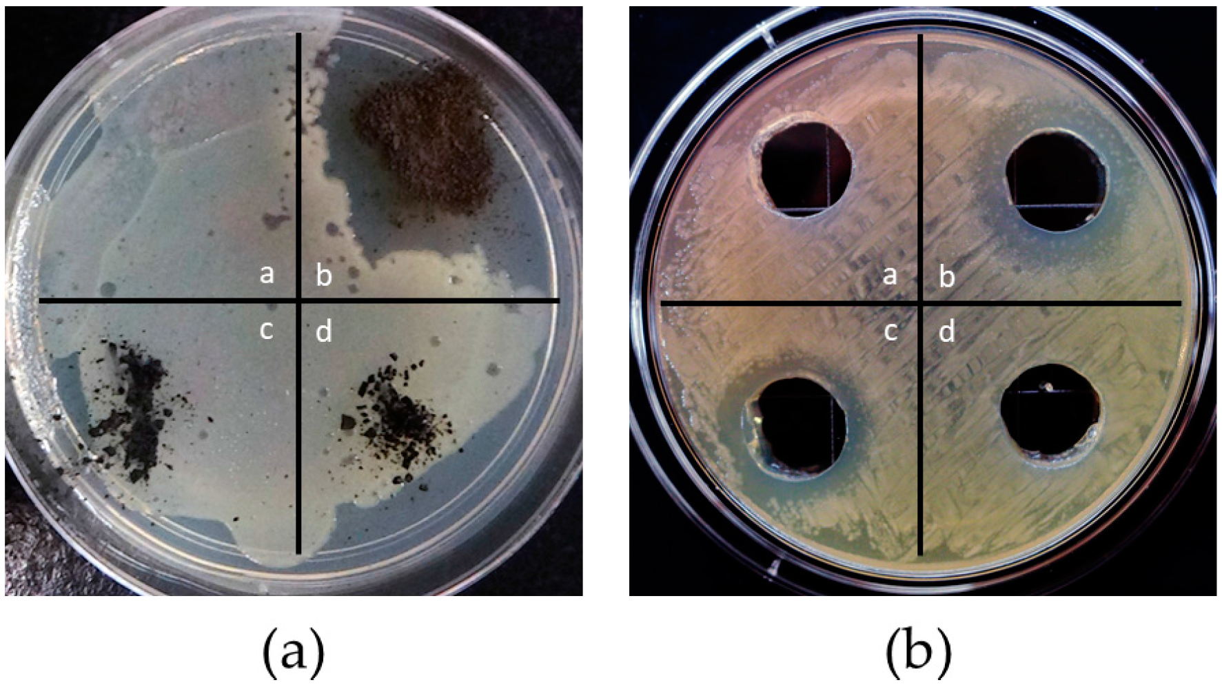
3.2.1. Zone of Inhibition

The zone of inhibition method was used to examine the antimicrobial activities of the relevant MACAg components against E. coli. Figure 3a shows the results [15] of solid samples of AgNPs (Aesar, 20–40 nm), AC, and MNPs containing 0.25 mmol atoms, while Figure 3b those of liquid samples having 2.5 × 10⁻⁵ mmol of Ag+ ions, Ag atoms in AgNPs (10 nm), Allura Red 40 ions, and BH4 ions. The Ag+ solution was used as a reference. Figure 3 clearly shows that only AgNPs and Ag+ ions produced zone of inhibition, revealing their remarkable ability at stopping the growth of E. coli. All other components (AC, MNPs, Allura Red 40, and BH4 ions) showed no influence on the growth of E. coli. The zone of Ag+ ions was larger than that of AgNPs and appeared less clear relative to that of AgNPs. These observations may be largely explained by the proposed mechanisms explaining the anti-microbial effect of Ag [32,33], and the relative sizes of AgNPs and Ag+ ions. AgNPs coming in contact with the microbes are thought to be oxidized into Ag+ ions which disrupt permeability and respiration functions of the cell, and penetrate inside the microorganisms. This leads to cell death or cellular inactivation. Since AgNPs are larger than Ag+ ions, they travel slower, and thus produce a smaller zone. When immobilized to a bacterium, one AgNP is believed to be able to release several tens of thousands of Ag+ ions in this vicinity, producing a local high concentration of antimicrobial ions. This seems to agree with the much clearer zone produced by AgNPs observed in this study.

3.2.2. Shaking Tests

Several tests were done to detect the antimicrobial efficiency of MACAg using 20 mL of E. coli suspensions with concentrations of 103–107 CFU/mL. These include the effect of treatment time, the E. coli CFU, and the Ag content of MACAg. Water disinfection was made much easier due to the magnetic properties of MACAg. The antimicrobial agent’s effectiveness was measured as the minimum treatment time required to achieve a 100% reduction in the number of surviving E. coli after MACAg treatment to the control sample. MACAg sediment after the treatment and wash were also tested for any viable bacteria that may remain attached to MACAg or be found in wash.
A 0.5 g quantity of MACAg (0.04% Ag) was placed in each of the beakers containing 20 mL of E. coli suspensions of 103, 104, 105 and 106 CFU/mL. The beakers were shaken for 30 min and samples were drawn, after settling MACAg magnetically, at 10 min time intervals to check microbe reduction as a function of time. The maximum silver concentration introduced in treating the suspensions was 0.01 mg/mL. Within 10 min, 100% bacterial reduction was achieved for E. coli suspensions containing 104 CFU/mL or less, and 30 min for suspension containing 105 CFU/mL. The microbial reduction for a suspension of 106 CFU/mL after 30 min treatment with MACAg was above 98%. No surviving E. coli were found on the sediment before or after wash and sediment washes after 30 min treating of E. coli suspensions containing 105 CFU/mL or less. This shows the antimicrobial effect of MACAg decreased with increasing E. coli concentration. Figure 4 illustrates the effectiveness of MACAg (0.04% Ag) against E. coli in a suspension of 2 × 10⁵ CFU/mL [29]. The results in Figure 4a demonstrated increased microbial reduction effect in the treated water with increasing treating time. A 99.9% reduction in the number of microbial colonies in the treated water was observed after 20 min treatment with MACAg. The colony numbers were reduced to zero after 30 min treatment. Figure 4b shows the incubated samples of MACAg magnetically separated from the treated water after 30-min treatment, 20 mL of deionized water used to wash this MACAg, and the MACAg after the wash, respectively. No viable microbes were found in these samples, indicating that the microbes, absent from the treated water, were not only removed from water but also killed. The MACAg (0.04% Ag)’s antimicrobial threshold against E. coli was therefore found to be 106–107 CFU in 30 min.
The effect of AgNPs content was investigated using three 0.5 g MACAg samples containing 0.06%, 0.15% and 0.22% silver. Each sample was placed in a 20 mL of E. coli suspension of 1 × 10⁵ CFU/mL, introducing silver in treating the suspension at a concentration of 0.02 mg/mL, 0.04 mg/mL or 0.05 mg/mL. The beakers were shaken for 15 min and samples were drawn at 3 min intervals. MAC without AgNPs was used as a control. Results in Figure 5 (top row) show that MAC removed nearly all (99.9%) microbes in the treated water in 3 min. MACAg (0.22% Ag) was able to achieve 100% microbial reduction in 12 min while MACAg (0.15% Ag) in 15 min. Some viable microbes (0.1%) still remained in water after 15 min treatment for MACAg (0.06% Ag). The sediment results in Figure 5 (bottom row) show that the microbes attached to MAC remained viable while no colonies were observed on the MACAg (0.06% Ag) sediment samples or in their washes. The sediment and wash results of MACAg (0.15% Ag) and MACAg (0.22% Ag) were identical to those of MACAg (0.06% Ag). This shows that although MAC removed the microbes the fastest, the microbes survived due to the absence of antimicrobial AgNPs. The microbes remained attached to MAC after the wash. The negative BH4 coating on the MACAg surface discouraged the adsorption of E. coli, which has a net negatively charged surface due to abundant phosphate groups, the same effect as observed in the Allura Red experiment. Nevertheless, the number of viable microbes in the treated water decreased as a function of time. Moreover, the microbes were not only removed, but also killed due to the presence of AgNPs, and the rate of removing and killing increased with increasing amount of AgNPs.

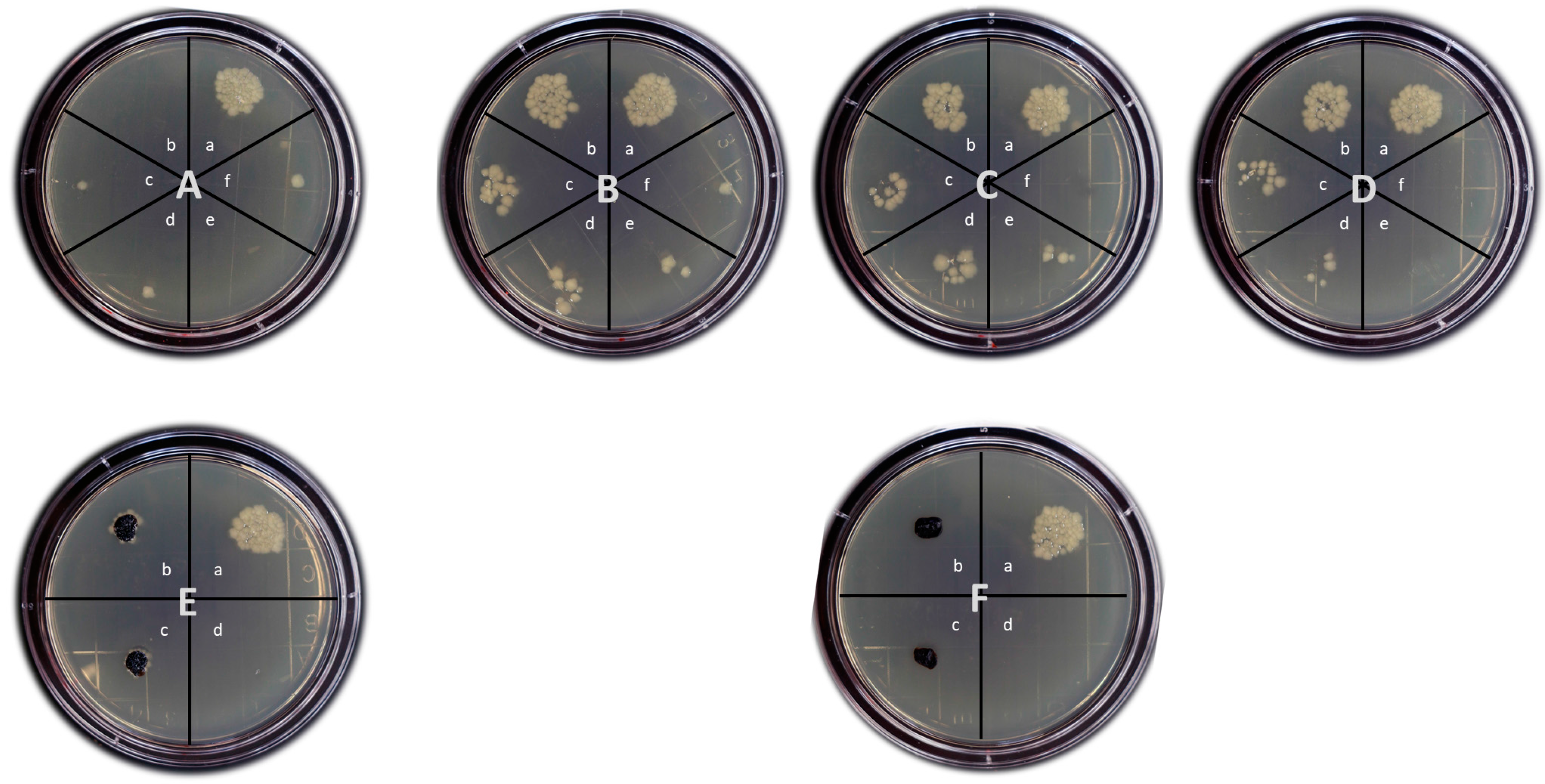
3.2.3. Repeated Use of MACAg Nanocomposite

Samples of MAC and MACAg were repeatedly employed in treating 20 mL of 2 × 10⁵ CFU/mL E. coli suspensions for up to five cycles. Each treatment was 15–20 min long. The sediments were magnetically separated after each cycle and rinsed with deionized water twice before the next cycle of use. The results of the reusability tests of MAC and MACAg (0.22%) are shown in Figure 6. It can be seen that MAC had the poorest performance for the test: once its surface was saturated with the microbes, it was no longer able to adsorb or remove additional microbes from the E. coli suspension. MACAg (0.22%), on the other hand, demonstrated great reusability as an antimicrobial agent and its antimicrobial efficiency was found to increase with increasing number of cycles. After the 4th and 5th cycles, the MACAg sediments and washes were checked for microbe growth on nutrient agar and no colonies were found. Reusability test results for MACAg (0.04% Ag), MACAg (0.06% Ag), and MACAg (0.15% Ag) were similar to those of MACAg (0.22% Ag). The increased antimicrobial activity of MACAg was likely due to an increase in the amount of more potent Ag+ ions formed, resulting from an increased surface oxidation of Ag atoms due to a prolonged exposure to water containing dissolved oxygen [34]. At the end of the 5th cycle, the samples were in water for nearly 2 h. The Ag+ ions formed may be released into E. coli suspension or may be adsorbed by AgNPs [34] or MACAg, both may result in an increased antimicrobial effect. This seems to agree with the preliminary results of silver release test, described in Section 3.3: consistently being at a level below 0.1 ppm, the silver concentrations in the treated water may remain relatively constant for repeatedly used MACAg containing low amount of Ag or may slightly increase with increasing number of uses for MACAg with higher silver content. No evidence was seen to indicate a noticeable degree of coverage of the active Ag surface by the killed bacterial cells, which would have decreased the MACAg antimicrobial efficiency after repeated uses. This may be due to the fact that the surface of MACAg, negatively charged with BH4 ions, repelled the net negative surface of the dead E. coli cells. The easy recoverability and excellent reusability of MACAg make the nanocomposite extremely desirable, especially in large-scale applications, since they greatly reduce material cost and effectively minimize secondary pollution relating to disposal of used materials.

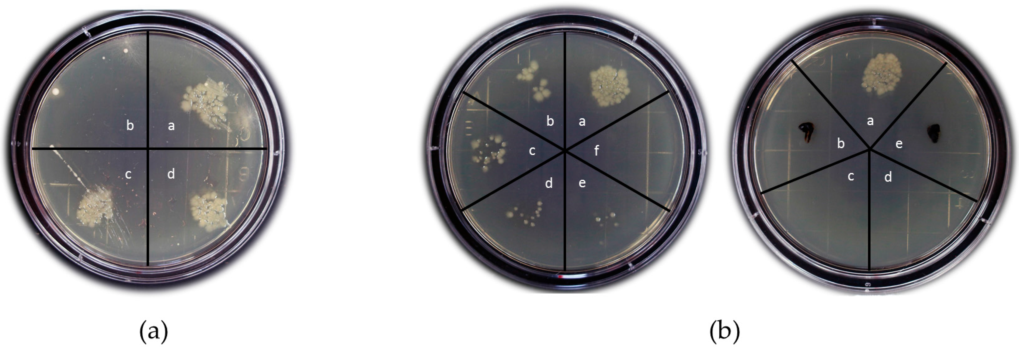
3.2.4. Disinfection of Long Island Sound (LIS) Surface Water

The feasibility of employing MACAg nanocomposite for disinfecting Long Island Sound (LIS) surface water was studied by performing the shaking test. The LIS surface water was obtained at the Academy’s waterfront site on 4 April 2017. The colony counts of the LIS surface water was found to vary with seasons, mostly due to temperature variations, with the summer water containing the most abundant cells and the winter water the least. The water sample obtained on 4 April 2017 was estimated to contain 104 CFU/mL. The colonies of cultivable microbes were shown to be of diversified nature with varied colors, shapes, sizes, and morphologies. Different types of bacteria may exhibit different levels of susceptibility to the bactericidal effect of silver. The reduction of LIS water bacteria following interaction with MACAg (0.22% Ag) was investigated by inoculating the water sample treated with MACAg (0.22% Ag) on nutrient agar plates. Twenty mL of LIS surface water was shaken with MACAg for 15 min and samples were drawn at 3 min intervals to observe the reduction in colony count in the treated water as a function of time. The maximum silver concentration introduced in treating the water samples was 0.05 mg/mL. The results of shaking tests are shown in Figure 7. Within 3 min, 100% bacteria removal rate was achieved and no colonies were detected in any of the water samples after 3 min treatment with MACAg (0.22% Ag). At the end of 15 min treatment, MACAg and its two washes were sampled for the presence of viable bacteria (results of two trials were shown in Figure 7b). No colonies were found on the MACAg sediment and the second wash. One small round yellow colony, however, appeared on the agar plate from 10 μL of one of the first washes. This estimates a more than 98.5% killing rate of all microbes present in LIS surface water after 15 min treatment with MACAg (0.22% Ag). The presence of surviving bacteria in the wash provided evidence that bacteria cells attached onto the MACAg can be washed off easily, likely due to the unfavorable forces between the BH4 anion-coated MACAg surface and the net negatively charged surface of the bacteria. This minimizes the possible dead cell blockage of the active Ag surface and favors the reusability of MACAg nanocomposite.

3.3. Release of Silver

The incorporation of magnetite nanoparticles in MACAg nanocomposite enabled the fast and effective removal of silver nanoparticles from “disinfected” water. A 0.50 g quantity of each MACAg sample containing 0.04% to 0.22% by mass of Ag was subjected to repeated use up to five cycles for treating 20 mL of E. coli suspensions containing 105 CFU/mL. Each cycle involved 15–20 min shaking in water. Non-detectable amounts of silver nanoparticles were found using UV-visible spectroscopy in any of the treated E. coli suspensions. The UV-visible spectrophotometer is able to show a clear peak at 399 nm if the AgNPs concentrations are 0.10 ppm or above. This demonstrates the general stability of the MACAg nanocomposite and the silver nanoparticles incorporated into the MACAg nanocomposite remained undetached after use. Atomic absorption (AA) spectroscopy analysis was used to estimate the leaching of total Ag, mostly in the oxidized Ag+ ion form, into the treated E. coli suspension of 105 CFU/mL, especially after repeated uses. Table 1 shows the preliminary results that indicate consistent low levels of silver, lower than the 0.1 ppm limit recommended by the Environmental Protection Agency (EPA), for all tested water samples. The replicates of our standards showed approximately 5% error in the measurements. For the water samples treated by MACAg containing less than 0.15% Ag, the silver concentrations were found to be less than 0.05 ppm and the repeated use of MACAg did not alter these values much. However, when the silver content of MACAg was at 0.22%, an increased silver release was observed and the release was found to increase with increasing number of repeated use, resulting in the silver levels changing from under 0.05 ppm (1st use) to above 0.05 ppm (5th use) in the treated water. These results suggest that MACAg (0.15% Ag) may offer the optimal and the most desirable performance: when compared to MACAg (0.22% Ag), although it took 3 min longer to produce 100% reduction/killing of the E. coli in a suspension containing 2 × 10⁶ CFU, it demonstrated excellent and similar reusability, and released much less silver into the treated water. The release of silver for each treatment was less than 0.2% of the total amount of Ag in MACAg for all samples. The results therefore support the view that AgNPs, acting as a Ag+ ion reservoir, provide a relatively low but sufficient concentration of silver antimicrobial species, and can thus remain active for a long period of time [29,30]. This in fact makes AgNPs very attractive in water disinfection applications, since higher-than-necessary levels of silver application not only are wasteful but also may pose health and environmental concerns. The lost AgNPs may be replenished by treating the used MACAg with sodium borohydride and silver nitrate solutions. The facile procedure developed here would therefore be considered “green” since it could be optimized to not release silver to a level harming mammalian cells, but be very potent to microbes with easy recoverability and excellent reusability. Additionally, the presence of less than 0.1 ppm of residual Ag in water may serve to prevent the regrowth of microbes in the treated water, eliminating the need for secondary disinfection [35,36]. Further investigations will center on establishing the silver release profiles of MACAg in water, in terms of the silver leaching rates and amounts, as a function of silver mass content and the number of repeated MACAg uses to more accurately determine the optimal MACAg compositions for water disinfection.

4. Conclusions

Using commonly available materials and equipment, we developed a simple and easy scale-up procedure for preparing a MACAg nanocomposite based on activated carbon, magnetite nanoparticles, and silver nanoparticles for water disinfection. The activated carbon was first impregnated with magnetite nanoparticles that imparted magnetic property to the nanocomposite. The resultant nanocomposite was then coated with negatively charged borohydride ions that provided an effective surface to first attract silver ions and then reduce them to form AgNPs. The AgNPs, intimately integrated into the nanocomposite, gave the nanocomposite the biocidal property. The as-prepared MACAg nanocomposite (0.5 g, 0.04–0.22% Ag) demonstrated high antimicrobial efficacy against E. coli in water, killing 106–107 CFU in 12–30 min, with higher antimicrobial efficacy corresponding to higher silver content. The nanocomposite enabled the easy recovery of AgNPs due to its magnetic behavior and was shown to be reusable for at least five cycles without losing its efficiency. The recoverability and reusability of MACAg are extremely desirable, especially in large-scale applications, since they greatly reduce material cost and minimize the need to dispose the used materials. The MACAg nanocomposite also demonstrated a high efficacy for disinfecting Long Island Sound surface water samples containing different microbes.

Acknowledgments

The authors would like to thank Dylan Franz, Midshipman 2nd Class, U.S. Merchant Marine Academy, for assisting in the methylene blue dye experiment. The work performed at Brookhaven National Laboratory is supported by the U.S. DOE under Contract No. DE-SC00112704.

Author Contributions

Ping Y. Furlan, Adam J. Fisher and Alexander Y. Furlan conceived and designed the experiments; Ping Y. Furlan, Adam J. Fisher and Alexander Y. Furlan performed the experiments, especially relating to synthesis and antimicrobial tests; Ping Y. Furlan, Adam J. Fisher, Alexander Y. Furlan and Michael E. Melcer analyzed the data; Michael E. Melcer and David W. Shinn performed atomic absorption (A.A.) experiments and analyzed A.A. data; John B. Warren performed scanning electron microscopy (SEM) experiments and analyzed SEM data; Ping Y. Furlan supervised the project and wrote the paper.

Conflicts of Interest

The authors declare no conflict of interest. The founding sponsors had no role in the design of the study; in the collection, analyses, or interpretation of data; in the writing of the manuscript, and in the decision to publish the results. The authors declare that the views expressed in this article are the authors’ own and not those of the U.S. Merchant Marine Academy, the Maritime Administration, the Department of Transportation or the United States government.

References

  1. World Health Organization (WHO). “Water, Sanitation and Hygiene Links to Health Facts and Figures”. 2004. Available online: http://www.who.int/water_sanitation_health/en/factsfigures04.pdf (accessed on 31 May 2017).
  2. Seiden, J.M.; Way, C.; Rivkin, R.B. Microbial hitchhikers: Dynamics of bacterial populations in a trans-Pacific voyage of a bulk carrier. Aquat. Invasions 2010, 5, 13–22. [Google Scholar] [CrossRef]
  3. First, M.R.; Drake, L.A. Approaches for determining the effects of UV radiation on microorganisms in ballast water. Manag. Biol. Invasions 2013, 4, 87–99. [Google Scholar] [CrossRef]
  4. Krasner, S.W.; Weinberg, H.S.; Richardson, S.D.; Pastor, S.J.; Chinn, R.; Sclimenti, M.J.; Onstad, G.D.; Thruston, A.D., Jr. Occurrence of a new generation of disinfection byproducts. Environ. Sci. Technol. 2006, 40, 7175–7185. [Google Scholar] [CrossRef] [PubMed]
  5. Centers for Disease Control and Prevention. “The Safe Water System—Disinfection by-Products”. Available online: www.cdc.gov/safewater/chlorination-byproducts.html (accessed on 31 May 2017).
  6. Shannon, M.A.; Bohn, P.W.; Elimelech, M.; Georgiadis, G.J.; Marinas, B.J.; Mayers, A.M. Science and technology for water purification in the coming decades. Nature 2008, 452, 301–310. [Google Scholar] [CrossRef] [PubMed]
  7. Marambio-Jones, C.; Hoek, E.M.V. A review of the antibacterial effects of silver nanomaterials and potential implications for human health and the environment. Nanopart. Res. 2010, 12, 1531–1551. [Google Scholar] [CrossRef]
  8. Tan, S.; Zou, W.; Jiang, F.; Tan, S.; Liu, Z.; Yuan, D. Facile fabrication of copper-supported ordered mesoporous carbon for antibacterial behavior. Mater. Lett. 2010, 64, 2163–2166. [Google Scholar] [CrossRef]
  9. Damm, C.; Munstedt, H.; Rosch, A.J. Long-term antimicrobial polyamide 6/silver nanocomposites. Mater. Sci. 2007, 42, 6067–6073. [Google Scholar] [CrossRef]
  10. Ivask, A.; Kurvet, I.; Kasemets, K.; Blinova, I.; Aruoja, V.; Suppi, S.; Viga, H.; Kakinen, A.; Titma, T.; Heinlaan, M.; et al. Size-dependent toxicity of silver nanoparticles to bacteria, yeast, algae, crustaceans and mammalian cells in vitro. PLoS ONE 2014, 9, e102108. [Google Scholar] [CrossRef] [PubMed]
  11. Panyala, N.R.; Pena-Mendez, E.M.; Havel, J. Silver or silver nanoparticels: A hazardous threat to the environment and human health? J. Appl. Biomed. 2008, 6, 47–59. [Google Scholar]
  12. Okafor, F.; Janen, A.; Kukhtareva, T.; Edwards, V.; Curley, M. Green synthesis of silver nanoparticles, their characterization, application and antibacterial activity. Int. J. Environ. Res. Public Health 2010, 10, 5221–5238. [Google Scholar] [CrossRef] [PubMed]
  13. Alt, V.; Bechert, T.; Steinrucke, P.; Wagener, M.; Seidel, P.; Dingeldein, E.; Domann, E.; Schnettler, R. An in vitro assessment of the antibacterial properties and cytotoxicity of nanoparticulate silver bone cement. Biomaterials 2004, 25, 4383–4391. [Google Scholar] [CrossRef] [PubMed]
  14. Zhu, C.; Xue, J.; He, J. Controlled in-situ synthesis of silver nanoparticles in natural cellulose fibers toward highly efficient antimicrobial materials. J. Nanosci. Nanotechnol. 2009, 9, 3067–3074. [Google Scholar] [CrossRef] [PubMed]
  15. Furlan, P.Y.; Melcer, M.E. Removal of aromatic pollutant surrogate from water by recyclable magnetite-activated nanocomposite: An experiment for general chemistry. J. Chem. Educ. 2014, 91, 1966–1970. [Google Scholar] [CrossRef]
  16. Furlan, P.Y.; Ackerman, B.M.; Melcer, M.E.; Perez, S.E. Reusable magnetic nanocomposite sponges for removing oil from water discharges. J. Ship Prod. Des. 2016, 32, 1–10. [Google Scholar]
  17. Dankovich, T.A.; Gray, D.G. Bactericidal paper impregnated with silver nanoparticles for point-of-use water treatment. Environ. Sci. Technol. 2011, 5, 1992–1998. [Google Scholar] [CrossRef] [PubMed]
  18. Ferreira, A.M.; Roque, E.B.; Fonseca, F.V.D.; Borges, C.P. High flux microfiltration membranes with silver nanoparticles for water disinfection. Desalin. Water Treat. 2015, 56, 3590–3598. [Google Scholar] [CrossRef]
  19. Ren, D.; Smith, J.A. Retention and transport of silver nanoparticles in a ceramic porous medium used for point-of use water treatment. Environ. Sci. Technol. 2013, 47, 3825–3832. [Google Scholar] [CrossRef] [PubMed]
  20. Quang, D.V.; Sarawade, P.B.; Jeon, S.J.; Kim, S.H.; Km, J.; Chai, Y.G.; Kim, H.T. Effective water disinfection using silver nanoparticle containing silica beads. Appl. Surf. Sci. 2013, 266, 280–287. [Google Scholar] [CrossRef]
  21. Zhang, Y.; Wan, Y.; Shi, Y.; Pan, G.; Yan, H.; Xu, J.; Guo, M.; Qin, L.; Liu, Y. Facile modification of thin-film composite nanofiltration membrane with silver nanoparticles for anti-biofouling. J. Polym. Res. 2016, 23, 105. [Google Scholar] [CrossRef]
  22. Karthik, C.; Radha, K.V. Silver nanoparticle loaded activated carbon: An escalated nanocomposite with antimicrobial property. Orient. J. Chem. 2016, 32, 735–741. [Google Scholar] [CrossRef]
  23. Eltugral, N.; Simsir, H.; Karagoz, S. Preparation of nano-silver-supported activated carbon using different ligands. Res. Chem. Intermed. 2016, 42, 1663. [Google Scholar] [CrossRef]
  24. Azmier, M.; Alrozi, R. Removal of malachite green dye from aqueous solution using rambutan peel-based activated carbon: Equilibrium, kinetic and thermodynamic studies. Chem. Eng. J. 2011, 171, 510–516. [Google Scholar]
  25. Cook, D.; Newcombe, G.; Sztajnbok, P. The application of powdered activated carbon for MIB and geosmin removal: Predicting PAC doses in four waters. Water Res. 2001, 35, 9. [Google Scholar] [CrossRef]
  26. Crittenden, J.; Vaitheeswaran, K.; Hand, D.; Howe, E.; Aieta, E. Removal of dissolved organic carbon using granular activated carbon. Water Res. 1993, 27, 7. [Google Scholar] [CrossRef]
  27. Removal of Inorganic Contaminants by Activated Carbon Adsorption. Available online: http://www.webapps.cee.vt.edu/ewr/environmental/teach/gwprimer/group23/inorganics.html (accessed on 31 May 2017).
  28. Rao, M.M.; Ramana, D.K.; Seshaiah, K.; Wang, M.C.; Chien, S.W.C. Removal of some metal ions by activated carbon prepared from Phaseolus aureus hulls. J. Hazard. Mater. 2009, 166, 1006–1013. [Google Scholar] [CrossRef] [PubMed]
  29. Furlan, P.Y.; Fisher, A.J.; Melcer, M.E.; Furlan, A.Y.; Warren, J.B. Preparing and testing a magnetic antimicrobial nanocomposite for water disinfection to gain experience at nanochemistry-microbiology interface. J. Chem. Educ. 2017, 94, 488–493. [Google Scholar] [CrossRef]
  30. Berger, P.; Adelman, N.B.; Beckman, K.J.; Campbell, D.J.; Eillis, A.B.; Lisensky, G.C. Preparation and properties of an aqueous ferrofluid. J. Chem. Educ. 1999, 76, 943–948. [Google Scholar] [CrossRef]
  31. Solomon, S.D.; Bahadory, M.; Jeyarajasingam, A.V.; Rutkowsky, S.A.; Boritz, C. Synthesis and study of silver nanoparticles. J. Chem. Educ. 2007, 84, 322–325. [Google Scholar]
  32. Franci, G.; Falanga, A.; Galdiero, S.; Palomba, L.; Rai, M.; Morelli, G.; Galdiero, M. Silver nanoparticles as potential antibacterial agents. Molecules 2015, 20, 8856–8874. [Google Scholar] [CrossRef] [PubMed]
  33. Le Quay, B.; Stellacci, F. Antibacterial activity of silver nanoparticles: A surface science insight. Nano Today 2015, 10, 339–354. [Google Scholar] [CrossRef]
  34. Liu, J.Y.; Hurt, R.H. Ion release kinetics and particle persistence in aqueous nano-silver colloids. Environ. Sci. Technol. 2010, 44, 2169–2175. [Google Scholar] [CrossRef] [PubMed]
  35. Shuval, H. Stabilized formulation of hydrogen peroxide with silver: A secondary disinfectant. In Providing Safe Drinking Water in Small Systems: Technology, Operations, and Economics; Cotruvo, J.A., Carun, G.F., Hearne, N., Eds.; Lewis Publishers: Boca Raton, FL, USA; London, UK; New York, NY, USA; Washington, DC, USA, 1999; p. 184. [Google Scholar]
  36. Water Technology: Silver, Coper Have Antibacterial Effects. Available online: http://www.watertechonline.com/silver-copper-have-anti-bacterial-effects/ (accessed on 31 May 2017).
Figure 1. Schematic representation for preparing MACAg nanocomposite. AC = activated carbon; MAC = magnetite-activated carbon nanocomposite; BH4 = borohydride; MACAg = magnetite-activated carbon-silver nanocomposite.
Inventions 02 00010 g001
Figure 2. (a) The molecular structure of Allura Red 40; (b) The colors of the Allura Red 40 dye solutions before (Control—D) and after the treatment with MAC (A), MAC/BH4 (B), and MACAg (C).
Inventions 02 00010 g002
Figure 3. Antimicrobial activity of relevant MACAg components against E. coli. (a) Solid samples containing 0.25 mmol atoms of AgNPs in space b, MNPs in space c, and AC in space d were placed on an agar plate covered with 100 μL of E. coli suspension of 2 × 10⁸ CFU/mL (control is in space a); (b) 100 μL of liquid samples containing 2.5 × 10⁻⁵ mmol of Allura Red 40 ions in space a, Ag+ ions in space b, Ag atoms in AgNPs (10 nm) in space c, and BH4 ions in space d were placed in the holes bored in an agar plate covered with 100 μL of E. coli suspension of 0.5 × 10⁸ CFU/mL. Only AgNPs and Ag+ ions produced zone of inhibition. Figure 3a was published in reference [29]. AgNPs = silver nanoparticles; MNPs = magnetite nanoparticles; CFU = colony–forming units.
Inventions 02 00010 g003
Figure 4. (a) The effectiveness of MACAg (0.5 g, 0.04% Ag) against E. coli in a suspension containing 4 × 10⁶ CFU: space a = control, space b = 10 min. MACAg treatment, space c = 20 min. MACAg treatment, space d = 30 min. MACAg treatment (absent of E. coli); (b) The absence of viable microbes on MACAg after treating E. coli solution—space b; in 20 mL wash—space c; and on MACAg after wash—space d (Space a shows the colonies formed from a sample of the E. coli solution before the MACAg treatment). This Figure was published in reference [29].
Inventions 02 00010 g004
Figure 5. Effect of silver content on MACAg’s antimicrobial effectiveness. Top row: The rate of removing E. coli from a suspension containing 2 × 106 CFU by 0.5 g of MAC (A), MACAg (0.06% Ag) (B), MACAg (0.15% Ag) (C), or MACAg (0.22%) (D). Sampling (6 μL) of treated water took place at: a = 0 min, b = 3 min, c = 6 min, d = 9 min, e = 12 min, and f = 15 min; Bottom row: Investigation of viable microbes on small samples of sediment before (b) and after (c) the wash and in the wash (6 μL) (d) for MAC (E) and MACAg (0.06% Ag) (F). Sediment and wash results of MACAg (0.15% Ag) and MACAg (0.22% Ag) were identical to those of MACAg (0.06% Ag). Space a in all agar plates represents the initial colony counts before the treatment in the sampled waters.
Inventions 02 00010 g005
Figure 6. Reusability test results for MAC and MACAg. (a) Remaining microbes in treated water after 15 min treatment by MAC (0.5 g) that was used 1st time—space b, 2nd time—space c, or 3rd time—space d; (b) Left: Remaining microbes in treated water after 20 min treatment by MACAg (0.5 g, 0.22% Ag) that was used 1st time—space b, 2nd time—space c, 3rd time—space d, 4th time—space e, or 5th time—space f; Right: No viable microbes were observed on small samples of MACAg (0.5 g, 0.22% Ag) sediment after the 4th (space b) and the 5th (space e) uses or in their washes (spaces c and d). Space a in all agar plates represents the initial colony counts before the treatment in the sampled waters.
Inventions 02 00010 g006
Figure 7. Disinfection of Long Island Sound (LIS) surface water using MACAg. (a) The rate of removing the cultivable microbes from a LIS surface water containing 105 CFU by 0.5 g of MACAg (0.22%). Sampling (10 µL) of treated water took place at: space a = 0 min, space b = 3 min, space c = 6 min, space d = 9 min, space e = 12 min, and space f = 15 min; (b) Investigation of viable microbes on small samples of MACAg sediment before (spaces d, e) the wash and in the 1st wash (10 μL) (spaces c, f), and 2nd wash (10 μL) (spaces a, b).
Inventions 02 00010 g007
Table 1. MACAg Silver Release into 20 mL of Treated Escherichia coli (E. coli) Suspension in Parts Per Million. Preliminary Estimates from Atomic Absorption Spectrophotometer (Perkin–Elmer AAnalyst 200).
No. Cycles 1MACAg (0.22% Ag)MACAg (0.15% Ag)MACAg (0.06% Ag)
10.03 ppm 0.01 ppm 0.02 ppm
30.05 ppm 0.01 ppm 0.01 ppm
50.09 ppm
1 MACAg samples were subjected to repeated use: MACAg (0.22% Ag) was 20 min/cycle; MACAg (0.15% Ag) and MACAg (0.06% Ag) 15 min/cycle.

