Editorial

Functional Anatomy in Knee Osteoarthritis: Patellofemoral Joint vs. Tibiofemoral Joint

by
Giuseppe Musumeci
Department of Biomedical and Biotechnological Sciences, Human Anatomy and Histology Section, School of Medicine, University of Catania, Via S. Sofia 87, 95123 Catania, Italy
J. Funct. Morphol. Kinesiol. 2017, 2(1), 8; https://doi.org/10.3390/jfmk2010008
Submission received: 16 November 2016 / Accepted: 6 January 2017 / Published: 22 February 2017

Abstract

:
“Knee Joint Osteoarthritis” is the topic chosen for the first editorial of the second volume of this journal. The aim of this editorial is to discuss this interesting but little analyzed topic in the current literature, in order to explain and help readers to better understand the functional anatomical aspects of knee joints affected by Osteoarthritis (OA). As the knee joint is tri-compartmental, numerous radiographic patterns of disease are possible and the differences between the two main compartments of the knee (patellofemoral joint versus tibiofemoral joint) are explored in this editorial.

Graphical Abstract

1. Introduction

Osteoarthritis (OA), the most common form of arthritis, is a degenerative disease characterized by a progressive deterioration of articular cartilage, resulting in pain and severe disability [1,2,3,4]. OA affects millions of people and is the most common cause of pain and physical disability in the world today, with huge social and economic costs. In Europe, over 40 million people are affected by OA and it is estimated that 130 million Europeans will suffer from OA by 2050. An estimated 52.5 million people in the US suffer from OA and the total costs exceed $100 billion per year [5]. OA is a complex multifactorial disease affecting more than half of the over 65 population throughout the world. Knee OA is a chronic joint disease affecting one-third of elderly people [6]. Nevertheless, joint injury also contributes to acute and long-term cartilage degradation in the younger population. Presently, there is no treatment that can reverse or halt OA, other than pain-relief, until joint replacement becomes necessary, with considerable economic impact. Abnormal mechanical loading plays an important role in the progression of cartilage degeneration after injury. Moreover, injured cartilage has limited intrinsic repair capacity and the current research in the field of pathophysiological mechanisms at cellular and tissue levels is essential for developing effective treatments to restore function and prevent disease progression [7].
The disease limits daily activities such as walking, stair-climbing and housekeeping, leading to a lack of functional independence and impairment of quality of life. Clinical guidelines recommend non-pharmacological strategies in the initial management of OA symptoms [8]. The need of tailored interventions is emphasized, given the heterogeneity of knee OA in etiology, clinical presentation and natural history, in order to optimize treatment outcome. Since the knee joint is a tri-compartmental (patellofemoral joint (PFJ), medial and lateral tibiofemoral joint (TFJ)) [9], tailored treatment based on compartmental involvement may be appropriate. The aim of this editorial is to discuss this interesting topic, one that is little analyzed in the current literature, in order to explain and help readers to better understand the functional anatomical aspects of the two main compartments of the knee joint (PFJ vs. TFJ) in joints affected by OA.

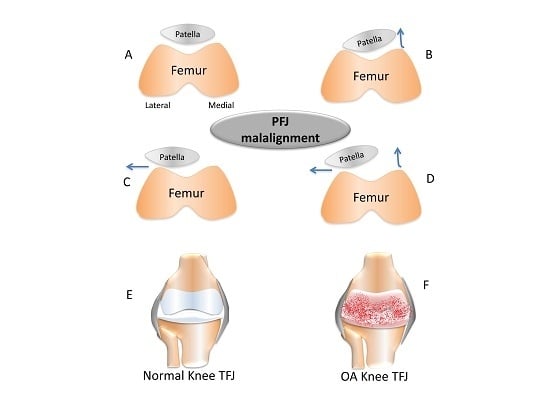
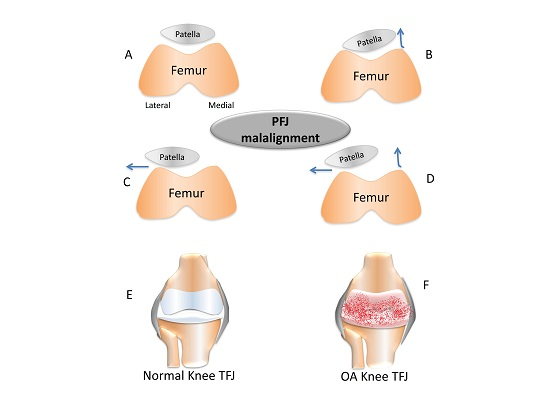
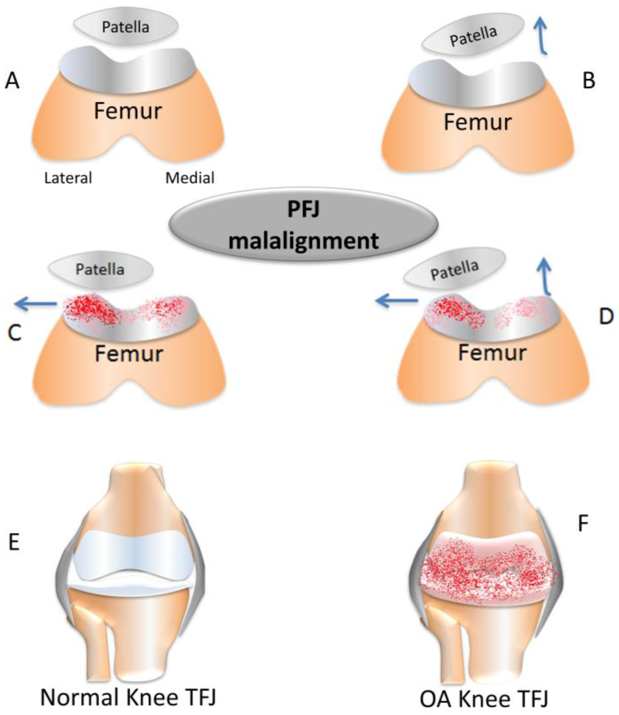
2. Short Overview of Patellofemoral Joint vs. Tibiofemoral Joint in the Osteoarthritis (OA) Process

Since the knee joint is tri-compartmental, there are various possible patterns of knee OA. In general, we see knee OA principally as a disorder of the TFJ and radiographic investigations tend to focus only on the antero-posterior X-ray, without exploration of the PFJ [10]. However, PFJ involvement in the OA process has become more apparent since the increased use of lateral and skyline X-rays. In truth, the PFJ is one of the most commonly affected compartments. Although the presence of osteophytes alone is not sufficient to diagnose OA, a higher frequency of radiographic osteophytes in the PFJ compared with the TFJ compartment has been observed [10]. A study on the most common radiographic pattern in patients suffering from knee pain evidenced a great prevalence of the combined TFJ and PFJ disease (40%), followed by isolated PFJ OA (24%), with isolated TFJ disease in just 4% of patients while the remaining 32% had normal radiographs [11]. Although there is a high prevalence of PFJ OA, little research has been carried out regarding this specific disease entity. The significance of PFJ OA has been demonstrated in recent data, showing that the presence of PFJ OA at baseline is an indicator of the progression of structural change in the TFJ compartment over 30 months [12]. The PFJ compartment is often significantly affected by symptoms associated with knee OA, and is possibly more important than the TFJ. Knee pain in PFJ is often associated with osteophytes, whereas the presence of osteophytes in the TFJ compartment is often not painful [13].
Radiographic and Magnetic Resonance Imaging (MRI) studies show that the PFJ is crucial to exacerbate knee OA symptoms, and might be more relevant than the TFJ compartment. Moreover, patella cartilage has different biochemical and mechanical properties from tibia and femur cartilage. Indeed, patella cartilage demonstrates more in vivo deformation in weight-bearing loaded activities [14], since it has higher water content than femoral cartilage and lower proteoglycan content. Moreover, biomarkers of cartilage damage/loss, as serum cartilage oligomeric matrix protein concentration, are higher in patients with TFJ OA than in patients with PFJ OA of similar seriousness [15]. We may deduce that this means qualitative differences in cartilage metabolism in the two compartments. The pathogenesis of OA has been associated with both systemic and local factors and several studies suggest that there is a difference in risk factors between the PFJ and TFJ [16,17]. The PFJ’s biomechanics are different from those of the TFJ and the elements affecting the magnitude and distribution of PFJ pressure have important pathogenetic implications. Unlike the biomechanics of the PFJ, the factors influencing the distribution and magnitude of TFJ force are exclusive to that compartment. Medial TFJ OA is more prevalent than lateral TFJ OA [18], due to the specific nature of the biomechanics of each compartment. Devices such as laterally wedged shoe insoles, varus knee bracing and kinesio tape are potentially very useful in the management of medial TFJ OA, because of their minimizing effects on the knee adduction moment [19,20]. Although there is limited available evidence, patients with PFJ OA show features and physical impairments that are sufficiently different from subjects with TFJ OA to warrant targeted intervention. An important feature of TFJ OA is reduced strength in muscles, often the quadriceps in particular. This is crucial for both pain severity and physical impairment in individuals with TFJ OA [21], and some studies indicate that quadriceps weakness may precede the development of OA [22,23]. Muscle weakness probably also accompanies PFJ OA, but it is not evident which muscles are primarily involved, due to the lack of studies comparing lower limb muscle strength in patients with PFJ OA to TFJ OA. Studies confirmed the relationship between quadriceps weakness and knee OA in all compartments [24], showing an important role of quadriceps strength in TFJ OA.
Kinematic changes during ambulation, caused by traumatic or chronic conditions, induce in the knee the shift of the load-bearing contact location of the joint to a region not conformed to the new loading, leading to osteoarthritis. The rate of progression of osteoarthritis is associated with increased load during ambulation. Recent studies have supported the earlier findings that a high adduction movement of the knee during ambulation is frequently reported to influence the progression of medial compartment osteoarthritis [25]. Varus but not valgus alignment increased the risk of incident TFJ OA. In knees with osteoarthritis, varus and valgus alignment increased the risk of progression in the biomechanically stressed compartment. Knee valgus and varus load during sports movement is viewed as an important predictor of non-contact anterior cruciate ligament injury risk, particularly in females. Formulating movement strategies that can reduce valgus loading during these movements therefore appears pertinent to reducing anterior cruciate ligament injury rates. Although knee malalignment is assumed to correlate with knee OA, it is still unknown whether malalignment precedes the development of OA or whether it is a result of OA or (even more likely) whether the relationship between malalignment and OA is bidirectional (Figure 1) [26].

3. Conclusions

This editorial explores the evidence suggesting that PFJ OA should be considered distinctly in respect to other knee compartments OA. Even if the PFJ involvement is underestimated with respect to TFJ, this compartment is commonly afflicted by OA, leading to considerable pain and disability. The osteophytes are more present with a higher frequency in the PFJ than in the TFJ compartment, while serum cartilage oligomeric matrix protein (COMP) and other biomarkers are higher in patients with TFJ OA than in PFJ OA [15]. The reduced serum COMP in PFJ disease compared with TFJ OA could be due to small articular cartilage volume in the latter or to a qualitative difference in cartilage metabolism. The clinical outcome for PFJ OA should be improved, increasing the study on conventional pharmacological and/or non-pharmacological treatment strategies oriented to the exclusive biomechanical functions of the PFJ and the specific impairments linked with disease in this compartment. Kinesio tape, bracing, cupping therapy, adapted physical activity, kinesiotherapy and physiotherapy are interventions that all offer possibilities in the improvement of symptoms and/or reduction of PFJ stress and pain in patients with PFJ OA [27,28,29]. There are few studies in literature that demonstrate these important differences in knee joint compartments. Such differences likely reflect distinct cellular and molecular responses to different biomechanical and/or biochemical signals and thus, understanding such processes opens new directions for therapeutic intervention, both pharmacological and non-pharmacological. Definitely, more basic and clinical studies are required to understand differences in OA pathophysiology affecting distinct knee compartments and to identify the therapeutic strategies most effective for each one.

Acknowledgments

A special thanks goes to our Editorial Advisors, eminent scientists in these fields that, with their experience and important suggestions, guide us in this great enterprise; our excellent Editorial Board members whose depth of experience cover a very broad spectrum on morphology and kinesiology; the Assistant Editors that, day after day, thanks to their valuable contributions, make the growth of this journal and the peer reviewer. On behalf of the Editorial Board we appreciate the voluntary contribution that each peer reviewer gives to maintain the high standard in his or her role for this journal and to ensure rapid publication. I sincerely thank them in advance for offering their precious time for the participation in the review process.

Conflicts of Interest

The author declares no conflict of interest.

References

  1. Castrogiovanni, P.; Musumeci, G. Which is the best physical treatment for osteoarthritis? J. Funct. Morphol. Kinesiol. 2016, 1, 54–68. [Google Scholar] [CrossRef]
  2. Warner, S.C.; Valdes, A.M. The genetics of osteoarthritis: A review. J. Funct. Morphol. Kinesiol. 2016, 1, 140–153. [Google Scholar] [CrossRef]
  3. Musumeci, G.; Szychlinska, M.A.; Mobasheri, A. Age-related degeneration of articular cartilage in the pathogenesis of osteoarthritis: Molecular markers of senescent chondrocytes. Histol. Histopathol. 2015, 30, 1–12. [Google Scholar] [CrossRef] [PubMed]
  4. Mobasheri, A.; Matta, C.; Zákány, R.; Musumeci, G. Chondrosenescence: Definition, hallmarks and potential role in the pathogenesis of osteoarthritis. Maturitas 2015, 80, 237–244. [Google Scholar] [CrossRef] [PubMed]
  5. Musumeci, G.; Loreto, C.; Carnazza, M.L.; Strehin, I.; Elisseeff, J. OA cartilage derived chondrocytes encapsulated in poly (ethylene glycol) diacrylate (PEGDA) for the evaluation of cartilage restoration and apoptosis in an in vitro model. Histol. Histopathol. 2011, 26, 1265–1278. [Google Scholar] [PubMed]
  6. Musumeci, G.; Castrogiovanni, P.; Leonardi, R.; Trovato, F.M.; Szychlinska, M.A.; di Giunta, A.; Loreto, C.; Castorina, S. Knee osteoarthritis. New perspectives for articular cartilage repair treatment through tissue engineering. A contemporary review. World J. Orthop. 2014, 5, 80–88. [Google Scholar] [CrossRef] [PubMed]
  7. Musumeci, G.; Loreto, C.; Imbesi, R.; Trovato, F.M.; di Giunta, A.; Lombardo, C.; Castorina, S.; Castrogiovanni, P. Advantages of exercise in rehabilitation, treatment and prevention of altered morphological features in knee osteoarthritis. A narrative review. Histol. Histopathol. 2014, 29, 707–719. [Google Scholar] [PubMed]
  8. Martel-Pelletier, J. Pathophysiology of osteoarthritis. Osteoarthr. Cartil. 2004, 12, 31–33. [Google Scholar] [CrossRef]
  9. Hinman, R.S.; Crossley, K.M. Patellofemoral joint osteoarthritis: An important subgroup of knee osteoarthritis. Rheumatology 2007, 46, 1057–1062. [Google Scholar] [CrossRef] [PubMed]
  10. Altman, R.; Asch, E.; Bloch, D.; Bole, G.; Borenstein, D.; Brandt, K.; Christy, W.; Cooke, T.D.; Greenwald, R.; Hochberg, M.; et al. Development of criteria for the classification and reporting of osteoarthritis. Classification of osteoarthritis of the knee. Arthritis Rheum. 1986, 29, 1039–1049. [Google Scholar] [CrossRef] [PubMed]
  11. Duncan, R.; Hay, E.; Saklatvala, J.; Croft, P. Prevalence of radiographic osteoarthritis: It all depends on your point of view. Rheumatology 2006, 45, 757–760. [Google Scholar] [CrossRef] [PubMed]
  12. McAlindon, T.E.; Snow, S.; Cooper, C.; Dieppe, P.A. Radiographic patterns of osteoarthritis of the knee joint in the community: The importance of the patellofemoral joint. Ann. Rheum. Dis. 1992, 51, 844–849. [Google Scholar] [CrossRef] [PubMed]
  13. Hunter, D.; March, L.; Sambrook, P. The association of cartilage volume with knee pain. Osteoarthr. Cartil. 2003, 11, 725–729. [Google Scholar] [CrossRef]
  14. Eckstein, F.; Lemberger, B.; Gratzke, C.; Hudelmaier, M.; Glaser, C.; Englmeier, K.H.; Reiser, M. In vivo cartilage deformation after different types of activity and its dependence on physical training status. Ann. Rheum. Dis. 2005, 64, 291–295. [Google Scholar] [CrossRef] [PubMed]
  15. Sharif, M.; Granell, R.; Johansen, J.; Clarke, S.; Elson, C.; Kirwan, J.R. Serum cartilage oligomeric matrix protein and other biomarker profiles in tibiofemoral and patellofemoral osteoarthritis of the knee. Rheumatology 2006, 45, 522–526. [Google Scholar] [CrossRef] [PubMed]
  16. Cooper, C.; McAlindon, T.; Snow, S.; Vines, K.; Young, P.; Kirwan, J.; Dieppe, P. Mechanical and constitutional factors for symptomatic knee osteoarthritis: Differences between medial and patellofemoral joint disease. J. Rheumatol. 1994, 21, 307–313. [Google Scholar] [PubMed]
  17. Cicuttini, F.M.; Spector, T.; Baker, J. Risk factors for osteoarthritis in the tibiofemoral and patellofemoral joints of the knee. J. Rheumatol. 1997, 24, 1164–1167. [Google Scholar] [PubMed]
  18. Ledingham, J.; Regan, M.; Jones, A.; Doherty, M. Radiographic patterns and associations of osteoarthritis of the knee in patients referred to hospital. Ann. Rheum. Dis. 1993, 52, 520–526. [Google Scholar] [CrossRef] [PubMed]
  19. Kerrigan, D.; Lelas, J.; Goggins, J.; Merriman, G.; Kaplan, R.; Felson, D. Effectiveness of a lateral-wedge insole on knee varus torque in patients with knee osteoarthritis. Arch. Phys. Med. Rehabil. 2002, 83, 889–893. [Google Scholar] [CrossRef] [PubMed]
  20. Pollo, F.; Otis, J.; Backus, S.; Warren, R.; Wickiewicz, T. Reduction of medial compartment loads with valgus bracing of the osteoarthritic knee. Am. J. Sports Med. 2002, 30, 414–421. [Google Scholar] [PubMed]
  21. O’Reilly, S.C.; Jones, A.; Muir, K.R.; Doherty, M. Quadriceps weakness in knee osteoarthritis: The effect on pain and disability. Ann. Rheum. Dis. 1998, 57, 588–594. [Google Scholar] [CrossRef] [PubMed]
  22. Slemenda, C.; Heilman, D.; Brandt, K.; Katz, B.P.; Mazzuca, S.A.; Braunstein, E.M.; Byrd, D. Reduced quadriceps strength relative to body weight: A risk factor for knee osteoarthritis in women? Arthritis Rheum. 1998, 41, 1951–1959. [Google Scholar] [CrossRef]
  23. Thorstensson, C.; Petersson, I.; Jacobsson, L.; Boegard, T.; Roos, E. Reduced functional performance in the lower extremity predicted radiographic knee osteoarthritis five years later. Ann. Rheum. Dis. 2004, 63, 402–407. [Google Scholar] [CrossRef] [PubMed]
  24. Baker, K.; Xu, L.; Zhang, Y.; Nevitt, M.; Niu, J.; Aliabadi, P.; Yu, W.; Felson, D. Quadriceps weakness and its relationship to tibiofemoral and patellofemoral knee osteoarthritis in Chinese. The Beijing osteoarthritis study. Arthritis Rheum. 2004, 50, 1815–1821. [Google Scholar] [CrossRef] [PubMed]
  25. Tanamas, S.; Hanna, F.S.; Cicuttini, F.M.; Wluka, A.E.; Berry, P.; Urquhart, D.M. Does knee malalignment increase the risk of development and progression of kneeosteoarthritis? A systematic review. Arthritis Rheum. 2009, 61, 459–467. [Google Scholar] [CrossRef] [PubMed]
  26. Brouwer, G.M.; van Tol, A.W.; Bergink, A.P.; Belo, J.N.; Bernsen, R.M.; Reijman, M.; Pols, H.A.; Bierma-Zeinstra, S.M. Association between valgus and varus alignment and the development and progression of radiographic osteoarthritis of the knee. Arthritis Rheum. 2007, 56, 1204–1211. [Google Scholar] [CrossRef] [PubMed]
  27. Musumeci, G. Effects of exercise on physical limitations and fatigue in rheumatic diseases. World J. Orthop. 2015, 6, 762–769. [Google Scholar] [CrossRef] [PubMed]
  28. Castrogiovanni, P.; di Giunta, A.; Guglielmino, C.; Roggio, F.; Romeo, D.; Fidone, F.; Imbesi, R.; Loreto, C.; Castorina, S.; Musumeci, G. The effects of exercise and kinesio tape on physical limitations in patients with knee osteoarthritis. J. Funct. Morphol. Kinesiol. 2016, 1, 355–368. [Google Scholar] [CrossRef]
  29. Musumeci, G. Could cupping therapy be used to improve sports performance? J. Funct. Morphol. Kinesiol. 2016, 1, 373–377. [Google Scholar] [CrossRef]
Figure 1. Graphic representation of the patellofemoral joint (PFJ) and the tibiofemoral joint (TFJ) in an axial view. The arrows represent the patella movements. The red spots represent the cartilage degeneration. (A) Normal relationship between the femoral trochlea and the patella; (B) Malalignment with an increased lateral tilt; (C) Malalignment with an increased lateral displacement (trochlear cartilage with osteoarthritis); (D) Malalignment with a combination of tilt and displacement (trochlear cartilage with osteoarthritis); (E) Normal knee tibiofemoral joint; (F) Osteoarthritic knee tibiofemoral joint.
Jfmk 02 00008 g001

