Open Tibial Fracture in a Non-Compliant Patient: A Case Report

 , ,
, , 
Abstract
:1. Introduction
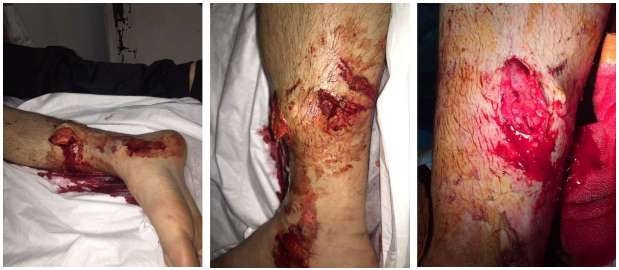
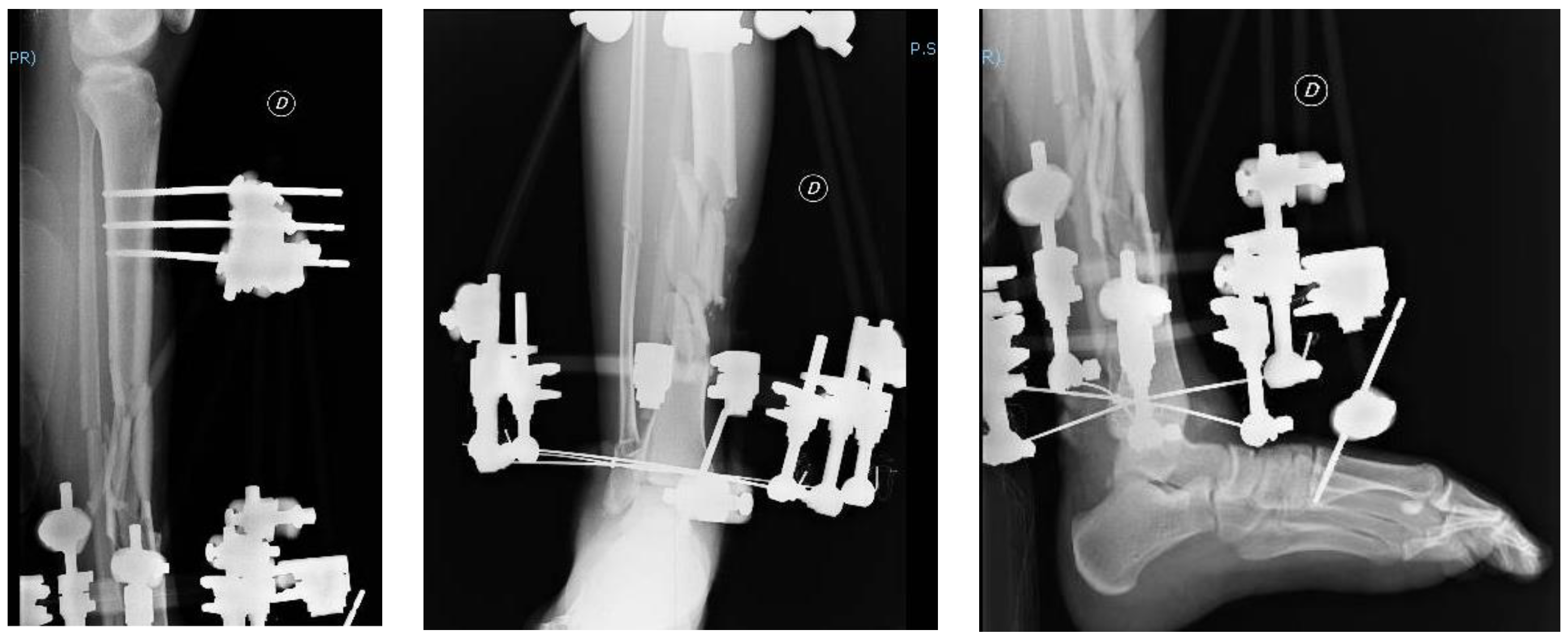
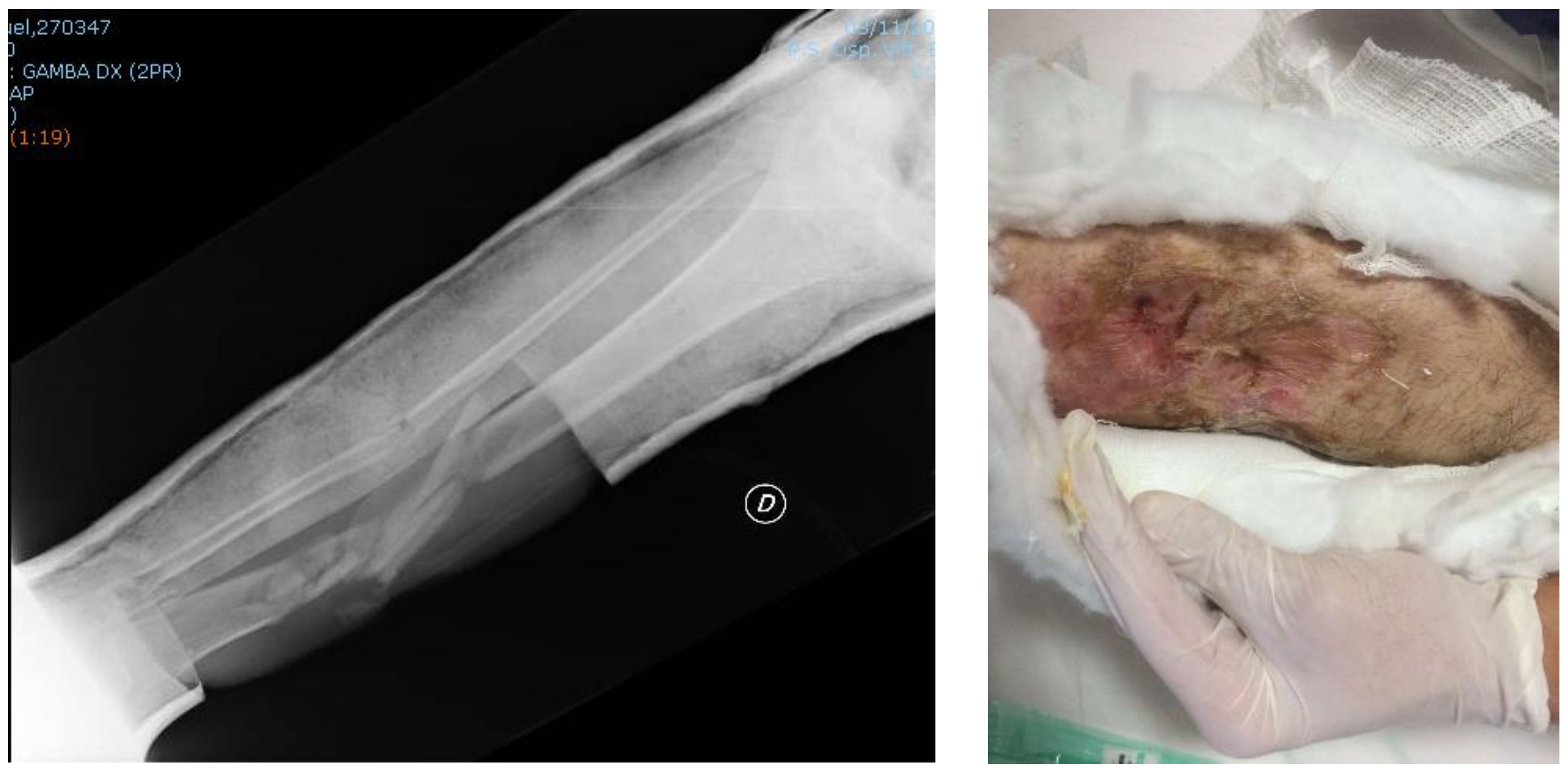
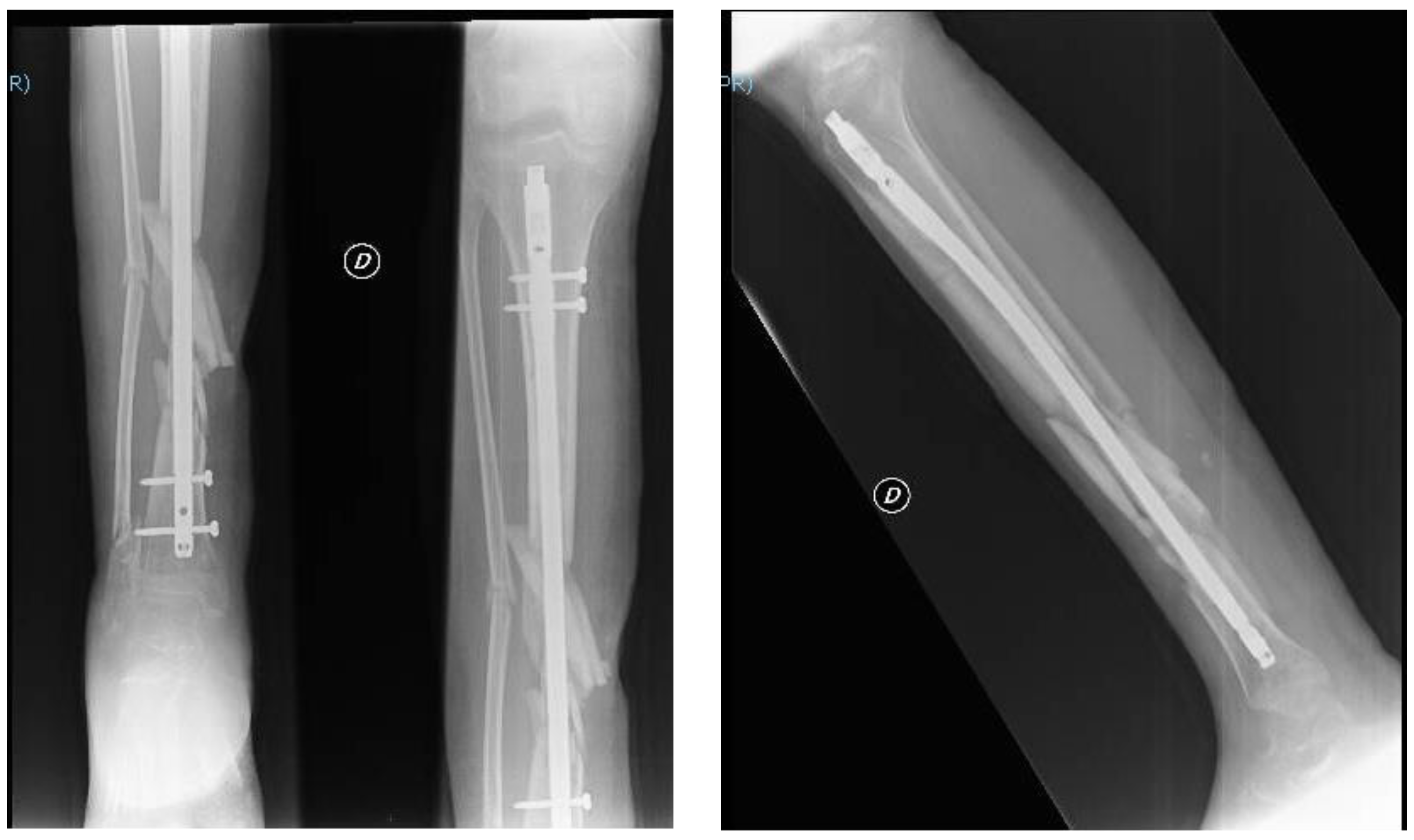
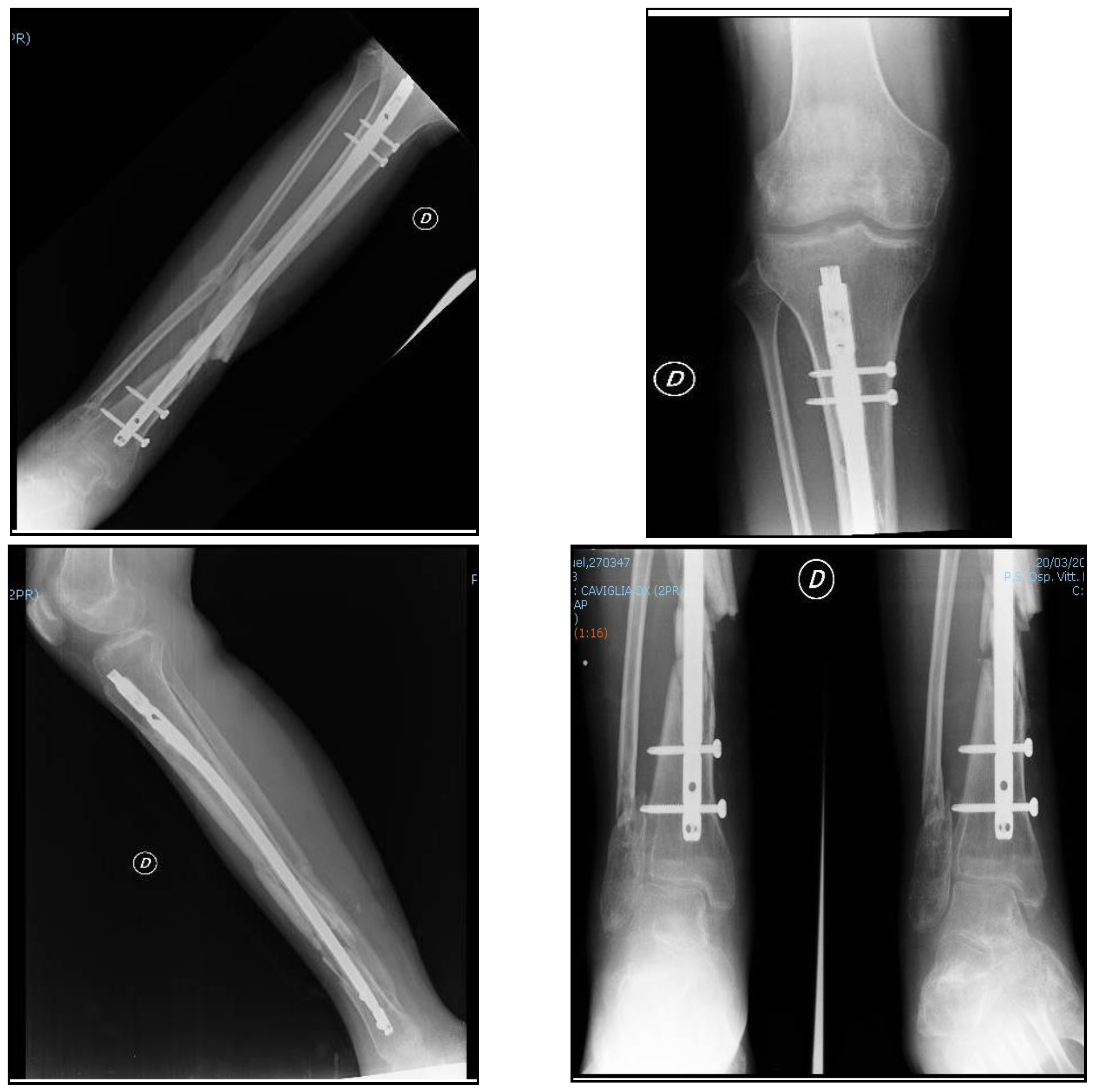
2. Clinical Case
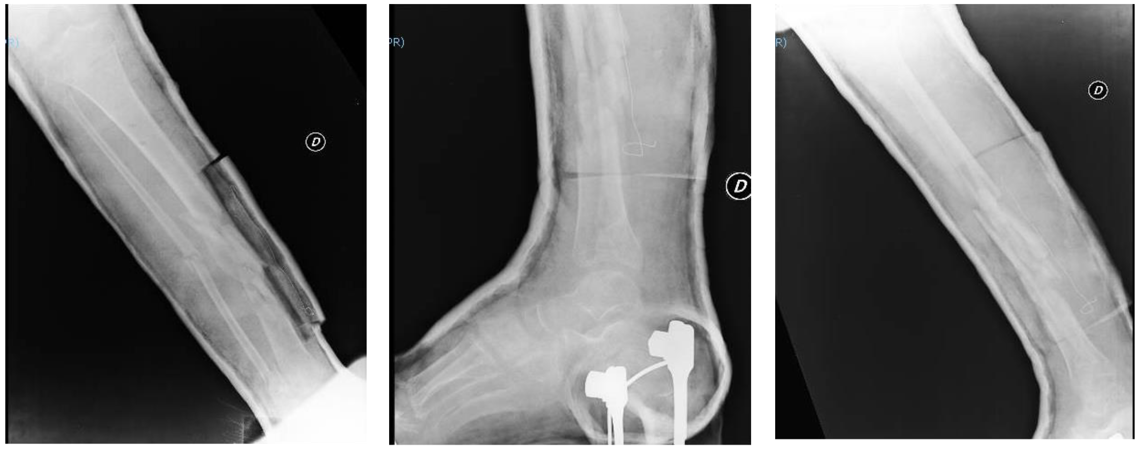
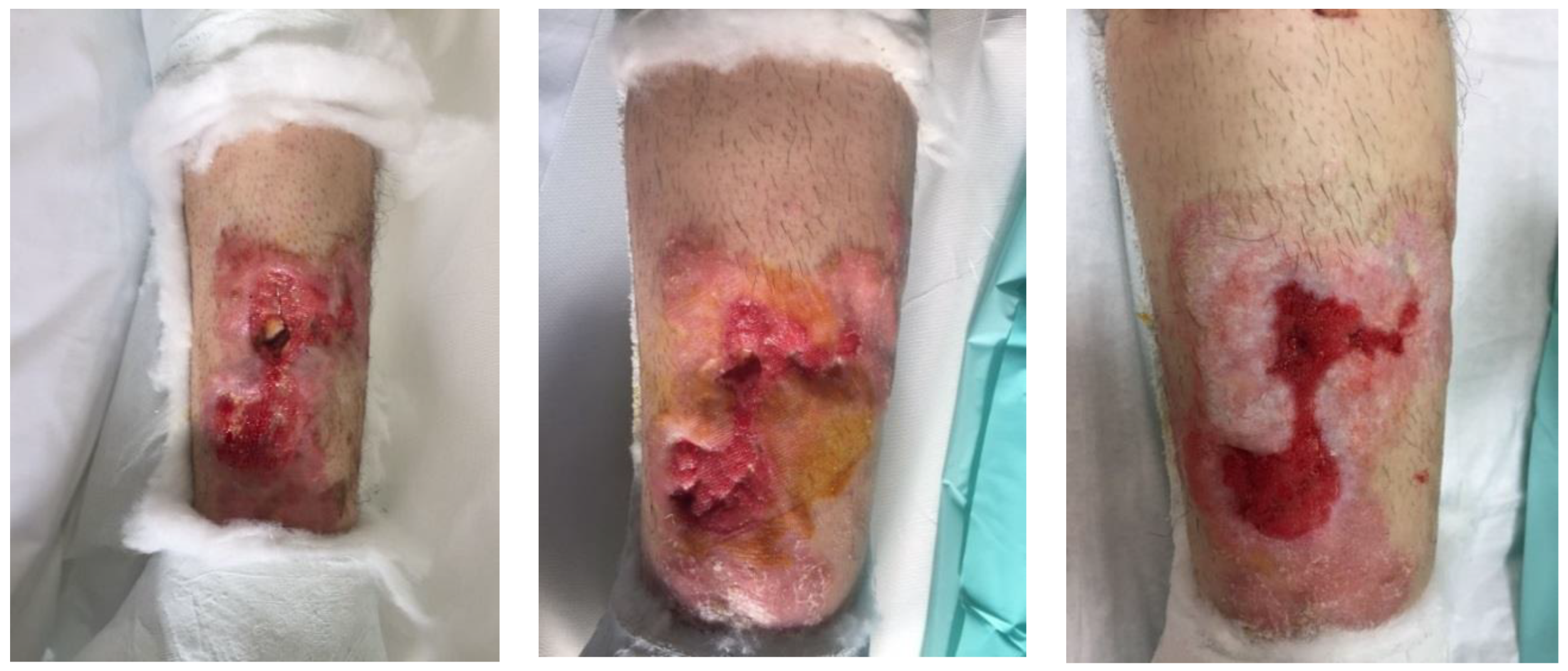
Post-Operative Care
3. Discussion
4. Conclusions
Author Contributions
Funding
Conflicts of Interest
References
- Marx, J. Rosen’s Emergency Medicine: Concepts and Clinical Practice; Mosby: Philadelphia, PA, USA, 2002. [Google Scholar]
- Court-Brown, C.M.; McBirnie, J. The epidemiology of tibial fractures. J. Bone Jt. Surg. Br. 1995, 77, 417–421. [Google Scholar] [CrossRef]
- Weiss, R.J.; Montgomery, S.M.; Ehlin, A.; Al Dabbagh, Z.; Stark, A.; Jannson, K.A. Decreasing incidence of tibial shaft fractures between 1998 and 2004: Information based on 10627 Swedish inpatients. Acta Orthop. 2008, 77, 526–533. [Google Scholar] [CrossRef] [PubMed]
- Müller, M.E.; Nazarian, S.; Koch, P.; Schatzker, J.; Heim, U. The Comprehensive Classification of Fractures of Long Bones; Spinger-Verlag: Berlin, Germany, 1990. [Google Scholar]
- Haas, N.; Krettek, C.; Schandelmaier, P.; Frigg, R.; Tscherne, H. A new solid unreamed tibial nail for shaft fractures with severe soft tissue injury. Injury 1993, 24, 49–54. [Google Scholar] [CrossRef]
- Socie, M.J.; Rovesti, G.L.; Griffon, D.J.; Elkhatib, O.; Mudrock, R.N.; Kurath, P. Biomechanical comparison of strategies to adjust axial stiffness of a hybrid fixator. Vet. Comp. Orthop. Traumatol. 2012, 25, 224–230. [Google Scholar] [CrossRef] [PubMed]
- Ilizarov, G.A.; Ledyaev, V.I. The replacement of long tubular bone defects by lengthening distraction osteotomy of one of the fragments. Clin. Orthop. Relat. Res. 1992, 280, 7–10. [Google Scholar] [CrossRef]
- Henley, M.B.; Chapman, J.R.; Agel, J.; Harvey, E.J.; Whorton, A.M.; Swiontkowski, M.F. Treatment of type II, IIIA, and IIIB open fractures of the tibial shaft: A prospective comparison of unreamed interlocking intramedullary nails and half-pin external fixators. J. Orthop. Trauma 1998, 12, 1–7. [Google Scholar] [CrossRef] [PubMed]
- Friedl, H.P.; Stocker, R.; Czermak, B.; Schmal, H.; Trentz, O. Primary fixation and delayed nailing of long bone fractures in severe trauma. Tech. Orthop. 1996, 11, 59–66. [Google Scholar] [CrossRef]
- Virani, S.R.; Dahapute, A.A.; Bava, S.S.; Muni, S.R. Impact of negative pressure wound therapy on open diaphyseal tibial fractures: A prospective randomized trial. J. Clin. Orthop. Trauma 2016, 7, 256–259. [Google Scholar] [CrossRef] [PubMed] [Green Version]


| A1 | A2 | A3 | B1 | B2 | B3 | C1 | C2 | C3 |
|---|---|---|---|---|---|---|---|---|
| Spiral | Oblique | Transverse | Spiral wedge | Oblique wedge | Transversal wedge | Comminuted | Segmental | Crush |
| I | Open fracture, clean wound, wound <1 cm in length. |
| II | Open fracture, wound >1 cm but <10 cm in length without extensive soft tissue damage, flaps, avulsions. |
| IIIA | Open fracture with adequate soft tissue coverage of a fractured bone despite extensive soft tissue laceration or flaps, or high-energy trauma regardless of the size of the wound. |
| IIIB | Open fracture with extensive soft tissue loss and periosteal stripping and bone damage. Usually associated with massive contamination. Will often need a further soft tissue coverage procedure. |
| IIIC | Open fracture associated with an arterial injury requiring repair, irrespective of degree of soft tissue injury. |
© 2018 by the authors. Licensee MDPI, Basel, Switzerland. This article is an open access article distributed under the terms and conditions of the Creative Commons Attribution (CC BY) license (http://creativecommons.org/licenses/by/4.0/).
Share and Cite
Pizzolo, S.; Testa, G.; Papotto, G.; Mobilia, G.; Di Stefano, G.; Sessa, G.; Pavone, V. Open Tibial Fracture in a Non-Compliant Patient: A Case Report. J. Funct. Morphol. Kinesiol. 2018, 3, 44. https://doi.org/10.3390/jfmk3030044
Pizzolo S, Testa G, Papotto G, Mobilia G, Di Stefano G, Sessa G, Pavone V. Open Tibial Fracture in a Non-Compliant Patient: A Case Report. Journal of Functional Morphology and Kinesiology. 2018; 3(3):44. https://doi.org/10.3390/jfmk3030044
Chicago/Turabian StylePizzolo, Samuele, Gianluca Testa, Giacomo Papotto, Giuseppe Mobilia, Giovanni Di Stefano, Giuseppe Sessa, and Vito Pavone. 2018. "Open Tibial Fracture in a Non-Compliant Patient: A Case Report" Journal of Functional Morphology and Kinesiology 3, no. 3: 44. https://doi.org/10.3390/jfmk3030044
APA StylePizzolo, S., Testa, G., Papotto, G., Mobilia, G., Di Stefano, G., Sessa, G., & Pavone, V. (2018). Open Tibial Fracture in a Non-Compliant Patient: A Case Report. Journal of Functional Morphology and Kinesiology, 3(3), 44. https://doi.org/10.3390/jfmk3030044