Toward the Analysis of Mitochondria Isolated from Leukemic Cells with Electrochemically Instrumented Microwell Arrays †

Abstract
:1. Introduction
1.1. Motivation
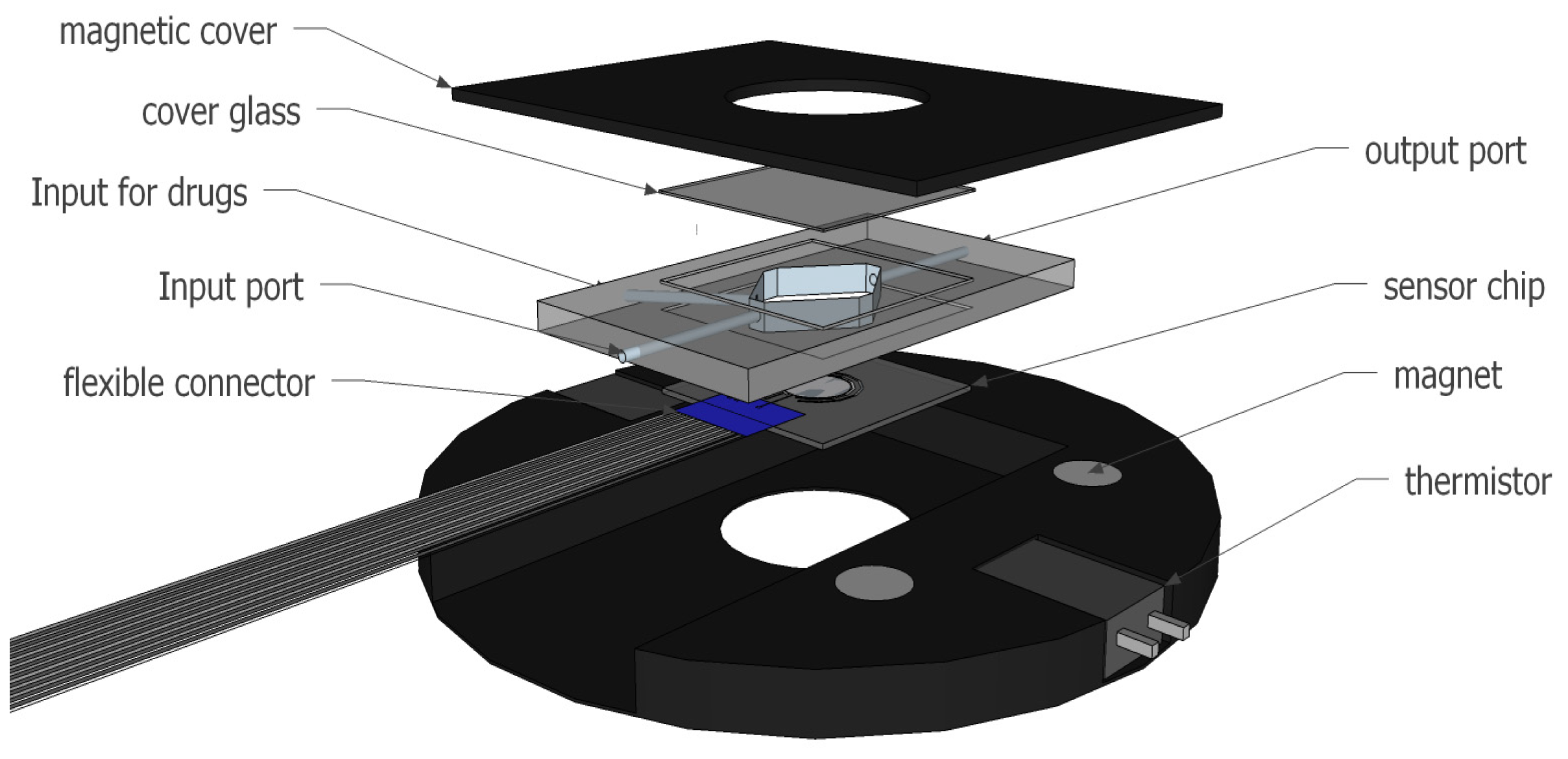
1.2. Concept
2. Materials and Methods
2.1. Device Manufacturing
2.2. Functionalization of Surfaces
2.3. Fluidics
3. Results
3.1. Measurements of Mitochondrial Activity
3.2. Functionalization of Surfaces
4. Discussion
Conflicts of Interest
References
- Farge, T.; aland, E.; de Toni, F.; Aroua, N.; Hosseini, M.; Perry, R.; Bosc, C.; Sugita, M.; Stuani, L.; Fraisse, M.; et al. Chemotherapy resistant human acute leukemia cells are not enriched for leukemic stem cells but require oxidative metabolism. Cancer Discov. 2017, 7, 716–735. [Google Scholar] [CrossRef] [PubMed]
- Sekli-Belaïdi, F.; Vanhove, E.; Tiddi, W.; Polverel, M.; Lemercier, G.; Lecestre, A.; Dubreuil, P.; Launay, J.; Arbault, S.; Temple-Boyer, P. Integration of ring nanoelectrodes into microwells for the bioelectrochemical analysis in sub-picolitre volumes. Sens. Actuators B Chem. 2016, 232, 345–356. [Google Scholar] [CrossRef]
- Ghilane, J.; Hapiot, P.; Bard, A.J. Metal/polypyrrole quasi-reference electrode for voltammetry in nonaqueous and aqueous solutions. Anal. Chem. 2006, 78, 6868–6872. [Google Scholar] [CrossRef] [PubMed]
- Ben-Amor, S.; Vanhove, E.; Belaïdi, F.S.; Charlot, S.; Colin, D.; Rigoulet, M.; Devin, A.; Sojic, N.; Launay, J.; Temple-Boyer, P.; et al. Enhanced detection of hydrogen peroxide with platinized microelectrode arrays for analyses of mitochondria activities. Electrochim. Acta 2014, 126, 171–178. [Google Scholar] [CrossRef]


© 2022 by the authors. Licensee MDPI, Basel, Switzerland. This article is an open access article distributed under the terms and conditions of the Creative Commons Attribution (CC BY) license (https://creativecommons.org/licenses/by/4.0/).
Share and Cite
Lemercier, G.; Sekli-Belaïdi, F.; Vajrala, S.; Descamps, E.; Foncy, J.; Arbault, S.; Sarry, J.-E.; Temple-Boyer, P.; Launay, J. Toward the Analysis of Mitochondria Isolated from Leukemic Cells with Electrochemically Instrumented Microwell Arrays. Proceedings 2017, 1, 289. https://doi.org/10.3390/proceedings1040289
Lemercier G, Sekli-Belaïdi F, Vajrala S, Descamps E, Foncy J, Arbault S, Sarry J-E, Temple-Boyer P, Launay J. Toward the Analysis of Mitochondria Isolated from Leukemic Cells with Electrochemically Instrumented Microwell Arrays. Proceedings. 2017; 1(4):289. https://doi.org/10.3390/proceedings1040289
Chicago/Turabian StyleLemercier, Gabriel, Fadhila Sekli-Belaïdi, Suresh Vajrala, Emeline Descamps, Julie Foncy, Stéphane Arbault, Jean-Emmanuel Sarry, Pierre Temple-Boyer, and Jérôme Launay. 2017. "Toward the Analysis of Mitochondria Isolated from Leukemic Cells with Electrochemically Instrumented Microwell Arrays" Proceedings 1, no. 4: 289. https://doi.org/10.3390/proceedings1040289