Proceeding Paper

Development of a MEMS Plate Based on Thin-Film Piezoelectric AlN Actuators for Biological Applications †

1
University Grenoble Alpes, F-38000 Grenoble, France
2
CEA-LETI, MINATEC Campus, 38054 Grenoble, France
3
INSERM U1205 Team ATIP/Avenir Mechanobiology Innate Immunity and Cancer, 38054 Grenoble, France
*
Author to whom correspondence should be addressed.
Presented at the Eurosensors 2017 Conference, Paris, France, 3–6 September 2017.
Proceedings 2017, 1(4), 386; https://doi.org/10.3390/proceedings1040386
Published: 9 August 2017
(This article belongs to the Proceedings of Proceedings of Eurosensors 2017, Paris, France, 3–6 September 2017)

Abstract

:
This paper presents the development of a lab-on-chip system based on the use of local vibrations to mechanically stimulate biological materials. It reports on the development and characterization of a piezoelectric actuators driven system designed to operate in liquid media. The microfluidic packaging of the Micro Electro Mechanical System (MEMS) is first presented. Then, electromechanical measurements done to calibrate our system are compared with Finite Element Method (FEM) simulations. These results are the first steps for implementation of piezoelectric MEMS to study mechanical response of biological cells at the population level.

1. Introduction

MEMS based sensors and microsystems offer significant potential for studying biological cells. Studies have already been done on single cell using MEMS to study how cells grow upon exposure of drugs [1,2] or cell fatigue [3]. MEMS devices with piezoelectric transducers can also be used for cell studies using acoustic waves [4]. These systems offer the possibility to study adhesive forces and cellular response to controlled mechanical stimuli. It has been recently recognized that cells do display differential mechanical behaviors according to their biological states. Our intended goal is the label free recognition of various cell types (e.g., cancerous cells) and their response to various drugs according to their mechanobiology. To achieve this, we developed a MEMS controlled mechanical waves generating system with submicronic displacements in liquid media [5,6] in order to study cells in cell culture setting [7].

2. Materials and Methods
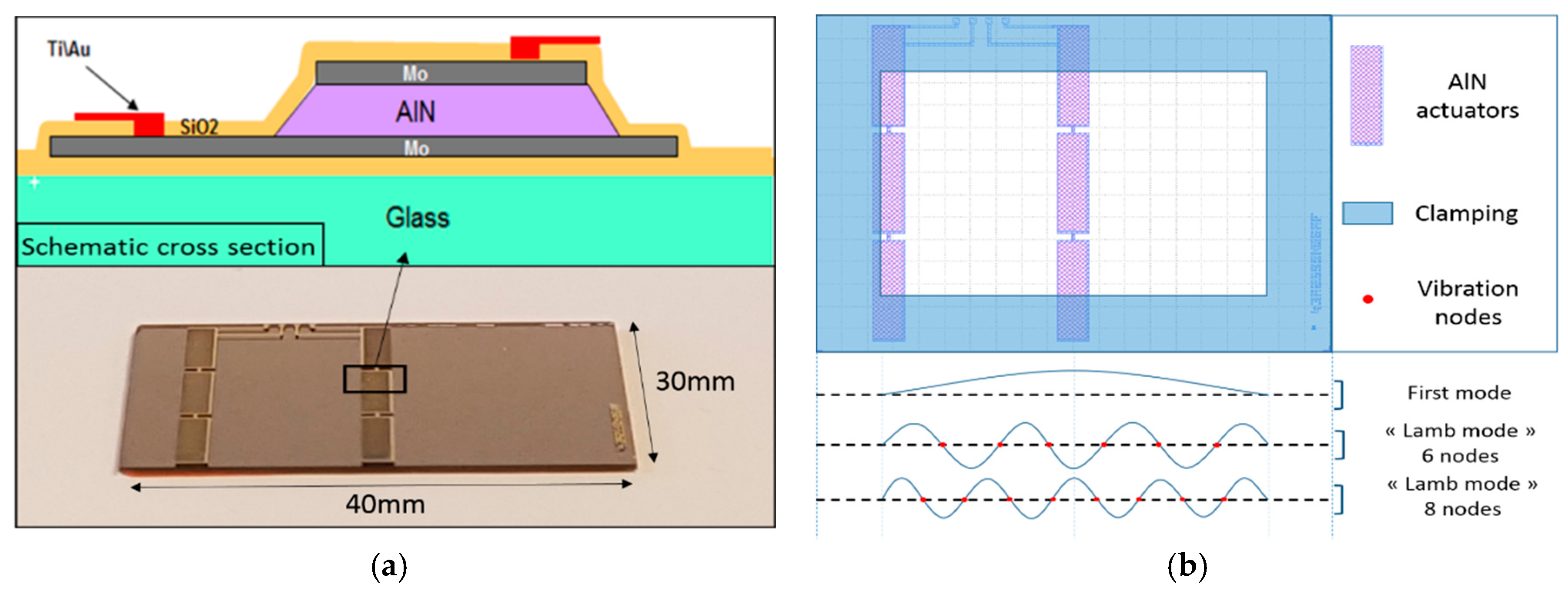
2.1. Design

The vibrating MEMS was developed based on thin film piezoelectric Aluminum Nitride (AlN) actuators, manufactured out of standard 200 mm glass substrate (EAGLE XG®) with a standard thickness of 700 μm. The piezoelectric stack of the actuators consists of 2 μm thick AlN, in between 220 nm thick Molybdenum bottom and top electrodes [8]. A 300 nm thick silicon dioxide passivation layer is deposited and the connection of the electrodes is enable by a gold (Au) layer of 500 nm above a 20 nm thick Titanium (Ti) adhesive layer. The AlN is deposited using reactive sputtering from an aluminum target under a nitrogen atmosphere. A reflective coating is added to perform optical measurements. Figure 1a gives a schematic view of the technological stack and a photography of a 40 × 30 mm2 plate diced from the glass substrate. The actuator is positioned at the center of the plate in order to fit the maximum out-of-plane vibration mode amplitude area as shown in Figure 1b. The second actuator column at the left of the plate is not used in this study.

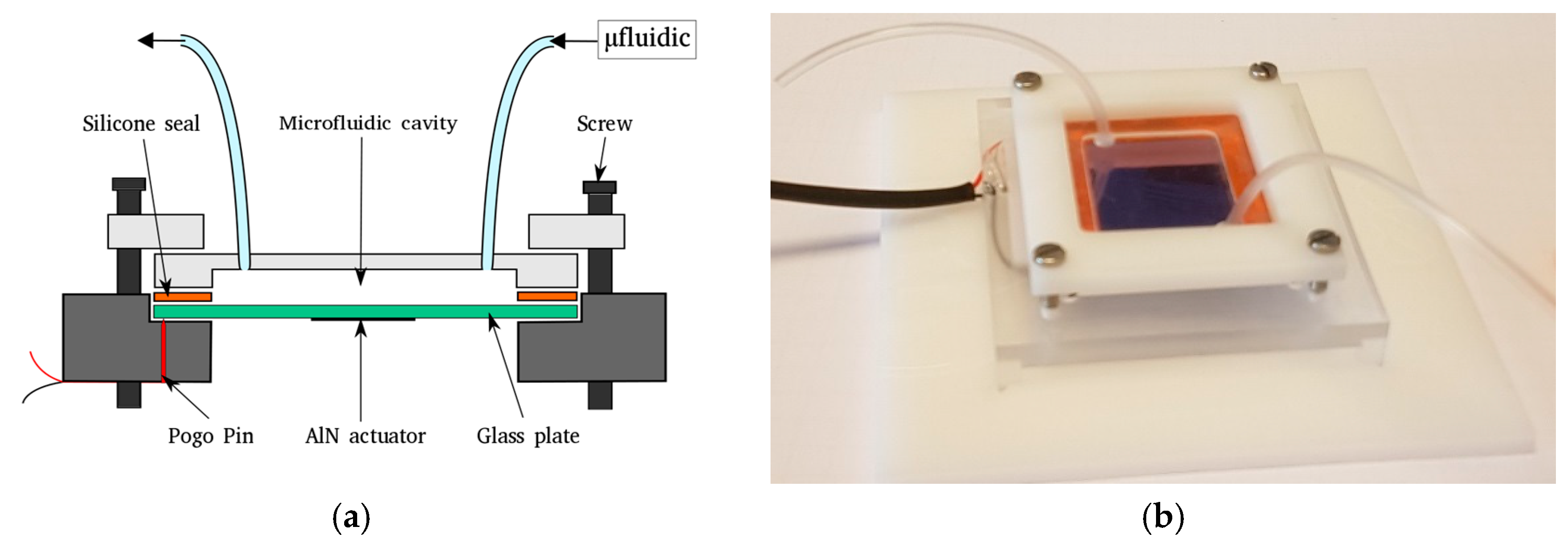
2.2. Microfluidic Packaging

A microfluidic cavity is made out of etched Poly(methyl methacrylate) (PMMA) plate placed on the back side of the glass plate. The dimension of the cavity are 30 mm × 20 mm × 1 mm and can be filled using Tygon tubing. In order to actuate the AlN actuator, the gold pad electrical contacts are connected by Pogo Pin. The whole packaging is clamped by screws. With this system the glass plate can be removed and changed easily in the perspective of testing different glass plates and actuation configurations. A schematic drawing of the microfluidic packaging is described in Figure 2a and a photography is presented in Figure 4b.

2.3. Electromechanical Characterizations and FEM Simulations
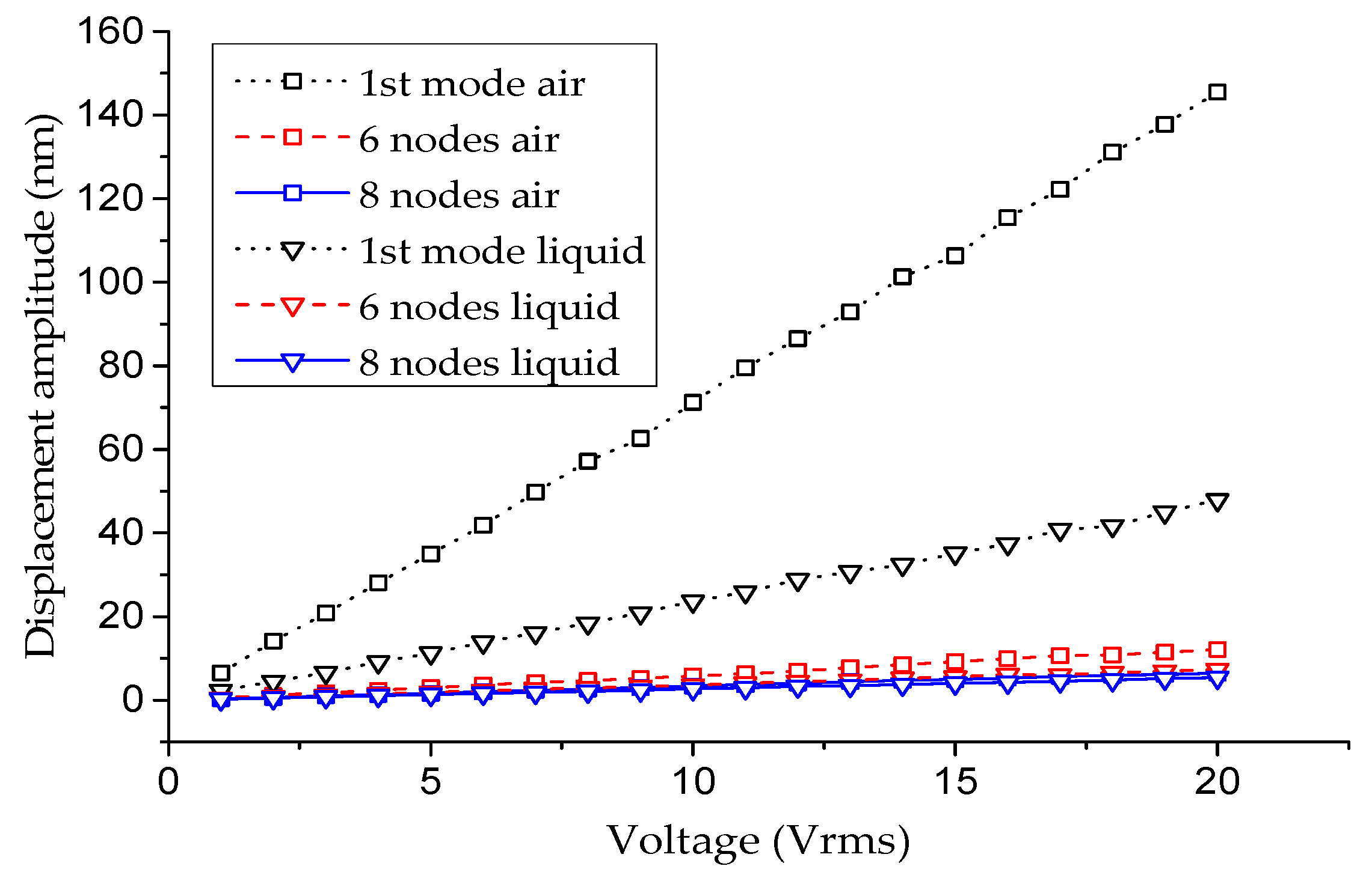
The electromechanical characterization of the device is performed using a Polytec Laser Doppler Vibrometer (LDV) MSA400. The out-of-plane resonant modes are studied between 500 Hz and 150 kHz. An electrical signal of 1 Vrms is applied to actuate the AlN actuator with the focus on three modes of interest: 1st flexural mode, Lamb mode with 6 nodes and Lamb mode with 8 nodes as shown in Figure 1b. The measurements are performed in air and with the cavity filled with water (Figure 3). The velocity obtained by the LDV in liquid as to be corrected by the refractive index of the liquid to take into account the fact that the vibration is measured in liquid. The different modes are identified in this two environments by several measurements on the whole plate. Displacement amplitudes are also measured for the focused resonant modes with different AC voltages up to 20 Vrms applied to the actuator.
Finite Element Method (FEM) simulations are carried out, using COMSOL Multiphysics, to model the behavior of the system under different environments and to study the impact of the clamping on the glass plate. 3D model was developed using the acoustic-structure interaction module to take into account the impact of the liquid media on the vibration modes.

3. Results

The three out-of-plane modes have been identified thanks to vibrometry measurements at the center of the plate (Figure 3). The results show an impact of the liquid media on the resonant frequencies of the interesting vibration modes. As predicted by FEM simulations, the presence of liquid in the microfluidic cavity induced a frequency shift towards lower values. As shown by Figure 3, unlike the first mode which has been importantly impacted by the filling of the cavity, the quality factors of Lamb resonant modes are only slightly affected by the presence of liquid. For example a quality factor calculated at −3 dB of 18 is observed for the Lamb 6 nodes mode in liquid compared to a value of 20 in air. These measurements make the different Lamb modes some good candidates for our further biological studies in liquid medium.
Now that the different resonant modes are identified, we are interested in the displacement amplitudes of these vibration modes in function of the applied voltage on the actuator for both environments. Figure 4 shows, as expected by Figure 3, that the first mode of resonance induced a higher displacement compared to Lamb mode but it is importantly influenced by the environment. At contrary, the displacement amplitudes of the Lamb modes stay relatively close to their value measured in air. For example, an interesting 5.4 nm is obtained for the Lamb mode with 8 nodes measured in liquid under only 20 Vrms for a reference value of 6.4 nm in air. It is a decrease of only 15% with displacement values sufficient to study the mechanical response of a population of biological cells under mechanical stimuli.
Table 1 sums up the results obtained by electromechanical characterization and FEM model. It is important to know that the resonant frequency of the first mode is mainly affected by the smaller dimension of the vibrating surface which corresponds to the larger side of the plate in our system. That is why the disagreement between simulation and measurement for the first mode (Table 1) is attributed to an imperfect clamping along the larger side of the plate in the packaging.

4. Conclusions

A lab on chip system based on a thin film AlN actuated vibrating plate was presented. Resonant modes, frequencies and displacement amplitudes of the packaged glass plate were studied under different conditions. This calibration step of our system paves the way for an easy to use system able to apply and control mechanical stimuli to biological cells.

Conflicts of Interest

The authors declare no conflict of interest.

References

  1. Cermak, N.; Olcum, S.; Delgado, F.F.; Wasserman, S.C.; Payer, K.R.; Murakami, M.A.; Knudsen, S.M.; Kimmerling, R.J.; Stevens, M.M.; Kikuchi, Y.; et al. High-throughput measurement of single-cell growth rates using serial microfluidic mass sensor arrays. Nat. Biotechnol. 2016, 34, 1052–1059. [Google Scholar] [CrossRef] [PubMed]
  2. Park, K.; Millet, L.J.; Kim, N.; Li, H.; Jin, X.; Popescu, G.; Aluru, N.R.; Hsia, K.J.; Bashir, R. Measurement of adherent cell mass and growth. Proc. Natl. Acad. Sci. USA 2010, 107, 20691–20696. [Google Scholar] [CrossRef] [PubMed]
  3. Fukui, W.; Kaneko, M.; Sakuma, S.; Kawahara, T.; Arai, F. μ-cell fatigue test. In Proceedings of the 2012 IEEE International Conference on Robotics and Automation (ICRA), Saint Paul, MN, USA, 14–18 May 2012; pp. 4600–4605. [Google Scholar]
  4. Wang, L.; Li, Y.-J.; Lin, A.; Choe, Y.; Gross, M.E.; Kim, E.S. A Self-Focusing Acoustic Transducer That Exploits Cytoskeletal Differences for Selective Cytolysis of Cancer Cells. J. Microelectromech. Syst. 2013, 22, 542–552. [Google Scholar] [CrossRef]
  5. Ruiz-Díez, V.; Hernando-García, J.; Ababneh, A.; Seidel, H.; Sánchez-Rojas, J.L. In-liquid characterization of in-plane and high order out-of-plane modes of AlN-based square microplates. Microsyst. Technol. 2016, 22, 1701–1708. [Google Scholar] [CrossRef]
  6. Ali, A.; Lee, J.E.-Y. Fully-differential AlN-on-Si wine glass mode resonator for enhanced characterization in water. In Proceedings of the 2016 IEEE SENSORS, Orlando, FL, USA, 30 October–3 November 2016; pp. 1–3. [Google Scholar]
  7. Debavelaere-Callens, D.; Peyre, L.; Campistron, P.; Hildebrand, H.F. On the use of ultrasounds to quantify the longitudinal threshold force to detach osteoblastic cells from a conditioned glass substrate. Biomol. Eng. 2007, 24, 521–525. [Google Scholar] [CrossRef] [PubMed]
  8. Casset, F.; Danel, J.; Renaux, P.; Chappaz, C.; Bernard, F.; Sednaoui, T.; Basrour, S.; Desloges, B.; Fanget, S. 4-inch transparent plates based on thin-film AlN actuators for haptic applications. Mechatronics 2016, 40, 264–269. [Google Scholar] [CrossRef]
Figure 1. (a) AlN-on-glass schematic technological stack cross section and photography; (b) Design of the 40 mm × 30 mm glass plate and schematic cross section of resonant mode shapes.
Proceedings 01 00386 g001
Figure 2. (a) Schematic drawing of the microfluidic packaging; (b) Photography of the microfluidic packaging.
Proceedings 01 00386 g002
Figure 3. Spectrum of the different modes of interest and identification of the resonant modes from simulation in air and liquid. (a) 1st mode; (b) Lamb mode with 6 nodes; (c) Lamb mode with 8 nodes.
Proceedings 01 00386 g003
Figure 4. Maximum displacement amplitudes for different modes in function of applied voltage.
Proceedings 01 00386 g004
Table 1. Displacement amplitudes, quality factors and comparison of the resonant frequencies.
EnvironmentAirLiquid
MethodDisplacement amplitudes @20 VrmsQuality factorExperimentalFEMRelative errorDisplacement amplitudes @20 VrmsQuality factorExperimentalFEMRelative error
1st mode146 nm225.47 kHz6.9 kHz20%48 nm133.75 kHz5.5 kHz31%
Lamb mode:
6 nodes
12 nm2098.92 kHz99.5 kHz0.6%7.3 nm1876.50 kHz80 kHz4.4%
Lamb mode:
8 nodes
6.4 nm30144.7 kHz145.7 kHz0.7%5.4 nm33121.4 kHz121 kHz0.3%
Publisher’s Note: MDPI stays neutral with regard to jurisdictional claims in published maps and institutional affiliations.

