Proceeding Paper

Comparison of ITO and IrOx-Modified ITO Interdigitated Electrodes for Electrical Cell-Substrate Impedance Sensing (ECIS) Applications †

Institut des Nanotechnologies de Lyon UMR 5270 CNRS, Université Claude Bernard Lyon 1, INSA, Villeurbanne, France
*
Author to whom correspondence should be addressed.
Presented at the Eurosensors 2017 Conference, Paris, France, 3–6 September 2017.
Proceedings 2017, 1(4), 532; https://doi.org/10.3390/proceedings1040532
Published: 9 August 2017
(This article belongs to the Proceedings of Proceedings of Eurosensors 2017, Paris, France, 3–6 September 2017)

Abstract

:
This study compares the sensitivity of interdigitated electrodes (IDEs) used in Electrical Cell-Substrate Impedance Sensing (ECIS). IDEs made of indium tin oxide (ITO) and ITO coated with iridium oxide (IrOx) were used for ECIS measurements with the human breast cancer cell line, MCF-7. The results indicate that IrOx-modified ITO electrodes provide both biocompatibility and higher cell sensitivity compared to ITO electrodes. We also found that the quantity of IrOx required to generate such a sensitivity improvement is sufficiently low to not interfere with the visualization of the cells under study. IrOx-modified ITO electrodes are therefore promising sensors for ECIS applications.

1. Introduction

Personalized medicine, which seeks to offer more effective treatments and cause fewer side effects on patients, is now playing an important role in cancer therapy. In this approach, in vitro tests are required for evaluating the effects of new chemotherapeutic drugs on living cells. As a result, several techniques have emerged in order to monitor and analyze cellular activities [1]. ECIS, introduced by Giaever and Keese [2], is one of them and it has become a well-known, label-free technique for real-time study of cancer cells proliferating on top of electrodes.
In ECIS, various studies have used gold as the standard electrode material for impedance measurements [3,4]. ITO is seen as an alternative material to gold due to its transparency [5]. Surprisingly, despite this advantage, ITO is largely under-used compared to gold. Moreover, the electrode coatings explored to enhance the performance of electrodes for neural applications [6] are mostly considered as a strategy to modify the sensitivity of ECIS electrodes [7]. In this paper, a comparison between IDEs of ITO and ITO coated with IrOx is presented and their sensitivities are studied in response to the proliferation of the human breast cancer cell line, MCF-7. The results indicate that IrOx-modified ITO electrodes provide both biocompatibility and higher cell sensitivity compared to ITO electrodes, which make them promising sensors for monitoring cell proliferation using both impedance and optical measurements.

2. Materials and Methods

2.1. Fabrication of Electrodes

The interdigitated electrodes used in this work comprised 39 electrode fingers of 20 μm width, 1 mm length and with inter-electrode gaps of 20 μm. The fabrication process of ITO-IDEs from glass slides coated with a 370 nm layer of ITO (76 × 26 mm, Solems) consisted in three steps: photolithography, evaporation of Cr layer (200 nm) and reactive ion etching (RIE). The IrOx-modified ITO-IDEs were fabricated using an additional step of electrodeposition by cyclic voltammetry. This was done by cycling 10 times the ITO electrode potential between 0 and 0.7 V/Ag/AgCl in 4.5 mM IrCl4·H2O solution (55.5 mM C2H2O4 as supporting electrolytic solution, pH = 10.5 in water) with a scan rate of 20 mV/s. Once the electrodes were fabricated, a reservoir (diameter of 5 mm) of PolyDiMethylSiloxane (PDMS) was bonded to the glass slides using O2 plasma treatment so that the IDEs could be in contact with the different solutions during impedance measurements.

2.2. Impedance Measurement Set-Up

A potentiostat (PGSTAT, Metrohm) and its associated software Nova 2.1 were used for the impedance measurements and to fit the experimental data. The set-up consisted of the IDEs connected in a two-electrode configuration and measurements were carried out from 100 mHz to 1 MHz with a sinusoidal signal amplitude of 10 mV (0 V offset).

2.3. Bioimpedance Measurements with MCF-7 Cells

The cell line used for the bioimpedance experiments was the human breast cancer cell, MCF-7. The cell culture medium contained Dubelcco’s Modified Eagle Medium (DMEM, ThermoFisher), 10% fetal bovine serum (v/v) (FBS, Dominique Dutscher) and 1% penicillin-streptomycin (v/v) (PS, ThermoFisher). Before addition of cells, the electrode surface was coated with a solution containing a mixture of type IV collagen (CN, Sigma-Aldrich) and bovine serum albumin (BSA, Sigma-Aldrich) (nCN/nBSA = 99). Trypsin (ThermoFisher) was used to collect the cells from the Petri dish before the electrode inoculation. After centrifugation and cell counting, the MCF-7 cells were seeded.

3. Results and Discussion

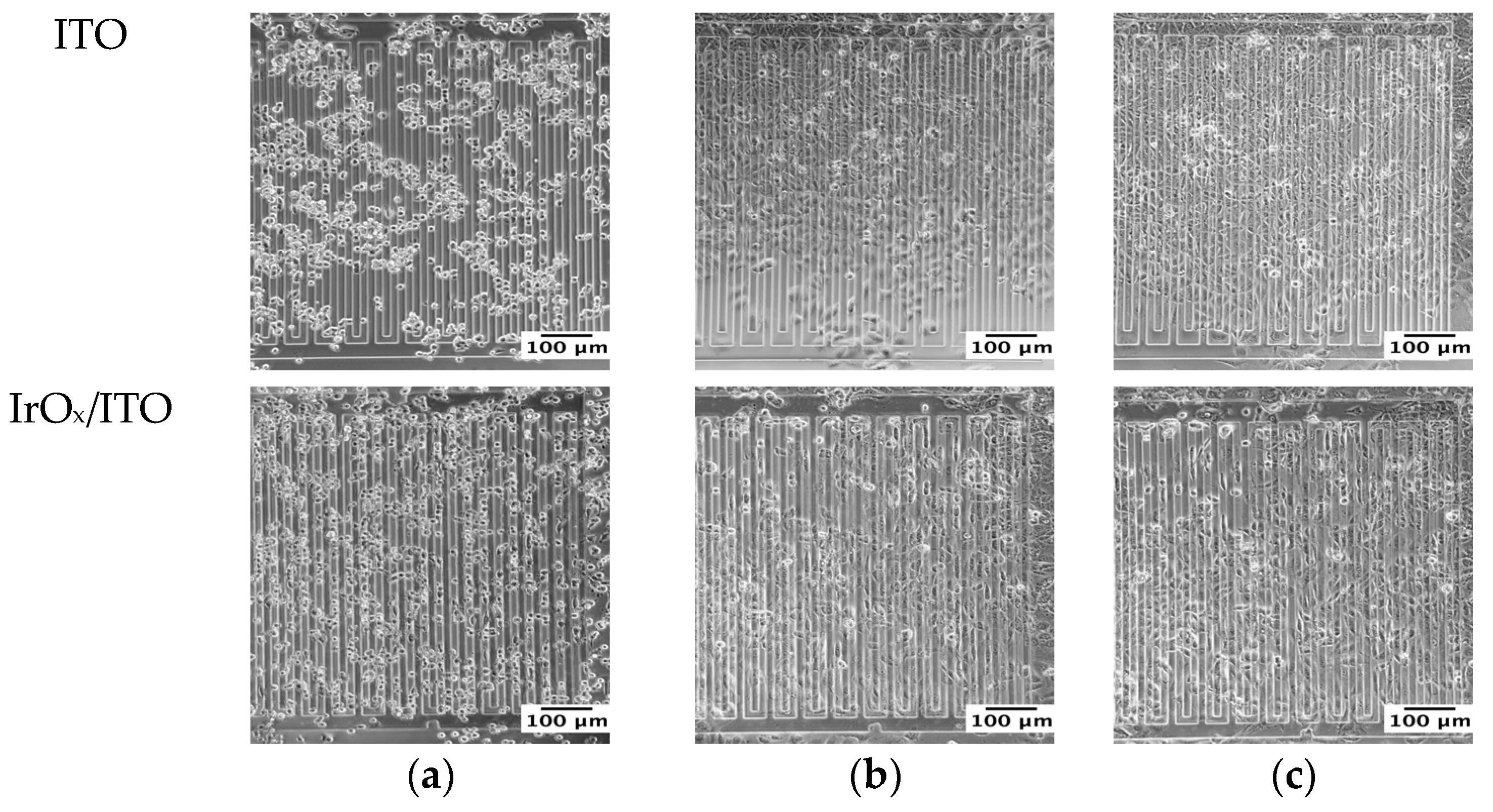
3.1. ECIS Measurements with MCF-7 Cells

Phase-contrast micrographs of the breast cancer cells, MCF-7, proliferating on top of electrodes were taken during an ECIS experiment. After sedimentation, cells started adhering onto the electrode surface (Figure 1a) and then proliferating (Figure 1b,c). As shown in Figure 1, modifying the surface of ITO electrodes with IrOx did not affect their transparency nor their biocompatibility. In order to compare the sensitivities of these materials, impedance results are presented as the normalized impedance Znormalized, which is given by [8]:
Z n o r m a l i z e d = Z t Z 0 Z 0
where Z0 is the modulus of the impedance before seeding the cells and Zt is the modulus of the impedance at a time t following addition of the cells. The evolution of Znormalized as a function of surface coverage ratio is presented in Figure 2, at specific frequencies of 3.2 kHz and 8 Hz for ITO and IrOx-modified electrodes, respectively. These frequencies were chosen because they represent the frequency of highest Znormalized variation [8]. As expected from the literature [1], the impedance increased with cell proliferation. It was also noted that the values of Znormalized for IrOx-modified ITO electrodes were four times higher than for bare ITO electrodes, suggesting that the modified electrodes seem to be more sensitive to the cell contributions.

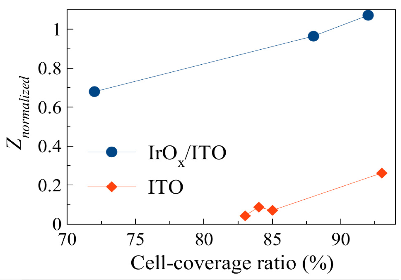
3.2. Interfacial Impedances of the IDEs

Equivalent circuit modeling was used in an effort to explain the difference of sensitivity between ITO and IrOx/ITO electrodes. Figure 3a represents the equivalent circuit of ITO-IDEs whereas IrOx/ITO electrodes were modeled with the equivalent circuit shown in Figure 3b. By fitting the experimental impedance measured in cell culture medium for both electrodes, we were able to compute the value of the pseudocapacitance of the electrode-electrolyte interface, Cinterface, given by [9]:
C i n t e r f a c e = Q 2 1 / α 2 R 1 / α 2 1
Figure 3c shows that Cinterface is higher for IrOx-modified electrodes, leading to lower interfacial impedances. This could explain the differences in sensitivity observed in Figure 2.

4. Conclusions

This study demonstrates that interdigitated electrodes made of ITO coated with IrOx show higher sensitivity for ECIS measurements, when using breast cancer cells MCF-7, than ITO electrodes. Moreover, fitting experimental data with equivalent circuit models revealed that IrOx coated ITO electrodes have lower interfacial impedance than ITO electrodes. This could explain the difference in electrode sensitivity for ECIS measurements. Therefore, ITO with IrOx might be a promising material in applications requiring both impedance and optical measurements.

Conflicts of Interest

The authors declare no conflict of interest.

References

  1. Hong, J.; Kandasamy, K.; Marimuthu, M.; Choi, S.; Kim, S. Electrical cell-substrate impedance sensing as a non-invasive tool for cancer cell study. Analyst 2011, 136, 237–245. [Google Scholar] [CrossRef] [PubMed]
  2. Giaever, I.; Keese, C.R. Micromotion of mammalian cells measured electrically. Proc. Natl. Acad. Sci. USA 1991, 88, 7896–7900. [Google Scholar] [CrossRef] [PubMed]
  3. Ceriotti, L.; Ponti, J.; Colpo, P.; Sabbioni, E.; Rossi, F. Assessment of cytotoxicity by impedance spectroscopy. Biosens. Bioelectron. 2007, 22, 3057–3063. [Google Scholar] [CrossRef]
  4. Mamouni, J.; Yang, L. Interdigitated microelectrode-based microchip for electrical impedance spectroscopic study of oral cancer cells. Biomed. Microdevices 2011, 13, 1075–1088. [Google Scholar] [CrossRef] [PubMed]
  5. Choi, C.K.; Margraves, C.H.; Jun, S.I.; English, A.E.; Rack, P.D.; Kihm, K.D. Opto-Electric Cellular Biosensor Using Optically Transparent Indium Tin Oxide (ITO) Electrodes. Sensors 2008, 8, 3257–3270. [Google Scholar] [CrossRef]
  6. Li, X.; Pei, W.; Tang, R.; Gui, Q.; Guo, K.; Wang, Y.; Chen, H. Investigation of flexible electrodes modified by TiN, Pt black and IrOx. Sci. China Technol. Sci. 2011, 54, 2305–2309. [Google Scholar] [CrossRef]
  7. Srinivasaraghavan, V.; Strobl, J.; Wang, D.; Heflin, J.R.; Agah, M. A comparative study of nano-scale coatings on gold electrodes for bioimpedance studies of breast cancer cells. Biomed. Microdevices 2014, 16, 689–696. [Google Scholar] [CrossRef] [PubMed]
  8. Jiang, W.G. Electric Cell-Substrate Impedance Sensing and Cancer Metastasis, 1st ed.; Springer: Dordrecht, The Netherlands; Heidelberg, Germany; New York, NY, USA; London, UK, 2012; p. 108. [Google Scholar]
  9. Hirschron, B.; Orazem, M.E.; Tribollet, B.; Vivier, V.; Frateur, I.; Musiani, M. Determination of effective capacitance and film thickness from constant-phase-element parameters. Electrochim. Acta 2010, 55, 6218–6227. [Google Scholar] [CrossRef]
Figure 1. Phase contrast micrographs of ITO and IrOx coated ITO electrodes for various breast cancer cell coverage ratios (a) 65%; (b) 88%; and (c) 92%.
Proceedings 01 00532 g001
Figure 2. Evolution of the normalized impedance of ITO electrodes (3.2 kHz) and ITO coated with IrOx (8 Hz) as a function of cell coverage ratio.
Proceedings 01 00532 g002
Figure 3. Equivalent circuit models used for (a) ITO-IDEs and (b) IrOx/ITO-IDEs. (c) Pseudocapacitance of the electrode-cell culture medium interface for ITO and ITO coated with IrOx electrodes. Celec represents the capacitive behavior of the electrode fingers; CPE1 and R1 model the IrOx on the surface of ITO while CPE2 and R2 are associated with the interface between the electrode and the cell culture medium.
Proceedings 01 00532 g003
Publisher’s Note: MDPI stays neutral with regard to jurisdictional claims in published maps and institutional affiliations.

