1. Introduction
In conventional diagnosis of leishmaniasis, a study of amastigotes in smear, culture for Leishmania, histopathological examination of lesions and identification of Leishmania by polymerase chain reaction (PCR) and enzyme-linked immunosorbent assay (ELISA) should be performed. Recently label-free surface plasmon resonance method has been employed as a viable alternative for leishmaniasis diagnosis providing fast response and comparable sensitivity. Besides, another important feature of the SPR based sensors is the need for very small quantity of analyte material, such as blood samples from a patient.
1.1. General Aspects of Leishmaniasis
Leishmaniasis represents endemic infections caused by a protozoan of the genus
Leishmania, which affects man and different animal species, wild and domestic, being transmitted by different species of mosquitoes and presenting a broad spectrum of clinical manifestations [
1]. Visceral leishmaniasis is caused by the species
Leishmania donovani and
Leishmania infantum (
Leishmania chagasi).
Leishmania infantum is the most common species on the American continents. It is a disease that has the mosquito of the genus Lutzomyia, phlebotomine, as main vector. Visceral leishmaniasis has raised concerns of the health authorities due to the great spread of this disease in the last decades.
According to the World Health Organization are 500,000 new cases of visceral leishmaniasis for year, being a protozoonosis fatal when left untreated [
2]. The forms of diagnosis are performed through clinical examination, but because of the difficulty found for safe diagnosis, laboratory methods are part of the research arsenal. The methods currently employed in visceral leishmaniasis clinical diagnoses are costly, requiring numerous steps to perform besides specialized labor, have also shown low sensitivity to detect the initial cases of the disease, generating false negatives for the disease. Therefore, it is clear the need to develop methodologies that allow a more sensitive and faster diagnosis, guaranteeing greater control of visceral leishmaniasis [
3,
4].
1.2. Surface Plasmon Resonance Biosensig Technique
According varying coupling mechanisms, two basic modes of operation normally are commonly used at the surface plasmon resonance (SPR) biosensors: Angular Interrogation Mode (AIM), where the wavelength of incident light is fixed and the angle of incidence is varied. The SPR resonance is recorded by an optical detector; Wavelength Interrogation Mode (WIM), where the angle of incidence is kept constant and incident light wavelength is varied. A spectrometer normally is employed to determine the SPR resonance.
A 4-layer SPR device is depicted in
Figure 1. The optical system is responsible for the adjustments on light source to get the optical conditions (polarization, focus, position) for phenomenon excitation; a multilayer system composed by the optical materials used in the sensor manufacturing including a bio-recognition layer that interacts with the substances contained in the analyte; a microfluidic system to confine and delivery the analyte to the sensitive surface; and one Acquisition Control Processing and Display unit (ACPDU) to analyze the sensor signal.
2. Materials and Methods
Two polymer SPR biochips have been employed in the experiments: one, proposed in 2004 [
5] and called VIRChip. It only works at the AIM case and has diffractive optical coupling elements (DOCEs), manufactured in nanometer dimensions by using holographic techniques; another, proposed in 2009 [
6], called PPBIO. It has a simple structure and can be applied for both AIM and WIM requiring relatively low design and manufacturing costs. At the experiments carried out, a 670-nm laser diode and a white light diode was employed at the PPBIO for AIM and WIM cases; correspondingly, a 670-nm laser diode and a CCD camera (AIM case) has been used with the VIRChip.
A highly specific and sensitive-constrained synthetic peptide (LC2) derived from a mimotope of
Leishmania chagasi antigen, selected by phage display (is a selection technique in which a library of peptide or protein variants is expressed as a genetic fusion to a bacteriophage coat protein), was used as probe for IgG detection [
4] were synthesized and pre-validated by the enzyme-linked immunosorbent assay (ELISA) method. Phosphate buffer saline solution (pH 7.4) was used as the baseline. This peptide LC2 was modified with BSA and cysteine [
7].
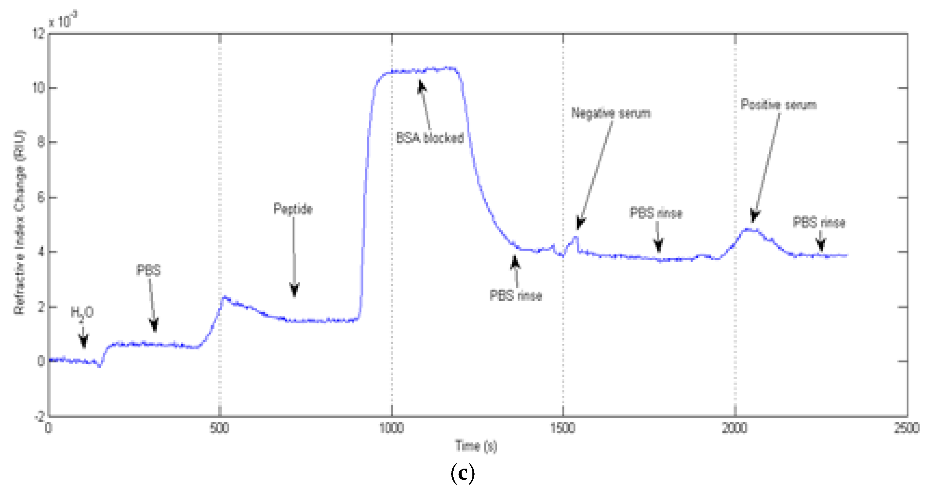
After biochip cleansing, -2 pg of PBS- diluted LC2 peptide was applied onto the biochips, and non-specific binding was blocked by bovine serum albumin (BSA). Diluted samples (1:1000) from healthy volunteers (negative controls) or visceral leishmaniasis infected patients (positive) were tested.
The protocol was made from the peptide LC2 modified with BSA and cysteine. Initially the gold surface was cleaned with sodium hypochlorite. 1000 μL of diluted peptide was placed in PBS, after using the same amount of μL for the other steps (BSA, control of a negative human patient and control of a seropositive human patient diluted in PBS). Among the stages, one would read PBS.
3. Results and Discussion
Visceral leishmaniasis diagnosis has been developed by VIRChip and PPBIO biosensing systems. Sensorgrams are depicted in the
Figure 2a–c, where both AIM and WIM cases are used in the PPBIO system.
Tests for positive and negative sera using the VIRChip (
Figure 2a) where developed when the antigen blocked layer was adsorbed on the metallic thin layer. It took 7 min approximately, which lesser than conventional methods, like ELISA. The fast and correct, response, almost in real time, was observed for both serum. A sensitivity of 3300°/RIU was achieved in these experiments.
Sensorgrams for AIM (
Figure 2b) and WIM (
Figure 2c) were also developed for negative and positive sera. Higher refractive indexes for positive samples were obtained when compared to negative controls. A sensitivity of 3800 nm/RIU has been achieved for the polychromatic source-based biochip.
4. Conclusions
Highly specific interactions between the LC2 peptide and circulating anti-Leishmania antibodies in sera of infected patients were detected. The current proof-of-concept for diagnosis of leishmaniasis using disposable biochips suggests this biosensor as a potential diagnostic tool that may be applied in other infectious and chronic diseases.
The efficacy of the protocol was observed in real-time confirmation of the negative and positive patients in relation to the specific peptide, showing that the immunosensor can be a highly rapid, sensitive, specific and cheaper diagnosis in relation to the other diagnoses.

 ,
, 


