1. Introduction
As optical and computing systems are improved as well as Micro Electro Mechanical Systems (MEMS), various types of optical imaging techniques are emerged. Some of these imaging methods are known as confocal microscopy, optical coherence microscopy and two photon microscopy.
Confocal microscopy is a well-known optical imaging technique which provides increased contrast and high optical resolution [
1]. Scanning the laser source over the sample point-by-point and collecting the reflected light by eliminating out-of-focus light using a pinhole is known as laser scanning confocal microscopy (LSCM). The scanner of a laser scanning confocal microscope plays a dominant role in the field-of-view (FOV). Electrostatic comb drivers, electrothermal actuators and magnetic actuators are preferred as actuation mechanisms in order to achieve the desired FOV. On the other hand, structural material of the scanning unit should be taken into consideration for the performance of the device. Typically, scanners are fabricated out of Silicon, polymer and structural steel.
In this work, a 2D micro-scanner is fabricated out of stainless steel (ss) (grade: 430) using laser-cutting technology. Laser micro-machining is preferred due to its low cost and it enables the fabrication of fine detailed structures with high accuracy and fast production speed. Moreover, stainless steel grade 430 is preferred as structural material because of its inherent high relative magnetic permeability to attain larger deflections in a magnetic actuation mechanism. The presented scanner delivers a Lissajous scan pattern, with high-fill rate. In order to maximize both slow and fast scan line lengths, a DC magnetic field with an inclination of 45 to the fast axis line is induced.
2. Optical Design
Fabricated scanner is integrated in our custom-built confocal microscope (
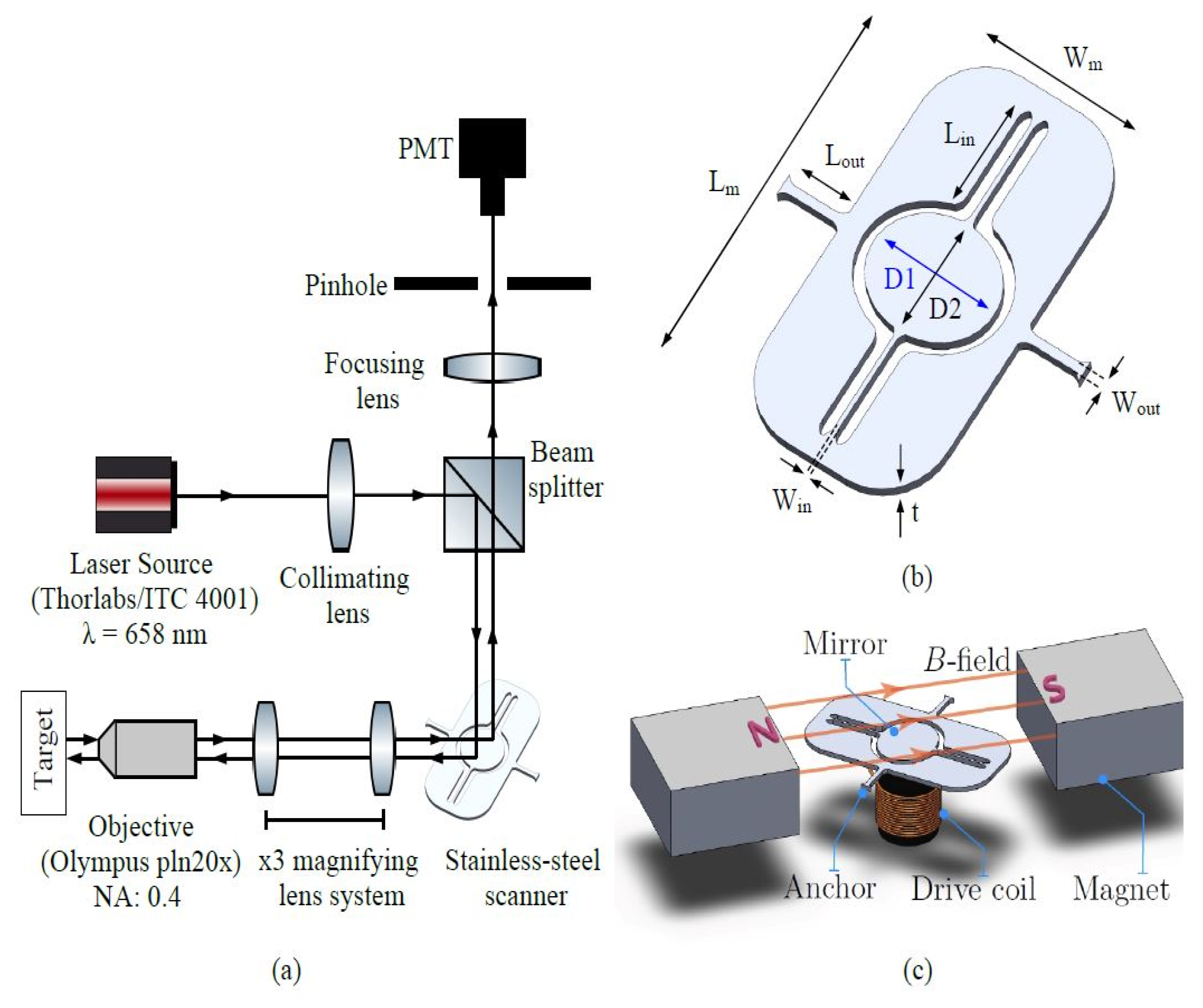
Figure 1a). The setup consists of a fiber-coupled laser (Thorlabs/ITC 4001) that is directed onto the scanning unit via a beam splitter. The scanner plane is relayed onto the objective lens (Olympus/PLN20x), which focuses and collects light to/from the target, via a lens pair that impose a ×3 magnification on the 1.1 mm input beam diameter. The light is finally epi-collected onto a photomultiplier unit (Hamatsu/H10721-20), after getting focused onto a 100 μm pinhole to eliminate out-of-focus light from the target.
Firstly, optical targets of our confocal system are determined as follows: (i) less than 10 μm resolution and (ii) 200 μm × 200 μm FOV that is comparable to state-of-the-art benchtop laser scanning microscopes [
2]. An objective lens having a numerical aperture (NA) of 0.4 and a clear aperture of 10 mm is chosen to achieve the desired lateral resolution. After ×3 magnification by the relay lens pair, the diameter of the input beam reaches 3.3 mm and fills less than half of the objective lens clear aperture. Hence, the effective NA reduces from 0.4 to 0.132. The spot full-width half-maximum (FWHM) diameter at the target can be calculated using Equation (1) [
3].
where,
is the wavelength input laser, NA is the effective numerical aperture of the objective lens. For the above mentioned conditions in our custom built setup and for the 0.658 μm wavelength input laser, we calculate the spot diameter to be 2.66 μm, satisfying our desired lateral resolution of less than 10 μm. Furthermore, in order to achieve the desired FOV of 200 μm, one needs a total optical scan angle of 3.82 degrees for the objective lens having a focal length of 9 mm using the equation below [
4]
where, F is the focal length of the objective lens and M denotes magnification amount of the relay lens pair.
3. Mechanical Design
The SS micro-scanner is developed to satisfy the FOV requirements that was set in the previous section. In
Figure 1a, CAD drawing of the device is given. The dimensions of the micro-scanner (
Table 1) is adjusted to achieve the desired TOSA that is determined in the previous section. Therefore, the gap between the inner flexure and gimbal is reduced to gain more magnetic area on the gimbal for the interaction with the external B-field. Another aim of this design is to keep the slow and fast resonance frequencies of the scanner mutually prime and as high as possible in order to increase the fill factor of the Lissajous pattern. As shown in
Figure 1a, two permanent magnets are located close to the mirror with an inclination of 45 degrees to the fast axis scan to get maximum displacement for both slow and fast scan.
4. Experimental Results
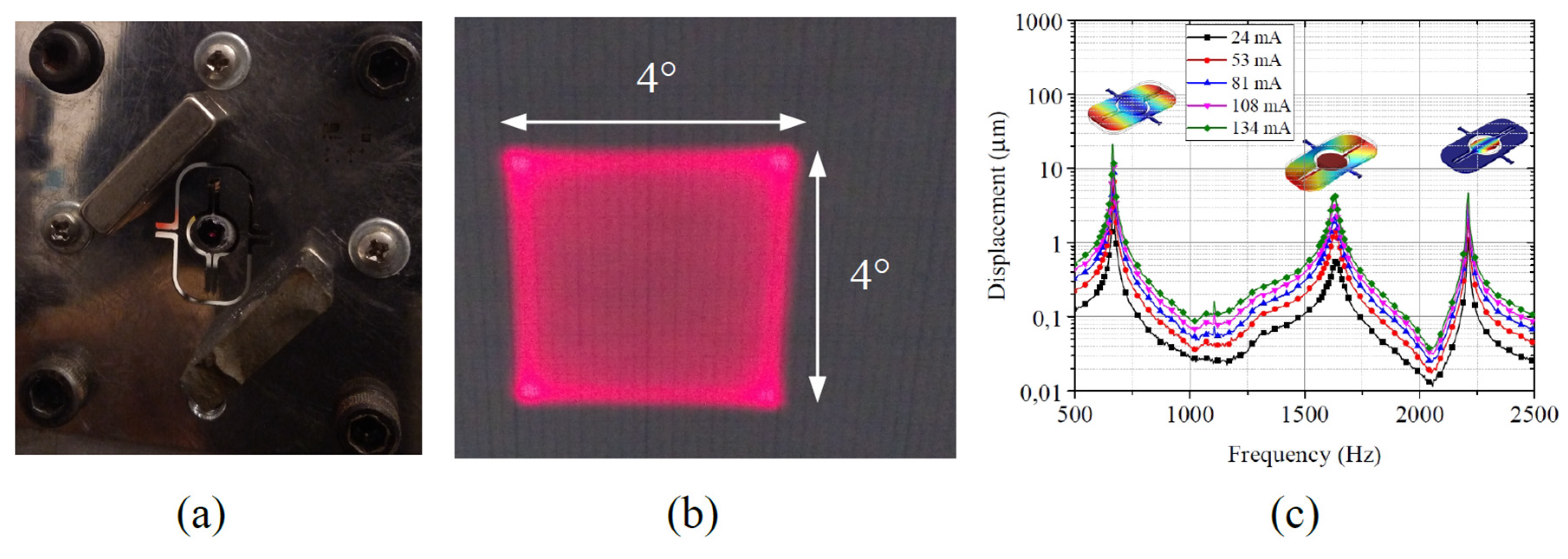
Laser machining technology allows us to create fine shapes on a given substrate, which is stainless-steel for our case, easily with high accuracy within a small amount of time. After fabrication process, two permanent magnets are attached close to the mirror with an inclination of 45 degress to obtain maximum displacement for both slow and fast scan axes (
Figure 2a).
Figure 2b shows the Lissajous pattern at 663 and 2209 Hz for the LSCM system. Total optical scan angle of 4 degrees is observed for both slow and fast scan movements for a 180 mA driving current. The deflection of the mirror as the frequency of the coil drive current is swept between 500 and 2500 Hz. Torsion around the slow and the fast axis is observed at 663 and 2211 Hz, respectively. An additional out-of-plane pumping mode occurs around 1625 Hz which could potentially be used for third dimension scan in future. Quality factors of slow and fast scan, in air operation, are measured as 110 and 275, respectively.
Figure 3 shows the USAF target, and the acquired images using custom-built confocal microscope setup, utilizing the steel-scanner presented in this study (test areas are shown in
Figure 3b,c, having 8.77 μm and 39.37 μm line width, respectively). The FOV is observed as 200 μm × 200 μm with the achieved scan angles. The profile of a line-pair is acquired in
Figure 3c and the derivative of this step corresponds to the impulse response of the system (optical resolution) [
5]. The FWHM resolution based on this calculation is found as 9.4 μm which satisfies our target provided in the optical design section.
5. Conclusions
A laser machined stainless steel micro scanner is designed and manufactured for confocal microscopy. The 2D scanning pattern is developed using Lissajous scan. Laser machining provided both rapid and precise manufacturing process. A FOV of 200 μm × 200 μm is obtained. The main purpose of this work is to produce a low cost micro-scanner for compact scanning microscopy technologies.

 ,
, 


