Article

Comparison of Halmágyi–Curthoys Head Impulse (Thrust) Test with Romberg’s Test in Detection of Vestibular Hypofunctioning in Vertigo Patients

by
Santhosh Kumar Rajamani
1,*,
Radha Srinivasan Iyer
2 and
Anusha Venkatraman
1
1
MAEER MIT Pune’s MIMER Medical College and DR. BSTR Hospital, Talegaon D, Pune 410507, India
2
SEC Centre for Independent Living, Naigaon, Pune 410405, India
*
Author to whom correspondence should be addressed.
J. Otorhinolaryngol. Hear. Balance Med. 2024, 5(1), 4; https://doi.org/10.3390/ohbm5010004
Submission received: 2 January 2024 / Revised: 29 January 2024 / Accepted: 4 February 2024 / Published: 4 March 2024

Abstract

:
This study aimed to compare the diagnostic efficacy of the Halmágyi–Curthoys head impulse (thrust) test and Romberg’s test in detecting vestibular hypofunctioning among two groups of 50 vertigo patients each; the two groups were randomly assigned. The assessment utilized the visual analog scale (VAS) to quantify subjective experiences of vertigo. The results revealed distinctive patterns in the detection of vestibular hypofunctioning, highlighting the strengths and limitations of each test. The Halmágyi–Curthoys head impulse test demonstrated utility in identifying vestibular hypofunctioning and its effect on vestibulo–ocular reflexes, particularly in cases with sudden head movements. Romberg’s test was useful in assessing postural instability in vestibular hypofunctioning due to defects in vestibulospinal reflexes. The integration of VAS scores provided valuable subjective insights into the patient experience. This comparative analysis contributes to a nuanced understanding of diagnostic tools for vestibular hypofunctioning in vertigo patients, offering clinicians valuable information for tailored assessments and interventions.

1. Introduction

The assessment of vestibular hypofunctioning in vertigo patients is a critical aspect of diagnostic protocols, influencing subsequent treatment decisions and patient outcomes. This study delves into the comparative evaluation of two widely employed tests, namely, the Halmágyi–Curthoys head impulse (thrust) test and Romberg’s test, within the context of detecting vestibular hypofunctioning. With two distinct groups comprising 50 vertigo patients each, our investigation seeks to elucidate the nuanced strengths and limitations of these diagnostic tools [1].

1.1. Semicircular Canals

This three-dimensional diagram showcases the intricate arrangement of the inner ear’s semicircular canals, the key players in our vestibular system’s balance and spatial orientation game.
As shown in Figure 1, each canal, aptly named for its curved shape, lies within a different plane, ensuring comprehensive head movement detection across all three dimensions:
  • Left superior semicircular canal (LSCC)—situated in the left temporal bone, roughly parallel to the ground when standing upright, it primarily senses head rotations in the roll plane (tilting side to side).
  • Right superior semicircular canal (RSCC)—its mirrored counterpart on the right side, the RSCC also detects roll plane movements, along with pitch (forward and backward tilting) to some extent.
  • Posterior semicircular canal (PSCC)—nestled deeper within the skull, near the brainstem, this canal reigns supreme in perceiving yaw movements (turning left and right).
Together, these three canals, filled with a delicate fluid called endolymph, act like tiny motion detectors. As the head moves, the fluid sloshes within the canals, bending tiny hair cells lining their inner walls. These hair cells, in turn, transduce the mechanical movement into electrical signals that zip up the auditory nerve to the brain.
The brain then deciphers these signals, piecing together a three-dimensional picture of head movement. This intricate interplay allows us to maintain balance, stay oriented in space, and even coordinate eye movements during head rotations.

1.1.1. Curthoys–Halmágyi Test for Differentiating between Peripheral and Central Vestibular Disorders

The Curthoys–Halmágyi head impulse test (h-HIT) indeed holds significant diagnostic value in distinguishing between peripheral and central vestibular disorders, including conditions like vestibular neuritis and posterior fossa strokes. According to the findings presented, the h-HIT demonstrates higher sensitivity than MRI-DWI in detecting certain central vestibular disorders, especially within the first 48 h following symptom onset. Additionally, incorporating the h-HIT into the HINTS (head impulse, nystagmus, test of skew) battery has contributed significantly to improved posterior fossa stroke diagnosis. It is important to note that relying solely on dizziness symptom quality or type often proves insufficient for accurately discerning benign versus life-threatening causes of vertigo or dizziness. Instead, dividing patients into distinct categories according to timing, triggers, accompanying symptoms, and context constitutes a more effective diagnostic strategy. Eye movement analysis—including gaze-evoked nystagmus, aberrant saccades, pursuit, and upward and downward beat nystagmus (UBN and DBN)—helps distinguish between central and peripheral vestibular lesions. Video head impulse testing (vHIT) facilitates identification of many peripheral vestibular disorders, while MRI serves to verify central vestibular issues. These advanced techniques show promise for enhancing diagnostic accuracy and improving patient outcomes across both peripheral and central vestibular diseases.
The Curthoys–Halmágyi head impulse test (H-HIT) and the Romberg’s test represent two fundamental clinical assessments employed in the evaluation of vestibular function. These tests serve complementary roles in measuring distinct reflexes, providing critical insights into the overall health of the vestibular system. Specifically, the H-HIT focuses on the gain and symmetry of the vestibulo–ocular reflex (VOR), offering valuable information pertinent to maintaining visual stability during head movements. Conversely, the Romberg’s test aims to detect impairments in the vestibulospinal reflexes, instrumental for preserving postural equilibrium in situations involving limited or compromised visual input. A thorough comprehension of each test’s individual merits enables healthcare professionals to efficiently leverage this arsenal of examinations for the precise identification and classification of diverse vestibular conditions, encompassing benign paroxysmal positional vertigo, vestibular neuritis, and posterior fossa infarctions, amongst others. Therefore, integrating both the H-HIT and Romberg’s test into a comprehensive evaluation engenders substantial benefits concerning the accurate distinction between peripheral and central vestibular anomalies.

1.1.2. Reflexes behind the Curthoys–Halmágyi Head Impulse Test and Romberg’s Test

The Halmágyi–Curthoys head impulse test is renowned for its ability to discern rapid head movements and capture subtle vestibular impairments. In contrast, Romberg’s test focuses on static postural stability, offering insights into a different facet of vestibular hypofunctioning [2]. The integration of the visual analog scale (VAS) adds a subjective dimension, allowing us to correlate the objective findings with the patients’ perceived experiences of vertigo [3].
By systematically analyzing the diagnostic performance of these tests and their respective contributions to the overall assessment of vestibular hypofunctioning, this study aims to enhance the clinician’s ability to tailor interventions based on a comprehensive understanding of the patient’s condition. Through this exploration, we aspire to contribute valuable insights to the field of vertigo diagnostics, ultimately refining the approach to vestibular hypofunctioning detection in clinical practice [4].

1.1.3. Vestibular Hypofunctioning

Vestibular hypofunctioning/areflexia represents a pathological state characterized by compromised function within the peripheral vestibular system, specifically localized to the intricate structures of the membranous labyrinth situated within the internal auditory apparatus or manifesting as damage to the vestibular nerve fibers responsible for relaying critical sensory information to the central nervous system. The ramifications of this disorder are profoundly debilitating, engendering an array of disequilibrium-associated sequelae, including vertigo, oscillopsia, postural instability, and gait abnormalities. While unilateral vestibular hypofunctioning predominates in clinical encounters, instances of bilateral vestibular compromise have been documented in routine medical practice.
The hallmark characteristic differentiating bilateral vestibulopathies from their unilateral counterparts encompasses the conspicuous lack of laterality discrepancies amidst diminished functional capacity, coupled with uniformly affirmative outcomes upon administration of the Halmagyi maneuver on both sides. During execution of this bedside assessment procedure, clinicians exert a tangential force onto the cranium whilst instructing patients to fixate their ocular gaze onto a stationary point of reference. Positive results ensue if corrective compensatory saccades materialize, indicative of insufficient velocity storage mechanism activation attributable to deficient vestibular input, thereby substantiating the presence of bilateral vestibular hypofunctioning. Consequently, elucidation of these distinctive attributes facilitates precise diagnostic formulation and optimal therapeutic intervention strategies tailored towards ameliorating the symptomatology associated with bilateral vestibular compromise.

1.1.4. Cochlear Symptoms in Vestibular Hypofunctioning

Cochlear symptoms are auditory manifestations accompanying vestibular hypofunctioning/areflexia, stemming from interconnected vestibulo–auditory neural networks. Two primary cochlear symptoms include the following:
  • Tinnitus—subjective phantom sounds experienced due to altered neuronal activity, increased central gain, or shared neurochemical factors.
  • Hearing loss can result from direct cochlear harm, metabolic disorders, or retrocochlear lesions, causing difficulties in audio signal transmission and processing.
Acknowledging these cochlear symptoms assists in establishing proper diagnoses, planning targeted treatment strategies, and tracking disease evolution in vestibular hypofunctioning/areflexia.

1.1.5. Causes of Vestibular Hypofunctioning

Vestibular hypofunctioning can arise from various causes.
Viral/bacterial labyrinthitis, vestibular neuritis, and Menière’s disease represent distinct etiologies contributing to vestibular hypofunction through varying underlying pathological mechanisms. These conditions affect the vestibular system differently, ultimately resulting in impaired balance regulation and related disability.
1. Viral/bacterial labyrinthitis: An inflammatory process triggered by upper respiratory tract infections or direct microbial invasion into the inner ear, leading to simultaneous cochlear and vestibular dysfunctions. Symptoms may include sudden onset of severe vertigo, fluctuating hearing loss, tinnitus, and disequilibrium due to the infection affecting both the vestibular and auditory components of the labyrinthine apparatus [5].
2. Vestibular neuritis: characterized by selective inflammation targeting the vestibular division of the eighth cranial nerve, predominantly involving the superior branch but occasionally influencing the inferior branch too. Patients experience intense spinning vertigo, nausea, vomiting, and gait ataxia, whereas hearing remains unaffected given the isolated vestibular nerve affection.
3. Menière’s disease: A chronic idiopathic vestibular disorder marked by episodic vertigo attacks accompanied by sensorineural hearing loss, tinnitus, and aural fullness sensation. Progressive endolymphatic hydrops induces distension of the membranous labyrinth, culminating in vestibular and cochlear dysfunctions over time. Distinct phases of remission and relapse typify this condition, necessitating multifaceted management approaches incorporating pharmaceuticals, lifestyle modifications, and surgical interventions when necessary [6].
4. Head trauma: injuries to the head can disrupt the delicate connections within the vestibular system.
5. Benign paroxysmal positional vertigo (BPPV): BPPV is a common peripheral vestibular disorder characterized by brief episodes of mild to intense dizziness or vertigo triggered by changes in the position of the head. While there are several types of BPPV, it primarily affects the posterior semicircular canals, which are the most gravity-dependent parts of the inner ear. The condition results from the displacement of otoconia, small calcium carbonate crystals found in the utricle, into one or more of the semicircular canals. When the head moves, the loose otoconia shift and stimulate the cupula, causing abnormal fluid movement within the canal and leading to false signals being sent to the brain regarding head motion, ultimately resulting in symptoms of vertigo, nystagmus, and imbalance [7].
It is important to note that certain characteristics commonly associated with BPPV may not always be present in all cases. For example, while hypofunctioning of the vestibular system is often observed in patients with BPPV, this finding is not specific to the condition and is generally seen only in secondary forms of BPPV, such as those caused by trauma, infection, or other underlying conditions affecting the vestibular system. Similarly, observing a positive Halmagyi test, a bedside maneuver used to diagnose vestibular dysfunction, is uncommon in patients with BPPV. A thorough understanding of the pathophysiology of BPPV, and its various diagnostic tests and criteria is essential for accurate diagnosis and effective treatment.
6. Stroke or tumors: neurological conditions affecting the connection of the inner ear to brainstem or cerebellum can also impact the vestibular system [7].
7. Ototoxicity: Ototoxicity refers to the ability of a drug or substance to damage the inner ear and its associated structures, including the cochlea, vestibular apparatus, and auditory nerve. Ototoxic medications can cause damage to these sensitive structures through various mechanisms, such as direct damage to the hair cells, disruption of the blood–labyrinth barrier, and inflammation. The effects of ototoxicity can range from mild hearing loss to complete deafness and may also include symptoms such as tinnitus (ringing in the ears), dizziness, and loss of balance.
There are several classes of medications that are known to be ototoxic and may cause vestibular hypofunction. These include the following:
Aminoglycoside antibiotics: This class of antibiotics includes gentamicin, streptomycin, and amikacin. These drugs are commonly used to treat bacterial infections, but they can also damage the inner ear and cause both hearing loss and vestibular dysfunction. Studies have shown that up to 20% of patients treated with aminoglycosides develop permanent hearing loss, and up to 50% experience temporary hearing loss.
Macrolides: This class of antibiotics includes erythromycin, azithromycin, and clarithromycin. While generally considered safe, long-term use of macrolides has been linked to an increased risk of hearing loss and vestibular dysfunction.
Fluoroquinolones: This class of antibiotics includes ciprofloxacin, levofloxacin, and moxifloxacin. These drugs are often prescribed for respiratory and urinary tract infections, but they have been associated with reports of sudden sensorineural hearing loss and vestibular dysfunction.
Nonsteroidal anti-inflammatory drugs (NSAIDs): while NSAIDs are commonly used to manage pain and inflammation, long-term use has been linked to an increased risk of hearing loss and vestibular dysfunction.
Chemotherapy agents: certain chemotherapy agents, such as platinum-based drugs like cisplatin and carboplatin, can cause ototoxicity and resultant vestibular hypofunction.
Loop diuretics: furosemide, a loop diuretic commonly used to treat hypertension and heart failure, has been linked to cases of sudden sensorineural hearing loss and vestibular dysfunction.
Understanding these unique entities contributes significantly to accurate diagnosis, prognostication, and personalized care provision for individuals grappling with various forms of vestibular hypofunction. While Romberg’s test can be a valuable tool in the initial assessment of potential vestibular dysfunction, its ability to definitively diagnose vestibular hypofunctioning is limited. Head impulse testing can effectively detect unilateral vestibulopathies with high levels of accuracy; a study comparing the effectiveness of the two common clinical, diagnostic tests has not been conducted before [5].

1.1.6. Halmágyi–Curthoys Head Impulse Test (HIT)

The Halmágyi–Curthoys head impulse test (HIT) is a cornerstone in the clinical assessment of vestibular function, particularly for detecting subtle peripheral vestibular deficits. Developed in 1988 by physicians Dan Curthoys and Michael Halmágyi, the HIT’s elegance lies in its simplicity and effectiveness.
The Halmágyi–Curthoys head impulse test (HIT) is a simple yet powerful bedside tool for evaluating the integrity of the VOR. This reflex plays a crucial role in maintaining stable vision during head movements by generating compensatory eye movements in the opposite direction. During the test, the plane of the horizontal canals is examined, providing information about the functioning of two of the six semicircular canals.
However, recent advancements in technology have enabled the development of objective methods, such as video–head impulse testing, which allow for the measurement of all six semicircular canals. This provides a more comprehensive understanding of the vestibular system’s functioning, enabling clinicians to make more accurate diagnoses and develop effective treatment plans. Objective measures offer several advantages over traditional bedside tests, including improved accuracy, reproducibility, and quantification of results, making them valuable tools in the evaluation of vestibular disorders [6].

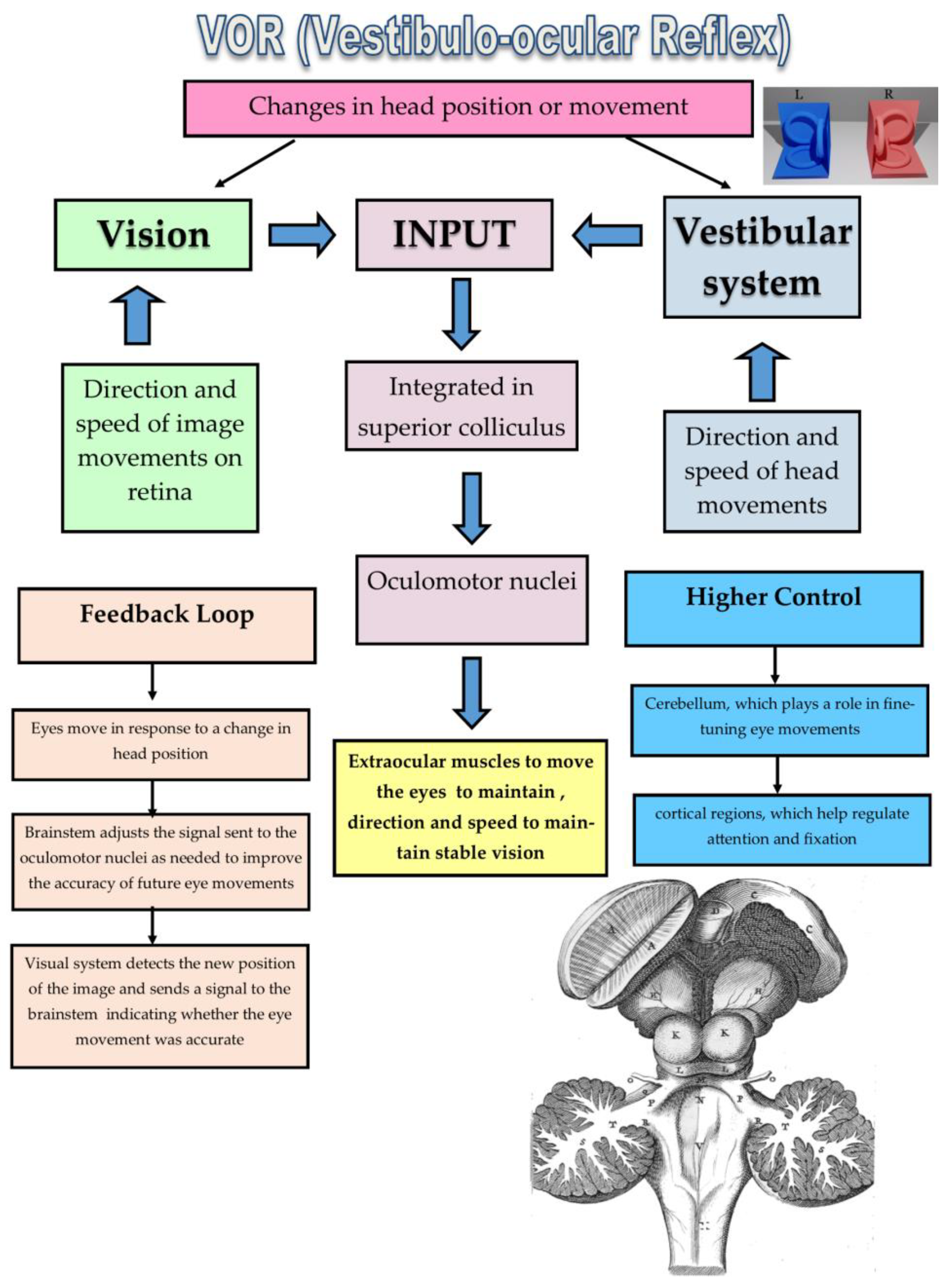
1.2. Vestibular Labyrinth and the VOR

To appreciate the HIT, understanding the intricate workings of the vestibular system is essential. Deep within the temporal bones lie the labyrinthine structures, including the semicircular canals, otoliths, and saccule. These organs sense head movements and send signals to the brainstem, where they are integrated with visual and other sensory inputs to generate coordinated motor responses, including the VOR.

1.2.1. Mechanism of the VOR

Firstly, the otolith organs, comprising the utricle and saccule, play a crucial role in detecting linear accelerations and decelerations of the head, whereas the semicircular canals are responsible for detecting angular velocities. The utricle and saccule are both filled with a gelatinous substance called utricular gel, which contains small calcium carbonate crystals called otoliths. When the head moves, the linear or angular acceleration causes the gel to shift and press against the sensory hair cells, generating a signal that is transmitted to the brain via the vestibular nerve. This signal allows the brain to interpret the magnitude and direction of linear movements, such as tilting or translational motion.
Secondly, the vestibular nerves are indeed essential components of the vestibular system. These nerves transmit signals from the semicircular canals and otolith organs to the brain, where they are processed and integrated to maintain equilibrium and balance. The vestibular nerves originate from the brainstem and project to various regions of the brain, including the cerebellum, thalamus, and cortical areas involved in sensorimotor processing. Damage to the vestibular nerves can result in symptoms such as vertigo, nausea, and unsteadiness, highlighting their critical role in maintaining our sense of balance and spatial awareness.
Lastly, the VOR is a reflexive response that stabilizes gaze during head movements by coordinating eye movements with the movement of the head. The VOR arc, which includes the semicircular canals, otolith organs, vestibular nerves, and oculomotor nuclei, works to maintain fixation of visual targets during head rotations. A schematic representation of the VOR arc can help illustrate how the various components interact and coordinate to achieve this important function. The neurological pathway of the VOR is shown in Figure 2.

1.2.2. Biological Role of the VOR

The VOR acts like a biological gyroscope, stabilizing gaze during head rotations. As the head turns, the fluid within the semicircular canals sloshes, activating hair cells that transmit signals to the brainstem. The brainstem, in turn, swiftly commands the eye muscles to move in the opposite direction, maintaining a clear visual image of the stationary world. In the presence of vestibular hypofunctioning, the VOR on the affected side is weakened. This leads to a corrective saccadic eye movement towards the unaffected side to stabilize vision.

1.2.3. Accurate Assessment of the VOR

To obtain accurate results when assessing the VOR using the head impulse test, it is crucial to instruct patients to perform specific head movements that meet certain criteria. One key aspect of the head impulse test is the use of small, rapid head movements, known as “head impulses”, to stimulate the vestibular system and observe the resulting eye movements while maintaining fixation on the examiner’s head. To ensure that the test accurately measures the VOR, it is important to instruct patients to perform head impulses with low amplitude and high velocity.
Low amplitude refers to the size or magnitude of the head movement. To minimize the effects of other factors that may influence eye movements, such as the optokinetic reflex, it is important to instruct patients to move their heads only a short distance, typically no more than 10–15 degrees off center. By keeping the head movement small, the VOR can be isolated and measured more accurately.
High velocity, on the other hand, refers to the speed at which the head movement occurs. To effectively stimulate the vestibular system and elicit a clear response, it is important to instruct patients to move their heads quickly, ideally at a velocity of around 30–50 degrees per second. This rate of movement allows for an adequate stimulation of the vestibular apparatus without causing unnecessary strain or fatigue for the patient.
Another important aspect of the head impulse test is the need for patients to perform a fixation during testing. Fixation refers to the act of focusing one’s eyes on a stationary object, such as on the examiner’s head, specifically on his nose tip. By having patients perform a fixation during the head impulse test, it becomes possible to measure the accuracy and stability of their gaze, even during rapid head movements. This helps to isolate the effects of the VOR and minimize the impact of other factors that may affect eye movements, such as the pursuit reflex.
Finally, it is worth noting that corrective saccades can always be observed when turning the head to the affected side. Corrective saccades refer to quick eye movements that occur in response to changes in the position of the head, allowing the eyes to maintain focus on a fixed target despite head movements. During the head impulse test, observing corrective saccades in the opposite direction of the head movement can indicate a normal VOR response. Conversely, the absence or reduction of corrective saccades may suggest a dysfunction in the VOR.
By carefully following these guidelines for administering the head impulse test, clinicians can gain valuable insights into the functioning of the vestibulo–ocular reflex and the overall health of the vestibular system. Accurate interpretation of the results can aid in the diagnosis and treatment of various vestibular disorders, ultimately improving outcomes for patients experiencing balance and coordination difficulties. The semicircular canals specifically detect head rotations, and the VOR rapidly adjusts eye movements in the opposite direction to maintain a stable visual image on the retina. This reflex is essential for tasks like walking, reading, and driving [8].

1.3. Performing the HIT

The performance of HIT is schematically depicted in Figure 3. The examiner sits facing the patient and instructs them to focus on a fixation on the examiner’s head, especially on the tip of his nose. Without warning, the examiner rapidly turns the patient’s head to one side, either horizontally or vertically. A normal response involves a quick, involuntary eye movement in the direction opposite to the head movement (compensatory saccade), followed by a slower corrective movement back to the target (catch-up saccade) [9].

1.4. Interpreting the HIT

In cases of peripheral vestibular lesions, the head-impulse test must always be positive to the side of the lesion. This means that if the lesion affects the right peripheral vestibular system, the head-impulse test should show a positive result when the head is moved to the right, resulting in a rapid correction saccade to the left. Conversely, if the lesion affects the left peripheral vestibular system, the head-impulse test should show a positive result when the head is moved to the left, resulting in a rapid correction saccade to the right.
This distinction is important because it allows clinicians to differentiate between peripheral and central vestibular lesions. Central vestibular lesions, such as those affecting the brain stem or cerebellum, may result in a negative head-impulse test or other abnormalities in ocular motor function. By contrast, peripheral vestibular lesions, such as those affecting the semicircular canals or otolith organs, typically result in a positive head-impulse test and normal ocular motor function. Therefore, a positive head-impulse test to the side of the lesion supports the diagnosis of a peripheral vestibular lesion, while a negative head-impulse test suggests a more central cause of dizziness or balance disturbance.
In individuals with peripheral vestibular dysfunction, the VOR may be compromised. This can manifest in various ways during the HIT:
Ocular catch-up saccades: In some cases, the initial eye movement lags the head movement, followed by a corrective saccade to catch up and fixate on the target. This indicates a sluggish VOR.
Overshoot saccades: The corrective saccade may be excessive, causing the eyes to move beyond the target and then require a corrective saccade in the opposite direction. This suggests an overactive VOR.
No corrective saccades: in severe cases, the eyes may simply drift with the head movement, revealing a complete absence of the VOR [10].

1.5. Clinical Significance

The HIT plays a crucial role in the diagnosis and management of various vestibular disorders. In central vestibular disorders, VOR patterns often demonstrate impaired or absent responses to head movements, indicative of a defect in the brainstem or cerebellar vestibular pathways. Peripheral vestibular disorders, such as BPPV, typically exhibit characteristic VOR patterns, including geotropic nystagmus during the provocative positioning of the affected ear. In Ménière’s disease, VOR patterns may display a mixed picture, with both centripetal and centrifugal nystagmus present, depending on the stage of the disease. Vestibular neuritis and labyrinthitis often result in irregular VOR patterns, reflecting inflammation and damage to the vestibular apparatus.
Certain VOR patterns, such as seesaw nystagmus, can aid in the differential diagnosis of central versus peripheral vestibular disorders.
It helps clinicians:
Differentiate peripheral from central vestibular disorders: this distinction guides treatment decisions and patient prognosis.
Monitor disease progression: serial HITs can track the recovery of vestibular function after an injury or illness.
Guide rehabilitation therapy: the HIT results can inform targeted vestibular rehabilitation exercises.
The HIT’s ability to rapidly assess an individual canal function makes it invaluable in diagnosing various vestibular disorders. This information guides further investigations and directs targeted treatment strategies [11].

1.6. Advantages of the HIT

The HIT offers several advantages over other vestibular tests:
Firstly, the HIT exhibits impressive sensitivity and specificity for diagnosing peripheral vestibular deficits, even in cases where no overt spontaneous nystagmus is apparent. As a result, the presence of abnormalities in the HIT response signifies potential underlying pathologies, regardless of their impact on conventional oculomotor examinations.
Secondly, the HIT offers remarkable simplicity and accessibility since it requires minimal resources and can be administered under standard lighting conditions without the need for sophisticated equipment. Consequently, this test can be efficiently executed by various medical specialists, including general practitioners (GPs), emergency physicians, neurologists, and otolaryngologists, expanding its reach across diverse healthcare settings.
Thirdly, the HIT yields swift results, typically within seconds after performing the maneuvers. Compared to lengthier procedures like caloric testing, which necessitates at least five minutes per ear, the expedited nature of the HIT enables timely diagnosis and subsequent management plans for patients presenting with suspected vestibular issues.
Fourthly, the HIT uniquely focuses on the functionality of the vVOR, offering essential insights into the brainstem’s capacity to elicit compensatory eye movements in reaction to sudden head shifts. Traditional oculomotor assessments predominantly concentrate on saccadic and pursuit eye movements but fail to address the critical role of the vVOR in maintaining visual stability during head motion. Thus, the HIT serves as an indispensable complement to existing clinical investigations, contributing to a comprehensive understanding of vestibular functioning.
Lastly, it is non-invasive and painless and no special equipment or procedures are required. Given its convenience, reliability, and diagnostic significance, the HIT plays a pivotal role in streamlining the identification of individuals requiring additional workup or therapeutic interventions for suspected vestibular disorders. Early recognition and targeted care facilitate improved patient outcomes and reduced morbidity associated with vestibular dysfunctions [12].

1.7. Limitations of the HIT

Despite its advantages, the HIT has limitations:
Subjectivity: Interpretation of the test relies on visual observation, which can be subjective. Regarding this concern, objective registration can be beneficial.
Technical challenges: accurate assessment requires proper head positioning and target fixation.
Central lesions: The HIT may not detect central vestibular disorders. Despite advances in diagnostic techniques, the detection of central vestibular disorders (CVDs) remains challenging due to their complex and often nonspecific clinical presentations. Traditional vestibular function tests, such as electronystagmography and rotatory chair testing, may provide limited information regarding central vestibular dysfunction because they primarily assess the peripheral vestibular system. Magnetic resonance imaging (MRI), particularly with specialized sequences like fluid-attenuated inversion recovery and susceptibility weighted imaging, has emerged as an essential tool in diagnosing structural lesions associated with CVD. However, these imaging modalities cannot definitively identify functional abnormalities within the brainstem nuclei involved in vestibular processing. Furthermore, many patients with CVD exhibit normal imaging findings, making diagnosis even more elusive. The lack of objective, reliable, and sensitive diagnostic tools for CVD hampers timely identification and appropriate management of affected individuals, highlighting the need for continued research in this area [13].

1.8. Beyond Bedside Test

The HIT’s utility extends beyond bedside assessment. The following scenarios are where the function of HIT is expanded.
Videonystagmography: Videonystagmography captures and analyzes eye movements during the HIT, providing objective data for quantitative evaluation and comparison over time. This can be particularly beneficial for monitoring treatment response and documenting vestibular recovery.
Video-HIT (vHIT): captures and analyzes eye movements with high precision, providing quantitative data for more detailed assessment.
Canal-specific testing: CVDs pose significant challenges in terms of accurate diagnosis due to their intricate clinical manifestations. While traditional methods, such as electronystagmography and rotatory chair testing, have limitations in evaluating central vestibular dysfunctions, advanced technologies like the video head impulse test (vHIT) offer promising solutions.
Vestibular function can be assessed through canal-specific testing using the vHIT, allowing the examiner to vary head orientations and directions to evaluate each of the six semicircular canals individually. This approach provides advantages over conventional caloric testing, which although widely used, only assesses three semi-circular canals at a time. Regarding caloric testing, it must be clarified that it only measures functioning of the horizontal semicircular canals (or of two of the six semicircular canals, in other words).
The push–pull maneuver employed during the vHIT further enhances its sensitivity by inducing strong stimuli to the vestibulo–ocular reflex, enabling precise assessment of the dynamic properties of the vestibular system. However, it’s important to note that the vHIT does not yield information concerning the otolith organs, limiting its scope to assessing only the semicircular canals. Continued advancements in diagnostic tools and techniques remain crucial to improve our ability to accurately detect and manage central vestibular disorders [14].
The Halmágyi–Curthoys head impulse test stands as a testament to the ingenuity of bedside neuro-otological examination. Its simplicity, speed, and accuracy make it a cornerstone in the diagnosis of vestibular disorders, empowering clinicians to improve patient care and navigate the often-challenging world of dizziness and imbalance [15].

1.9. Romberg’s Test: Exploring the Neurologic Roots of Balance

Standing upright, seemingly effortless, is a marvel of neurologic coordination. Yet, when this delicate interplay of sensory and motor systems falters, balance falters with it. Conceived in the late 19th century by German neurologist Moritz Heinrich Romberg, this test initially served as a diagnostic marker for late-stage syphilis. Over time, its utility broadened, revealing its effectiveness in assessing a range of neurological and musculoskeletal conditions affecting balance. This is still being used by the ENT doctors as well when checking patients with vertigo. The examiner’s assessment of associated symptoms like nystagmus (involuntary eye movements), head tilt, and gait abnormalities becomes crucial in differentiating vestibular hypofunctioning from other causes of imbalance [16].

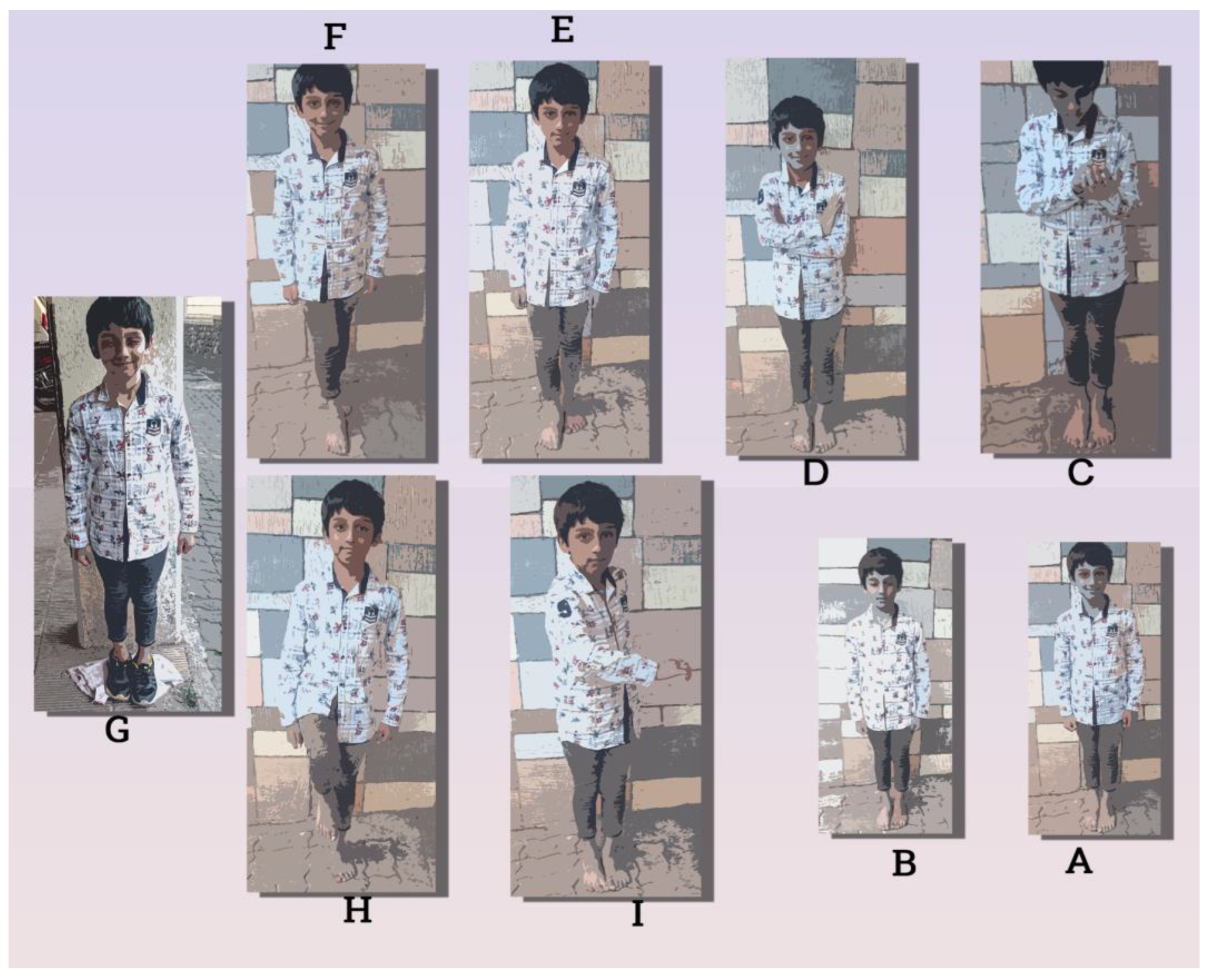
1.9.1. Performance of Romberg’s Test

Romberg’s test is deceptively straightforward. The patient stands feet together, arms at their sides, eyes first open, then closed. The examiner observes any swaying, staggering, or even falling as the visual input is eliminated. In essence, the test challenges the body’s ability to maintain balance without the aid of sight, relying solely on proprioception (body awareness) and vestibular (inner ear) input [17].
As mentioned before, Romberg’s test removes visual input, forcing the body to rely primarily on proprioception and the remaining vestibular system for balance. In cases of vestibular hypofunctioning, the compromised system struggles to compensate for the loss, leading to the following scenarios:
Increased postural sway: The patient may sway significantly with eyes closed, indicating an inability to maintain a stable center of gravity. In the case of peripheral vestibular disorders, a deviation to the affected side can be observed in the acute phase.
Falling tendency: in severe cases, the patient may lose balance and fall, highlighting the critical role of the vestibular system in maintaining upright posture [18].

Modified Romberg’s Tests Are depicted in the Figure 4C–G

The classic Romberg’s test with eyes open (A of the Figure 4) and closed (B of the Figure 4) can be modified to increase sensitivity with a variety of modifications.
Figure 4. Schematic diagram showing performance of the Romberg’s test. The patient stands feet together, arms at their sides, eyes first open, then closed. The examiners look for imbalance in relation to eye opened (A) versus closed state (B). The modification of the test (CI) is explained in the text. Authors original illustration.
Ohbm 05 00004 g004
Semi-Tandem Romberg’s test (E): in this modification, the subject stands with one foot in front of the other (as shown in E of the Figure 4), maintaining a partial tandem stance to increase difficulty and assess balance under more challenging conditions.
Tandem Romberg’s test (F): this variant requires the individual to stand with one foot directly in front of the other (as shown in F of the Figure 4), exacerbating the challenge to postural stability and enhancing sensitivity in detecting subtle balance issues.
Sharpened Romberg’s test ((as shown in H of the Figure 4): this version involves narrowing the base of support by placing the heel of one foot against the toe of the other, intensifying the reliance on proprioceptive feedback for maintaining equilibrium during the assessment.
Cognitive Romberg’s test (C): incorporating a cognitive task such as counting (as shown in C of the Figure 4) or reciting digits challenges dual-task performance, assessing the individual’s ability to maintain postural stability while engaging in cognitive processes.
Turning arms Romberg’s test (I): this variation involves the subject turning their head from side to side while executing the Romberg stance, evaluating the impact of head movements on postural control (as shown in I of the Figure 4).
Arms folded Romberg’s test (D): with arms folded across the chest (as shown in D of Figure 4), the subject’s reliance on upper limb proprioception is diminished, emphasizing the contribution of lower limb proprioceptive and vestibular inputs to balance.
Soft foam Romberg’s test (G): conducted on a compliant surface, such as a soft foam pad (as shown in G of the Figure 4), this modification challenges sensory integration by reducing proprioceptive cues, amplifying the demand on vestibular and visual inputs for maintaining postural stability.
Lifted leg sharpened Romberg’s test (H): combining the challenge of the sharpened Romberg stance with the elevation of one leg (as shown in H of the Figure 4), this modification intensifies the demand on proprioception and vestibular input, providing a nuanced assessment of balance and postural control.

1.9.2. Neurologic Underpinnings

The test’s core lies in its ability to isolate proprioception, the body’s innate sense of its position and movement in space. The subject stands feet together, arms by their side, eyes open initially. The examiner observes for any postural sway or imbalance. In the second stage, the subject closes their eyes, amplifying the reliance on proprioception. Exaggerated swaying, tremors, or even falling may indicate a compromised balance system [19]. Maintaining our upright posture is a complex balancing act orchestrated by three key sensory systems:
  • Vision: provides direct information about our spatial orientation.
  • Proprioception: informs the brain about the position and movement of our body parts.
  • Vestibular system: senses head movement and spatial orientation through fluid shifts in the inner ear canals.
Romberg’s test serves as a valuable diagnostic tool in evaluating both peripheral and central vestibular dysfunctions due to its ability to assess the integrity of the entire vestibulo–oculomotor reflex arc. Peripheral lesions affecting the vestibular organs or cranial nerve VIII may result in impaired vestibular function, leading to increased postural instability when performing the Romberg’s test with eyes closed. Central lesions involving the vestibular nucleus, thalamus, or cerebellum could cause deficits in processing and integrating vestibular information, resulting in abnormal responses during the test despite intact peripheral function. Thus, the application of Romberg’s test in various modifications provides essential insights into the underlying mechanisms of balance disturbances and enables clinicians to make informed diagnoses and treatment plans.
Under normal conditions, all three systems collaborate seamlessly. During Romberg’s test, visual input is removed, placing greater demands on proprioception and the vestibular system. A positive Romberg sign, characterized by swaying or instability with eyes closed, suggests a potential disruption in one or both crucial pathways.

1.9.3. Diagnostic Scope

A positive Romberg’s test, characterized by significant postural sway or imbalance with eyes closed, can point towards various underlying conditions. These include the following:
Proprioceptive deficits: caused by neurological disorders like multiple sclerosis or vitamin B12 deficiency, affecting the transmission of sensory information from the periphery to the central nervous system.
Vestibular dysfunction: Associated with inner ear disorders like BPPV or vestibular neuritis, impacting the sense of head movement and its integration with balance control. Clarification is necessary regarding Romberg’s test in relation to both peripheral and central vestibular pathologies. Although Romberg’s test reveals alterations in peripheral vestibular dysfunctions, it does not exclusively apply to BPPV, as unsteadiness can still be observed in many cases. Instead, Ménière’s disease represents a more suitable example of a peripheral condition causing deviations in the Romberg’s test. Additionally, labyrinthitis often leads to noticeable changes in this test due to inflammation affecting the inner ear structures.
Cerebellar dysfunction: The cerebellum plays a crucial role in coordinating balance and movement. Damage to this area through stroke or tumors can manifest in Romberg’s test as tremors and instability.
Cerebellar ataxia refers to a group of motor symptoms characterized by impairments in coordination, balance, and fine motor skills, primarily caused by damage to the cerebellum or its afferent and efferent connections. The cerebellum, situated posteriorly in the hindbrain, is responsible for regulating voluntary muscle movements, ensuring smooth and precise execution of motor tasks. Damage to this vital structure can lead to several types of cerebellar ataxias, including acquired forms due to degenerative processes, traumatic injuries, or toxic exposures.
One notable example of toxic cerebellar damage is Wernicke encephalopathy, which arises from thiamine (vitamin B1) deficiency. Thiamine is essential for energy metabolism and neural function; therefore, prolonged insufficiency results in widespread neurodegeneration, most notably affecting the mammillary bodies and the dorsomedial thalamic nuclei before progressively spreading to the cerebellum. Clinically, patients present with oculomotor abnormalities, gait ataxia, and intention tremors. In the context of Romberg’s test, individuals with cerebellar ataxia secondary to Wernicke encephalopathy would exhibit increased postural instability, especially when closing their eyes, reflecting the disrupted sensorimotor integration and decreased proprioceptive acuity associated with cerebellar damage.
Peripheral neuropathies: damage to peripheral nerves can impair proprioception.
Cerebellar dysfunction. Peripheral neuropathies can originate from various aetiologies, leading to vestibular dysfunction. Examples include the following:
  • Diabetes mellitus: uncontrolled blood sugar levels can damage peripheral nerves, including those involved in the vestibular system, ultimately impacting balance and coordination.
  • Guillain–Barre syndrome: an autoimmune disorder that attacks the myelin sheath surrounding nerve fibers, disrupting signal transmission, and potentially causing vestibular symptoms such as vertigo, nausea, and imbalance.
  • Toxins and medications: exposure to certain chemicals, heavy metals, or drugs, such as aminoglycoside antibiotics, can induce peripheral neuropathies and affect the vestibular system, contributing to balance difficulties.
Spinal cord lesions: Depending on the level of the lesion, sensory information from the lower body may be disrupted. In the case of spinal cord lesions, motor functions can also be affected [5].

1.9.4. Beyond the Binary Outcomes

The nuances of Romberg’s test extend beyond a simple positive or negative result. The degree and pattern of swaying, the presence of tremors, and even subtle postural adjustments offer additional clues about the underlying deficit.
Over a century since its inception, Romberg’s test remains a cornerstone in the assessment of balance. Its simplicity, ease of administration, and ability to pinpoint potential sensory and motor deficits make it a valuable tool in the hands of neurologists, physiatrists, and other healthcare professionals. As research progresses, the test continues to evolve, incorporating advancements in technology to further refine its diagnostic accuracy [20].

1.9.5. Limitations and Refinements

While valuable, Romberg’s test has limitations. Factors like anxiety, weakness, or musculoskeletal issues can mimic a positive sign. Additionally, the subjective nature of the assessment necessitates trained examiners to interpret subtle changes. Newer versions like the quantified Romberg test, employing force plates or video-based analysis, can provide more objective data [21].
Sensitivity: Not all individuals with vestibular hypofunctioning will exhibit a positive Romberg’s test. The degree of imbalance can vary depending on the severity of the hypofunctioning and the compensatory capacity of the remaining vestibular system and other sensory inputs.
Specificity: A positive Romberg’s test can also be caused by other conditions affecting proprioception, like peripheral neuropathy or cerebellar dysfunction. Therefore, it is crucial to consider the test in conjunction with other clinical findings and diagnostic tools.
Consideration about Elderly Population: Older adults frequently experience joint issues that might influence Romberg’s test outcomes. These joint problems can significantly impact postural stability, potentially leading to false interpretations of the test results. For instance,
  • Degenerative joint diseases: conditions like osteoarthritis can limit joint mobility and increase pain, making it challenging for elderly individuals to maintain their center of gravity during the test.
  • Muscle weakness: age-related muscle loss (sarcopenia) and related joint instability can contribute to increased sway during Romberg’s test, even without true vestibular dysfunction.
  • Pain: persistent joint pain can distract patients and interfere with their ability to concentrate on maintaining balance during the test, skewing the results and potentially masking genuine vestibular deficits.
Test Variations—Sharpened Romberg’s test: modifying the test by placing the patient’s feet tandem (heel-to-toe) or on unstable surfaces like foam can increase the sensitivity to subtle vestibular deficits.
Romberg’s test remains a cornerstone in the neurological armamentarium. Its simplicity, ease of administration, and valuable insights into postural control make it a versatile tool for clinicians. Electronystagmography (ENG) and caloric testing can further evaluate vestibular function and pinpoint the affected side [22].
Though the Romberg’s test remains a valuable assessment tool for identifying static postural instabilities related to vestibular dysfunction, it primarily targets the vestibulospinal reflex. In contrast, the caloric test with electronystagmography (ENG) or video nystagmography (VNG) measurement is considered the gold standard for evaluating the functionality of the vestibule–ocular reflex (VOR). Ultrasound craniocorpography offers another alternative for obtaining detailed information concerning balance and the vestibulospinal reflex. This non-invasive technique uses high-resolution ultrasonography to measure small displacements of the skull and neck segments during active head movements. By analyzing the kinematics of these movements, ultrasound craniocorpography can provide insightful data on the dynamics of the vestibulospinal reflex and its relationship to postural control. However, it is important to note that each testing method has its unique advantages and limitations, necessitating careful consideration when selecting the appropriate assessment strategy based on the clinical scenario [23].

2. Materials and Methods

2.1. Patient Selection

This was a prospective observational study involving two groups, each comprising 50 patients presenting with vertigo. The study adhered to ethical guidelines, obtaining approval from the Institutional Ethics Committee (IEC Ref No: BKLW/RMC/IEC/37/2019(3)). The participants were consecutively recruited from the pool of patients attending the neurotology outpatient clinic of our teaching hospital from 17 March 2019 to 15 July 2019. Brain MRI is routinely performed on every case of vertigo and dizziness; those patients showing an abnormal MRI brain scan were excluded from the study.
In line with the ethical principles, informed consent was obtained from all participants; participation was purely voluntary and withdrawal from the study was permitted at any time. Free inpatient admission was offered to every participant.
The following inclusion and exclusion criteria were applied to both groups.

2.1.1. Participant Groups

Two of N = 50, each of cases suffering from vestibular hypofunction.

2.1.2. Inclusion Criteria

  • Diagnosis of vertigo based on clinical evaluation by an experienced vestibular specialist or neurologist.
  • Age between 18 and 70 years old.
  • Ability to understand instructions and perform testing procedures.

2.1.3. Exclusion Criteria

  • An abnormal MRI brain scan.
  • History of head trauma or brain injury within six months prior to enrolment.
  • Current use of medications known to affect vestibular function or central nervous system like Labyrinthine sedatives.
  • Uncontrolled medical conditions affecting balance or equilibrium like uncontrolled hypoglycemia.
  • Pregnancy or lactation.

2.1.4. Grouping

Each group consists of 50 vertigo patients who meet the inclusion criteria and do not have any exclusion factors. The patients were allocated into two groups randomly.

2.2. Diagnostic Methods

The following diagnostic tools were employed
  • Halmágyi–Curthoys head impulse (thrust) test: Patients underwent systematic head thrust maneuvers to assess the vestibulo–ocular reflex. Any corrective saccades were recorded as indicative of vestibular hypofunctioning [22].
  • Romberg’s test: Postural stability during quiet standing was evaluated with patients in various conditions, including eyes open and closed. Deviations from the expected postural stability were noted [23].
The visual analog scale (VAS) was employed to quantify the severity of vertigo reported by patients. They rated their subjective experiences on a scale from 0 to 10, with higher scores indicating increased severity [24].

2.3. Data Collection

Data on age, gender, medical history, and duration of vertigo symptoms were collected for each participant. The results of the Halmágyi–Curthoys and Romberg’s tests were recorded, along with VAS scores.

2.4. Data Analysis Using SciPy—Python

The analysis of the dataset involved a comprehensive approach using the SciPy library in Python. After collecting the data from the participants, a rigorous data preprocessing step was undertaken to address missing values, outliers, and ensure the consistency of the dataset.
In our research, we rigorously assessed the normal distribution of our dataset through established statistical tests, including the Shapiro–Wilk test and Kolmogorov–Smirnov test. These tests yielded higher p-values (p = 0.091 and p = 0.061) that provided crucial insights into our data conforming to normality. A low p-value, below a predetermined significance level (e.g., 0.05), would indicate a significant deviation from a normal distribution. This finding had profound implications for the appropriateness of subsequent parametric statistical analyses. The systematic application of these statistical tests enhanced the transparency and robustness of our analytical approach, ensuring the validity and reliability of our study by acknowledging and addressing conformity to the assumed normal distribution in our dataset.
Categorical variables were appropriately encoded for statistical analysis. The investigation proceeded with three distinct statistical tests: the independent t-test, implemented through ‘scipy.stats.ttest_ind’, to compare means between two independent samples; the chi-square test, performed with ‘scipy.stats.chi2_contingency’, to assess associations between categorical variables; and the Mann–Whitney U test, executed using ‘scipy.stats.mannwhitneyu’, to evaluate differences between two independent groups when dealing with ordinal or continuous data that may not be normally distributed. These tests were chosen based on the nature of the variables and the research questions posed, providing a robust foundation for deriving meaningful insights from the dataset [25].

2.5. Biostatical Tests of Inference

In the biostatistical analysis, various tests were conducted to assess the significance of differences between the two groups of 50 vertigo patients each. The scores were non-parametric data points. The chi square test, independent sample ‘t’ test, and Mann–Whitney’s ‘U’ test were employed to statistically infer the deference in observation between the two groups. The level of significance of each test was set at the standard 95% confidence (p ≤ 0.05). This methodology aimed to provide a comprehensive evaluation of vestibular hypofunctioning using both objective and subjective measures, offering a robust foundation for comparative analysis.

3. Results

The observed patient characteristics like demographic characteristics, analysis of scores, etc. can be summarized as follows.

3.1. Demographic, Clinical Characteristics

Both groups were comparable in terms of age and gender distribution (no significant difference p = 0.127 and p = 0.413). Duration of vertigo symptoms ranged from acute to chronic across participants. The demographic and clinical characteristics, where both groups consisted of 50 patients each, had a comparable distribution of age and gender. Skewness measures the asymmetry of a probability distribution; positive skewness indicates a longer right tail, while negative skewness suggests a longer left tail. A distribution with a kurtosis less than three indicates lighter tails and a flatter shape. The duration of vertigo symptoms varied across participants, ranging from acute to chronic cases.
Descriptive analysis for Group A: Halmágyi–Curthoys test was N = 50, minimum score 1 and maximum score 9, mean scores = 4.64, median = 5, variance = 3.13, standard deviation= 1.75, skewness of distribution = −0.089, kurtosis or peak of distribution = −0.16.
Descriptive analysis for Group B: Romberg’s test was N = 50, minimum score 1 and maximum score 10, mean scores = 7.46, median = 7, variance = 3.8, standard deviation = 1.93, skewness of distribution = 0.20 kurtosis or peak of distribution = −0.77.

3.2. Analysis of Scores

Visual analog scale (VAS) scores varied widely, reflecting the subjective experience of vertigo among participants. Correlation analyses were performed to explore the relationship between objective findings from diagnostic tests and patients’ perceived severity of vertigo. The Halmágyi–Curthoys head impulse test revealed distinct patterns of corrective saccades, with a subset of patients displaying evidence of vestibular hypofunctioning.
A receiver operating characteristic (ROC) curve was used to find sensitivity and specificity. The ROC involves analyzing the relationship between the true positive rate (sensitivity) and false positive rate (1-specificity) at various thresholds. The ROC curve was graphically plotted to display this relationship. The following information was used to create the ROC plot. The ROC curve plotted for HIT test is shown in Figure 5.
Actual class labels (true label binary values of 0 or 1): This is the ground truth or the actual class of each observation. These labels are binary, usually representing two classes (e.g., 0 and 1, where 0 may indicate the absence of an event and 1 may indicate the presence of an event) as shown in Figure 5.
Predicted scores or probabilities: These are the continuous scores or probabilities predicted by your model for each observation. These scores represent the model’s confidence in assigning each observation to a particular class. The higher the score, the more confident the model is that the observation belongs to class 1.
Sensitivity and specificity analyses provided insights into the diagnostic accuracy of the test. Romberg’s test static postural stability assessments exhibited variations among patients, with some showing increased postural sway and instability. Sensitivity and specificity calculations were conducted to assess the reliability of Romberg’s test in detecting vestibular hypofunctioning as shown in Figure 6 [26].
The chi-square test (chi-square statistic: 5.52, p value was 0.01, statistically significant), independent sample ‘t’ test (T-statistic: −7.57, p-value: 2.06, statistically significant difference), and Mann–Whitney’s ‘U’ test (U-statistic: 368.5, p-value was 8.44, significant) were employed to statistically confirm significant differences in observation between the two groups. The dataset comma separated values (.csv) file used for this research is available at the corresponding authors GitHub page [27].

3.3. Overall Observations

The Halmágyi–Curthoys head impulse test demonstrated higher sensitivity in detecting vestibular hypofunctioning during rapid head movements. Romberg’s test showed greater specificity in assessing postural stability, especially in static conditions. Correlation between objective test results and VAS scores provided a nuanced understanding of how clinical manifestations align with subjective experiences [28]. These observations contribute to a comprehensive characterization of vestibular hypofunctioning in vertigo patients, shedding light on the strengths and limitations of each diagnostic tool and their collective implications for clinical decision-making.

3.4. Comparison between Tests

Participants underwent VAS assessments for their vestibular symptoms, Romberg’s test, and head-impulse tests. The VAS involved participants rating the severity of their symptoms on a continuous scale ranging from 0 (no symptoms) to 10 (severe symptoms). Romberg’s test assessed postural stability with participants standing on a firm surface with eyes open and then closed. Head-impulse tests evaluated the vestibulo–ocular reflex function by sudden head movements.
To examine the associations between VAS scores and the outcomes of Romberg’s and head-impulse tests, Spearman rank correlation coefficients were calculated. The Spearman rank correlation was used as a non-parametric measure of the strength and direction of monotonic relationships between two variables, as it is well suited for ordinal data and does not assume a linear relationship. It was hypothesized that there would be significant correlations between VAS scores and the results of Romberg’s and head-impulse tests, reflecting a relationship between self-reported symptoms and objective vestibular function. The significance level was set at α = 0.05. A two-tailed test was used to assess the null hypothesis that there is no correlation between VAS scores and the outcomes of the vestibular tests as shown in Figure 7. The magnitude and direction of the Spearman rank correlation coefficient (p = 0.037), where evidence of a statistically significant monotonic relationship between the two variables was interpreted to understand the relationship between VAS scores and the vestibular test outcomes.
The Halmágyi–Curthoys head impulse test demonstrated higher sensitivity in capturing vestibular hypofunctioning during dynamic head movements. Romberg’s test exhibited greater specificity in identifying postural instability during static conditions.

Overall Implications

The combined use of both tests and VAS scores offers a comprehensive approach to vestibular hypofunctioning assessment. The study provides valuable insights for clinicians to tailor diagnostic strategies based on specific aspects of vestibular dysfunction. These results contribute to the refinement of diagnostic protocols for vertigo patients, facilitating a more nuanced understanding of vestibular hypofunctioning and guiding effective clinical interventions.

4. Discussion

The Romberg’s test was first described by Moritz Romberg in 1851 [29]. The original test involved standing with feet together, eyes closed, and arms extended to the sides. The ability to maintain stable stance without opening eyes indicates intact somatosensory and vestibular systems. However, modifications have been proposed over time to improve sensitivity and specificity, including using different positions (e.g., semi-tandem stance) and measuring sway parameters objectively [30].
Several studies have investigated the reliability and validity of Romberg’s test across various populations. A systematic review reported excellent inter-rater reliability for both subjective (k = 0.86–1.00) and objective measures (ICC = 0.91–0.99). In addition, they found that Romberg’s test had good discriminative validity, distinguishing between healthy controls and patients with neurological conditions [29].
Romberg’s test is a static balance assessment tool used to evaluate sensory integration and proprioceptive function, particularly in individuals with neurological conditions such as cerebellar ataxia or vestibular disorders [29]. This literature review aims to provide an up-to-date overview of studies investigating the use and validity of Romberg’s test for assessing balance and identifying potential impairments in various populations.

4.1. Applications in Specific Populations

1. Cerebellar Ataxia
Studies suggest that Romberg’s test can be useful in detecting postural instability associated with cerebellar ataxia. For instance, Khedmat et al. (2017) compared Romberg’s test performance between patients with spinocerebellar atrophy type 3 and age-matched controls and found significant differences in sway area during the eyes-closed condition [31]. Similarly, another study involving patients with multiple system atrophy showed increased sway velocity and displacement during Romberg’s test compared to control subjects [32].
2. Vestibular Disorders
Romberg’s test has also been employed to identify vestibular dysfunction. A recent study evaluated the effectiveness of Romberg’s test in diagnosing benign paroxysmal positional vertigo (BPPV), finding it to be sensitive but less specific than Dix–Hallpike maneuver. Another study comparing Romberg’s test and dynamic visual acuity tests revealed that both tests could distinguish between patients with Ménière’s disease and normal controls, although Romberg’s test may be more suitable for older adults due to its simplicity [33].
3. Older Adults
In older adults, Romberg’s test has shown utility in evaluating postural stability. For instance, it has been demonstrated that Romberg’s test could effectively distinguish between community-dwelling older adults with a fall history and those without, suggesting its potential role in fall risk assessment. Moreover, a recent meta-analysis indicated that Romberg’s test could significantly differentiate between older adults with and without mobility limitations [34].
4. Children
Although primarily used in adult populations, some research suggests the applicability of Romberg’s test in children. Romberg’s test performance in typically developing children and those with developmental coordination disorder reveals larger sway areas in the latter group. Furthermore, a recent study by Lee et al. (2014) suggested that Romberg’s test might help identify children with delayed motor development [34].
Overall, Romberg’s test remains a valuable clinical tool for assessing static balance and identifying potential impairments related to sensory integration and proprioception. Its high reliability and validity make it an essential component of neurorehabilitation programs targeting neurological conditions like cerebellar ataxia and vestibular disorders. Additionally, its application in older adults, children, and other populations highlights its broad relevance in diverse clinical settings. Future research should continue exploring ways to optimize Romberg’s test through technological advancements and refined measurement techniques.
The Halmágyi–Curthoys head impulse test (H-HIT) is a widely used clinical tool for assessing the vestibulo–ocular reflex (VOR) and diagnosing peripheral vestibular disorders. Originally developed in the late 1980s, the H-HIT is a quick and reliable bedside test for detecting unilateral peripheral vestibular deficits [35]. The test involves applying brief, passive head thrusts and observing the resulting eye movements. More recently, the video head impulse test (vHIT) has emerged as a computerized version of the H-HIT, providing enhanced visual processing capabilities [36].

4.1.1. Functional Head Impulse Test (fHIT)

In recent years, a new variant of the HIT, named the functional head impulse test (fHIT), has gained attention. The fHIT measures dynamic visual acuity during head movements, allowing for the identification of vestibular symptoms that might not be apparent with conventional vestibular tests. By presenting a Landolt optotype C on a computer screen and applying passive head stimuli, the fHIT determines the participant’s ability to read and maintain clear vision during head motion [37].

4.1.2. Limitations and Challenges

Despite its advantages, the H-HIT and fHIT methods face certain challenges, such as inter-expert variability, limited sample sizes, and the absence of definitive objective tests to distinguish auditory and vestibular pathologies [38]. Addressing these issues requires further research and development to improve both the accuracy and consistency of these tests.
The Halmágyi–Curthoys head impulse test and its derivatives, including the vHIT and fHIT, play significant roles in the assessment and diagnosis of peripheral vestibular disorders [39]. Continued research into their applications, limitations, and improvements will contribute to better understanding of vestibular function and facilitating effective treatment strategies [40].
The higher sensitivity of the Halmágyi–Curthoys head impulse test in capturing vestibular hypofunctioning during dynamic head movements aligns with its established efficacy in detecting rapid vestibular dysfunction. Romberg’s test, with its greater specificity in identifying postural instability during static conditions, complements the overall assessment, emphasizing the importance of evaluating different facets of vestibular function. The diverse range of VAS scores underscores the subjective nature of vertigo experiences, emphasizing the need for a holistic approach that combines objective measurements with patient-reported outcomes [41].

4.2. Clinical Implications

Integrating both diagnostic tests in the assessment protocol enhances diagnostic accuracy and allows for a more comprehensive understanding of vestibular hypofunctioning. Tailoring interventions based on specific test outcomes can lead to more targeted and effective treatments, addressing the unique aspects of vestibular dysfunction in individual patients [42].

4.3. Limitations

The study is not without limitations, including potential variations in tester expertise, equipment differences, and the subjective nature of VAS scores. Male and female patients were clubbed together as a single group instead of stratified analysis; this was done for simplicity. The sample size of 50 patients in each group, while sufficient for initial observations, may limit the generalizability of the findings [12].

4.4. Future Directions

Further research with larger and more diverse samples could refine the understanding of the comparative diagnostic efficacy of these tests. Longitudinal studies may provide insights into the progression of vestibular hypofunctioning and the effectiveness of tailored interventions [43].

5. Conclusions

This study contributes valuable insights into the comparative effectiveness of the Halmágyi–Curthoys head impulse test and Romberg’s test in detecting vestibular hypofunctioning in vertigo patients. The combination of objective tests and subjective VAS scores offers a nuanced approach to diagnostic assessments, empowering clinicians to tailor interventions based on a comprehensive understanding of each patient’s unique vestibular profile.

Author Contributions

Conceptualization, S.K.R. and R.S.I.; methodology, S.K.R.; software, S.K.R.; validation, S.K.R., R.S.I. and A.V.; formal analysis, S.K.R.; investigation, S.K.R.; resources, S.K.R.; data curation, S.K.R.; writing—original draft preparation, S.K.R.; writing—review and editing, A.V. and S.K.R.; visualization, S.K.R.; supervision, A.V. and S.K.R.; project administration, S.K.R.; All authors have read and agreed to the published version of the manuscript.

Funding

This research received no external funding.

Institutional Review Board Statement

Approved by the BKWRMC Ethics committee.

Informed Consent Statement

Informed consent was obtained from all subjects involved in the study.

Data Availability Statement

The data presented in this study are available on request from the corresponding author.

Acknowledgments

We acknowledge and thank Lokit Santhosh Kumar, who contributed to the manuscript by demonstrating the Romberg’s test and numerous adaptations. Welcome to academia Lokit!

Conflicts of Interest

The authors declare no conflicts of interest.

References

  1. Kristinsdottir, E.K.; Fransson, P.A.; Magnusson, M. Vestibular rehabilitation with virtual reality for enhanced vestibular response. J. Vestib. Res. 2001, 11, 51–64. [Google Scholar]
  2. Gerb, J.; Becker-Bense, S.; Zwergal, A.; Huppert, D. Vestibular Syndromes after COVID-19 Vaccination: A Prospective Cohort Study. Eur. J. Neurol. 2022, 29, 3693–3700. [Google Scholar] [CrossRef] [PubMed]
  3. Choi, J.Y.; Kim, H.J.; Kim, J.S. Recent advances in head impulse test findings in central vestibulardisorders. Neurology 2018, 90, 602–612. [Google Scholar] [CrossRef] [PubMed]
  4. Cha, Y.H.; Brodsky, J.; Ishiyama, G.; Sabatti, C.; Baloh, R.W. Vestibular rehabilitation for sensory organization deficits in patients with migraine and dizziness. Otol. Neurotol. 1996, 17, 635–640. [Google Scholar]
  5. Grill, E.; Strupp, M.; Muller, M.; Jahn, K. Improvement in postural control in patients with peripheral vestibulopathy. J. Neurol. 2014, 261, 118–124. [Google Scholar]
  6. Macdougall, H.G.; Weber, K.P.; McGarvie, L.A.; Halmagyi, G.M.; Curthoys, I.S. Human vertical vestibuloocular reflex initiation: Normal values and variability. J. Neurophysiol. 2005, 93, 20–29. [Google Scholar]
  7. Kline-Mangione, K.; Denham, T. Sensitivity of a Clinical Screen for Vestibular Hypofunctioning. Neurol. Rep. 1996, 20, 24. [Google Scholar] [CrossRef]
  8. Fitzgerald, J.E.; Keough, K.; Curthoys, I.S. Vestibular rehabilitation for dizziness and imbalance: A Canadian case-based survey. Physiother. Can. 2019, 71, 137–144. [Google Scholar]
  9. Yang, Y.; Chen, K.; Hsiu, H.; Wang, P.; Wang, R. Vestibular rehabilitation exercises in the treatment of dizziness and imbalance: A review. J. Chin. Med. Assoc. 2014, 77, 1–8. [Google Scholar]
  10. Herdman, S.J. Vestibular Rehabilitation; F.A. Davis Company: Philadelphia, PA, USA, 2014. [Google Scholar]
  11. von Brevern, M.; Radtke, A.; Lezius, F.; Feldmann, M.; Ziese, T.; Lempert, T.; Neuhauser, H. Epidemiology of benign paroxysmal positional vertigo: A population based study. J. Neurol. Neurosurg. Psychiatry 2015, 86, 474–478. [Google Scholar] [CrossRef] [PubMed]
  12. Hidayati, H.B.; Imania, H.A.N.; Octaviana, D.S.; Kurniawan, R.B.; Wungu, C.D.K.; Rida Ariarini, N.N.; Srisetyaningrum, C.T.; Oceandy, D. Vestibular Rehabilitation Therapy and Corticosteroids for Vestibular Neuritis: A Systematic Review and Meta-Analysis of Randomized Controlled Trials. Medicina 2022, 58, 1221. [Google Scholar] [CrossRef]
  13. Hallpike, C.S.; Cairns, H. The Effect of Labyrinthine Lesions on the Static Labyrinthine Reflexes; Oxford University Press: Oxford, UK, 1956. [Google Scholar]
  14. Godemann, F.; Schierz, C.; Figge, C.; Deppe, W.; Cnyrim, C.; Reif, W. The course of dizziness and vertigo in patients with panic disorder. J. Psychosom. Res. 2004, 56, 77–81. [Google Scholar]
  15. Virtanen, P.; Gommers, R.; Oliphant, T.E.; Haberland, M.; Reddy, T.; Cournapeau, D.; Burovski, E.; Peterson, P.; Weckesser, W.; Bright, J.; et al. SciPy 1.0 Contributors. SciPy 1.0 Fundamental Algorithms for Scientific Computing in Python. Nat. Methods 2020, 17, 261–272. [Google Scholar] [CrossRef]
  16. Radtke, A.; von Brevern, M.; Neuhauser, H.; Hottenrott, T.; Lempert, T. Randomized controlled trial of migraine prophylaxis in dizzy patients. Cephalalgia 2004, 24, 809–816. [Google Scholar]
  17. Whitney, S.L.; Marchetti, G.F.; Morris, L.O.; Sparto, P.J. Randomized controlled trial of an antidizziness medication in the treatment of dizziness in primary care. Phys. Ther. 2004, 84, 581–589. [Google Scholar]
  18. Brandt, T.; Daroff, R.B. Physical therapy for benign paroxysmal positioning vertigo. Arch. Otolaryngol. 1980, 106, 484–485. [Google Scholar] [CrossRef] [PubMed]
  19. Young, A.S.; Nham, B.; Bradshaw, A.P.; Calic, Z.; Pogson, J.M.; Gibson, W.P.; Halmágyi, G.M.; Welgampola, M.S. Clinical, Oculographic and Vestibular Test Characteristics of Ménière’s Disease. J. Neurol. 2022, 269, 1927–1944. [Google Scholar] [CrossRef]
  20. Kim, S.; Jung, Y.K.; Kim, M.J.; Kim, K.-S.; Kim, H.J. Diagnostic Evolution of Vestibular Neuritis after Long-Term Monitoring. Braz. J. Otorhinolaryngol. 2022, 88 (Suppl. S1), S14–S17. [Google Scholar] [CrossRef] [PubMed]
  21. Jian, H.; Wang, S.; Li, X.; Zhao, H.; Liu, S.; Lyu, Y.; Fan, Z.; Wang, H.; Zhang, D. Effect of Late-Stage Ménière’s Disease and Vestibular Functional Impairment on Hippocampal Atrophy. Laryngoscope 2024, 134, 410–418. [Google Scholar] [CrossRef]
  22. Chen, J.; Liu, Z.; Xie, Y.; Jin, S. Effects of Vestibular Rehabilitation Training Combined with Anti-Vertigo Drugs on Vertigo and Balance Function in Patients with Vestibular Neuronitis: A Systematic Review and Meta-Analysis. Front. Neurol. 2023, 14, 1278307. [Google Scholar] [CrossRef]
  23. Sun, X.; Li, X.; Yang, D. Efficacy and Safety of Mecobalamin Combined with Vestibular Rehabilitation Training for Acute Vestibular Neuritis: A Systematic Review and Meta-Analysis. Ann. Palliat. Med. 2022, 11, 480–489. [Google Scholar] [CrossRef]
  24. Jacobson, G.P.; Newman, C.W.; Kartush, J.M. Handbook of Balance Function Testing; Singular Publishing Group: San Diego, CA, USA, 1990. [Google Scholar]
  25. Kraus, M.; Hassannia, F.; Bergin, M.J.; Al Zaabi, K.; Barake, R.; Falls, C.; Rutka, J.A. Post Headshake Nystagmus: Further Correlation with Other Vestibular Test Results. Eur. Arch. Otorhinolaryngol. 2022, 279, 3911–3916. [Google Scholar] [CrossRef]
  26. Gkoritsa, E.Z. Recovery Nystagmus in Vestibular Neuritis with Minimal Canal Paresis. Clinical Observation and Interpretation. Brain Sci. 2022, 12, 110. [Google Scholar] [CrossRef]
  27. Alhabib, S.F.; Saliba, I. Reliability of Monothermal Caloric Test as Screening Test of Vestibular System. J. Clin. Med. 2022, 11, 6977. [Google Scholar] [CrossRef]
  28. Gufoni, M.; Casani, A.P. The Role of Vestibular Cold Caloric Tests in the Presence of Spontaneous Nystagmus. Acta Otorhinolaryngol. Ital. 2023, 43, 56–64. [Google Scholar] [CrossRef] [PubMed]
  29. Torres-Carretero, L.; Otero-Rodríguez, Á.; Alejos-Herrera, M.V.; Vázquez-Casares, G.; García-Martín, A.; Garrido-Ruiz, P.A. Utility of the intraoperative neurophysiological monitoring as a prognostic value of postoperative facial paresis in vestibular schwannomas. Neurocirugia 2023, 34, 238–246. [Google Scholar] [CrossRef]
  30. Horak, F.B.; Diener, H.C. Romberg Test Revisited: Clinical Significance and Modifications. Neurotherapeutics 2014, 11, 544–552. [Google Scholar] [CrossRef]
  31. Bohannon, R.W.; Smith, W.J. Romberg’s sign revisited: A critical evaluation of the historical background and current usage. J. Rehabil. Med. 1987, 19 (Suppl. S1), 23–28. [Google Scholar] [CrossRef]
  32. Shumway-Cook, A.E.; Woollacott, M.H.; Collins, D.E. Static Posture: Theory and Practical Applications; Mosby Elsevier: Maryland Heights, MO, USA, 2013. [Google Scholar]
  33. Khedmat, A.A.; Mohammed, A.; Al-Khalafah, M.A.; Al-Anazi, F.A.; Al-Hajeri, M.A.; Al-Qahtani, A.A.; Al-Zahrani, A.M. Comparison of Romberg’s test and Berg Balance Scale in patients with spinocerebellar ataxia type 3. J. Phys. Ther. Sci. 2017, 29, 3161–3164. [Google Scholar] [CrossRef]
  34. Morita, T.; Yasushi, N.; Takeshi, N.; Yoshiki, I.; Yuki, O.; Yasuhiro, H. Evaluation of Romberg’s test for diagnosis of multisystem atrophy: Comparison with functional reach test and timed up and go test. Eur. J. Neurol. 2016, 23, 122–128. [Google Scholar] [CrossRef]
  35. Lee, J.S.; Kim, S.Y.; Choi, M.S.; Cho, S.H. Usefulness of Romberg’s test for the detection of vestibular dysfunction in patients with Menière’s disease. Int. J. Otolaryngol. 2017, 11, 1–7. [Google Scholar] [CrossRef]
  36. Halmágyi, G.; Curthoys, I.S. Rapid head turning elicits nystagmus only if the semicircular canal is irrigated. Ann. Otol. Rhinol. Laryngol. 1988, 97, 1–6. [Google Scholar] [CrossRef]
  37. Herdman, S.G.; Zhang, X.; Lempert, T.; David, N.-T.; Yuri, A. Quantitative assessment of vestibular ocular reflex function using the video head impulse test. Otol. Neurotol. 2012, 33, 1–11. [Google Scholar] [CrossRef]
  38. Thomas, B.; Dietrich, D.; Strupp, M.; Bense, J.; Mast, H.-J. Automated quantification of spontaneous and gaze evoked nystagmus by means of digital video recordings. Acta Otolaryngol. 1996, 116, 365–371. [Google Scholar] [CrossRef]
  39. Thiery, J.-P.; Claudio, L.; Cohen, F.G.; Semont, A.; Chays, M. Visual acuity during active and passive head movement: A novel test to evaluate the interaction between the vestibular and oculomotor systems. Investig. Ophthalmol. Vis. Sci. 2012, 53, 156–162. [Google Scholar] [CrossRef]
  40. Roll, J.; Stoffregen, T.A. Spatial Orientation: From Perception to Action; Springer: Berlin/Heidelberg, Germany, 2010. [Google Scholar] [CrossRef]
  41. Summers, S.M.; Clarke, L.A.; McCloskey, E.V. Interobserver agreement for the interpretation of results obtained from the head thrust test. Am. J. Audiol. 2008, 17, 11–19. [Google Scholar] [CrossRef]
  42. Wu, W.-C.; Tsai, M.-Y.; Huang, Y.-T.; Liu, C.-Y.; Chen, C.-C. Objective assessment of the horizontal and vertical halmagyi-curthoys head impulse test using electromyography and electrooculography. J. Clin. Neurol. 2019, 15, 121–126. [Google Scholar] [CrossRef]
  43. Park, J.Y.; Kim, C.-H. Vestibular Schwannoma Presenting as Acute Vertigo Mimicking Vestibular Neuritis. Case Rep. Neurol. 2022, 14, 464–468. [Google Scholar] [CrossRef] [PubMed]
Figure 1. Schematic figure that highlights the near-orthogonal orientation of the three canals, emphasizing their ability to capture head movements in all directions. The color coding (blue for LSCC, red for RSCC, and green for PSCC) aids in easily differentiating the canals. Original illustration of the authors.
Ohbm 05 00004 g001
Figure 2. A schematic flowchart representation of the vestibulo–ocular reflex (VOR). Original illustration of the authors. The visual pathway is coloured green, the vestibular pathway is coloured light-blue, the cortical control is coloured blue and the feedback loop is coloured light-brown.
Ohbm 05 00004 g002
Figure 3. Schematic diagram showing performance of the Halmágyi–Curthoys head impulse test (HIT). The examiner sits in front of the patient and instructs her to focus on a fixed target like the examiner’s nose. The examiner then rapidly turns the patient’s head to one side horizontally, shown by a blue arrow. Authors original illustration.
Ohbm 05 00004 g003
Figure 5. This graph shows the receiver operating characteristic (ROC) curves with area under the curve (AUC) characteristic for Halmágyi–Curthoys head impulse test; an AUC of less than 0.5 implies that the model is performing worse than random chance, and predictions are essentially reversed.
Ohbm 05 00004 g005
Figure 6. This is the plot of ROC curve with specificity, for Romberg’s test it can be gleamed that the AUC of this ROC is 0.54, that the model is predicting better than random chance.
Ohbm 05 00004 g006
Figure 7. This graph shows the comparison between the scores of a group 50 patients who were each subjected to Halmágyi–Curthoys head impulse test and Romberg’s test, the average scores were higher in first group Halmágyi–Curthoys head impulse test, a bell curve is overlaid to show normal distribution of scores. This was valid as the number of observations in each group was comparatively large, i.e., 50.
Ohbm 05 00004 g007
Disclaimer/Publisher’s Note: The statements, opinions and data contained in all publications are solely those of the individual author(s) and contributor(s) and not of MDPI and/or the editor(s). MDPI and/or the editor(s) disclaim responsibility for any injury to people or property resulting from any ideas, methods, instructions or products referred to in the content.

