Article

Rare-Earth Doped Gd3−xRExFe5O12 (RE = Y, Nd, Sm, and Dy) Garnet: Structural, Magnetic, Magnetocaloric, and DFT Study

1
Department of Physics and Materials Science, The University of Memphis, Memphis, TN 38152, USA
2
Department of Physics, SUNY Buffalo State, Buffalo, NY 14222, USA
3
Department of Health and Natural Science, Union College, Barbourville, KY 40906, USA
*
Author to whom correspondence should be addressed.
Ceramics 2023, 6(4), 1937-1976; https://doi.org/10.3390/ceramics6040120
Submission received: 21 July 2023 / Revised: 11 September 2023 / Accepted: 13 September 2023 / Published: 22 September 2023
(This article belongs to the Special Issue Advances in Ceramics, 2nd Edition)

Abstract

:
The study reports the influence of rare-earth ion doping on the structural, magnetic, and magnetocaloric properties of ferrimagnetic Gd3−xRExFe5O12 (RE = Y, Nd, Sm, and Dy, x = 0.0, 0.25, 0.50, and 0.75) garnet compound prepared via facile autocombustion method followed by annealing in air. X-Ray diffraction (XRD) data analysis confirmed the presence of a single-phase garnet. The compound’s lattice parameters and cell volume varied according to differences in ionic radii of the doped rare-earth ions. The RE3+ substitution changed the site-to-site bond lengths and bond angles, affecting the magnetic interaction between site ions. Magnetization measurements for all RE3+-doped samples demonstrated paramagnetic behavior at room temperature and soft-ferrimagnetic behavior at 5 K. The isothermal magnetic entropy changes (−ΔSM) were derived from the magnetic isotherm curves, M vs. T, in a field up to 3 T in the Gd3−xRExFe5O12 sample. The maximum magnetic entropy change ( S M m a x ) increased with Dy3+ and Sm3+substitution and decreased for Nd3+ and Y3+ substitution with x content. The Dy3+-doped Gd2.25Dy0.75Fe5O12 sample showed S M m a x ~2.03 Jkg−1K−1, which is ~7% higher than that of Gd3Fe5O12 (1.91 Jkg−1K−1). A first-principal density function theory (DFT) technique was used to shed light on observed properties. The study shows that the magnetic moments of the doped rare-earths ions play a vital role in tuning the magnetocaloric properties of the garnet compound.

Graphical Abstract

1. Introduction

Magnetic refrigeration (MR) technology based on the magnetocaloric effect (MCE) principle has been considered a promising alternative to replace conventional vapor compression cooling technology [1,2,3,4]. It is an intrinsic magneto-thermal response of magnetic materials [5]. Materials exhibit MCE by inducting adiabatic heating or cooling in the applied magnetic field. One of the quantitative parameters to characterize magnetocaloric materials (MCM) is the isothermal magnetic entropy change (ΔSM), which is induced by a change in an applied magnetic field (ΔH) [1]. Recently, a Gd-based garnet, Gd3Fe5O12, has received attention due to its high MCE at low temperatures (below 50 K), making it suitable for liquefaction processes, cryogenic technology, and space applications [2,3,4]. Gd3Fe5O12 belongs to an essential class of iron garnet materials due to their significant magnetocaloric [6], magneto-optic [7,8], recording device [9], microwave device [10], sensing [11], and magnetic properties [10,11].
Gd3Fe5O12 is a complex ceramic oxide, having the chemical formula A3B2C3O12 (where A = RE3+ ion, B and C = Fe3+ ions). The unique crystal symmetry of a garnet plays a crucial role in its physical properties. The garnet structure holds a wide variety of cations. The structure consists of three different crystallographic sites, namely, dodecahedral (c), octahedral (a), and tetrahedral (d), where 24A ions reside in the (c) site, 16B ions in the (a) site, and 24C ions in the (d) site. The unit cell of the garnet structure contains eight formula units of {Gd3}[Fe12](Fe23)O12 arranged as a framework of metal-oxygen polyhedra formed from (a) and (d) site cations. Here, { }, [ ], and ( ) represent the three different cationic sublattices. These cations are located at the centers of the corresponding polyhedrons, as shown in Figure 1. Gd3+ ions occupy the dodecahedral (Figure 1a) site with position 24c, while Fe1 and Fe2 ions occupy octahedral and tetrahedral sites with positions 16a and 24d (Figure 1b,c). The arrangements of different polyhedral and oxygen ions are given in Figure 2a for the garnet structure. The crystal structure of Gd3Fe5O12 with eight formula units per cell is shown in Figure 2b.
In Gd3Fe5O12, two sub-lattices of ferric ions couple anti-ferromagnetically in the superexchange interaction via oxygen anions. The formula unit consists of three Fe3+ cations on tetrahedral sites and two Fe3+ cations on octahedral sites. The Gd3+ ions are also anti-ferromagnetically coupled to the net moment of the Fe3+ ions, but this coupling is weaker than that between Fe3+ ions. Since the Gd3+ ions are disordered at room temperature, the ferri-magnetic properties of the material at high temperatures are governed by the moments of the Fe3+ ions [12,13,14]. It is known that Fe3+ ions at octahedral and tetrahedral sites provide a positive and negative contribution to the compound’s net magnetic moment. At low temperatures, however, the Gd3+ lattice becomes ordered and dominates the material’s magnetic properties due to the more significant magnetic moment of Gd3+ (below 90 K [3]) compared with Fe3+ ions. The magnetic and magnetocaloric properties largely depend on the total angular momentum quantum number (J). The increase in the J value is expected to increase the magnetic moment of each magnetic cluster in the garnet and lead to a rise in the ΔSM value. The bulk garnet magnetization as a function of temperature can be written as
M T = M c T [ M d T M a T ]
where M c T is the magnetization of the Gd3+ sublattice, and M d T and M a T are the magnetizations of Fe3+ at tetrahedral and octahedral sublattices, respectively. At low temperatures, the magnetization of RE3+ sublattices is more than that of the Fe3+ sublattices. As T increases, the magnetization of the RE3+ sublattices decreases faster than Fe3+ sublattices and reaches a point where the net moment is zero. The temperature at this point is called the compensation temperature (Tcomp). Above the compensation temperature, the net magnetization of the iron sublattices [ M d T M a T ] exceeds that of the RE3+ sublattice, resulting in a rise in the magnetization [15]. This is because the rare-earth and iron sublattice moments randomize at different temperatures. For example, Ho3Fe5O12 shows Tcomp~127 K [16], and Er3Fe5O12 shows Tcomp at 186 K [17].
The Gd3Fe5O12 compound displays high magnetocaloric properties at low temperatures associated with intrinsic magnetic frustration and magnetic ordering of the Gd3+ sublattice [3]. The intrinsic magnetic properties of the garnet are affected by the partial substitution for Gd3+ or Fe3+ sites or both. Nguyet et al. studied the crystallization and magnetic characterization of (Dy, Ho)3Fe5O12 nanopowders prepared using a sol-gel technique [18]. They reported a sizeable magnetic susceptibility and coercivity compared to the corresponding values for bulk samples, a trend attributed to the disordered nature of the surface spin of single-domain particles. Jie et al. studied the structural and magnetic properties of Ca- and Sr-doped Nd3Fe5O12 nanopowders prepared using a hydrothermal method [19]. The particle size of Nd3−x(Ca, Sr)xFe5O12 decreased with the concentration of Ca and Sr, while the saturation magnetization value decreased due to the weak exchange interaction. Li et al. studied the MCE in heavy rare-earth iron garnets (Ho3Fe5O12 and Er3Fe5O12) [20]. Ho3Fe5O12 and Er3Fe5O12 displayed a compensation effect characterized by a zero magnetization at 134 K and 80 K, respectively. The reported maximum magnetic entropy change value at the 5 T field is 4.72 Jkg−1K−1 for Ho3Fe5O12 at 34 K and 4.94 Jkg−1K−1 for Er3Fe5O12 at 24 K, respectively. Aparnadevi et al. studied the structural and magnetic behavior of Bi-doped Gd3Fe5O12 prototype garnet synthesized via the ball milling method [21]. A shift in the Curie point towards the high-temperature region was observed and ascribed to the stabilizing effect of Bi ion on magnetic ordering. Canglong Li et al. studied the magnetocaloric effect in RE3Fe5O12 (RE = Gd, Dy) synthesized using a sol-gel method [22]. The maximum value of ΔSM achieved 3.40 Jkg−1K−1 at 40 K and 3.51 Jkg−1K−1 at 58 K, for RE = Gd and Dy, respectively, reflecting the influence of the difference in magnetic moments of Gd3+ and Dy3+.
The ionic radii of these rare-earth ions are Dy3+~0.912 Å, Nd3+~0.983 Å, Sm3+~0.958 Å, and Y3+~0.90 Å [23], and their corresponding magnetic moments are 10 µB for Dy3+ [24], 1.14 µB for Nd3+ [25], 0.74 µB for Sm3+ [26], and 0 for Y3+ [27]. Considering these ionic radii and magnetic moment trends, the rare-earth substitution in Gd garnet is expected to bring a new magnetic order in the compound. A detailed study of the RE3+ doping effect on the structural, magnetic, and magnetocaloric properties of Gd3Fe5O12 garnet is lacking. Suitable RE3+ substitution in Gd3Fe5O12 is expected to bring changes in the lattice structure, magnetic moment, and exchange-coupling, affecting the compound’s magnetic and magnetocaloric properties. The present work reports a detailed study on the effect of RE3+ substitution in Gd3−xRExFe5O12, (RE3+ = Y, Nd, Sm, and Dy, x = 0.0, 0.25, 0.50, and 0.75) garnet compound. For example, in Gd3+-rich Gd3Fe5O12, the Gd3+ ion is an 8S7/2 -state (J = 7/2, L = 0), and the magnetic moment per ion is 7 μB. Thus, the Gd3+ ion is not affected by the crystalline field. The system is isotropic, with the Gd3+ moment following the applied magnetic field. Therefore, it is easy to align other substituted anisotropic ions towards the hard direction of magnetization when Gd3+ is replaced with other rare-earth ions in a small amount. However, in RE3Fe5O12 compounds other than Gd, with RE = Ho3+ (a non-S state ion), the crystalline electric field causes quenching of the orbital angular momentum L. The exchange field plus the crystal electric field will cause the RE3+ moments to assume a conical arrangement relative to the easy magnetization direction, which is also possible [28,29,30].
In the present work, we report the results of detailed structural, magnetic, magnetocaloric, and Mossbauer spectral studies of rare-earth ion substituted Gd3−xRExFe5O12 (RE3+ = Y, Nd, Sm, and Dy) garnet, with the compounds being synthesized using an autocombustion technique. The chosen rare-earth ions Y3+ (zero magnetic moments), Sm3+ and Dy3+ (positive magnetic moment), and Nd3+ (negative moment) are expected to have a marked influence on the exchange-interaction and dipole–dipole interaction in the ferrimagnetic Gd3−xRExFe5O12 compound.

2. Experimental Details

2.1. Synthesis

Gd3−xRExFe5O12, RE3+ = Y, Nd, Sm, and Dy, x = 0.0, 0.25, 0.50, and 0.75 samples were synthesized via an autocombustion method using glycerin as a chemical reagent. Nitrate salts of rare-earth, Gd(NO3)3·6H2O and Fe(NO3)3·9H2O, were mixed in the stoichiometric amount into deionized water. Glycine-to-metal nitrate molar ratios of 1:8 were mixed as a combustion reagent fuel. The solution was ultrasonicated for 60 min. Glycine complexes the metal cations, thereby preventing selective precipitation and oxidizing by nitrate anions, thereby serving as a fuel for combustion [31]. The mixture was heated to 80 °C until a brown viscous gel formed. Instantaneously, the gel ignited, forming copious amounts of gas, resulting in a lightweight, voluminous powder. The resulting “precursor” powder was calcined at 1100 °C for 12 h to obtain pure RE3+ doped Gd3−xRExFe5O12 iron garnet. Table 1 provides the stoichiometry of the chemicals used in the synthesis.

2.2. Characterization

X-ray diffraction (XRD) experiment was conducted with CuKα1 (λ~1.5406 Å) radiation using a D8 Advance diffractometer (Bruker, Germany) to examine the phase purity and structural characteristics of the prepared sample. The powder X-ray data were collected in the 2θ range from 20° to 70° with a step size of 0.042° and collection time of 0.2 s/step using a solid-state Vantec detector (Bruker). The morphology of the samples was analyzed using scanning electron microscopy, SEM (Phenom model number PW-100-015 at 10 kV. The magnetic properties of samples viz. hysteresis and field-cooled (FC) and zero-filed-cooled (ZFC) measurements were performed using a physical property measurement system (PPMS, Quantum Design, San Diego, CA, USA) as a function of temperature in the range 5–300 K and field up to 3 T. The sample was cooled down to 5 K for the ZFC measurement without an external field. Then, the magnetic field of 100 Oe was applied to the system, followed by magnetization measurement as a function of temperature from 5 K to 300 K. The FC measurement was performed by lowering the system temperature to 5 K in a 100 Oe field. The FC magnetization as a function of temperature was recorded in warming-up conditions from 5 to 300 K. To calculate magnetic entropy change, isothermal magnetization curves were collected in a field up to 3 T in a temperature step of 7 K.

2.3. Density Functional Theory

First-principles density functional theory (DFT) calculations were performed for the self-consistent calculations and geometry optimizations. The DFT+U method [32] was used with the Perdew–Burke–Ernzerhof (PBE) [33] version of the exchange-correlation functional. The calculations were performed using the Vienna ab initio simulation package (VASP) [34] under the projected-augmented wave (PAW) [35] pseudo-potential. The structure Gd3−xRExFe5O12 considered in the calculation has a size of 80 atoms in total. Respective structures with the variable x ranging from 0.0 to 1.0 along with a step of 0.25, were considered in the calculations. All the data is taken from the relaxed structures after optimization. Note that in the Dudarev approach of DFT+U [32], the parameters U and J do not enter calculations separately; instead, an effective Coulomb-exchange interaction Ueff = U − J is used.

3. Results and Discussion

3.1. Structural Properties

3.1.1. Phase Analysis

The XRD was performed on a powder sample and finally spread on a zero background sample holder. Figure 3 shows the room temperature XRD pattern of Gd3−xRExFe5O12 (RE = Y, Nd, Sm, and Dy, x = 0.0, 0.25, 0.50, and 0.75). Single-phase garnet structure (ICDD card no. 01-083-1027) with a cubic crystalline phase group Ia 3 d was evident for x < 0.75. An impurity, GdFeO3, appears at higher doping content, x = 0.75, for Nd3+, Sm3+, and Y3+. The RE3+ substitution shows a gradual shift in the XRD peaks compared to pure Gd3Fe5O12 (inset Figure 3). The observed shifts are in accordance with the difference in ionic radii of substituted RE3+ compared to Gd3+ (r~0.938 Å) in octahedral symmetry where Dy3+ (r = 0.912 Å) and Y3+ (r = 0.90 Å) ions are smaller, and Nd3+ (r = 0.983 Å) and Sm3+ (r = 0.958 Å) are bigger than Gd3+ ion [23].
The structural analysis was carried out via the Rietveld [36] refinement technique using GSAS [37] software. The fitted powder profile of Gd3-xRExFe5O12 is presented in Figure 4, Figure 5, Figure 6 and Figure 7, and the structural parameters derived from Rietveld refinement are listed in Table 2.

3.1.2. Bond Angle and Bond Length

The Rietveld refinement reveals a good match (R-weighted parameter (Rwp %) < 2.0%) of the observed and calculated profiles for all the samples. The lattice parameters, density, oxygen coordinate, and Rwp (%) obtained from the Rietveld refinement are listed in Table 2. It can be seen that the value of the lattice parameter a and unit cell volume V changes with the RE3+ content x. The changes in the lattice parameter with x obey Vegard’s law [38].
Each of the three positive ion positions in the garnet structure is associated with a different coordination polyhedron of oxygen ion. In Gd3Fe5O12, Fe3+ Octa (Figure 8a), Fe3+ tetra (Figure 8b), and Gd3+ dodeca (Figure 8c) have a regular polyhedral with respect to edge length. The atom-to-atom angles (Gd–O–Fe1(Oct.), Gd–O–Fe2(Tetra.), and Fe1–O–Fe2 and site-to-site bond distances (Gd–Fe1, Gd–Fe2, Fe1–O, Fe2–O, and Fe1–Fe2) are listed in Table 3 and plotted in Figure 9a,b, Figure 10a,b, respectively. The bond lengths Gd–Fe1 decreased with the Dy3+ and Y3+ substitution, whereas Nd3+ and Sm3+ substitution increased. The bond length of RE–Fe1 is similar to the bond length calculated by S. Geller et al. for the Y3Fe5O12 garnet [39], as listed in Table 3. Due to the high centrosymmetric nature of the cubic garnet structure, the bond angles, such as Fe1–Gd–Fe2 (56.8°) and Gd–O–Gd, etc., remain largely unaltered. In contrast, the bond angle Fe1–O–Fe2 decreases, and the bond angle Gd–O–Fe1 increases with RE3+ substitution following the ionic size difference between the Gd3+ and RE3+ ions. The magnetic interactions between ions largely depend on bond-angle and bond-length values. Magnetic interactions cannot occur via the conduction of electrons in garnet due to their insulating nature. The magnetic ions of garnet are separated from each other by the large oxygen ions, and this separation is too large to give rise to an appreciable direct exchange [40]. However, this situation can give rise to important superexchange interactions. These indirect exchange interactions depend on the bond length and bond angle. Moreover, indirect exchange interactions decrease with the magnetic ions separation and increase as the angle formed by triplet Fe3+O2−Fe3+ tends to 180° [41]. Table 3 shows the bond angle between Fe13+–O2−–Fe23+ decreases from 129.04° to 126.67° for Dy3+, 127.67° for Nd3+, 127° for Sm3+, and 127.69° for Y3+ doped Gd garnet samples at x = 0.75. This arrangement favors weak superexchange interaction between the tetrahedral and octahedral sublattices. The bond length Fe1-Fe2 decreased for Dy3+ and Y3+ and increased for Nd3+ and Sm3+ doped samples from 3.485 Å (x = 0.0) to 3.483 Å for Dy3+ (x = 0.75), 3.478 Å for Y3+ (x = 0.75), 3.494 Å for Nd3+ (x = 0.75) and 3.492 Å for Sm3+ (x = 0.75), respectively. Thus, the strength of superexchange interaction between Fe3+–O2−–Fe3+ increased for Dy3+ and Y3+ doped and decreased for Nd3+ and Sm3+ doped garnet samples.
The angle between Fe13+–O2−–Fe23+ is greater than between Gd3+–O2−–Fe13+ and Gd3+–O2−–Fe23+. Therefore, the superexchange interaction Fe13+–O2−–Fe23+ could be weaker than Gd3+–O2−–Fe13+ and Gd3+–O2−–Fe23+. The exchange interaction of two magnetic ions and Gd3+ ions is through an oxygen ion bridging between them [42]. In this interaction, the overlap of the 2p electrons (with dumbbell-shaped distribution) of the oxygen ion with the electronic distribution of the magnetic ions is an important feature. The interaction increases with the overlap and, accordingly, will be greatest for short Fe3+/Gd3+-O2− distances and Gd3+/Fel3+-O2−-Fe23+ angles near 180°. Therefore, in Gd3Fe5O12, the strongest interaction (Table 3A) will probably occur between Fe3+(octa) and Fe3+(tetra), for which the Fe13+-O2−-Fe23+ angle is 129.04° and bond length is ~3.485 Å. In Gd3−xRExFe5O12, the RE3+-O2−-Fe3+(tetra) angle is 121.36° (Table 3A), and the bond length is 3.117 Å (Table 3B). Table 4 lists the atomic site occupancy for Gd3+, RE3+, Fe3+, and O2− determined from the Rietveld refinement. As reported in Reference [43], the crystallographic structure is defined in the space group Ia-3d (#230) with four independent atoms: gadolinium, oxygen, and the two iron atoms successively at 24c, 96h, 16a, and 24d sites. Gd (dodecahedral site) has a maximum occupancy at x = 0.0, which decreases with the x content of RE3+. The compound’s chemical formula derived from the atomic site occupancy is listed in Table 4. The derived composition of the compound matches the assumed starting stoichiometry of the compound.

3.2. Structural Parameters

The site radii (rA and rB), bond length (RA and RB), shared edges (dAE and dBE) length, and unshared edges (dBEU) length for tetrahedral and octahedral of Gd3−xRExFe5O12 compound are calculated using Bertaut method [44]. The subscripts A and B refer to octahedral and tetrahedral sites.
r A = u 1 4 a 3 R o
r B = 5 8 u a R o
R A = a 3 Ƃ + 1 8
R B = a 1 16 Ƃ 2 + 3 Ƃ 2
d A E = a 2 2 u 1 2
d B E = a 2 1 2 u
d B E U = a 4 u 2 3 u + 11 / 16 ,
where Ro is the radius of the oxygen ion (1.32 Å), u is a positional parameter, uideal is 0.375 Å, and ƃ = uuideal, ƃ is the deviation of oxygen parameters [45]. The positional parameter (u) is calculated from the relation [46];
u = 1 2 R 2 11 12 + 11 48 R 2 1 18 2 2 R 2 2 ,
where R = (Fe2-O)/(Fe1-O). The calculated rA, rB, RA, RB, dA, dBE, and dBEU values for RE3+ doped garnets are listed in Table 5. The unshared edges, dAE, and dBE for Dy3+ and Y3+ decreased, while for Nd3+ and Sm3+ doping, the value increased [47]. Similarly, site radii rA and bond-length RA decreased, and rB and RB increased for Dy3+ and Y3+ doping, while for Nd3+ and Sm3+, the opposite trend is observed. These variations in structural parameters are per ionic radii differences between doped RE3+ and Gd3+ ions. The u value is observed to remain unaffected by doping due to the centrosymmetric structure of the compounds.

3.3. Crystallite Size and Density

The crystallite size and strain were also calculated using Halder-Wagner-Langford’s (HWL) method [48]. The HWL equation relates the FWHM of peaks, β, with the mean crystallite size,“D,” and the micro-deformation of a grain, ε (strain parameter), as follows,
β * d * 2 = 1 D β * d * 2 + ε 2 2 ,
where β* is given by β* = (β/λ) cos (θ), where λ is the X-ray wavelength and d* is given as d* = (2/λ) sin (θ).
The plot of (β*/d*)2 vs. β*/d*2 is a straight line, for which the intercept and the slope allow the values of the microstrain (ε) and the crystallite size (D) to be determined. The HWL plot has the advantage that data for reflections at low and intermediate angles are given more weight than those at higher diffraction angles, which are often less reliable. Figure 11 shows the HWL plots to compute the crystallite size and strain of the sample using Equation (2). The average crystallite size of the doped Gd3−xRExFe5O12 samples obtained from HWL plots is listed in Table 6. The crystallite size of the pure sample obtained from Scherrer’s method was 67 nm and decreased with x content from 67 nm to 64 nm for Dy3+ (x = 0.75), 61 nm for Nd3+ (x = 0.75), 63 nm for Sm3+ (x = 0.75) and 53 nm for Y3+ (x = 0.75) doped compound. The observed grain refinement upon RE3+ substitution can result (1) from the increased microstrain due to the size difference between Gd3+ and RE3+ [49,50], (2) RE3+ diffusion to the boundaries, which could restrain the grain growth [51], and (3) the reduction in the unit cell volume accompanied by shortening the diffusion path between nearby grains could result in smaller grains during calcination. Similar grain refinement is observed upon RE3+ substitution in other ferrites [52,53]. The HWL strain increased with RE3+ content and reached a value of 2.77 × 10⁻⁴, 1.68 × 10⁻⁴, 7.11 × 10⁻⁴, and 2.77 × 10⁻⁴ for x = 0.75. The observed positive slopes in the HWL plots in all samples indicate the presence of strain. Due to the complex and inhomogeneous nature of the substituted oxide sample, the single origin of strain is difficult to pin. The observed strain may have its origin in ionic size differences, vacancies, and random distribution of ions on the atomic sites.
The X-ray density was calculated using the relation,
ρx = 8 M/NAa3
where M is the relative molecular mass, NA is Avogadro’s number, and a is the lattice parameter. Table 6 listed the X-ray density for Gd3−xRExFe5O12. The calculated density increased for the Dy3+ (from 6.487 g/cm3 to 6.496 g/cm3), whereas it decreased for the Nd3+, Sm3+, and Y3+ doped compounds. The change in density observed in the RE3+ doped garnet is due to the doped element’s different atomic radii and mass. The observed variation in X-ray density is due to the lower atomic mass of Nd (144.24 u), Sm (150.40 u), and Y (88.91 u), replacing Gd (157.20 u), and the higher atomic mass of Dy (162.50 u) replacing Gd in Gd3−xRExFe5O12. However, the contribution of defects to the X-ray density cannot be ignored.

3.4. Microstructural Analysis

The surface morphologies of Gd3−xRExFe5O12, x = 0.0, and 0.75 obtained via SEM are shown in Figure 12. The parent compound consists of irregularly shaped grains with well-defined boundaries and voids. The grain size measurement histogram is also shown in Figure 12. The average grain size, listed in Table 6, is obtained by fitting the size distribution histogram to the log-normal distribution function as reported by Odo [54].
f d , μ , σ = 1 d σ 2 π e x p ln d μ 2 2 σ 2 ,
where d is the cross-sectional length of the particle, µ and σ are the logarithmic mean and standard deviation, respectively. It is noted that the Dy3+ and Y3+ substitution garnet has a grain size similar to that of the parent compound Gd3Fe5O12, whereas the grain size decreased upon Nd3+ and increased with Sm3+ substitution. The average length of particles for the Gd3Fe5O12, Dy3+ (x = 0.75), and Y3+ (x = 0.75) substituted samples is ~1.0 μm, and the average length reduced to ~500 nm for the Nd3+ (x = 0.75) doped samples. The observed decrease in particle size with Nd3+ substitution can result from the increased microstrain due to the higher ionic radii of Nd3+. Moreover, the diffusion of Nd3+ to the boundaries restrains grain growth. The average length of particles increased to 1.5 μm for the Sm3+ (x = 0.75) samples. The increased grain size with Sm3+ substitution is due to nearly the same ionic radii as Gd3+, allowing the easy long-range diffusion of Sm3+. However, the grain size also depends on the porosity, sintering temperature, and grain boundaries found in the substituted garnet compounds.

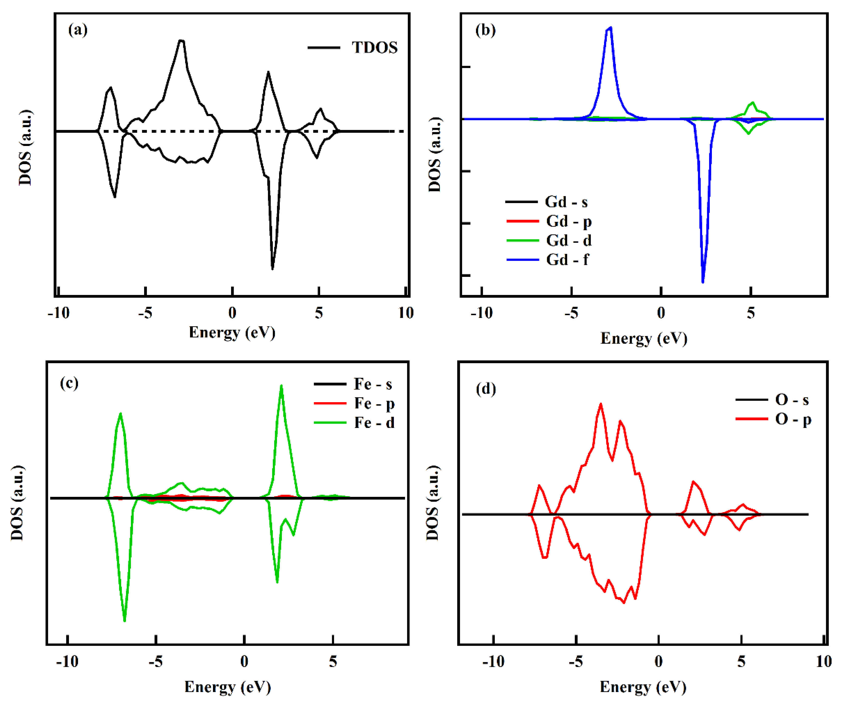
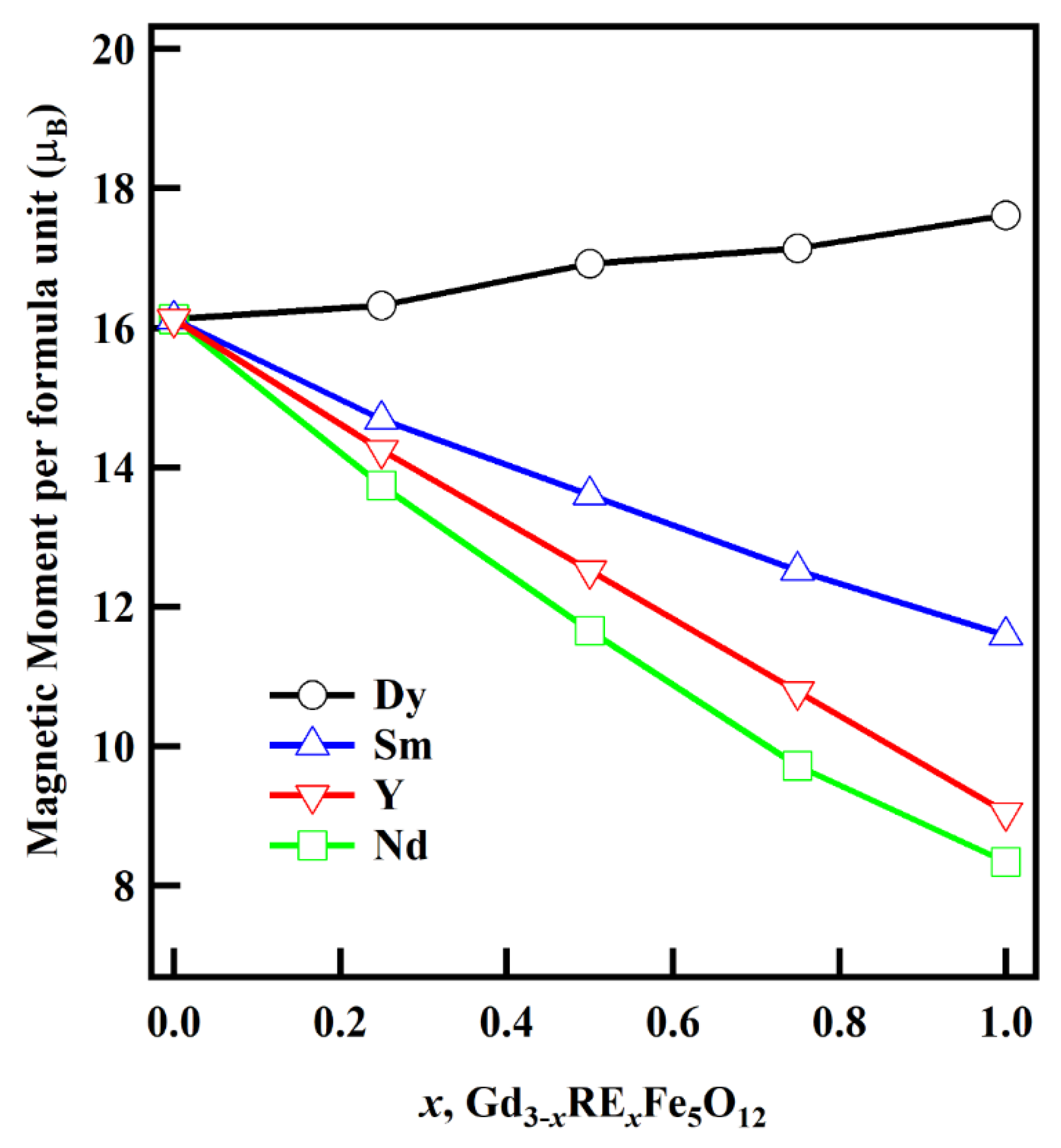
3.5. Theoretical Study

Ab initio density functional theory (DFT) calculations are performed using the VASP [33] simulation package for geometry optimization and post-processing calculations. The pseudo-potential constructed under the projector augmented wave (PAW) [55] method describes the valence electrons. The exchange-correlation functional of the Perdew–Burke–Ernzerhof (PBE)+U [32,56] type is considered in the total energy calculations, where the wave function expansion is carried out by considering the plane-wave basis set having the energy cutoff of 400 eV. The spin and the orbital part of the total magnetic moment are calculated by considering the spin–orbit interactions. A gamma-centered k-point mesh sampled at 2 × 2 × 2 is used to integrate the Brillouin zone. We used the total energy criteria for both electronic self-consistency and geometry optimization. The electronic self-consistency is achieved when the total energies of two consecutive electronic steps are smaller than 10−4 eV. The structures are allowed to relax along with the lattice parameters until the total energies of two consecutive ionic steps are smaller than 10−3 eV.
The total density of states (TDOS) of Gd3Fe5O12 is shown in Figure 13a, along with the spin-up and spin-down components, and the orbital contributions from Gd, Fe, and O to the TDOS are shown in Figure 13b–d, respectively. From Figure 13b, it is clear that the f-orbital of the Gd atom has a major contribution to the spin-up component of the conduction band and the spin-down component of the valence band in TDOS, along with the small contribution from its d-orbital. Similarly, in Figure 13c,d, the d-orbitals of Fe3+ atoms and the p-orbitals of the oxygen (O) seem to have a small contribution to the TDOS as well. The magnetism in the material arises because of the asymmetric nature of spin-up and spin-down components in the density of states, which is seen in Figure 14.
The Gd3−xRExFe5O12 structure considered in the calculations consists of 80 atoms (Figure 1a), which is four times larger than its functional unit (f. u). The calculations are carried out for the four rare-earth (RE) elements, namely, Dy, Nd, Sm, and Y, which are used as a dopant on the Gd sites with values ranging from x = 0 to 1, with a step size of 0.25. The corresponding values for the total magnetic moments (spin and orbital) per formula unit of the optimized structures are obtained from the calculations. The effective Coulomb-exchange interaction (Ueff) value is set to be 6 eV for the 4f orbitals in Dy, Nd, Sm, and Gd, whereas it is 4 eV for the d orbitals in Fe and Y [57,58]. The initial values of the magnetic moment of each element were taken from the literature [5,59,60]. The calculated values of the individual elements’ orbital and spin magnetic moments after the optimization are listed in Table 7 below.
From Table 7, the orbital magnetic moment of Dy3+ has a positive value, whereas Nd3+ and Sm3+ have negative values. The higher negative μL value in Nd3+ makes the total moment (μT) negative. Moreover, Fe3+, devoid of the orbital moment, has only the spin magnetic moment (μS), with octahedral Fe3+ having a positive value and tetrahedral Fe3+ having a negative value. The elements Y3+ and O2− have zero magnetic moments. At low temperatures with a 3d5, S = 5/2 configuration, we expect the magnetic moment to be 5 μB per iron ion. The Fe sublattices are anti-ferromagnetically coupled, giving a maximum net magnetization of 3 × (−5 μB) = −15 μB for the tetrahedral sublattice and 2 × (5 μB) = 10 μB for the octahedral sublattice per formula unit. The spontaneous magnetization direction of the Gd sublattice is taken to be positive. From the Gd sublattice, with Gd3+, S = 7/2 ions, we can expect a maximum magnetization of 3 × 7μB = 21 μB. This gives the expected saturation magnetization of 16 μB for Gd garnet, a value confirmed experimentally earlier [28].
The variation of the magnetic moment formula unit as a function of doping concentration, x, is shown in Figure 14 for four different RE3+ ions mentioned above. When x = 0.0, i.e., in the Gd3Fe5O12 sample, the μ B ~16 agrees with the previous calculations [41,61,62]. With the increase in the x value, the total magnetic moment increases in the case of Dy3+ doping. This is because the magnetic moment of Dy3+ is larger than that of Gd3+. Meanwhile, the magnetic moment decreases in the other three doping cases (Nd, Sm, and Y). The decreasing rate is consistent with the order of their magnetic moments (Nd < Y < Sm), which is also consistent with the experimental results. Furthermore, structural parameters for Gd3−xRExFe5O12, Table 3A, B, derived from DFT calculations, validate the values obtained from the XRD Rietveld refinement

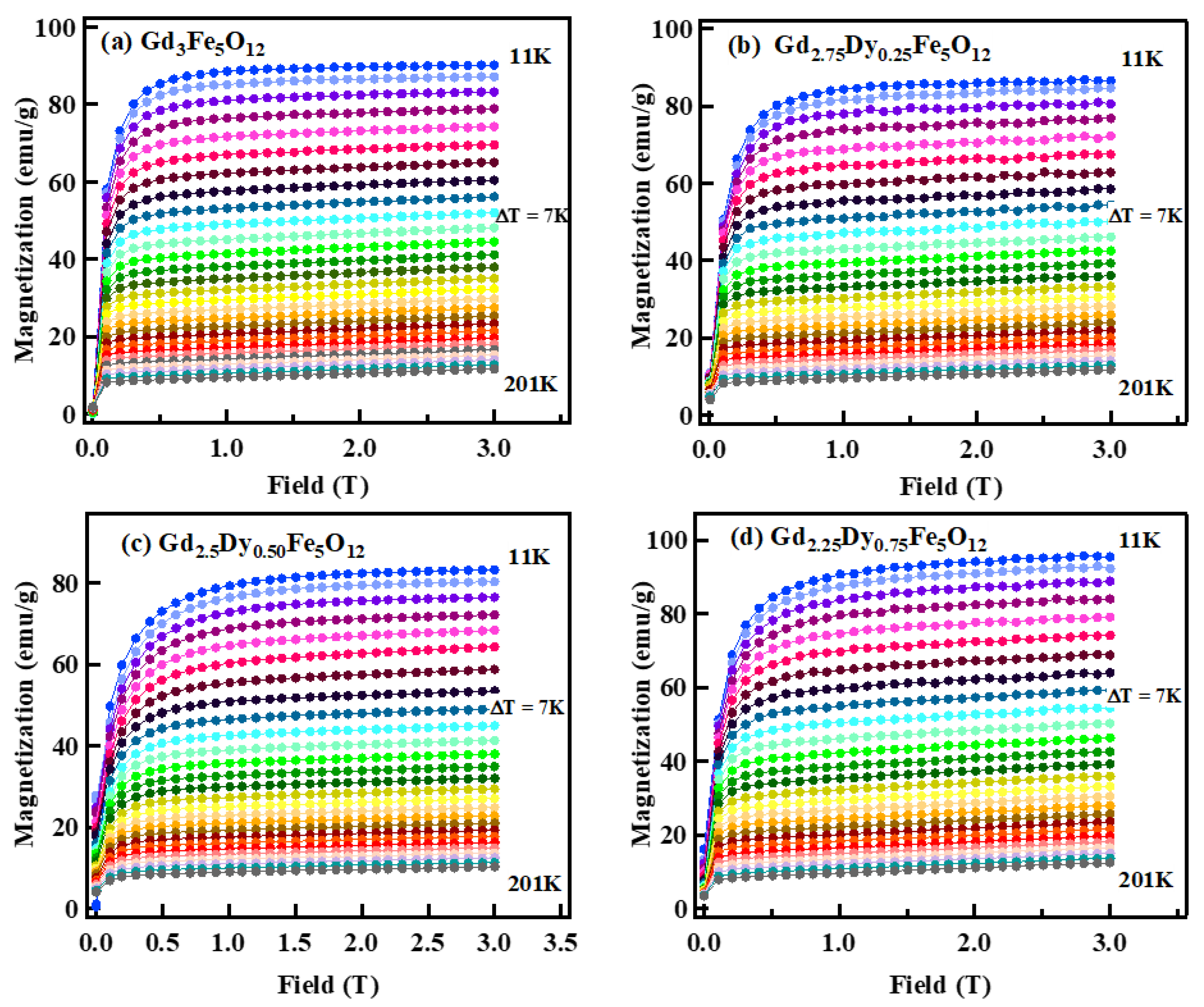
3.6. Magnetic Properties

The Curie temperature Tc for Gd3−xRExFe5O12 compounds was measured using a thermogravimetric analyzer (TGA) with a permanent magnet (Figure 15a–d). Figure 15 shows that the weight of the sample increased with temperature due to increased magnetic force on the sample. This implies that the net magnetic moment of the sample increased with the temperature up to Tc. The tetrahedral site iron ions have a positive moment, while the octahedral site has a negative moment. At elevated temperatures due to thermal energy, these moments are canted with respect to the z-axis. The sum of the projection of these moments on the z-axis determines the net moment of the compound. The observed increase in magnetization with temperature indicates that the octahedral site moment dominates the net moment. At temperature Tc, thermal energy dominates the magnetic spins, and the material exhibits paramagnetic behavior. The observed TC value is 570 K for x = 0.0 and remains unaltered upon RE3+ doping. The Tc value is dictated by the strength and the number of superexchange Fe3+O2−Fe3+ interactions, which largely remain unaffected by the RE3+ doping. The observed Tc with RE3+ substitution is listed in Table 8.
The magnetization as a function of temperature for RE3+ doped Gd3−xRExFe5O12 was investigated in the temperature range below room temperature. Figure 16 displays the temperature dependence of magnetization M(T) curves for Gd3−xRExFe5O12 under zero-field cooled (ZFC) and field-cooled (FC) conditions at a 100 Oe field. A distinct characteristic of M(T) curves is the presence of a compensation temperature, Tcomp. The temperature at which the magnetization crosses zero is called the compensation temperature, Tcomp. This occurs because the magnetization of RE3+ ion at the c sites is equal and opposite to the net magnetization of Fe3+ ion sublattice at the a and d sites. i.e., 3Fe(d)-[2MFe(a) + 3MRE(c)]. Table 8 lists the comparison of Tcomp values for different doped garnet compounds. In the case of Gd3−xRExFe5O12, Tcomp values vary between 280 K and 238 K, depending upon the type of RE3+ doping.
This behavior may be explained in terms of the temperature dependence of the magnetization of the three magnetic sublattices (dodecahedral, octahedral, and tetrahedral) balance each other [61,62]. Neel et al. [41] reported that the magnetic properties of RE3Fe5O12 can be explained by assuming that the three sublattice ferrimagnetism is due to positive RE3+ spin on dodecahedral sites, positive Fe3+ spin on octahedral site, and negative Fe3+ ion tetrahedral sites. The exchange interaction between RE3+ ions is almost negligible, so the moment of RE3+ ions should be aligned with the exchange interaction with Fe3+ ions. Structural evidence favors the strong magnetic interactions of the rare-earth ions with the tetrahedral Fe3+ ions (down magnetic moment) [41]. The strong interaction of the Gd3+ ion moments with those of the tetrahedral Fe3+ ion moments leads to random canting of the Gd3+ ion moments, thereby reducing the contribution of the dodecahedral sublattice to the net spontaneous magnetization of the garnet [67]. This could be the reason for the slow increase in the net magnetization value with temperature lowering.
A cusp is observed in the ZFC curves at a temperature where the RE3+ moment aligns parallel to the net Fe3+ moment. For Gd3Fe5O12, the cusp is observed at temperature (TF) = 50 K, while for other RE3+ substituted compounds, TF shifts to higher temperatures. Further, a decrease in magnetization value is observed at a temperature below TF. This decrease in magnetization value occurs because of a strong RE3+-O2−-Fe3+(Tetra.) interaction, which flips RE3+ moments parallel to the Fe3+(Tetra.) in a negative direction. The exchange interaction between the 4f rare-earth electrons and the 3d iron electrons is not direct but occurs indirectly via the oxygen ions [68]. This interaction is strengthened in the presence of RE3+ ions with non-zero orbital angular momenta, such as Nd3+, Sm3+, and Dy3+. This conclusion is further corroborated by noticing the absence of a cusp in M vs. T for Y3+ doped garnet, where Y3+ does not possess any orbital angular momentum. The increase in TF value with RE3+ substitution results from the increased number and strength of superexchange interactions RE3+O2−Fe3+(Tetra.) ensuing from increased bond-angle in favor of strengthening the interaction, Figure 10b. The M vs. T curve cusp is more prominent for the Dy3+ doped sample. Because of negative moments of Nd3+ (−1.54 μB), the magnetization attains a negative value below Tcomp. Meanwhile, Sm3+ (2.55 μB) shows a positive magnetization value with a cusp below Tcomp. This discussion concludes that RE3+ with a finite orbital angular momentum couple strongly with Fe3+ sublattice moment via superexchange interaction.
The temperature-dependent magnetization process is depicted in Figure 17. At Tcomp, the three sublattice moment adds to zero moments. Below Tcomp, magnetization slowly peaks with rare-earth contributing positively to the net moment, while at low temperatures below TF, increased RE3+ moments canting due to strong RE3+O2−Fe3+(Tetra.) superexchange interaction leads to net negative moments. The TF value shifts to a higher temperature depending on the strength and number of superexchange interaction pairs. At x = 0.75, at low temperatures, below Tcomp, the magnetic anisotropy of Nd3+, Sm3+, and Gd3+ exceeds that of iron, with their moment being aligned along the easy axis, thus increasing the net moment of the compound.
Figure 18a–d shows the magnetization vs. field curves, M vs. H, for Gd3−xRExFe5O12 measured at 5 K. All samples show the ferromagnetic behavior at 5 K. The magnetic curve tends to saturate below the applied field of 0.5 T for all samples. The saturation magnetization, Ms, value reached ~92.3 emu/g for x = 0.0 and decreased with x content for Nd3+, Sm3+, and Y3+ except for Dy3+. The saturation magnetization value for all samples matched the trend of theoretically derived values in Table 9. The total magnetic moment in the Gd3−xRExFe5O12 garnet is due to the contribution of three different magnetic sublattices. The total magnetic moment is represented as:
3MFe(tetra) − [2MFe(octa) + (3 − x)MGd(dodec) + xMRE(dodec)]
As obtained from the DFT study, the magnetic moment of the Dy3+ ion has a maximum value (10.48 µB), and low values for Nd3+ (3.62 µB), Sm3+ (0.65 µB), and Y3+ (0 µB) compared to Gd3+ (6.9 µB). Therefore, the net moment for the Gd3−xRExFe5O12 compound decreases for Nd3+, Sm3+, and Y3+ doped garnet but improves with Dy3+ doping. The experimental magnetic moment of the RE3+ substitution Gd3−xRExFe5O12 sample is calculated in terms of Bohr magneton using the equation below and listed in Table 9.
Bohr   magneton   μ B = M × M s 5585 × ρ x r a y
where ρx−ray is the X-ray density Equation (11), where M is the molecular weight of the samples, and Ms is the saturation magnetization in emu/g of Gd3−xRExFe5O12. Bohr magneton value for Gd3Fe5O12 is observed at 2.40 µB and changes with RE3+ substitution. The magnetic moment of Dy3+ doped garnet increases from 2.4 µB to 2.59 µB, whereas Nd3+, Sm3+, and Y3+ doped samples show a decreasing trend. Change in Bohr magneton value with RE3+ substitution is due to the different magnetic moment of RE3+ ions and matches the theoretical study’s trend.
Yafet and Kittle (Y-K) angles describe the direction of spin of iron ions in ferrites. The Yafet and Kittle ( α Y K ) angles of RE3+ doped Gd3−xRExFe5O12 are calculated by using the following equation:
μ B = 6 + x C o s α Y K 5 1 x ,
where μ B is the Bohr magnetons calculated experimentally. The α Y K arises due to the non-collinear direction of the moment between tetrahedral and octahedral sublattices. Table 9 shows the linear increase in the α Y K angle with RE3+ substitution. In addition, RE3+ doped samples show Y-K type canting of local moments. The linear trend in the α Y K angle with Rex is due to the split of sublattices having magnetic moments equal in magnitude and each making an angle α Y K with the direction of net magnetization.
Figure 19a–d shows the M vs. H curves for Gd3−xRExFe5O12 powder at 300 K. With the increase in temperature to 300 K, due to thermal fluctuation, the ferrimagnetic order is lost, and the Gd3−xRExFe5O12 system attains paramagnetic order (it is not PM, it is unusual that there is remanence magnetization). To further investigate the effect of RE3+ on the magnetic and magnetocaloric behaviors of Gd3−xRExFe5O12, isothermal magnetization as a function of the applied field, M(H), was measured from 11 K to 210 K with a temperature step of 7 K up to 3T field. The isothermal plots for Dy3+, Nd3+, Sm3+, and Y3+ substitution Gd3−xRExFe5O12 are shown in Figure 20, Figure 21, Figure 22 and Figure 23. The isothermal magnetization curve shows the ferromagnetic ordering at low temperatures and paramagnetic at elevated temperatures. The magnetization increases sharply and saturates immediately at the low field, which is a sign of ferromagnetic behavior. Magnetization increases gradually with an increasing field and does not show any sign of saturation, thus displaying the paramagnetic behavior.

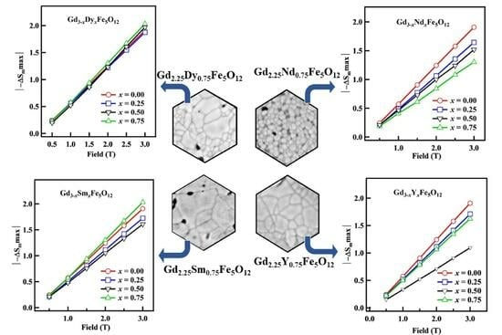
3.7. Magnetocaloric Study

Our primary focus is to study the magnetocaloric effect of RE3+ doped Gd3−xRExFe5O12. The change in magnetic entropy (ΔSM) is the most recommended parameter to evaluate the efficiency of magnetocaloric materials. It is calculated using the magnetic isothermal data (Figure 20, Figure 21, Figure 22 and Figure 23) near the vicinity of the transition temperature. The isothermal magnetic entropy change has been computed using the thermodynamic Maxwell relation [69],
S M = μ o H i H f ( M T ) H d H
S M = μ o T 0 H f M T + T , H d H 0 H f M T , H d H
It is numerically calculated as;
S M H , T = M i M i + 1 T i + 1 T i H i ,
where Hi and Hf are the initial and final external applied fields, and µo is the permeability of free space. −ΔSM is calculated from the isothermal magnetization curve of Figure 20, Figure 21, Figure 22 and Figure 23.
The magnetic entropy change has a maximum value near transition temperature, Tc, and its value decreases with a further increase or decrease in temperature. The sign of the magnetic entropy change is negative, which means heat is released when the magnetic field is changed adiabatically [70].
Figure 24, Figure 25, Figure 26 and Figure 27 show the magnetic entropy change curve −ΔSM(T) as a function of temperature for Gd3−xRExFe5O12. The maxima ( S M m a x ) for the ΔSM(T) curve is observed to be independent of temperature and field, as shown in Figure 28. As shown in Figure 24a–d, the S M m a x value for Dy3+ doped garnet increases with x content. The optimum value of magnetic entropy change reached 2.04 J.Kg−1K−1 for x = 0.75, which is ~7% higher than that of the x = 0.0 sample. An increase in magnetic entropy change with Dy3+ substitution is due to the replacement of Gd3+ (6.99 μ B ) having a smaller magnetic moment by Dy3+ (9.05 μ B ) having a significant magnetic moment (from the DFT calculation above). The variation in S M is mainly due to the superexchange interaction between Fe–O–Fe ions.
By doping Dy3+, the Dy-Fe superexchange interactions become strong, enhancing the −ΔSM value [71]. The ( S M m a x ) value decreases with x content for the Nd, Sm, and Y doped garnet except for Sm (x = 0.75), as shown in Figure 25, Figure 26 and Figure 27. The decreasing behavior of the magnetocaloric effect with RE3+ doped garnet can be explained based on the magnetic moment of an individual element. From the theoretical observations, the magnetic moment of Nd (−1.54 μ B ), Sm (2.55 μ B ), and Y (0 μ B ) are smaller than the magnetic moment of Gd (6.99 μ B ). The S M m a x increases for Sm3+ doped garnet for x = 0.75. The S M ( T ) plots show the broad curve covering a large temperature range with RE3+ doped samples. Figure 28a–d shows the summary of the S M m a x value of Gd3−xRExFe5O12 as a function of field. The maxima value ( S M m a x ) shows the proportional relation with the applied field. The S M m a x values of some garnets are summarized in Table 10 to compare our results.
The relative cooling power (RCP) is a metric that quantifies the performance of magnetocaloric materials. The RCP value depends on the −ΔSM and magnetocaloric materials’ operating temperature range. The RCP is calculated as follows,
R C P = S M m a x × δ T F W H M
where δTFWHM is the full-width-half-maxima obtained from the SM vs. T plots of Figure 24, Figure 25, Figure 26 and Figure 27.
Figure 29 shows the evolution of the RCP of Gd3−xRExFe5O12 as a function of the applied magnetic field. The RCP value for x = 0.0 is ~31 J/kg at H = 0.5 T, which increases with the applied field and becomes ~219 J/kg at H = 3.0 T. The calculated RCP value for Gd3−xRExFe5O12 is higher even at low fields than the other garnets reported in the literature [20,74,75]. The low field high RCP value of the Gd3−xRExFe5O12 is very promising for the magnetic refrigeration application for low-temperature applications. The influence of the magnetic field on RCP may be estimated according to the formula,
RCP = A HR
The R exponents obtained from the numerical fit of RCP are listed in Table 10. The R-value for Gd3Fe5O12 is 1.10 and increases with RE3+ substitution. The maximum R-value is obtained for the Nd3+ (0.75) doped garnet. The R-value describes the field dependency of RCP. An R-value close to 1 implies the linear increase of RCP with the applied field.

4. Conclusions

The synthesis of RE3+ doped Gd3−xRExFe5O12 (x = 0.0, 0.25, 0.50, and 0.75, RE3+ = Y, Nd, Sm, and Dy) was conducted successfully via the sol-gel autocombustion method. The substitution of RE3+ ions on the Gd3+ site of garnet brings in a structural and magnetic change in the compound. The XRD analysis shows the formation of a garnet structure with the Ia-3d space group. The Rietveld refinement shows that the lattice parameter decreased with Dy3+ and Y3+ substitution and increased with Nd3+ and Sm3+ substitution in accordance with the ionic radii of corresponding RE3+ ionic radii. The bond angle between RE3+-O2−-Fe3+ increased, Fe(Oct.)3+O2−Fe(Tetra.)3+ decreased, and the bond length between RE3+-O−2 decreased with the Dy3+ and Y3+ doped sample. These structural changes have an essential influence on the magnetic structure of the compound. Magnetic studies reveal that the Dy3+ substitution garnet shows higher saturation magnetization with a maximum value of 99 emu/g for x = 0.75, whereas all other RE3+ show a decrease in saturation magnetization value. The temperature-dependent magnetization study reveals that RE3+ ions with non-zero magnetic moments couple strongly with the Fe3+(Tetra.) site. The Dy3+ doped garnet shows the highest magnetic entropy change value compared to other RE3+ doped garnets. The maxima value for Dy3+ doped garnet achieved (ΔSMmax~2.00 Jkg−1K−1) is due to the compound’s sizeable magnetic moment. In summary, a substantial change in magnetic entropy value shows that rare-earth doped garnets could be suitable magnetocaloric materials for low-temperature cooling technology.

Author Contributions

Conceptualization, S.R.M.; methodology, S.R.M., D.N., A.K.P., N.K. and C.H.; software, X.S. and R.B.; validation, S.R.M. and A.K.P.; formal analysis, D.N.; investigation, D.N.; resources, S.R.M., A.K.P. and X.S.; data curation, D.N. and S.K.; writing—original draft preparation, D.N., S.R.M. and S.K.; writing—review and editing, S.R.M., S.K. and D.N.; visualization, S.R.M., A.K.P. and X.S.; supervision, S.R.M.; project administration, S.R.M.; funding acquisition, None. All authors have read and agreed to the published version of the manuscript.

Funding

This research received no external funding.

Institutional Review Board Statement

Not applicable.

Informed Consent Statement

Not applicable.

Data Availability Statement

The data presented in this study are available on request from the corresponding author.

Acknowledgments

Magnetic measurements were performed at the State University of New York (SUNY), Buffalo State, and supported by the National Science Foundation Award No. DMR-2213412. N.K. acknowledges financial support from the Office of Undergraduate Research, SUNY, Buffalo State. Computational resources were provided by the University of Memphis High-Performance Computing Center (HPCC).

Conflicts of Interest

The authors declare no conflict of interest.

References

  1. Phan, M.H.; Yu, S.C. Review of the magnetocaloric effect in manganites materials. J. Magn. Magn. Mater. 2007, 308, 325. [Google Scholar] [CrossRef]
  2. Neupane, D.; Hulsebosch, L.; Ali, K.S.; Bhattarai, R.; Shen, X.; Pathak, A.K.; Mishra, S.R. Enhanced Magnetocaloric Effect in Aluminum doped Gd3Fe5−xAlxO12 Garnet: Structural, Magnetic, and Mössbauer Study. Materialia 2021, 21, 101301. [Google Scholar] [CrossRef]
  3. Phan, M.H.; Morales, M.B.; Chinnasamy, C.N.; Latha, B.; Harris, V.G.; Srikanth, H. Magnetocaloric effect in bulk and nanostructured Gd3Fe5O12 materials. J. Phys. D Appl. Phys. 2009, 42, 115007. [Google Scholar] [CrossRef]
  4. Petrov, D.N.; Koshkid’Ko, Y.S.; Ćwik, J.; Nenkov, K. Large magnetocaloric effect in LiLnP4O12 (Ln = Gd, Tb, Dy) single crystals. J. Phys. D Appl. Phys. 2020, 53, 495005. [Google Scholar] [CrossRef]
  5. Lassri, H.; Hlil, E.K.; Prasad, S.; Krishnan, R. Magnetic and electronic properties of nanocrystalline Gd3Fe5O12 garnet. J. Solid State Chem. 2011, 184, 3216–3220. [Google Scholar] [CrossRef]
  6. Jiang, L.; Yang, S.; Zheng, M.; Chen, H.; Wu, A. Synthesis and magnetic properties of nanocrystalline Gd3Fe5O12 and GdFeO3 powders prepared by sol–gel auto-combustion method. Mater. Res. Bull. 2018, 104, 92–96. [Google Scholar] [CrossRef]
  7. Zhang, G. Preparation and Characterization of New Magneto-Optical Crystals for Optical Communication. Ph.D. Thesis, NUS Singapore, Singapore, 2005. Available online: http://scholarbank.nus.edu.sg/handle/10635/14540 (accessed on 10 September 2020).
  8. Fu, Q.; Xu, Q.; Zhao, Z.; Liu, X.; Huang, Y.; Hu, X.; Zhuang, N.; Chen, J. New magneto-optical film of Ce, Ga: GIG with high performance. J. Am. Ceram. Soc. 2016, 99, 234–240. [Google Scholar] [CrossRef]
  9. Bahadur, D. Current trends in applications of magnetic ceramic materials. Bull. Mater. Sci. 1992, 15, 431–439. [Google Scholar] [CrossRef]
  10. Schreier, M.; Chiba, T.; Niedermayr, A.; Lotze, J.; Huebl, H.; Geprägs, S.; Takahashi, S.; Bauer, G.E.; Gross, R.; Goennenwein, S.T. Current-induced spin torque resonance of a magnetic insulator. Phys. Rev. B 2015, 92, 144411. [Google Scholar] [CrossRef]
  11. Chaturvedi, A. Novel Magnetic Materials for Sensing and Cooling Applications. Ph.D. Thesis, University of South Florida, Tampa, FL, USA, 2011. [Google Scholar]
  12. Opuchovic, O.; Beganskiene, A.; Kareiva, A. Sol–gel derived Tb3Fe5O12 and Y3Fe5O12 garnets: Synthesis, phase purity, micro-structure and improved design of morphology. J. Alloys Compd. 2015, 647, 189–197. [Google Scholar] [CrossRef]
  13. Uemura, M.; Yamagishi, T.; Ebisu, S.; Chikazawa, S.; Nagata, S. A double peak of the coercive force near the compensation temperature in the rare earth iron garnets. Philos. Mag. 2008, 88, 209–228. [Google Scholar] [CrossRef]
  14. Serier-Brault, H.; Thibault, L.; Legrain, M.; Deniard, P.; Rocquefelte, X.; Leone, P.; Perillon, J.L.; Le Bris, S.; Waku, J.; Jobic, S. Thermochromism in yttrium iron garnet compounds. Inorg. Chem. 2014, 53, 12378–12383. [Google Scholar] [CrossRef] [PubMed]
  15. Gilleo, M.A. Ferromagnetic insulators: Garnets. Handb. Ferromagn. Mater. 1980, 2, 1–53. [Google Scholar]
  16. Kalashnikova, A.M.; Pavlov, V.V.; Kimel, A.V.; Kirilyuk, A.; Rasing, T.; Pisarev, R.V. Magneto-optical study of holmium iron garnet Ho3Fe5O12. Low Temp. Phys. 2012, 38, 863–869. [Google Scholar] [CrossRef]
  17. Bsoul, I.; Olayaan, R.; Lataifeh, M.; Mohaidat, Q.I.; Mahmood, S.H. Structural and magnetic properties of Er3Fe5−xGaxO12 garnets. Mater. Res. Express 2019, 6, 076114. [Google Scholar] [CrossRef]
  18. Nguyet, D.T.T.; Duong, N.P.; Satoh, T.; Hien, T.D.; Anh, L.N.; Loan, T.T. Crystallization and magnetic characterizations of DyIG and HoIG nanopowders fabricated using citrate sol–gel. J. Sci. Adv. Mater. Devices 2016, 1, 193–199. [Google Scholar] [CrossRef]
  19. Wu, J.; Yuan, L.; Wang, S.; Hou, C. Nd3−xAExFe5O12: Hydrothermal synthesis, structure and magnetic properties. Chem. Res. Chin. Univ. 2017, 33, 869–875. [Google Scholar] [CrossRef]
  20. Li, C.; Qiu, Y.; Barasa, G.O.; Yuan, S. Spin reorientation, normal and inverse magnetocaloric effects in heavy rare-earth iron garnets. Ceram. Int. 2020, 46, 18758–18762. [Google Scholar] [CrossRef]
  21. Aparnadevi, N.; Kumar, K.S.; Manikandan, M.; Kumar, B.S.; Punitha, J.S.; Venkateswaran, C. Structural properties, optical, electrical and magnetic behavior of bismuth doped Gd3Fe5O12 prototype garnet. J. Mater. Sci. Mater. Electron. 2020, 31, 2081–2088. [Google Scholar] [CrossRef]
  22. Li, C.; Barasa, G.O.; Qiu, Y.; Yuan, S. Magnetocaloric effect and sign reversal of magnetic entropy change across the spin reorientation temperature in R3Fe5O12 (R = Gd, Dy). J. Alloys Compd. 2020, 820, 153138. [Google Scholar] [CrossRef]
  23. Shannon, R.D. Revised effective ionic radii and systematic studies of interatomic distances in halides and chalcogenides. Acta Crystallogr. Sect. A Cryst. Phys. Diffr. Theor. Gen. Crystallogr. 1976, 32, 751–767. [Google Scholar] [CrossRef]
  24. Hutchison, W.D.; Stewart, G.A.; Cadogan, J.M.; Princep, A.; Stewart, R.; Ryan, D.H. Magnetic ground state of Dy3+ in DyNiAl4. AIP Adv. 2017, 7, 055702. [Google Scholar] [CrossRef]
  25. Przeniosło, R.; Sosnowska, I.; Fischer, P. Magnetic moment ordering of Nd3+ ions in NdFeO3. J. Magn. Magn. Mater. 1995, 140, 2153–2154. [Google Scholar] [CrossRef]
  26. Cevallos, F.A.; Guo, S.; Cava, R.J. Magnetic properties of lithium-containing rare earth garnets Li3RE3Te2O12 (RE = Y, Pr, Nd, Sm-Lu). Mater. Res. Express 2018, 5, 126106. [Google Scholar] [CrossRef]
  27. Alves, T.E.P.; Pessoni, H.V.S.; Franco, A., Jr. The effect of Y3+ substitution on the structural, optical band-gap, and magnetic properties of cobalt ferrite nanoparticles. Phys. Chem. Chem. Phys. 2017, 19, 16395–16405. [Google Scholar] [CrossRef] [PubMed]
  28. Geller, S.; Remeika, J.P.; Sherwood, R.C.; Williams, H.J.; Espinosa, G.P. Magnetic study of the heavier rare-earth iron garnets. Phys. Rev. 1965, 137, A1034. [Google Scholar] [CrossRef]
  29. Lataifeh, M.S.; Al-Sharif, A. Magnetization measurements on some rare-earth iron garnets. Appl. Phys. A 1995, 61, 415–418. [Google Scholar] [CrossRef]
  30. Lataifeh, M.S.; McCausland, M.H. On the molecular field seen by Gd3+ in iron garnets. Mu’tah Lil-Buhooth Wa Al-Dirasat 1994, 9, 23–30. [Google Scholar]
  31. Prasad, D.H.; Jung, H.Y.; Jung, H.G.; Kim, B.K.; Lee, H.W.; Lee, J.H. Single step synthesis of nano-sized NiO–Ce0.75Zr0.25O2 composite powders by glycine nitrate process. Mater. Lett. 2008, 62, 587–590. [Google Scholar] [CrossRef]
  32. Dudarev, S.L.; Botton, G.A.; Savrasov, S.Y.; Humphreys, C.J.; Sutton, A.P. Electron-energy-loss spectra and the structural stability of nickel oxide: An LSDA+U study. Phys. Rev. B 1998, 57, 1505. [Google Scholar] [CrossRef]
  33. Kresse, G.; Furthmüller, J. Efficient iterative schemes for ab initio total-energy calculations using a plane-wave basis set. Phys. Rev. B 1996, 54, 11169. [Google Scholar] [CrossRef] [PubMed]
  34. Perdew, J.P.; Burke, K.; Ernzerhof, M. Generalized gradient approximation made simple. Phys. Rev. Lett. 1996, 77, 3865. [Google Scholar] [CrossRef] [PubMed]
  35. Kresse, G.; Joubert, D. From ultrasoft pseudopotentials to the projector augmented-wave method. Phys. Rev. B 1999, 59, 1758–1775. [Google Scholar] [CrossRef]
  36. Rietveld, H.M. The Rietveld method. Phys. Scr. 2014, 89, 09800. [Google Scholar] [CrossRef]
  37. Toby, B.H.; Von Dreele, R.B. GSAS-II: The genesis of a modern open-source all purpose crystallography software package. J. Appl. Crystallogr. 2013, 46, 544–549. [Google Scholar] [CrossRef]
  38. Vegard, L. Die konstitution der mischkristalle und die raumfüllung der atome. Z. Für Phys. 1921, 5, 17–26. [Google Scholar] [CrossRef]
  39. Geller, S.; Gilleo, M.A. The crystal structure and ferrimagnetism of yttrium-iron garnet, Y3Fe2(FeO4)3. J. Phys. Chem. Solids 1957, 3, 30–36. [Google Scholar] [CrossRef]
  40. Coronado, E.; Tsukerblat, B.S.; Georges, R. Exchange Interactions I: Mechanisms. In Molecular Magnetism: From Molecular Assemblies to the Devices; Springer: Dordrecht, The Netherlands, 1996; pp. 65–84. [Google Scholar]
  41. Néel, L.; Pauthenet, R.; Dreyfus, B. Chapter VII—The rare earth garnets. In Progress in Low Temperature Physics; Elsevier: Amsterdam, The Netherlands, 1964; Volume 4. [Google Scholar]
  42. Kramers, H.A. L’interaction entre les atomes magnétogènes dans un cristal paramagnétique. Physica 1934, 1, 182–192. [Google Scholar] [CrossRef]
  43. Rovani, P.R.; Ferreira, A.S.; Pereira, A.S.; de Lima, J.C. Effect of pressure on nanostructured Gd3Fe5O12. J. Appl. Phys. 2017, 122, 035904. [Google Scholar] [CrossRef]
  44. Weil, L.; Bertaut, F.; Bochirol, L. Propriétés magnétiques et structure de la phase quadratique du ferrite de cuivre. J. Phys. Radium 1950, 11, 208–212. [Google Scholar] [CrossRef]
  45. Akhtar, M.N.; Ali, K.; Umer, A.; Ahmad, T.; Khan, M.A. Structural elucidation, and morphological and magnetic behavior evaluations, of low-temperature sintered, Ce-doped, nanostructured garnet ferrites. Mater. Res. Bull. 2018, 101, 48–55. [Google Scholar] [CrossRef]
  46. Sickafus, K.E.; Hughes, R. Spinel compounds: Structure and property relations. J. Am. Ceram. Soc. 1999, 82, 3277–3278. [Google Scholar]
  47. Akhtar, M.N.; Khan, S.N.; Ahmad, H.; Nazir, M.S.; Khan, M.A. Structural elucidation and magnetic behaviour evaluation of gallium substituted garnet ferrites. Ceram. Int. 2018, 44, 22504–22511. [Google Scholar] [CrossRef]
  48. Halder, N.C.; Wagner, C.N.J. Separation of particle size and lattice strain in integral breadth measurements. Acta Crystallogr. 1996, 20, 312–313. [Google Scholar] [CrossRef]
  49. Cureton, W.F.; Palomares, R.I.; Walters, J.; Tracy, C.L.; Chen, C.H.; Ewing, R.C.; Lang, M.; Baldinozzi, G.; Lian, J.; Trautmann, C. Grain size effects on irradiated CeO2, ThO2, and UO2. Acta Mater. 2018, 160, 47–56. [Google Scholar] [CrossRef]
  50. Dipesh, D.N.; Wang, L.; Adhikari, H.; Alam, J.; Mishra, S.R. Influence of Al3+ doping on structural and magnetic properties of CoFe2−xAlxO4 Ferrite nanoparticles. J. Alloys Compd. 2016, 688, 413–421. [Google Scholar] [CrossRef]
  51. Zulueta, Y.A.; Dawson, J.A.; Mune, P.D.; Froeyen, M.; Nguyen, M.T. Oxygen vacancy generation in rare-earth-doped SrTiO3. Phys. Status Solidi (B) 2016, 253, 2197–2203. [Google Scholar] [CrossRef]
  52. Poudel, T.P.; Rai, B.K.; Yoon, S.; Guragain, D.; Neupane, D.; Mishra, S.R. The effect of gadolinium substitution in inverse spinel nickel ferrite: Structural, Magnetic, and Mössbauer study. J. Alloys Compd. 2019, 802, 609–619. [Google Scholar] [CrossRef]
  53. Zhou, X.; Zhou, Y.; Zhou, L.; Wei, J.; Wu, J.; Yao, D. Effect of Gd and La doping on the structure, optical and magnetic properties of NiZnCo ferrites. Ceram. Int. 2019, 45, 6236–6242. [Google Scholar] [CrossRef]
  54. Odo, E.A. Morphology and Elemental Study of Silicon Nanoparticles Produced Using a Vibratory Disc Mill. Nanosci. Nanotechnol. 2015, 5, 57–63. [Google Scholar]
  55. Blöchl, P.E. Projector augmented-wave method. Phys. Rev. B 1994, 50, 17953. [Google Scholar] [CrossRef] [PubMed]
  56. Harmon, B.N.; Antropov, V.P.; Liechtenstein, A.I.; Solovyev, I.V.; Anisimov, V.I. Calculation of magneto-optical properties for 4f systems: LSDA + Hubbard U results. J. Phys. Chem. Solids 1995, 56, 1521–1524. [Google Scholar] [CrossRef]
  57. Zhang, L.; Meng, J.; Yao, F.; Zhang, W.; Liu, X.; Meng, J.; Zhang, H. Insight into the mechanism of the ionic conductivity for Ln-doped ceria (Ln = La, Pr, Nd, Pm, Sm, Gd, Tb, Dy, Ho, Er, and Tm) through first-principles calculation. Inorg. Chem. 2018, 57, 12690–12696. [Google Scholar] [CrossRef] [PubMed]
  58. Wurmehl, S.; Fecher, G.H.; Kandpal, H.C.; Ksenofontov, V.; Felser, C.; Lin, H.J.; Morais, J. Geometric, electronic, and magnetic structure of Co2 FeSi: Curie temperature and magnetic moment measurements and calculations. Phys. Rev. B 2005, 72, 184434. [Google Scholar] [CrossRef]
  59. Taneja, S.P. On the Magnetic Properties of Dy3+ Ion in Dysprosium Ethylsulfate Single Crystal. Phys. Status Solidi (B) 1969, 36, 525–529. [Google Scholar] [CrossRef]
  60. Sugiyama, J.; Miwa, K.; Nozaki, H.; Kaneko, Y.; Hitti, B.; Arseneau, D.; Morris, G.D.; Ansaldo, E.J.; Brewer, J.H. Magnetic moment of rare-earth elements in R2Fe14B estimated with μ+ SR. Phys. Rev. Mater. 2019, 3, 064402. [Google Scholar] [CrossRef]
  61. Liu, C.; Kan, X.; Liu, X.; Zhang, Z.; Hu, J. Magnetic compensation and critical behavior in spinel Co2TiO4. Phys. Chem. Chem. Phys. 2020, 22, 20929–20940. [Google Scholar] [CrossRef]
  62. Ivanov, S.A.; Tellgren, R.; Porcher, F.; Andre, G.; Ericsson, T.; Nordblad, P.; Sadovskaya, N.; Kaleva, G.; Baldini, M.; Mathieu, R. Structural and magnetic properties of nickel antimony ferrospinels. Mater. Chem. Phys. 2015, 158, 127–137. [Google Scholar] [CrossRef]
  63. Mahmood, S.H.; Bsoul, I.; Hawamdeh, K. Effect of Al-substitution on Structural and Magnetic Properties of Er3Fe5−xAlxO12 Garnets. Mater. Res. Found. 2020, 83, 21–40. [Google Scholar]
  64. Lahoubi, M.; Guillot, M.; Marchand, A.; Tcheou, F.; Roudault, E. Double umbrella structure in terbium iron garnet. IEEE Trans. Magn. 1984, 20, 1518–1520. [Google Scholar] [CrossRef]
  65. Fadly, M.; Feldmann, P.; Le Gall, H.; Guillot, M.; Makram, H. Magnetooptical coefficients of Ho3+ ions induced by electric and magnetic dipole transitions in single crystal HoIG. IEEE Trans. Magn. 1978, 14, 448–450. [Google Scholar] [CrossRef]
  66. Rudolf, P.; Sette, F.; Tjeng, L.H.; Meigs, G.; Chen, C.T. Magnetic moments in a gadolinium iron garnet studied by soft-X-ray magnetic circular dichroism. J. Magn. Magn. Mater. 1992, 109, 109–112. [Google Scholar] [CrossRef]
  67. Geller, S. Magnetic behavior of substituted ferrimagnetic garnets. J. Appl. Phys. 1966, 37, 1408–1415. [Google Scholar] [CrossRef]
  68. Levy, P.M. Rare-earth-iron exchange interaction in the garnets. I. Hamiltonian for anisotropic exchange interaction. Phys. Rev. 1964, 135, A155. [Google Scholar] [CrossRef]
  69. Pecharsky, V.K.; Gschneidner, K.A., Jr. Magnetocaloric effect and magnetic refrigeration. J. Magn. Magn. Mater. 1999, 200, 44–56. [Google Scholar] [CrossRef]
  70. Gavrilova, T.P.; Gilmutdinov, I.F.; Deeva, J.A.; Chupakhina, T.I.; Lyadov, N.M.; Faizrakhmanov, I.A.; Milovich, F.O.; Kabirov, Y.V.; Eremina, R.M. Magnetic and magnetocaloric properties of (1 − x) La0.7Sr0.3MnO3/xNaF composites. J. Magn. Magn. Mater. 2018, 467, 49–57. [Google Scholar] [CrossRef]
  71. Rogers, P.D.; Choi, Y.J.; Standard, E.; Kang, T.D.; Ahn, K.H.; Dubroka, A.; Marsik, P.; Wang, C.; Bernhard, C.; Park, S.; et al. Optical Identification of Hybrid Magnetic and Electric Excitations in Dy3Fe5O12 Garnet. arXiv 2011, arXiv:1101.2675. [Google Scholar]
  72. Provenzano, V.; Li, J.; King, T.; Canavan, E.; Shirron, P.; DiPirro, M.; Shull, R.D. Enhanced magnetocaloric effects in R3(Ga1−xFex)5O12 (R = Gd, Dy, Ho; 0 < x < 1) nanocomposites. J. Magn. Magn. Mater. 2003, 266, 185–193. [Google Scholar]
  73. McMichael, R.D.; Ritter, J.J.; Shull, R.D. Enhanced magnetocaloric effect in Gd3Ga5−xFexO12. J. Appl. Phys. 1993, 73, 6946–6948. [Google Scholar] [CrossRef]
  74. Hamilton, A.S.; Lampronti, G.I.; Rowley, S.E.; Dutton, S.E. Enhancement of the magnetocaloric effect driven by changes in the crystal structure of Al-doped GGG, Gd3Ga5−xAlxO12 (0 ≤ x ≤ 5). J. Phys. Condens. Matter 2014, 26, 116001. [Google Scholar] [CrossRef]
  75. Mukherjee, P.; Dutton, S.E. Enhanced magnetocaloric effect from Cr substitution in Ising lanthanide gallium garnets Ln3CrGa4O12 (Ln = Tb, Dy, Ho). Adv. Funct. Mater. 2017, 27, 1701950. [Google Scholar] [CrossRef]
Figure 1. Different polyhedral arrangements of cations in Gd3Fe5O12 (a) dodecahedral, (b) octahedral, and (c) tetrahedral.
Ceramics 06 00120 g001
Figure 2. (a) The arrangement of different polyhedral cells in a unit cell of Gd3Fe5O12. (b) The crystal structure of Gd3Fe5O12 with eight formula units per unit cell.
Ceramics 06 00120 g002
Figure 3. XRD pattern of the Gd3−xRExFe5O12 compound. The inset shows an expanded view of the XRD pattern between 31–33°.
Ceramics 06 00120 g003
Figure 4. (ad): Rietveld refinement profile for Gd3-xDyxFe5O12 compound.
Ceramics 06 00120 g004
Figure 5. (ad): Rietveld refinement profile for the Gd3−xNdxFe5O12 compounds.
Ceramics 06 00120 g005
Figure 6. (ad): Rietveld refinement profile for the Gd3-xSmxFe5O12 compounds.
Ceramics 06 00120 g006
Figure 7. (ad): Rietveld refinement profile for the Gd3-xYxFe5O12 compounds.
Ceramics 06 00120 g007
Figure 8. Position of the oxygen and magnetic ions at the (a) octahedral, (b) tetrahedral, and (c) dodecahedral sites.
Ceramics 06 00120 g008
Figure 9. (a) Lattice parameter and (b) Gd–Fe2 bond length of the Gd3−xRExFe5O12 compounds.
Ceramics 06 00120 g009
Figure 10. Bond angle (a) Fe1–O–Fe2 and (b) Gd–O–Fe2 for Gd3−xRExFe5O12 compounds.
Ceramics 06 00120 g010
Figure 11. (ad): HWL plots for the Gd3−xRExFe5O12 compounds.
Ceramics 06 00120 g011
Figure 12. (aj) SEM images and corresponding length distribution of the Gd3−xRExFe5O12 compounds.
Ceramics 06 00120 g012aCeramics 06 00120 g012b
Figure 13. The partial and total density of states of pure Gd3Fe5O12: (a) TDOS and (bd) orbital contributions of individual elements to the total DOS. Both the spin-up and spin-down components are shown.
Ceramics 06 00120 g013
Figure 14. The magnetic moment of Gd3−xRExFe5O12 as a function of doping content, x, was derived from the DFT study.
Ceramics 06 00120 g014
Figure 15. (ad) Thermogravimetric curves of Gd3−xRExFe5O12 compounds.
Ceramics 06 00120 g015
Figure 16. (ad) FC/ZFC magnetization vs. temperature curves for the Gd3-xRExFe5O12 compound.
Ceramics 06 00120 g016
Figure 17. Schematic of three sublattice magnetizations as a function of temperature for RE3Fe5O12 garnet compound.
Ceramics 06 00120 g017
Figure 18. (ad) Magnetization vs. field (M vs. H) curves for RE3+ doped Gd3−xRExFe5O12 compounds at 5 K.
Ceramics 06 00120 g018
Figure 19. (ad) M vs. H plots for the Gd3-xRExO12 compound measured at 300 K.
Ceramics 06 00120 g019
Figure 20. (ad) Isothermal magnetization curves for the Gd3-xDyxFe5O12 compound.
Ceramics 06 00120 g020
Figure 21. (ad) Isothermal magnetization curves for the Gd3-xNdxFe5O12 compound.
Ceramics 06 00120 g021
Figure 22. (ad) Isothermal magnetization curves for the Gd3-xSmxFe5O12 compound.
Ceramics 06 00120 g022
Figure 23. (ad) Isothermal magnetization curves for the Gd3-xYxFe5O12 compound.
Ceramics 06 00120 g023
Figure 24. (ad): Change in magnetic entropy −ΔSM, as a function of temperature up to 3 T fields for the Gd3-xDyxFe5O12 compound.
Ceramics 06 00120 g024
Figure 25. (ad): Change in magnetic entropy −ΔSM, as a function of temperature up to 3 T fields for the Gd3-xNdxFe5O12 compound.
Ceramics 06 00120 g025
Figure 26. (ad): Change in magnetic entropy −ΔSM, as a function of temperature up to 3 T fields for the Gd3-xSmxFe5O12 compounds.
Ceramics 06 00120 g026
Figure 27. (ad): Change in magnetic entropy −ΔSM, as a function of temperature up to 3 T fields for the Gd3-xYxFe5O12 compound.
Ceramics 06 00120 g027
Figure 28. ΔSMmax vs. field for the Gd3-xRExFe5O12 compound.
Ceramics 06 00120 g028
Figure 29. (ad): Relative cooling power (RCP) of the Gd3-xRExFe5O12 compound as a function of the applied field.
Ceramics 06 00120 g029
Table 1. Stoichiometry of chemicals used in the synthesis of the Gd3−xRExFe5O12 compound.
Gd3−xRExFe5O12Gd(NO3)3.
6H2O
Fe(NO3)3.
9H2O
Dy(NO3)3.
H2O
Nd(NO3)3.
6H2O
Sm(NO3)3.
6H2O
Y(NO3)3.
5H2O
Glycine
RE3+x
Weight in gm.
Dy0.000.7181.0710.000- - - 0.318
0.250.6571.0690.046---0.318
0.500.5961.0680.092---0.317
0.750.5361.0660.138 - - -0.317
Nd0.000.7181.071 -0.000 - -0.318
0.250.6611.075-0.058--0.319
0.500.6021.078-0.117--0.321
0.750.5441.082 -0.176 - -0.321
Sm0.000.7181.071 - -0.000 -0.318
0.250.6591.073--0.059-0.319
0.500.6011.075--0.118-0.319
0.750.5411.077 - -0.177 -0.321
Y0.000.7181.071 - - -0.0000.318
0.250.6711.091---0.0490.324
0.500.6211.111---0.1000.33
0.750.5691.132 - - -0.1530.336
Table 2. Structural parameters derived from Rietveld refinement of powder XRD data of the Gd3−xRExFe5O12 compound.
Gd3−xRExFe5O12 a (Å) V3) O(x) O(y) O(z) Density Rwp (%) χ2
x (g/cm3)
Dy 0.00 12.4693(11) 1938.629(5) −0.0296 0.0538 0.1467 6.487 1.194 1.64
0.25 12.4676(14) 1937.697(6) −0.0283 0.0562 0.1492 6.489 1.622 2.82
0.50 12.4647(12) 1936.299(5) −0.0297 0.0563 0.1470 6.485 1.351 2.55
0.75 12.4616(13) 1935.367(18) −0.0279 0.0561 0.1496 6.497 1.515 4.21
Nd 0.00 12.4693(11) 1938.629(5) −0.0296 0.0538 0.1467 6.487 1.194 1.64
0.25 12.482213) 1944.699(6) −0.0292 0.0524 0.1483 6.419 1.269 1.88
0.50 12.4899(12) 1948.422(5) −0.0306 0.0545 0.1472 6.430 1.207 1.80
0.75 12.5018(13) 1953.594(6) −0.0273 0.0567 0.1449 6.346 1.575 2.67
Sm 0.00 12.4693(11) 1938.629(5) −0.0296 0.0538 0.1467 6.487 1.194 1.64
0.25 12.4746(13) 1940.963(6) −0.0282 0.0558 0.1481 6.474 1.249 1.97
0.50 12.4848(14) 1946.077(7) −0.0298 0.0554 0.1461 6.423 1.586 3.50
0.75 12.4925(19) 1949.377(9) −0.0307 0.0573 0.1492 6.480 1.982 4.38
Y0.00 12.4693(11) 1938.629(5) −0.0296 0.0538 0.1467 6.487 1.194 1.64
0.25 12.4648(13) 1936.298(6) −0.0284 0.0558 0.1460 6.392 1.406 1.72
0.50 12.4550(11) 1932.136(5) −0.0295 0.0537 0.1481 6.235 1.458 2.24
0.75 12.4466(32) 1928.386(2) −0.026 0.0580 0.1460 6.100 1.256 1.64
Table 3. (A) Rietveld refinement and DFT derived bond-angle for Gd3−xRExFe5O12 compound. (B) Rietveld refinement and DFT derived bond-distance for the Gd3−xRExFe5O12 compound.
(A)
Gd3−xRExFe5O12Bond Angle (°)
Gd–O–Fe2(tetra)Gd–O–Fe2(tetra)Gd–O–Fe1(Octa)Gd–O–Fe1(Octa)Fe1–O–Fe2
xTheory Expt.Theory Expt.Theory Expt.Theory Expt.Theory Expt.
Dy3+0.0093.3792.36122.95121.36101.54101.78104.17104.43126.19129.04
0.2593.1993.10123.3122.05102.02101.94103.99104.12125.86127.82
0.5093.1891.87123.11122.51101.52102.46103.99103.66125.92127.66
0.7593.0893.23122.04122.85101.43102.55103.65104.15126.6126.67
Nd3+0.0093.3792.36122.95121.36101.54101.78104.17104.43126.19129.04
0.2593.3893.36122.72121.19101.71100.82103.95104.73127.26128.36
0.5093.5592.28121.62121.24100.35101.66102.56103.73128.73127.92
0.7593.4191.21122.03121.29100.88101.66102.67104.88128.2127.03
Sm3+0.0093.3792.36122.95121.36101.54101.78104.17104.43126.19129.04
0.2593.4692.60123.42122.10101.02102.21103.89104.32127.44128.75
0.5093.2191.71122.11122.32100.84102.48103.36104.04128.13128.34
0.7593.3792.43122.04122.71100.79101.71103.47102.72128.16127.92
Y3+0.0093.3792.36122.95121.36101.54101.78104.17104.43126.19129.04
0.2593.2291.72123.52121.61101.02101.83104.25104.51127.44128.93
0.5093.5392.95123.56121.71100.86101.25103.83104.33127.64128.82
0.7593.6393.06123.58122.21100.85101.34103.88103.92127.53127.69
(B)
Gd3−xRExFe5O12 Bond Lengths (Å)
Gd–Fe2(Tetra)Gd–Fe1(Octa)Fe2(Tetra)–OFe1–Fe2Fe1(Octa)–O
xTheory Expt.Theory Expt.Theory Expt.Theory Expt.Theory Expt.
Dy3+0.003.1173.1173.4853.4851.8781.8763.4843.4852.0291.984
0.253.1113.1173.4823.4851.8751.8773.4673.4852.0431.993
0.503.1043.1163.4563.4831.8671.8843.4623.48382.0281.997
0.753.1023.1153.4563.4831.8641.8753.4543.4832.0152.022
Nd3+0.003.1173.1173.4853.4851.8781.8763.4843.4852.0291.984
0.253.1123.1213.4733.48881.8761.8723.4693.48882.0021.997
0.503.1213.1233.4663.49111.8631.8713.4743.49111.9961.998
0.753.113.1263.4723.49441.8631.8893.4643.49442.0431.928
Sm3+0.003.1173.1173.4853.4851.8781.8763.4843.4852.0291.984
0.253.1133.1193.4623.48681.8651.8863.4653.48681.9972.007
0.503.1143.1213.4693.48961.8651.8913.4713.48962.0031.986
0.753.1153.1233.4723.49181.8651.8673.4723.49181.9952.034
Y3+0.003.1173.1173.4853.4851.8781.8763.4843.4852.0291.984
0.253.1113.1163.4673.48461.8661.873.4663.48461.9991.981
0.503.1133.1143.4633.48131.8651.8643.4753.48131.9951.996
0.753.1083.1113.4633.47891.8641.8633.4773.47891.9982.012
Table 4. Atomic site occupancy derived from Rietveld refinement for the Gd3−xRExFe5O12 compound.
Gd3−xRExFe5O12GdREFe2(tetra.)Fe1(Octa.)Chemical Formula
REx
Dy3+0.001.0052-0.99821.0093Gd3.02Fe2.99Fe2.02O12
0.250.92640.08990.99231.0587Gd2.78Dy0.27Fe2.98Fe2.12O12
0.500.82880.14921.00061.0067Gd2.49Dy0.45Fe3.00Fe2.01O12
0.750.77840.21811.00331.0108Gd2.34Dy0.65Fe3.01Fe2.02O12
Nd3+0.001.0052-0.99821.0093Gd3.02Fe2.99Fe2.02O12
0.250.92450.08060.99000.9917Gd2.77Nd0.24Fe2.97Fe1.98O12
0.500.8490.16490.99851.0022Gd2.55Nd0.49Fe3.00Fe2.00O12
0.750.74380.25261.00221.0111Gd2.23Nd0.76Fe3.01Fe2.02O12
Sm3+0.001.0052-0.99821.0093Gd3.02Fe2.99Fe2.02O12
0.250.91110.08291.0111.0186Gd2.73Sm0.25Fe3.03Fe2.04O12
0.500.82150.16040.99461.0073Gd2.47Sm0.48Fe2.98Fe2.01O12
0.750.76880.22691.05761.049Gd2.31Sm0.68Fe3.17Fe2.10O12
Y3+0.001.0052-0.99821.0093Gd3.02Fe2.99Fe2.02O12
0.250.90200.08731.05121.0705Gd2.71Y0.26Fe3.15Fe2.01O12
0.500.81710.16820.99170.9977Gd2.45Y0.51Fe2.98Fe2.00O12
0.750.78900.21561.02200.9900Gd2.37Y0.65Fe3.07Fe1.98O12
Table 5. Site-radii (r), bond-length (R), and share-edges (d) of the Gd3−xRExFe5O12 compound.
Site RadiiBond LengthShared EdgesUnshared EdgesTolerance Factor
RExurA (Å)rB (Å)RA (Å)RB (Å)dAE (Å)dBE (Å)dBEU (Å)t
Dy3+0.000.3821.5421.7042.8623.0264.6734.1434.4120.669
0.250.3811.5091.7212.8293.0434.6204.1954.4100.658
0.500.3821.5361.7052.8563.0284.6644.1494.4100.668
0.750.3811.5031.7232.8233.0454.6114.2014.4080.656
Nd3+0.000.3821.5421.7042.8623.0264.6734.1434.4120.669
0.250.3821.5281.7162.8483.0384.6514.1754.4160.663
0.500.3821.5271.7202.8473.0424.6494.1824.4180.662
0.750.3851.6171.6722.9372.9984.7964.0434.4270.694
Sm3+0.000.3821.5421.7042.8623.0264.6734.1434.4120.669
0.250.3821.5311.7122.8513.0344.6564.1644.4130.665
0.500.3831.5591.7002.8783.0234.7014.1274.4180.674
0.750.3801.4911.7412.8113.0634.5914.2424.4180.649
Y3+0.000.3821.5421.7042.8623.0264.6734.1434.4120.669
0.250.3831.5691.6862.8893.0104.7174.0964.4120.679
0.500.3821.5151.7142.8353.0364.6294.1774.4060.661
0.750.3811.4971.7212.8173.0434.6014.2014.4030.655
Table 6. Average crystallite size, strain, and X-ray density for the Gd3−xRExFe5O12 compound.
Gd3−xRExFe5O12Average Crystallite
Size (nm)
Grain Size from SEM (nm)Strain from HWL MethodX-ray Density, ρ (g/cm3)
RExHWL Method ×10−4
Dy3+0.0070.8210003.76.46
0.2568.55 12.56.47
0.5066.34 5.66.48
0.7565.4810002.86.49
Nd3+0.0070.8210003.76.46
0.2559.32 22.36.42
0.5058.68 23.16.39
0.7560.7850016.86.34
Sm3+0.0070.8210003.76.46
0.2562.46 6.06.44
0.5067.24 7.16.41
0.7561.0315007.16.39
Y3+0.0070.8210003.76.46
0.2570.81 17.66.35
0.5070.88 12.76.25
0.7574.70100013.46.14
Table 7. DFT calculated values of orbital and spin magnetic moments of RE3+, Fe3+, and O2− ions.
IonsOrbital Moment
(μL)
Spin Moment
(μS)
Total Moment
(μT)
Dy3+4.074.989.05
Nd3+−4.432.89−1.54
Sm3+−2.434.982.55
Y3+0.000.000.00
Gd3+0.006.996.99
Fe3+ (Tetra)0.00−4.06−4.06
Fe3+ (Octa)0.004.164.16
O2−0.000.000.00
Table 8. Tcomp and Tc values of garnet compounds.
CompoundsTcomp (K)TC (K)Reference
Er3Fe5O1287 [63]
Er3Fe4.2Al0.8O12139 [63]
Tb3Fe5O12244 [64]
Ho3Fe5O12137 [65]
Gd3Fe5O12288 [66]
Gd3Fe5O12280570Present work
Gd2.25Dy0.75Fe5O12287572Present work
Gd2.25Nd0.75Fe5O12263574Present work
Gd2.25Sm0.75Fe5O12260572Present work
Gd2.25Y0.75Fe5O12238567Present work
Table 9. Magnetic properties of Gd3−xRExFe5O12. K1 and a2 are calculated using Equations (14) and (15).
Gd3−xRExFe5O12Ms (emu/g)Bohr Magneton (α)Y-K (Degrees)
x (µB)
0.0092.32.40-
Dy3+0.2590.12.3512.70
0.5094.82.4740.08
0.7599.32.5955.34
Nd3+0.2579.42.0821.11
0.5070.61.8448.13
0.7561.71.6264.83
Sm3+0.2588.02.2914.89
0.5076.82.0146.07
0.7573.61.9162.15
Y3+0.2585.62.2217.23
0.5081.02.1144.81
0.7570.51.8462.74
Table 10. Comparison of the magnetocaloric parameters, ∆SMmax and RCP, of a selection of garnets.
CompoundTpeakΔSMmaxH (T)RCPRReference
Ho3Fe5O12344.725136 [16]
Er3Fe5O12244.945103 [16]
Gd3Fe5O12401.993193 [22]
Dy3Fe5O12582.033165 [18]
Gd3Ga2.5Fe2.5O1212~1.701 [72]
Gd2.25Dy0.75Ga2.5Fe2.5O1210~1.551 [18]
Gd3Fe5O12 (bulk)400.451 [73]
Gd3Fe5O12 (50 nm)251.493 [28]
Gd3Fe5O12401.9132191.10Present work
Gd2.25Dy0.75Fe5O12542.0432341.13Present work
Gd2.25Nd0.75Fe5O12401.2531401.14Present work
Gd2.25Sm0.75Fe5O12542.0332341.13Present work
Gd2.25Y0.75Fe5O12441.6231801.12Present work
Disclaimer/Publisher’s Note: The statements, opinions and data contained in all publications are solely those of the individual author(s) and contributor(s) and not of MDPI and/or the editor(s). MDPI and/or the editor(s) disclaim responsibility for any injury to people or property resulting from any ideas, methods, instructions or products referred to in the content.

