Article

Preparation and Characterization of Freeze-Dried β-Tricalcium Phosphate/Barium Titanate/Collagen Composite Scaffolds for Bone Tissue Engineering in Orthopedic Applications

by
Dwi Fortuna Anjusa Putra
1,
Bramantyo Bayu Aji
1,
Henni Setia Ningsih
1,
Ting-Wei Wu
1,
Akihiro Nakanishi
1,2,
Toshihiro Moriga
2 and
Shao-Ju Shih
1,*
1
Department of Materials Science and Engineering, National Taiwan University of Science and Technology, No. 43, Sec. 4, Keelung Road, Taipei 10607, Taiwan
2
Department of Chemical Science and Technology, Graduate School of Advanced Technology and Science, Tokushima University, 2-1 Minami-Josanjima, Tokushima 770-8506, Japan
*
Author to whom correspondence should be addressed.
Ceramics 2023, 6(4), 2148-2161; https://doi.org/10.3390/ceramics6040132
Submission received: 20 September 2023 / Revised: 13 October 2023 / Accepted: 9 November 2023 / Published: 11 November 2023
(This article belongs to the Special Issue Advances in Ceramics, 2nd Edition)

Abstract

:
The freeze-drying method creates a scaffold with a composite mesoporous structure with many advantages. However, everyday materials such as β-tricalcium phosphate (β-TCP) have been used as an orthopedic implant for canine tribal bone defects for decades, for instance, for grafting material of even shapes to form an implant for our teeth. However, this material is still not entirely expected to be the best implant due to its high biodegradability. Besides that, using the piezoelectric effect on the bone can lead to more efficiency in cell growth and a faster healing time for patients. Based on this phenomenon, a scaffold composite with a piezoelectric material such as barium titanate (BaTiO3/BT) has been tested. Based on the BT/β-TCP ratio, the scaffold composite of BT and β-TCP produces a porous structure with porosity ranging from 30.25 ± 11.28 to 15.25 ± 11.28 μm. The BT/β-TCP ratio influences the samples’ pore type, which affects each sample’s mechanical properties. In our result, the scaffold of 45.0 wt% BT/45.0 wt% β-TCP/10.0 wt% collagen has achieved a significant value of 0.5 MPa for maximum stress with a sufficient pore size of 25.32 ± 8.05 μm. Finally, we performed a viability test to see the sample’s piezoelectric effect, which showed that the piezoelectric effect does increase bone healing time when tested by growing MC3T3-E1 cells on the samples.

1. Introduction

Infections, injuries, tumors, cysts, and clefts of the alveolar bone and palate can cause bone abnormalities in oral and maxillofacial surgery [1,2,3]. Periodontal disorders that promote bone resorption lead to tooth loss [2]. Although autologous bone grafts have been implanted into these bone deficiencies or resorption sites, these autografts have inherent donor site constraints, such as inadequate bone volume, donor site morbidity, and pain. Artificial biomaterials for bone grafting have been developed, and the grafting materials should be formed into some of the bases, such as scaffold material. These scaffold materials have been generated from many kinds of materials, for instance, polymer, collagen, chitosan, gelatin, and silk [1,2,3]. Those are the candidates for making a scaffold that required a porous structure scaffold for differentiating and regenerating [4]. Based on the recent results, collagen is one of the best candidates for combining its biocompatibility, promoting cell adhesion on the surface, and good biodegradability compared to polymers, chitosan, gelatin, and silk. However, the main problem with these collagen-based materials is that they need more mechanical strength, which can cause many problems, such as fractures occurring due to insufficient mechanical strength following their usage as implants [5,6] and slow regenerating speed [6]. To overcome those problems, bioceramic-based materials have been inserted to support the scaffold structure, which β-tricalcium phosphate (the β-phase of Ca3(PO4)2/β-TCP) is frequently used as bone graft replacements due to its osteoconductivity, biodegradability, and biocompatibility [7,8], another problem needs to be resolved the time-consuming healing time that spent about 3 to 6 months to fully healing of our wound [1], as a time goes by, the piezoelectric effect of bone that found by Eiichi Fukada [9], he showed an increment of healing time for increase recovery time due to piezoelectric and based on this phenomenon, inserting a non-toxic biocompatibility material that has piezoelectric effect can be used to the reduced healing time of the scaffold, which barium titanate (BaTiO3/BT) is one of the candidates to support the scaffold. Both grafting materials are beneficial for collagen scaffolds because they improve the healing time and increase mechanical properties significantly with the bioactivity offered by this composite material.
Many procedures, including electrospinning [10], phase separation [11], solvent casting [12], and freeze-drying [13], have been devised for the preparation of collagen scaffolds. Electrospinning can produce collagen scaffolds with a high surface area, which is beneficial for cell attachment and proliferation [10]. In another case, phase separation can produce scaffolds with a homogeneous structure, which is important for consistent mechanical properties and cell distribution [11]. Meanwhile, solvent casting is a straightforward and easy-to-implement method for collagen scaffold preparation, requiring minimal equipment [12]. While solvent casting is a straightforward technique for synthesizing collagen scaffold, the remaining organic solvent inside the finished product may harm cells [14]. The disadvantages of electrospinning and phase separation were that the pore size (around 50–80 nm) could be unsuitable for the cell to grow (the pore is too small), uneven distribution, and complexity for pore connection [11]. On the other hand, freeze-drying has the advantages of non-toxicity, simplicity, and controlled pore size, which might mitigate the aforementioned disadvantages [15]. Currently, the benefits of freeze-drying collagen-based scaffolds have significantly attracted attention. T.Y. Animut et al. [13] tried to use a single ceramic material of β-TCP in the freeze-drying method on a collagen scaffold and showed significant achievements, such as high biodegradability, high mechanical properties, and low toxicity. Lastly, since the material of β-TCP on collagen scaffold has already passed the criteria as commercial standards, we developed bioceramic materials on β-TCP and barium titanate (BT) that can significantly increase the healing time due to the effect of piezoelectric on the BT material [9].
By using the freeze-drying method, we intended to produce collagen/β-TCP scaffolds, collagen/BT scaffolds, and collagen/β-TCP/BT composite scaffolds. In addition, the phase compositions, morphologies, and mechanical properties were analyzed using X-ray diffraction (XRD), scanning electron microscopy (SEM), and a universal testing machine, respectively. Biodegradability was determined using immersion tests. Last but not least, the MTT assay (MC3T3-E1) was utilized to differentiate in vitro cell viability.

2. Materials and Methods

2.1. Preparation of β-TCP Powder

The powders were synthesized by using a spray pyrolysis technique with a molar ratio of 1:1 and a total of 1 M of precursor solution. The sources of calcium and phosphate were calcium nitrate (Ca(NO3)2, 98.5%, Showa, Gyoda, Japan) and diammonium phosphate ((NH4)2HPO4, 98%, Avantor, Radnor Township, PA, USA). Initially, all the sources were put in 1000 mL of deionized (DI) water, including 23 mL of 1 M nitric acid for hydrolysis, and stirred for 12 h at room temperature. Secondly, the following solution was directly put on the ultrasonic humidifier to perform spray pyrolysis: there are 3 steps of calcination: Approximately 300 °C at the beginning for evaporating the solution, after the calcination process using 1050 °C to make the powder firm; and 350 °C for cooling purposes. Lastly, the powders were collected using an electrical statistic pole for the electrostatic deposition process with the help of a vacuum pump for the deposition of powders.

2.2. Fabrication of Composite Collagen Scaffolds

The freeze-drying process was used to create the collagen and composite scaffolds. Originally, collagen with a purity of up to >95% was isolated from porcine tendons. The first solutions were made by dissolving 2% of the total weight of collagen in DI water to make a pure collagen scaffold. In contrast, the first solutions were created for the composite scaffolds by combining collagen with β-TCP, BT (BaTiO3, 99.5%, Sigma Aldrich, St. Louis, MA, USA), or β-TCP/BT powders. The solutions were then put into a 1 × 1 × 1 cm3 mold for freeze-drying, which took 3 h at −45 °C with a pressure of about 6 × 10³ Pa on primary drying, and to firm the scaffold, the samples continued to be lyophilized on secondary drying for 10 h at −45 °C (FDS5B, BioTek, Berlin, Germany). Lastly, the scaffolds underwent dehydrothermal treatment to cross-link them and then underwent 25–30 kGy, including using γ-radiation to sterilize them.

2.3. Characterization

By using an X-ray diffractometer (D2 Phaser, Bruker, Karlsruhe, Germany) outfitted with Cu-K radiation and a Ni-filter, the phase compositions of the composite collagen/β-TCP scaffold, the composite of collagen/BT, and the composite of collagen/β-TCP/BT were analyzed. At a rate of 0.5° per step from 20° to 80°, the diffraction angle was scanned from 20° to 80°. Next, using a field-emission scanning electron microscope (JSM 6500F, JEOL, Tokyo, Japan), the surface morphologies of the scaffolds were analyzed by energy dispersive spectroscopy (X-Max 50 mm2, Oxford Instrument, Abingdon, UK). Before observation, the scaffolds were coated with platinum using a sputter coater (E-1030, Hitachi, Tokyo, Japan). In addition, the statistically averaged particle sizes and distributions were obtained by measuring over 100 particles extracted from many SEM images using the software J. For pore size measurement purposes, random pores were chosen by counting over 100 pores and calculating the average pore size, including the standard deviation.

2.4. Mechanical Properties

The mechanical parameters of composite collagen scaffolds were determined using a universal testing machine (QC513PC, Yang-Yi Technology, Tainan, Taiwan). Each scaffold has been partitioned into 1 × 1 × 1 cm3 cubes. Three distinct samples were investigated for each scaffold. The stress and strain diagrams were used to determine the compressive modulus with the error, which was calculated by the force required to compress to 10% strain based on the following international standard.

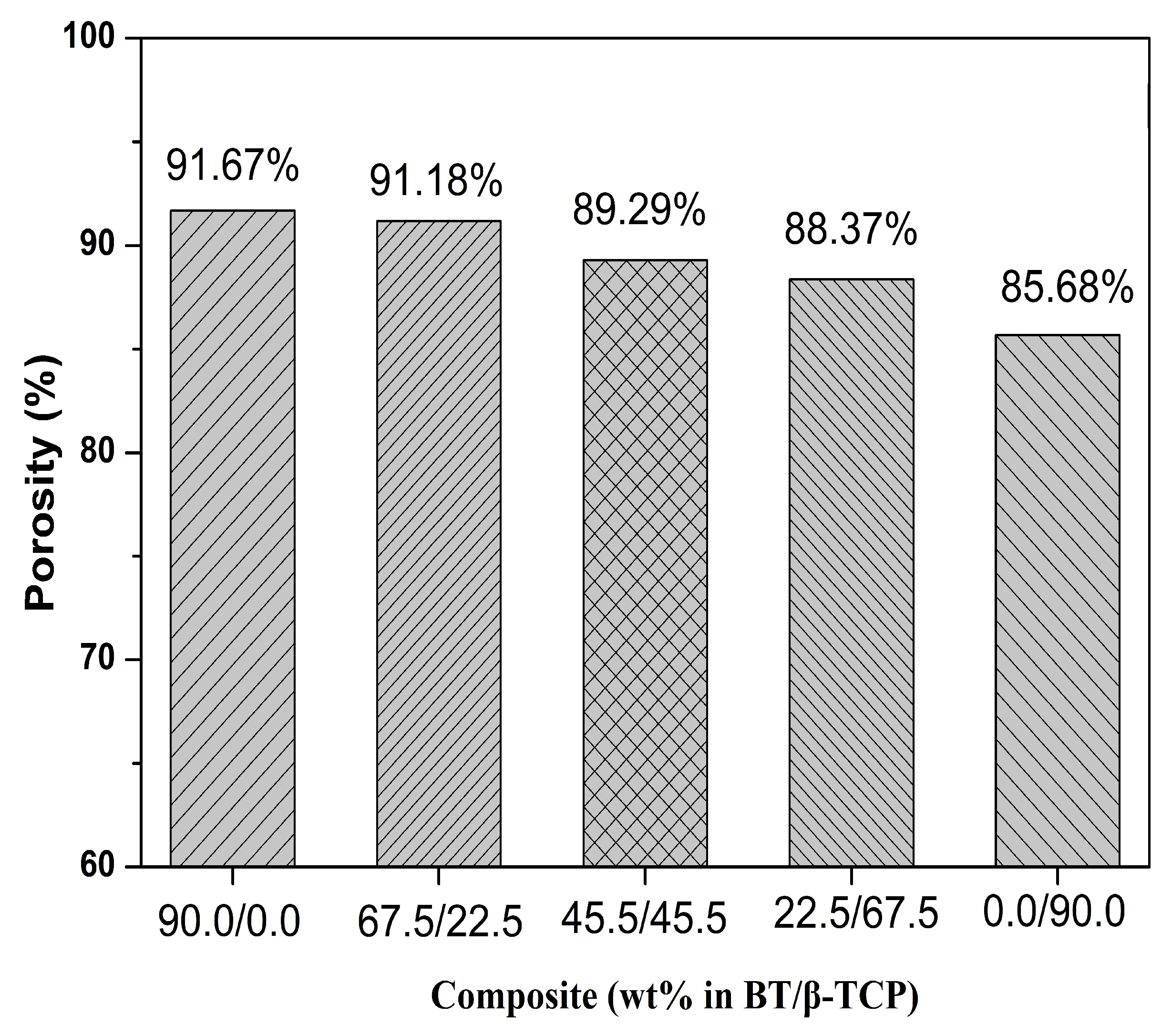
2.5. Porosity Measurement

Porosity measurement was performed by using a mercury porosimeter from the AutoPore IV9520 machine (Micromeritics, Norcross, GA, USA), for each scaffold has been shaped to 1 × 1 × 1 cm3 with a pressure measurement range from 0.1 to 60,000 psia.

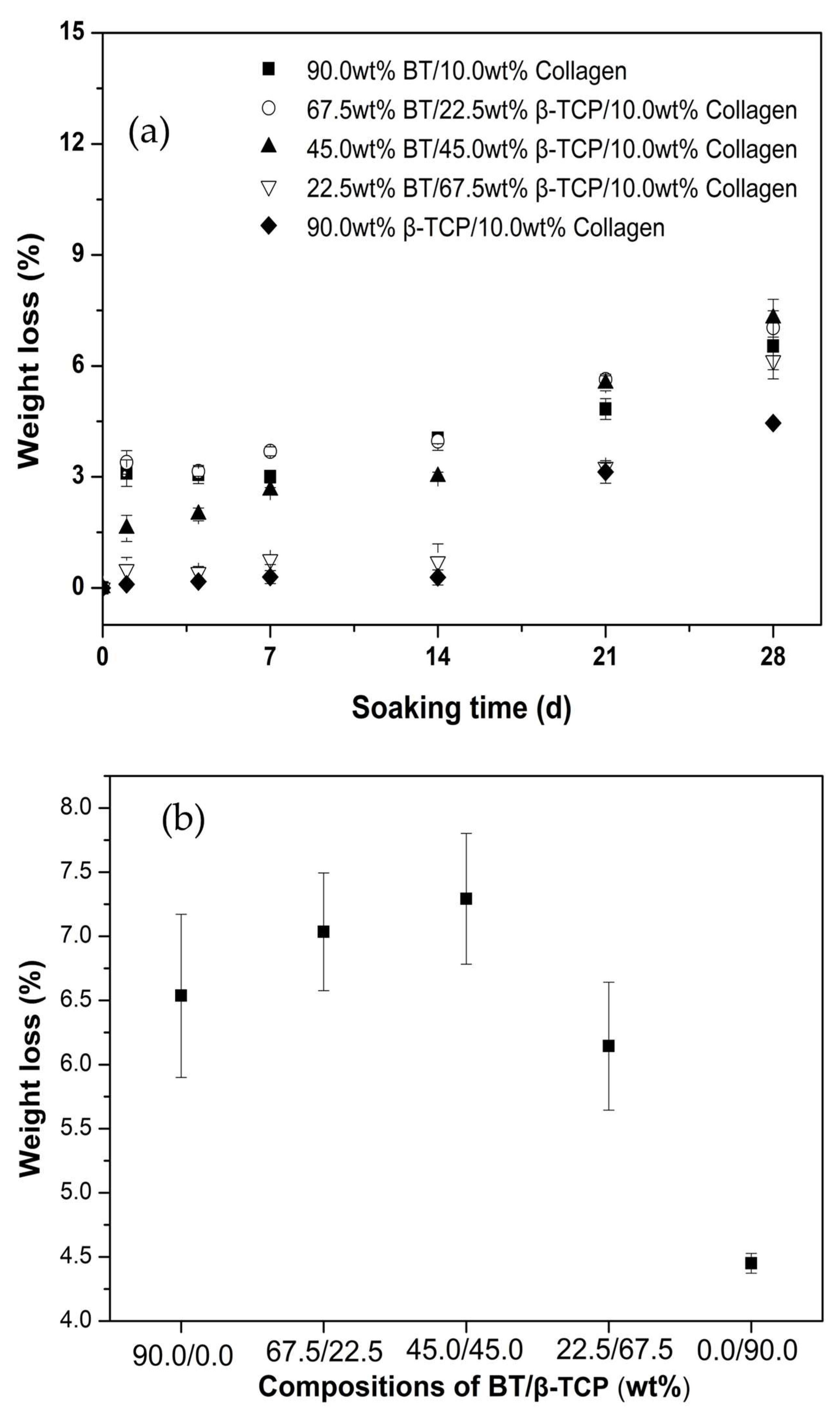
2.6. In Vitro Biodegradation

According to the ISO 10993-14 standard protocol [16], the biodegradability of all composite scaffolds was examined by immersing the specimens in the simulated body fluid (SBF) solution synthesized based on Kokubo et al. [17,18]. The 1 × 1 × 1 cm3-sized specimens were soaked in a 5 mL SBF solution. The pH of the solution was controlled at 7, while the ambient temperature was maintained at 37 °C for 28 d. The biodegradation condition of the scaffold was 0, 4, 7, 14, 21, and 28 d. To observe the weight loss, the degradation rate was calculated using the following equation:
Degradation   rate   ( % ) = W 0 W t   W 0 × 100
where W0 and Wt were represented as the initial weight and the incubated weight of the scaffold, respectively.

2.7. In Vitro Cytotoxicity

The MTT assay was used to assess cytotoxicity as a final step. All scaffolds were evaluated using an osteoblast cell line derived from rodents (MC3T3-E1, ATCC CRL-2542, Virginia, USA). Minimum Essential Medium (MEM, Gibco, Thermo Fisher, Waltham, MA, USA) containing 10% Foetal Bovine Serum (FBS, Gibco, Thermo Fisher, Waltham, MA, USA) and 1% Penicillin-Streptomycin (Thermo Fisher, Waltham, MA, USA) was initially used to culture MC3T3-E1 cells. On 24-well plates, the cells were seeded at a density of 2 × 10⁴ cells/mL, and composite scaffolds were added to each well. After three days of incubation, 200 μL of MTT solutions were added and incubated in an incubator for 4 h at 37 °C, 95% humidity, and 5% CO2. The crystal formazan was subsequently dissolved in dimethyl sulfoxide (DMSO, Fisher Chemical, Waltham, MA, USA), and the optical density (OD) at 570 nm was determined using a microplate reader (Multiskan Go, Thermo Scientific, Waltham, MA, USA). The cell viability of each scaffold was compared to that of the control group.

3. Results

Figure 1 shows the SEM images of commercial barium titanate and SP-derived β-TCP. The observing images of the SEM show the particle distribution of commercial BT with a size of 0.95 ± 0.24 μm and β-TCP with a size of 1.30 ± 0.59 μm, respectively. However, the shape of both has a significant difference, as shown in the commercial BT, which has been observed as an irregular structure; otherwise, the SP-derived β-TCP has been observed as a regular spherical structure.
Figure 2 shows the XRD result of the sample. When we composed the composite scaffold, the diffraction peaks of barium titanate and β-TCP onto the polymer collagen could make the signal cladding that makes the signals of barium titanate and β-TCP less pronounced. However, the barium titanate crystal grain is more extensive than ꞵ-TCP and has a stronger diffraction signal. The phase of β-TCP has been observed, and the peak has been noted by JCPDS 09-0169, which showed a whitlockite structure. However, the peak for BT (JCPDS 05-0626) could be observed clearly in all of the composite scaffolds, except the sample of 90.0 wt% β-TCP/10.0 wt% collagen composite scaffold; in brief, the particle size and properties of the powder may affect the macroscopic morphology of the composite scaffold structure. This experiment defines that the crystal size of ꞵ-TCP dominates the high concentration of ꞵ-TCP, and the BT signals have the same behavior applied to that.
We observed the surface of the porous structure and the porous alignment of the composite scaffold using an SEM. Based on the surface structure, there were alterations, particularly in the porosity. Approximately 90.0 wt% BT/10.0 wt% collagen, 67.5 wt% BT/22.5 wt% β-TCP/10.0 wt% collagen, 45.0 wt% BT/45.0 wt% β-TCP/10.0 wt% collagen, 22.5 wt% BT/67.5 wt% β-TCP/10.0 wt% collagen, and 90.0 wt% β-TCP/10.0 wt% collagen were the sample designations. Micrographs of composites (Figure 3) were then observed, and typically, the collagen from the freeze-drying process has been revealed and observed [13]. In the shown images of Figure 3, the pore distribution of the samples has been evaluated, which is sequentially described as 30.25 ± 11.28 µm, 28.25 ± 10.05 µm, 25.32 ± 8.05 µm, 17.38 ± 9.60 µm, and 15.25 ± 11.28 µm, respectively, where we put an order for pore size distribution as 90.0 wt% BT/10.0 wt% collagen > 67.5 wt% BT/22.5 wt% β-TCP/10.0 wt% collagen > 45.0 wt% BT/45.0 wt% β-TCP/10.0 wt% collagen > 22.5 wt% BT/67.5 wt% β-TCP/10.0 wt% collagen > 90.0 wt% β-TCP/10.0 wt%. In advance, the result shows that the addition of β-TCP particles will significantly decrease the average pore size. The other observation also showed the β-TCP particle around ~1 µm (as seen in Figure 3), similar to the SEM observation in Figure 1b; in another way, the size of BT particles has a homogenous size lower than ~1 µm, which means the distribution of the BT will cover more pore size compared to β-TCP, and another thing is that due to the irregular shape of commercial BT, the porosity of collagen composite has been increased significantly. In the end, the SEM images have briefly indicated that the porous collagen scaffolds with BT and β-TCP variations have been successfully synthesized.
The stress-strain test provided a toughness characterization of the composite samples. Figure 4a shows that the stress-strain curves are correlated to the modulus diagram. However, all the collagen scaffolds have similarities to staying at 80% of total strains, which were different BT and β-TCP ratios, making different results for the compressive modulus. As a result, all significant effects of BT and β-TCP have an impact on the stress and strain results. Based on that, the maximum stress of composite scaffolds was followed by 0.5 MPa for the 45.0 wt% BT/45.0 wt% β-TCP/10.0 wt% collagen composite sample, followed by 90.0 wt% β-TCP/10.0 wt% collagen with 0.48 MPa, 0.45 MPa for the 22.5 wt% BT/67.5 wt% β-TCP/10.0 wt% collagen, 0.3 MPa for the 67.5 wt% BT/22.5 wt% β-TCP/10.0 wt% collagen, and 0.15 MPa for the 90.0 wt% BT/10.0 wt% collagen for the condition of 80% strains. In the end, the compressive moduli have been observed, as shown in Figure 4b, which was calculated from the 10% of the strain with the linear slopes of the stress and strain curves. Based on the result, the values of compressive modulus of all the samples were evaluated as 1.23 Mpa for the 90.0 wt% BT/10.0 wt% collagen, 1.98 Mpa for the 67.5 wt% BT/22.5 wt% β-TCP/10.0 wt% collagen, 2.21 MPa for the 45.0 wt% BT/45.0 wt% β-TCP/10.0 wt% collagen, 1.94 MPa for the 22.5 wt% BT/67.5 wt% β-TCP/10.0 wt% collagen, and 2.09 MPa for the 90.0 wt% β-TCP/10.0 wt% collagen, which makes the 45.0 wt% BT/45.0 wt% β-TCP/10.0 wt% collagen have achieved significant value compared to all of those composite scaffolds. Ultimately, the composite scaffolds’ dependent structure will determine the substantial value of the compressive modulus.
Figure 5 represents the porosity percentage (%) from the porosimeter; the pore percentage for all scaffold samples was a sufficient amount of porosity to be applied for implant applications, with an average porosity of around 85 ± 5%. As we can see, additional β-TCP was decreasing the total porosity, which made the trend of adding β-TCP decrease a significant amount of porosity in the scaffold composite samples.
Figure 6a,b showed in vitro biodegradation test of all the composite scaffolds where all of those samples were immersed in the SBF solution (pH of 7) in the camber with a temperature of 37 °C on the shaking condition to mimic our body condition as followed by the international standard of ISO 10993-14 [16], on Figure 6a as shown the degradation behavior of all the composite scaffolds, as we can see the significant degradation appears after 14 d, which the value of degradation weight loss is about 3% except for the 90.0 wt% β-TCP/10.0 wt% collagen, in another hand the value of the degradation was related to the total amount of the BT percentage, which BT was given a more beneficial thing on the degradation rate. After immersing all the composite scaffolds, the value has increased to nearly 8% in 28 d, except for the 90.0 wt% β-TCP/10.0 wt% collagen. Based on Figure 6b, the weight loss has been observed highly on the composition BT/β-TCP of 45.0/45.0 wt%, confirming the piezoelectric effect of the BT has been affected by the influences of the degradation rate that have previously been observed by E. Fukada [9].
Lastly, to ensure the composite scaffolds were non-toxic to our bodies, MTT assays were performed to determine the cell viability of the collagen scaffolds. The test was performed by osteoblast MC3T3-E1 cells, which have generally been used to test the toxicity of composite scaffolds. The graph (Figure 7) showed that the samples’ composite scaffold ranged from about 100 ± 5.2% to 104 ± 5.5%, respectively. The composite scaffolds have been tested as non-toxic for the MC3T3-E1 cell, and based on statistical analysis, the sample that can be performed on the MC3T3-E1 cell could be categorized as safe to use in our body as an implant. In brief, the result showed that all those composite scaffolds that performed better than 70% had been indicated to have no cytotoxic effect, respectively.

4. Discussion

First, all scaffolds’ phase composition and pore structure are discussed below. Regarding phase composition, the XRD patterns confirmed the absence of any impurity phase, indicating that all composite scaffolds were effectively synthesized by freeze-drying. Physically simple to shape composite scaffold with injection molding and freeze-drying method as 1 × 1 × 1 cm3 test samples; samples can be used in various conventional applications, which will be sufficient to use as an implant on our hard tissue part. Moreover, according to the SEM images (Figure 3), adding β-TCP and BT particles to the collagen matrix will reduce the size of the scaffold’s pores. Since the pore size within the freeze-dried scaffold was determined by ice crystal formation and water sublimation [19], the pore size of the freeze-dried collagen scaffold would be affected by solubility, collagen concentration, freeze-drying rate, water absorption ability of the samples, and collagen-based slurry viscosity [16]. This study maintained constant values for solution solubility, collagen concentration, freeze-drying rate, and collagen-based slurry viscosity, and the remaining parameter was water absorption ability from the samples, which significantly depended on the materials. The freeze-drying method is a method that really depends on the water molecule absorbed on the grains; more absorbing water on the grain than low pore will form due to the water being formed as an ice filler on the scaffold structure [13]. From the references, both the BT and β-TCP are hydrophilic materials, and both of them easily absorb water molecules. However, based on the result, β-TCP could excess more water molecules compared to BT [20,21]; as a result, increasing β-TCP on the scaffold will remain low porosity and vice versa. Figure 8a showed a significant decrease due to the β-TCP composition of the scaffolds. There were closed-pore and continuous-pore structures for each scaffold structure; the pore structure type will determine the compressive strength value of each composite scaffold sample. The combination of BT and β-TCP will make a random macrostructure of the composite scaffold structure. The variety of open pores and closed pores would be beneficial for the cell to grow efficiently; under other conditions, the closed pores will give better mechanical test results, but that makes the cell have no space to grow, which makes the cell harder to attach to the composite scaffold implants. Conversely, the piezoelectric effect of BT particles has also played an essential role in biodegradation.
The recovery time depends on the composite scaffolds’ pore size, and based on the result, the BT concentration would increase the pore size diameter. Since the BT has a piezoelectric effect that would elevate the recovery time for the patient, that could be a good term for “kill two birds with one stone”. However, from the previous finding, the ideal porosity for facilitating cell growth was around 100 to 400 μm [22]. In that case, the normal collagen scaffold would give a good pore diameter, but since the collagen was too fragile, adding β-TCP and BT would be beneficial to facilitate mechanical strength. For example, Goodarzi et al. [23] found that increasing compressive strain with additional β-TCP in collagen composite scaffolds could give an additional 18% of the total average of composite scaffolds compared to pristine composite scaffolds. Based on the previous results, the variation of both BT, β-TCP, and collagen should be matched ideally. Based on the experiment, the result would be in perfect condition on between 50% of BT and 50% of β-TCP on the composite scaffold that is noted as 45.5 wt% BT/45.5 wt% β-TCP/10.0 wt% collagen (with 7.5 ± 0.5% absorption and 25.32 ± 8 µm pore size). However, all the composite scaffolds’ correlations related to compositions and pore size have been shown in Figure 8a; the increasing of β-TCP amounts has a significant decrease that has two major factors: First, the perfect round shape of β-TCP particles has to play a role in determining the total porosity of scaffold composite samples, and vice versa. The irregular shape of BT particles has been one of the causes of the increment in the total size of the porosity in the composite scaffold of samples; secondly, the water absorption on the β-TCP is greater than that of the BT, which makes the β-TCP swollen compared to the BT particles. This swelling effect of the β-TCP contributes to the pore size and porosity percentage shown in Figure 8b [24]. On the other hand, the pore size is also one of the causes of high in vitro biodegradation due to the high absorbance of composite scaffolds with the SBF solution (as seen in Figure 9), respectively.
Finally, cell viability was discussed. Based on Figure 6, the MTT assay showed that no cytotoxicity had been observed for all composite scaffolds. However, several studies reported that incorporating β-TCP and BT particles into collagen scaffolds facilitated cell growth and fast recovery [25]. Based on the result, the higher biodegradation percentage was in the sample of 45.0 wt% BT/45.0 wt% β-TCP/10.0 wt% collagen, which is near to 8%; on the other hand, from another previous result of Fei Lin et al. [26], their β-TCP/collagen could achieve around 10% weight loss in 28 days for the biodegradation test, which means our scaffold materials could still possibly compete with another result in terms of commercially used In conclusion, freeze-dried scaffold composite mesoporous BT/β-TCP/collagen would be beneficial and suitable for the reconstructive orthopedic field, respectively.

5. Conclusions

In this study, we synthesized a composite scaffold sample using a freeze-drying technique with the various BT, β-TCP on the 10% collagen concentrations (90.0 wt% BT/10.0 wt% collagen, 67.5 wt% BT/22.5 wt% β-TCP/10.0 wt% collagen, 45.0 wt% BT/45.0 wt% β-TCP/10.0 wt% collagen, 22.5 wt% BT/67.5 wt% β-TCP/10.0 wt% collagen, and 90.0 wt% β-TCP/10.0 wt% collagen composites), the physical observation of scaffold composite after freeze-drying, the phase composition, surface morphology, and mechanical properties were characterized using XRD, SEM, and tensile strength test, respectively. Based on the figure of composite scaffold microstructures, no prominent color changed, and the composite scaffold microstructures have a similar appearance to the naked eye. On the other hand, the XRD observed the structure of the β-TCP in the whitlockite structure. Based on SEM figures, all the BT/β-TCP scaffold composite microstructures showed different pore counts. The pore size value ranged between 30.25 ± 11.28 and 15.25 ± 11.28 μm, indicating a successful synthesis of the mesoporous structure of the scaffold composite. The tensile strength test shows the different capabilities of holding each sample’s compressive strength due to the charge affinity of BT and β-TCP. The piezoelectric effect after adding BT to the composite scaffolds has significantly improved the pore-type difference and the biodegradation rate due to the piezoelectric effect.

Author Contributions

Conceptualization, S.-J.S.; methodology, T.-W.W.; formal analysis, B.B.A., H.S.N., and A.N.; data curation, T.-W.W.; writing—original draft preparation, D.F.A.P.; writing—review and editing, D.F.A.P.; supervision, S.-J.S.; funding acquisition, S.-J.S., and T.M. All authors have read and agreed to the published version of this manuscript.

Funding

The authors acknowledge the financial support from the Taiwan National Science and Technology Council (grant numbers of NSTC 111-2221-E-011-117- and NSTC 112-2221-E-011-040-).

Institutional Review Board Statement

Not applicable.

Informed Consent Statement

Not applicable.

Data Availability Statement

The data presented in this study are available on request from the corresponding author.

Conflicts of Interest

The authors declare no conflict of interest.

References

  1. Boyce, S.T.; Lalley, A.L. Tissue engineering of skin and regenerative medicine for wound care. Burn. Trauma 2018, 6, 4. [Google Scholar] [CrossRef] [PubMed]
  2. Visser, R.; Rico-Llanos, G.A.; Pulkkinen, H.; Becerra, J. Peptides for bone tissue engineering. J. Control. Release 2016, 244, 122–135. [Google Scholar] [CrossRef] [PubMed]
  3. Yamada, S.; Yamamoto, K.; Ikeda, T.; Yanagiguchi, K.; Hayashi, Y. Potency of fish collagen as a scaffold for regenerative medicine. BioMed Res. Int. 2014, 2014, 302932. [Google Scholar] [CrossRef] [PubMed]
  4. Karageorgiou, V.; Kaplan, D. Porosity of 3D biomaterial scaffolds and osteogenesis. Biomaterials 2005, 26, 5474–5491. [Google Scholar] [CrossRef] [PubMed]
  5. Clarke, K.; Graves, S.; Wong, A.; Triffitt, J.; Francis, M.; Czernuszka, J. Investigation into the formation and mechanical properties of a bioactive material based on collagen and calcium phosphate. J. Mater. Sci. Mater. Med. 1993, 4, 107–110. [Google Scholar] [CrossRef]
  6. Dong, C.; Lv, Y. Application of collagen scaffold in tissue engineering: Recent advances and new perspectives. Polymers 2016, 8, 42. [Google Scholar] [CrossRef]
  7. Aichelmann-Reidy, M.E.; Yukna, R.A. Bone replacement grafts: The bone substitutes. Dent. Clin. N. Am. 1998, 42, 491–503. [Google Scholar] [CrossRef]
  8. Bissada, N.F.; Hangorsky, U. Alveolar bone induction: Alloplasts. Dent. Clin. N. Am. 1980, 24, 739–749. [Google Scholar] [CrossRef] [PubMed]
  9. Fukada, E.; Yasuda, I. On the piezoelectric effect of bone. J. Phys. Soc. Jpn. 1957, 12, 1158–1162. [Google Scholar] [CrossRef]
  10. Liang, D.; Hsiao, B.S.; Chu, B. Functional electrospun nanofibrous scaffolds for biomedical applications. Adv. Drug Deliv. Rev. 2007, 59, 1392–1412. [Google Scholar] [CrossRef]
  11. Keshaw, H.; Thapar, N.; Burns, A.J.; Mordan, N.; Knowles, J.C.; Forbes, A.; Day, R.M. Microporous collagen spheres produced via thermally induced phase separation for tissue regeneration. Acta Biomater. 2010, 6, 1158–1166. [Google Scholar] [CrossRef] [PubMed]
  12. Xie, Y.; Lee, K.; Wang, X.; Yoshitomi, T.; Kawazoe, N.; Yang, Y.; Chen, G. Interconnected collagen porous scaffolds prepared with sacrificial PLGA sponge templates for cartilage tissue engineering. J. Mater. Chem. B 2021, 9, 8491–8500. [Google Scholar] [CrossRef]
  13. Animut, T.Y.; Ningsih, H.S.; Shih, H.-H.; Wu, M.-H.; Shih, S.-J. Effect of Calcium Silicate and β-Tricalcium Phosphate Reinforcement on the Mechanical–Biological Properties of Freeze-Dried Collagen Composite Scaffolds for Bone Tissue Engineering Applications. Ceramics 2023, 6, 548–560. [Google Scholar] [CrossRef]
  14. Mabrouk, M.; Beherei, H.H.; Das, D.B. Recent progress in the fabrication techniques of 3D scaffolds for tissue engineering. Mater. Sci. Eng. C 2020, 110, 110716. [Google Scholar] [CrossRef]
  15. Finoli, A.; Ostrowski, N.; Schmelzer, E.; Nettleship, I.; Gerlach, J. Multiscale porous ceramic scaffolds for in vitro culturing of primary human cells. Adv. Appl. Ceram. 2012, 111, 262–268. [Google Scholar] [CrossRef]
  16. Wallin, R.F.; Arscott, E. A practical guide to ISO 10993-5: Cytotoxicity. Med. Device Diagn. Ind. 1998, 20, 96–98. [Google Scholar]
  17. Kokubo, T.; Takadama, H. How useful is SBF in predicting in vivo bone bioactivity? Biomaterials 2006, 27, 2907–2915. [Google Scholar] [CrossRef]
  18. Kokubo, T.; Takadama, H. Simulated body fluid (SBF) as a standard tool to test the bioactivity of implants. In Handbook of Biomineralization: Biological Aspects and Structure Formation; Wiley: Hoboken, NJ, USA, 2007; pp. 97–109. [Google Scholar]
  19. Liapis, A.I.; Bruttini, R. Freeze drying. In Handbook of Industrial Drying; CRC Press: Boca Raton, FL, USA, 2020; pp. 309–343. [Google Scholar]
  20. Spaniol, K.; Caldas, S.; Peres, A.; Dos Santos, E.; Acchar, W. β-TCP/PVA sheets crosslinked with citric acid produced via aqueous tape casting for bone regeneration. Ceram. Int. 2019, 45, 12417–12422. [Google Scholar] [CrossRef]
  21. Vázquez Lasa, B.; Pau Ginebra, M.; Gil, X.; Antón Planell, J.; San Román, J. Acrylic bone cements modified with b-TCP particles encapsulated with poly (ethylene glycol). Biomaterials 2005, 26, 4309–4316. [Google Scholar] [CrossRef]
  22. Perez, R.A.; Mestres, G. Role of pore size and morphology in musculo-skeletal tissue regeneration. Mater. Sci. Eng. C 2016, 61, 922–939. [Google Scholar] [CrossRef]
  23. Goodarzi, H.; Hashemi-Najafabadi, S.; Baheiraei, N.; Bagheri, F. Preparation and characterization of nanocomposite scaffolds (collagen/β-TCP/SrO) for bone tissue engineering. Tissue Eng. Regen. Med. 2019, 16, 237–251. [Google Scholar] [CrossRef] [PubMed]
  24. Lau, C.; Al Qaysi, M.; Owji, N.; Bayazit, M.; Xie, J.; Knowles, J.; Tang, J. Advanced biocomposites of poly (glycerol sebacate) and β-tricalcium phosphate by in situ microwave synthesis for bioapplication. Mater. Today Adv. 2020, 5, 100023. [Google Scholar] [CrossRef]
  25. Busuioc, C.; Voicu, G.; Jinga, S.-I.; Mitran, V.; Cimpean, A. The influence of barium titanate on the biological properties of collagen-hydroxiapatite composite scaffolds. Mater. Lett. 2019, 253, 317–322. [Google Scholar] [CrossRef]
  26. Lin, F.; Wang, X.; Wang, Y.; Yang, Y.; Li, Y. Preparation and biocompatibility of electrospinning PDLLA/β-TCP/collagen for peripheral nerve regeneration. RSC Adv. 2017, 7, 41593–41602. [Google Scholar] [CrossRef]
Figure 1. SEM images of (a) commercial barium titanate and (b) SP-derived ꞵ-TCP particles.
Ceramics 06 00132 g001
Figure 2. XRD patterns of various compositions for the BT/β-TCP/collagen composite scaffolds.
Ceramics 06 00132 g002
Figure 3. SEM images of (a) 90.0 wt% BT/10.0 wt% collagen, (b) 67.5 wt% BT/22.5 wt% β-TCP/10.0 wt% collagen, (c) 45.0 wt% BT/45.0 wt% β-TCP/10.0 wt% collagen, (d) 22.5 wt% BT/67.5 wt% β-TCP/10.0 wt% collagen, and (e) 90.0 wt% β-TCP/10.0 wt% collagen composite scaffolds.
Ceramics 06 00132 g003
Figure 4. (a) Stress-strain curve of various compositions of BT/β-TCP/collagen composite scaffolds under continuous load and (b) compressive modulus of various compositions of BT/β-TCP/collagen composite scaffolds.
Ceramics 06 00132 g004
Figure 5. The porosity measurement of BT/β-TCP/collagen composite scaffolds with different wt%.
Ceramics 06 00132 g005
Figure 6. (a) In vitro biodegradation of various compositions of BT/β-TCP/collagen composite scaffolds after soaking in SBF solution from 0 to 28 d and (b) weight loss after 28 days of biodegradation of all composites.
Ceramics 06 00132 g006
Figure 7. Cell viability of MC3T3-E1 cells in contact with BT/β-TCP/collagen composite scaffolds by MTT assay (* p < 0.05 as compared to control).
Ceramics 06 00132 g007
Figure 8. Correlation between (a) β-TCP concentration and pore size of composite scaffolds and (b) porosity percentage and pore size of composite scaffolds.
Ceramics 06 00132 g008
Figure 9. Correlation between weight loss and pore size of composite scaffolds.
Ceramics 06 00132 g009
Disclaimer/Publisher’s Note: The statements, opinions and data contained in all publications are solely those of the individual author(s) and contributor(s) and not of MDPI and/or the editor(s). MDPI and/or the editor(s) disclaim responsibility for any injury to people or property resulting from any ideas, methods, instructions or products referred to in the content.

