Review

High-Performance Ceramics in Musculoskeletal Surgery: Current Use and Future Perspectives

1
Department for Trauma and Reconstructive Surgery, BG Hospital Bergmannstrost, 06112 Halle (Saale), Germany
2
Department for Trauma and Reconstructive Surgery, University Hospital Halle, 06120 Halle (Saale), Germany
3
Department for Orthopaedics, Trauma and Reconstructive Surgery, RWTH Aachen University Hospital, 52074 Aachen, Germany
4
Department of Orthopaedic Surgery, Klinikum Wels-Grieskirchen, 4600 Wels, Austria
5
Department of Oral, Maxillofacial and Plastic Facial Surgery, University Hospital Aachen, 52074 Aachen, Germany
6
Department for Orthopaedic and Trauma Surgery, Academic Hospital Bolzano, Teaching Hospital of the Paracelsus Medical University, 5110 Oberndorf, Italy
*
Author to whom correspondence should be addressed.
Ceramics 2024, 7(1), 310-328; https://doi.org/10.3390/ceramics7010020
Submission received: 28 December 2023 / Revised: 12 February 2024 / Accepted: 19 February 2024 / Published: 23 February 2024

Abstract

:
Osteoarthritis (OA) is a prevalent disease among the elderly population, necessitating effective treatment options. Total joint arthroplasty (TJA) is a reliable surgical procedure that has shown good long-term clinical outcomes for OA. However, certain challenges, such as implant failure caused by particle-induced aseptic loosening or hypersensitivity to metal ions, remain unresolved in TJA. High-performance ceramic implants have emerged as a promising solution to address these persistent implant-related issues. This review article provides an overview of the composition and characteristics of ceramics used in TJA, highlighting their potential advantages and associated risks. While ceramic implants have demonstrated excellent performance in vivo for hip and knee arthroplasty, their bioinert behaviour is still considered a crucial factor regarding cementless options. Therefore, novel methods are investigated that seem to be able to combine the benefits of ceramic materials with an excellent osseointegration behaviour, which makes ceramics as implant materials an even stronger option for future applications.

1. Introduction

Osteoarthritis (OA) is common among elderlies, affecting predominantly the knee and hip joints [1,2,3,4,5]. Total joint arthroplasty (TJA) is the gold standard surgical intervention when conservative treatments fail or are impossible. Musculoskeletal surgery (orthopaedic and trauma surgery) is highly dependent on medical implants. By implanting, e.g., industrially prefabricated endoprostheses, TJA can effectively alleviate joint pain, correct deformities, and restore joint function [6]. TJA procedures, such as total knee arthroplasty (TKA) and total hip arthroplasty (THA), are the most frequently performed orthopaedic interventions, with 173,625 primary cases of TKA and 227,851 primary cases of THA recorded in Germany alone in the year 2020 [7]. Additionally, about 25% of the number of the primary cases came on top because of revision interventions [7]. These procedures have demonstrated exceptional success rates, with implant survival exceeding 80% after 15 years, e.g., for hip implants [8].
For bearing couple in TKA/THA and TJA, respectively, the most frequently used combination consists of cobalt-chrome (CoCr) or titanium (Ti) metal alloys articulating against ultrahigh-molecular-weight polyethylene (UHMWPE) and its variations [9,10,11,12,13,14,15]. Cross-linked polyethylene (XLPE) and highly cross-linked polyethylene (HXLPE), respectively, were introduced in the late 1990s to reduce wear and debris-induced osteolysis following TJA [16]. The first generation of XLPE/HXLPE is developed by exposing UHMWPE to gamma radiation, which breaks up intramolecular bonds and produces free radicals that promote cross-linking across multiple polymer chains [15,16]. This leads to increased density and improved wear characteristics [16]. The second generation of HXLPE underwent a modification by stabilising the residual-free radicals with vitamin E, a well-known chain-breaking antioxidant [15]. Vitamin E hinders the peroxidation of lipids in the body [15,17].
While most patients can be successfully treated with these standard joint implants made of CoCr or Ti alloys, complications such as aseptic loosening and implant intolerance remain significant challenges [10,18,19,20]. Approximately 25% of THA and TKA are revised in the long run due to failure of the artificial joints by aseptic loosening [10,18,19]. Aseptic loosening is a common complication of TJA, where the implant becomes loose due to the breakdown of the bond between the implant and bone, without the presence of an infection [21]. The causes of aseptic loosening can be attributed to several factors, e.g., wear and tear that occurs with joint use [21]. This can cause small particles of the implant material to break off and accumulate in the surrounding tissues and can trigger an inflammatory response that leads to bone loss and loosening of the implant. Particle-induced aseptic loosening is a major reason, especially in younger active (=higher wear rate?) patients [6]. It is usually caused by particle debris released by wear, which can still be increased by non-optimal positioning of the endoprosthesis components [21]. Also, the design and materials used in TJA implants are important in their longevity. Furthermore, patient factors such as age, weight, and activity level can also contribute to implant loosening [22]. Patients who are overweight or have a higher level of activity may put more stress on their (artificial) joints, causing the implant to loosen more quickly [22,23]. Additionally, the skill and technique of the surgeon performing the TJA intervention can also affect the success of the procedure [24]. Poor alignment or positioning of the implant during surgery can lead to uneven stress on the implant and cause it to loosen over time. Finally, certain implant-related factors, such as corrosion, implant mismatch, and implant size, can also contribute to aseptic loosening [21].
Another reason for implant failure is implant intolerance. This condition describes a variety of complaints related to allergy and unspecific inflammatory reactions to metal particles released by the implants. Allergies, e.g., chromium, cobalt, and nickel, have been found to cause implant failure [6]. This can lead to persistent pain, functional limitations, bone loss, and aseptic loosening. Of note, the reported incidence of metal allergies as a potential factor causing implant failure is on the rise [25].
Ceramic implants are already widely used, and their applications have increased in the medical field in the 20th century due to processing technology advancements. They have been used as an alternative to metal-on-polyethylene in TJA since the 1970s [9]. Notably in THA, the use of a ceramic femoral head is now considered a standard of care, and especially in younger patients, the implanted cup/insert is also made of ceramics [18]. The first ceramic knee prosthesis was implanted in 1972 by G. Langer at the Orthopaedic Clinic at the University of Jena [8,26].
Ceramic materials are in general defined as inorganic, non-metallic materials, which consist of metallic and non-metallic elements; they are most frequently oxides, phosphates, nitrides, and carbides [27]. Ceramics as biomaterials have advantageous characteristics in comparison to traditional metal-bearing components like higher hardness, resulting in reduced wear rates, and a negligible amount of ion release [27]. First attempts at the use of ceramics in arthroplasty were disappointing, for example, due to the high brittleness of the components, but following progress in endoprosthesis design and optimisation of ceramics, has turned these materials into a promising solution. While already established in THA, TKA ceramic components remain presently an option exclusively for patients with confirmed allergies to metal [28,29].
Ceramics that are used in the context of osseointegration and musculoskeletal restoration are in general called bio ceramics [30]. Bio ceramics are often used in orthopaedic surgery as articulation components in THA or TKA to treat OA or as bioactive coatings to enhance osseointegration in vivo. To date, to our knowledge, there is only one full ceramic TKA implant on the market (BPK-S INTEGRATION Ceramic, Peter Brehm GmbH, see Figure 1) [31].
This review intends to give an overview of high-performance (bio) ceramics in musculoskeletal surgery, especially in orthopaedics with a focus on TKA and THA, and it describes their properties to osseointegration based on the current state of the art. The intention is also to illuminate the interface between the surface of the ceramic implant and the bone in that way to discuss novel methods/possibilities for (better) osseointegration of ceramic implants.

2. General Definition and Classification of High-Performance Ceramics

High-performance ceramics are inorganic non-metallic materials [32,33]. They are formed from fine-grained raw material, and they obtain their final properties by a sintering process (>800 °C) [33]. Ceramics include materials based on metal oxides. The final microstructure of the ceramics depends on many factors including the used primary granulate, the applied thermal process, the maximum temperature reached, and the duration of the thermal steps [32].
According to Hamadouche and Sedel, in the medical context, there exists five types of ceramics: glasses, plasma-sprayed polycrystalline ceramics, vitrified ceramics, solid-state sintered ceramics, and polycrystalline glass ceramics [32,34]. Additionally, different factors such as the purity of the powder, the size and distribution of the grains, and the porosity are important issues in the context of the resulting mechanical and biological properties [32,34]. Ceramics possess excellent chemical and mechanical stability, corrosion resistance, and tribological properties [20,34,35].
For the sake of completeness, in the context of medical ceramics, three subclasses are classified: bioactive, bioinert, and bioresorbable ceramics [34]. The bioactivity of a material can be defined as its ability to bond biologically to native tissue [32]. Bioactive ceramics are employed as coatings to enhance the fixation of a device or as bone graft substitutes because of their osteoconductive properties [32,36]. Bioactive materials have a positive effect on living tissues, and they can induce a response that helps in the regeneration, repair, and reconstruction of body tissues [34]. Bioinert materials come with stable physicochemical properties and make good biocompatibility with native tissues [34]. These materials do not cause any substantial physiological reaction or immunological rejection [34]. An inert ceramic elicits at most a minor fibrous reaction [32]. In clinical practice, inert ceramics are used as bearing surfaces, e.g., in TJA, because of their exceptional resistance to wear and their tribological properties [32,37]. Bioresorbable or biodegradable materials completely degrade in the body while being replaced by native tissue [38]. The chemical structure of these materials is broken down by the body and removed by the surrounding fluid [39]. Bioresorbable ceramics are prone to react and break down (rapidly) when coming in contact with body tissue fluid [34]. They do not require a second surgical intervention for their removal [40]. These materials are completely absorbed by the body tissues, and the resulting chemicals from the ceramic resorption should be able to be treated by the normal metabolic pathways of the body without producing any toxic effect [34].
In the following, the focus will be on bioactive and bioinert ceramics.
In the context of this classification, the functionalisation of bioinert ceramics is a research field with a high future potential. To functionalise bioinert materials in the way that they show osseointegration behaviour and, therefore, do not need bone cement as adhesive would be a step forward in implant technology.

3. Ceramics in Musculoskeletal Surgery

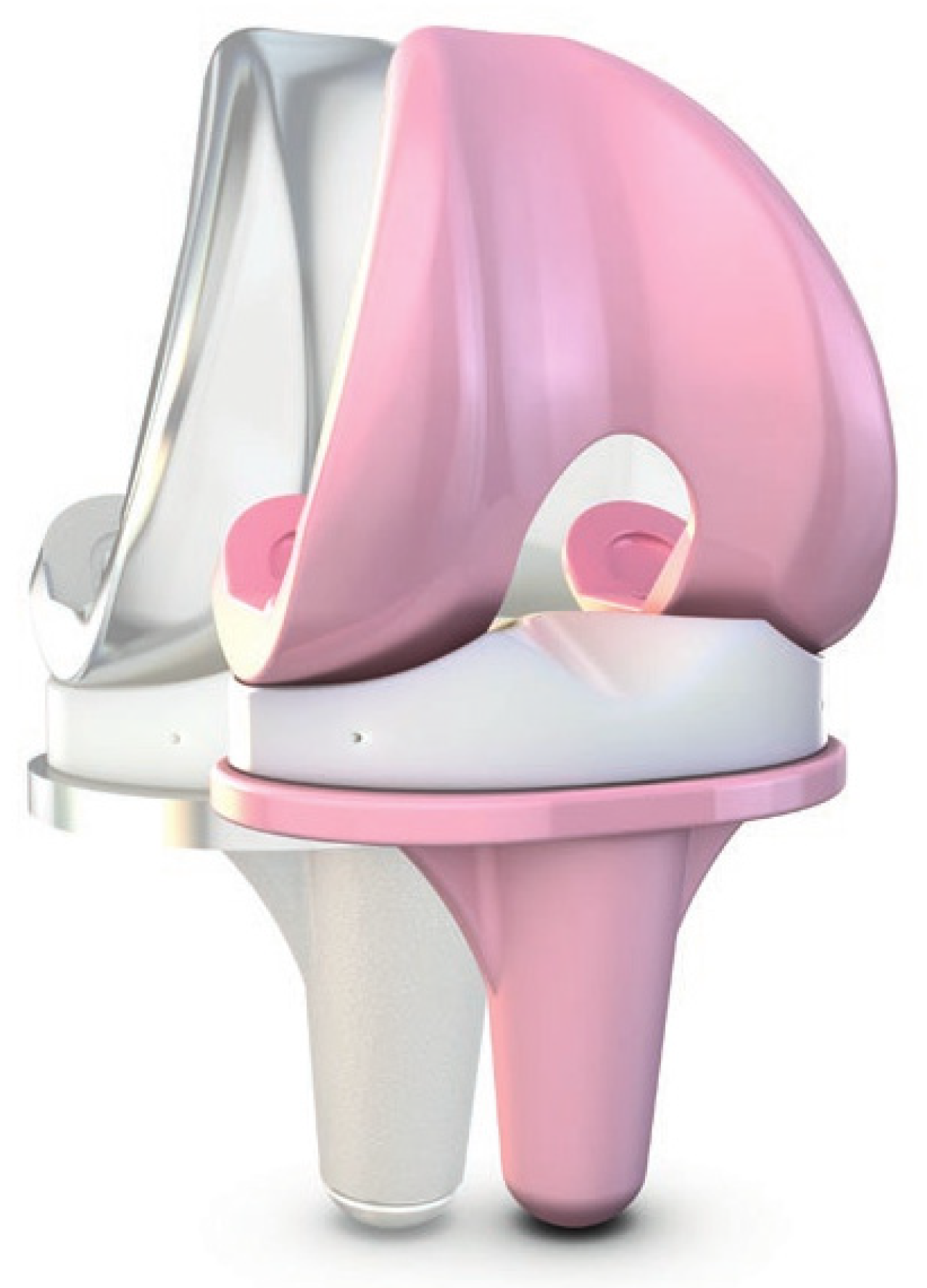
3.1. Full-Thickness Ceramic Implant Components

3.1.1. General Introduction to Oxide Ceramics

Alumina and zirconia are traditional bioinert materials that have been used for musculoskeletal applications since 1960 [34,35].
In the context of the implantation of (ceramic) endoprostheses, a clarification of the terms osseointegration, cell adhesion, and osteoconduction is important to understand the interaction of the implant surface and the surrounding soft tissue.
Osseointegration was first defined by Albrektsson as an existing direct contact at the light microscope level between living bone and the implant [41]. James defined osseointegration as a direct structural and functional link between organised living bone and the surface of a load-bearing implant [42]. Osseointegration is also histologically defined as the direct anchorage of an implant by the formation of bony tissue around the implant without the growth of fibrous tissue at the bone–implant interface [42,43]. A more biomechanically oriented definition of osseointegration has been suggested as a process whereby clinically asymptomatic rigid fixation of alloplastic materials is achieved and maintained in bone during functional loading [43,44]. The first step, after insertion and fixing of cementless implants, includes a hematoma formation and mesenchymal tissue development, woven bone formation through the intramembranous pathway, and lamellar bone formation on the spicules of woven bone [45]. Blood is the first biological component that comes into contact with an endosseous implant [45]. Getting in contact with the implant surface, the blood cells will be entrapped there and then activated and will release cytokines and other soluble, growth, and differentiation factors [45,46,47]. The cell adhesion of osteoblasts is then the initial step of osseointegration in response to bone material implants [48]. It is an important event in initiating and regulating cell survival, migration, recruitment, and osteogenic differentiation [48], which are important events during early osseointegration.
Cell adhesion refers to the ability of cells to bind to one another or extracellular matrix (ECM) components in their environment [49]. This process is critical for maintaining the structure and function of tissues and organs in multicellular organisms [50]. Cell adhesion molecules (CAMs) mediate the interactions between cells and their surroundings [51]. CAMs are transmembrane proteins that can either bind to other CAMs on adjacent cells or to ECM proteins such as collagen or laminin. There are several types of cell adhesion, including tight junctions, desmosomes, hemidesmosomes, and gap junctions [52,53].
Osteoconduction means that bone grows/binds on a surface. An osteoconductive material permits bone growth on its surface or down into pores, channels, or pipes [43]. Osteoconduction is a process of ingrowth of capillaries, perivascular tissue, and osteoprogenitor cells from a bony bed to the 3D structure of a porous implant [54].

3.1.2. Alumina (Al2O3)

Alumina ceramics are based on α-Al2O3. It is a highly stable metal oxide because of the ionic and covalent bond present between aluminium and oxygen [34,55]. The standard specifications of alumina for medical applications are suggested by the ASTM standard F603-12 [30] and the ISO 6474-1:2019—Implants for surgery—Ceramic materials—Part 1: Ceramic materials based on high-purity alumina [56].
Alumina ceramics are widely used in THA and TKA, although low fracture toughness has always been a concern [30]. Advancements in ceramic processing, along with better raw materials, have resulted in higher densification of alumina at low temperatures with finer grain sizes [30]. Furthermore, it has good corrosion resistance, low wear, and friction coefficient [55].

3.1.3. Zirconia (Y-TZP)

Another frequently used oxide ceramic is yttrium oxide-stabilised zirconia (Y-TZP). Y-TZP has found the most promising use in femoral head and cup implants in THA along with implants for TKA. The special thing about zirconium oxide (ZrO2) is its polymorphism, i.e., it occurs in three crystal modifications depending on the temperature: monoclinic below 1170 °C, tetragonal between 1170 °C and 2370 °C, and cubic above 2370 °C [57]. The increased mechanical properties of zirconia are the consequence of a phenomenon known as “transformation toughening” [58]. Zirconium oxide has good wear resistance and high fracture toughness. Compared to other high-performance ceramic materials, Y-TZP has the lowest thermal conductivity and highest strength and offers very good tribological properties [59].

3.1.4. Dispersion Ceramics

These dispersion ceramics are distinguished in those where the main phase is alumina (ZTA = zirconia-toughened alumina) and those where zirconia is the dominating phase (ATZ = alumina-toughened zirconia).
To further increase the strength of alumina, zirconia can be added: this particularly tough ceramic is known as ZTA. Through this process, material fracture toughness and strength can be significantly increased [60]. There are different variants with zirconium oxide contents of 5–20% with the following properties [61]. Here is one possible example of material performance:
  • Hardness HV10:            17 GPa;
  • Fracture toughness:         7 MPa*m1/2 (vs. Al2O3: 4.3 MPa*m1/2);
  • Compressive strength:       2600 MPa;
  • Four-point bending strength:    440–800 MPa.
The counterpart to ZTA is ATZ. Here, the properties of pure zirconia are positively influenced by the addition of alumina [61]. Here is also one possible example of the performance of ATZ:
  • Hardness HV10:            14 GPa;
  • Compressive strength:       2100 MPa;
  • Four-point bending strength:    820 MPa.
Here, different material test results are represented for ZTA and ATZ. The differences depend on the mixture of the materials itself. Both material types are available for biomedical applications. Table 1 provides additional orthopaedic biomaterials and their mechanical properties.

3.1.5. Pros

One advantage of ceramic bearings in TJA is the reduction in, e.g., polyethylene wear [63]. The reason for that is the lower coefficient of friction of ceramic surfaces. That means that ceramics will produce fewer (polyethylene) particles by abrasive wear than metal alloys [63]. In addition to their wear advantages, ceramics are biologically inert in bulk form, thermodynamically stable, and insoluble in aqueous environments, and ceramic particles are less (bio)reactive compared to polyethylene particles [63]. Ceramics have been shown to offer high immunological compatibility [31]. Stipulated, or shown further, advantages of such an implant are that it is/has
  • tissue friendly and very well-tolerated by the organism;
  • reduced risk of toxicity: compared to metal, ceramic particles are less cytotoxic to histiocytes and fibroblasts than metal particles at equivalent particle volumes [63];
  • lower biofilm formation.
The effect of wettability of ceramics based on the strong hydrogen bonds between ceramic and synovial fluid is a feature that hugely improves the lubrication of joints by a uniform distribution of the synovial film on articular surfaces [64]. Especially in THA, this feature is useful in causing gravitational effects. In this case, the gravitation increases the effect to leave articulating surfaces dry, and ceramics hydrophilic resists tendency [64].

3.1.6. Cons

A strong concern in the use of ceramics in TJA is that ceramic components may break. The construction of the ceramic implant is also often very complicated because of the material behaviour itself. The geometry of standard implants is often not ideal for ceramics, and they still have a high brittleness. There is no plastic deformation in contrast to ductile metallic or polymer materials and low tensile strength [6]. Therefore, stress concentration (for example, sharp edges) must be avoided during implantation, and the implant design must consider weak spots as subcritical crack growth is a known issue of ceramics [6]. That means the geometry of the implant must be adapted based on the general constraints of the used material. A surprising finding was that in the case of ceramic–ceramic articulations of THAs, ceramic wear particles could even be found in infra-diaphragmatic and para-iliac lymph nodes [63,65]. In a 13-year follow-up of almost six million ceramic hip implants (ceramic heads and balls), it was shown that the risk of implant failure related to fractures is extremely low with modern ceramics and implant designs [66].
However, osseointegration of ceramic surfaces remains a challenge. The first generation of TKAs with alumina-on-polyethylene articulations suffered from a high incidence of early loosening [63]. They consisted of cementless alumina components that relied on bone ingrowth for skeletal fixation [63].
One problem is that there is no direct bone–material interface created, and a soft tissue interlayer always shields the bone from the (ceramics) implant [27]. This biological shielding unfortunately leads to mechanical shielding, known to promote micro-motion and subsequent aseptic implant loosening [27].
Another form of shielding that has a major impact on ceramics as materials in orthopaedic implants is the so-called stress shielding. This occurs when an implanted medical device, such as an artificial joint or bone plate, bears a significant portion of the mechanical load that the surrounding bone tissue would normally experience. This phenomenon is often exacerbated by the extraordinarily high stiffness of certain materials, such as ceramics, used in implants. The huge stiffness mismatch between the implant and the natural bone leads to a transfer of load away from the bone, resulting in reduced mechanical stress on the bone tissue itself. However, this can have adverse effects on bone health, including bone resorption, weakened bone structure, and potential implant failure. Additionally, stress shielding disrupts the crucial process of natural bone remodelling, which relies on mechanical stimulation. Researchers are actively exploring alternative implant designs and materials that more closely match the mechanical properties of natural bone, aiming to mitigate stress shielding and promote healthier bone remodelling.

3.2. Ceramics as Coating or Finishing

In the field of ceramic coatings and finishings, nitride ceramics are often used as surface modifications of CoCr implants and offer significant benefits for orthopaedic implants, enhancing their performance and longevity. These most common modifications are the application of titanium nitride (TiN) or zirconium nitride (ZrN).
Nitride ceramic surface modifications offer notable benefits for patients who are sensitive or allergic to metals. By covering the metal surface with a ceramic layer, the critical contact between the metal of the implant and the patient’s tissues is minimised. Furthermore, nitride ceramic surface modifications offer superior corrosion resistance. As the ceramic layer acts as a protective barrier in both directions, it not only prevents the release of metal ions in the body of the patient but also protects the implant against corrosive substances present in the body, such as body fluids and salts. By preventing corrosion, the implant maintains its structural integrity over time, ensuring long-term functionality and reducing the risk of implant-related complications.
There are not only biological benefits, but also nitride ceramic coatings offer significant benefits in terms of wear resistance. The high hardness and low friction coefficients of nitrides such as TiN or ZrN reduce abrasive wear, extending the lifespan of orthopaedic implants. This translates to improved durability and reduced need for revision surgeries.
Another known surface-finishing process is the oxidisation of zirconium alloys. This modification has a significant advancement in orthopaedic implant materials. Its unique process results in a hard, wear-resistant surface, potentially extending the lifespan of implants beyond traditional materials. The smooth surface provides less friction, allowing for more natural joint movement post-surgery. Implants with such a modification are hypoallergenic, releasing fewer metal ions into the body and reducing the risk of metal hypersensitivity. Despite its hardness, it is fracture-resistant, making it a durable choice for weight-bearing joint replacements.
Overall, ceramic surface modifications for orthopaedic implants provide enhanced biocompatibility, wear resistance, and corrosion resistance. As research and technology continue to advance, ceramic coatings hold great promise for further enhancing orthopaedic implant performance.

4. Current Reasons for Using Ceramic Implants

4.1. General

Ceramic implants offer numerous advantages. They possess exceptional hardness and wear resistance while being lightweight. Additionally, they have a low modulus of elasticity, demonstrate outstanding resistance to creep and compressive stress, and do not produce artefacts in imaging [67]. The global utilisation of ceramics in THA has already been well-established owing to their remarkable chemical and mechanical stability, exceptional resistance to corrosion, absence of electrochemical reactions, and excellent tribological properties.

4.2. Wear

Wear is an inevitable problem for TJA. Relative motion between two surfaces under loading results in a successive release of wear debris. Depending on their size, debris particles can cause adverse biological reactions in the human body. When high numbers of particles are released, clinical problems can occur [9]. A major reason for the limited survival rate of TJA remains a high wear rate, which can result in particle-induced aseptic loosening [20].
The study of wear and the biological response to wear debris is a multidisciplinary approach. Different disciplines, e.g., tribology (the study of friction, lubrication, and wear), materials science, mechanical engineering, histopathology, biochemistry, and molecular biology, are involved in wear analysis [68]. Bearing wear and microscopic particulate debris in the joint space can result in periprosthetic inflammation, which, in turn, leads to bone loss and premature failure of the prosthetic joint [9,21,69,70].
The advantage of ceramic surfaces in prosthetic hip and knee joints is the drastic reduction in wear rates of the bearings [9,71].

4.3. Allergies

Hypersensitivity has been proposed as a possible mechanism of aseptic loosening in arthroplasty [6,28,29,72,73]. Hypersensitivity reactions are still poorly understood. After TJA, in case of sensitisation against specific implant materials, patients can develop exanthema, urticaria, and swelling. Still, the significance of the immune response in the outcome of arthroplasty is unclear [72]. Aseptic loosening due to metal hypersensitivity should be taken into consideration, however, when low-grade infection and other mechanical problems have been excluded [73]. Nickel, cobalt, and chromium have a relevant potential to trigger hypersensitivity reactions [20,74]. Ions released by corrosion of metallic wear debris may play a critical role, and metal particles can be found in the soft tissues surrounding the implant [73].
According to the 2016 Australian Arthroplasty Register, approximately 2% of revision TKAs are attributed to “metal-related pathology” [75]. Since hypersensitivity can cause implant failure, bioinert ceramics are a desirable alternative material for THA and TKA [76].

4.4. Biofilm Formation

Additionally, an advantage of ceramic implants is that they are less susceptible to bacterial adhesion and biofilm formation compared to traditional titanium implants [77,78]. Biofilm formation can lead to inflammation and bone loss around the implant. Biofilm refers to layers of mucus formed by microorganisms that are themselves embedded in this mucus layer. Bacteria that form biofilms pose a medical problem.
The formation of biofilms on implants is influenced by various factors such as surface roughness, surface chemistry, and the presence of organic contaminants [77]. Studies have shown that the surface characteristics of ceramic implants can inhibit bacterial adhesion and biofilm formation [79]. The smooth and hydrophilic surface of ceramic implants can reduce bacterial adhesion, and their chemical stability can prevent the formation of organic films that can promote bacterial growth.
Overall, ceramic implants show the potential to reduce the risk of biofilm formation and associated complications.

5. Clinical Success

5.1. Full-Thickness Implants

5.1.1. Ceramic Elements

The outstanding mechanical resistance of ceramics is the reason why elements like femoral heads and hip sockets are already standard as full-thickness parts made of oxide ceramics. In Germany, ceramic-on-polyethylene bearings are most used due to their longevity and strength, exhibiting lower wear rates compared to metal-on-polyethylene; while their wear rate is higher than ceramic-on-ceramic, they provide a balanced choice for most orthopaedic procedures. Their superior mechanical strength, high hardness, and impressive scratch resistance make them ideal for bearing the significant loads involved in human movement. Furthermore, they demonstrate low friction against polyethylene, reducing wear and tear and consequently prolonging the lifespan of the implant. Hence, the advancements in oxide ceramics have had a transformative effect on implant technology, enhancing the overall durability and functionality of implants and improving patient outcomes [80,81].
Table 2 shows some typical applications for oxide ceramics.
Ceramic elements are not only used in THA but also TKA.
Bergschmidt et al. [76] present their mid-term results of ceramic femoral components in TKA showing good clinical and radiological results. Survivorship was comparable with that of the metallic femoral component of the same total knee system and with that of other commonly used metallic TKA systems with a similar or different design [76].
Nakamura et al. presented a study in which the clinical and radiological outcomes and the long-term durability of a ceramic tri-condylar implant were evaluated over 15 years [85]. The tri-condylar ceramic knee implant showed excellent clinical results, low rates of radiological failure, and excellent survival rates [85]. They concluded that ceramic knee implants are comparable with metal knee implants in terms of clinical results and durability, rendering ceramics a promising alternative material for TKA [85].
In a systematic review from Xiang et al., 14 studies that met the inclusion criteria regarding ceramic TKAs were analysed [86]. Satisfactory mid- and long-term survival of the ceramic components were noted, which were comparable to that of the conventional metal alloy components reported [86]. Knee Society—Knee Scoring System (KSS) values and survival rates of the ceramic femoral components in their study appeared comparable with those of the conventional metallic femoral components [86].
In the study of Solarina et al., it was shown that even cementless ceramic-on-ceramic implants show excellent long-term stability [87]. For this, 200 patients under the age of 50 were treated with press-fitted alumina or ZTA ceramics. In the follow-up of 5–24 years, the implants showed very good clinical results and only 1% revision due to aseptic loosening [86].
The utilisation of oxide ceramics in implants, as reflected by the robust and promising outcomes of various studies, demonstrates their potential to be a viable, if not superior, alternative to traditional metal alloys not only in the hip but also in total knee arthroplasty, paving the way for future advancements in this field.

5.1.2. Metal-Free TKA

While ceramic elements are successfully implemented in the market, up-to-date metal-free implants remain a niche, especially for TKA. So in Japan, zirconia ceramic for TKA has been used since 2001 (KU type, Kyocera Corp, Japan) [88,89]. The two TKA systems currently manufactured by Kyocera Low Friction Anatomic and Bi-Surface are available for clinical use only in Japan [73]. These TKA endoprosthesis systems are both using BIOLOX delta components (CeramTec GmbH, Plochingen, Germany) [82]. In both devices, the ceramic components are designed to be cemented in the host bone. The Multigen Plus Total Knee Replacement made by LIMA Corporate (San Daniele del Friuli, Italy) has only ceramic femoral condyles [82]. The companies Lima Corporate (Villanova di San Daniele, Udine, Italy) in collaboration with CeramTec GmbH developed a femoral TKA component made of a ZTA ceramic (BIOLOX delta, Multigen Plus Ceramic Knee) that is geometrically identical to those made of CoCrMo alloy (MultigenPlusKnee System) [20]. Because of the material properties of the BIOLOX delta ceramic as a composite matrix material containing alumina (Al2O3) and zirconia (Y-TZP), a reduction in polyethylene wear in TKA is realised. Furthermore, it minimises the potentially detrimental effects of wear particles and reduces the risk of breakage by a higher resistance against bending stresses in contrast to pure alumina [20]. The BPK-S (Peter Brehm, Weisendorf, Germany) (Figure 1) has both the femoral component and the tibial tray made of the ceramic BIOLOX delta [73,82]. The ceramic tibial tray hosts a semi-constrained XLPE insert [73]. To our knowledge, the company PETER BREHM GmbH is the only manufacturer worldwide that offers metal-free primary knee endoprosthesis (BPK-S INTEGRATION Ceramic, see Figure 1) for the care of patients with proven metal allergies to provide patients with long-term freedom from symptoms [31].
Unlike THA, in which ceramic bearings have already been used in countless patients, much less information is available about ceramic bearings in TKA [63].
Numerous references report on the outcome of using ceramic implants in the case of TKA. Only a little information is available, however, regarding the long-term clinical results of ceramic knee implants. Oonishi et al. [90,91,92] reported on their patients treated with alumina TKA implants, the use of which was started in 1982 [63]. In a study analysing the results of these patients after 26 years of use, the alumina ceramic TKA performed well even after such a long period [93].
Breuer et al. showed their mid-term results of a completely metal-free ceramic total knee endoprosthesis [94]. The used ceramic knee system (metal-free BPK-S (Peter Brehm)) offers a suitable option for patients with known metal hypersensitivity/metal intolerance reactions [94]. No revision surgery had to be performed within the entire follow-up period of four years. They concluded that the fully metal-free BPK-S Integration ceramic knee replacement system exhibits excellent immuno-allergological compatibility, offering a safe option for patients with prior hypersensitivity reactions to metallic materials [94].
Meier et al. investigated in a prospective, non-controlled single-arm, open-label, observational study the efficacy and safety of the complete metal-free BPK-S Integration ceramic knee replacement system in 34 patients [95]. The authors report that the patients treated with the BPK-S ceramic TKA device achieve the functional performance of other established primary standard metal TKA systems [95]. They stipulate that the implantation of the BPK-S ceramic implant does neither require any differential inclusion nor exclusion criteria [95].
Trieb et al. performed an open-label, prospective study comparing 40 patients treated with the ceramic Brehm BKS Integration knee arthroplasty system (Peter Brehm GmbH, Weisendorf, Germany) with 40 patients receiving a standard metal implant [96]. The authors reported a good clinical outcome with no revision surgery during follow-up and no recorded complications.

5.2. Ceramic Coatings

As an alternative, there are not only full-thickness ceramic implants and elements but also metal implants coated with a ceramic layer available. Up to today, these implants remain a possible solution for metal-sensitive patients only (see also Table 3).
Laskin et al. showed their results at a 2-year follow-up after TKA with an oxidised Zr femoral component [97]. They evaluated 73 patients clinically and radiologically and no adverse effects were observed. All patients showed an adequate outcome.
Thienpont showed in a mid-term follow-up that TiN implants are not inferior compared to known metal alloy implants [98].
In another 10-year follow-up, Louwerens et al. came to a similar result when investigating the difference between TiN and CoCrMo implants in a double-blinded randomised controlled study [99]. For this study, 101 patients underwent surgery, after which different scoring systems, such as the Knee Society Score (KSS) and the visual analog scale (VAS), were analysed as well as radiological investigations. In none of the measured values, a significant difference was found [99].

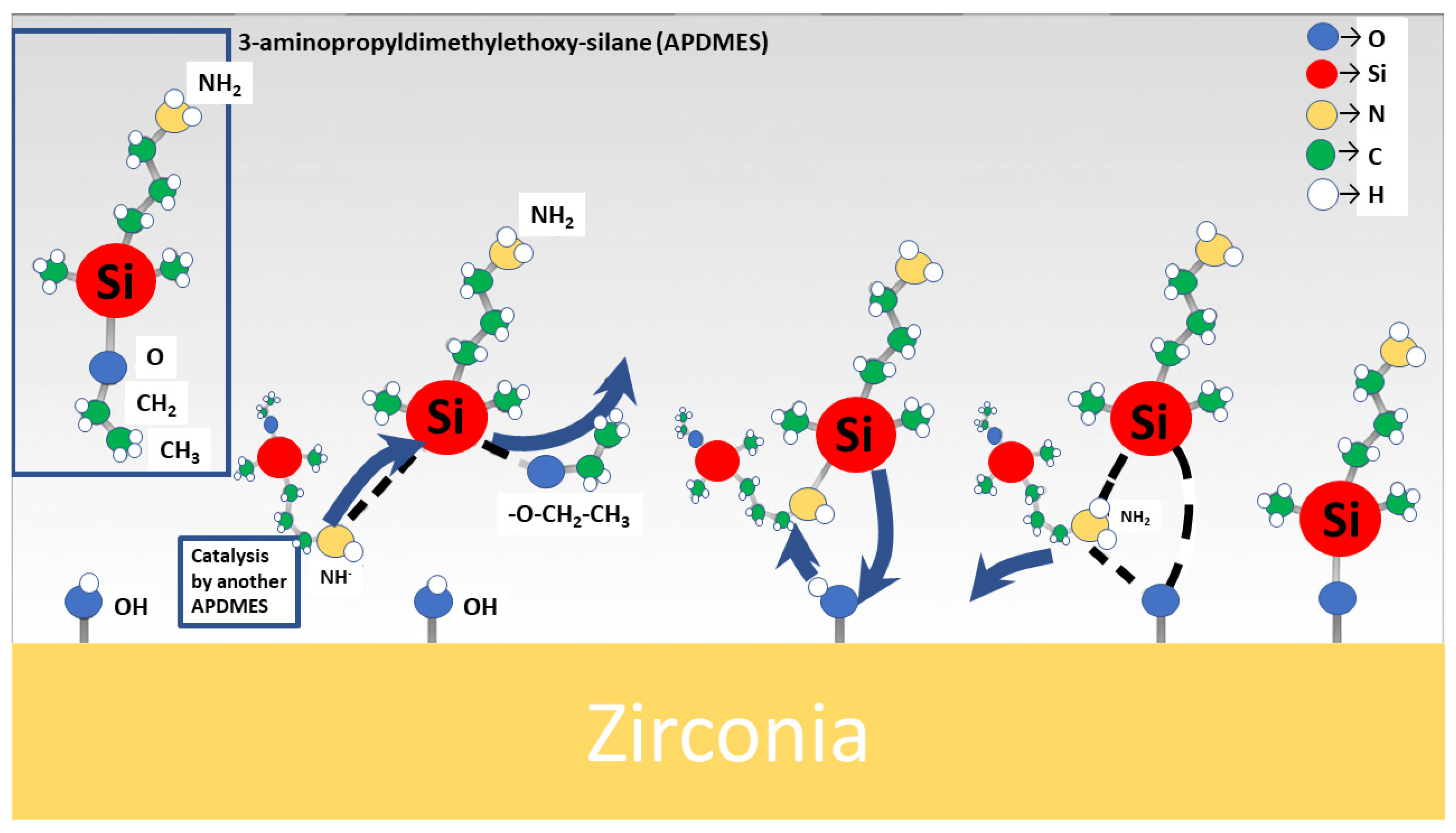
6. Biofunctionalisation of Ceramic Surfaces Using Biomolecules—An Alternative Approach

Inert high-performance ceramics are widely used in biomedical applications due to their biocompatibility and durability [100]. However, these ceramics are bioinert, meaning that they do not actively participate in biological processes. This can limit their effectiveness in medical use [101].
To overcome this limitation, exploring ways to bioactivate inert ceramics is an interesting approach in current research [102,103,104]. Bioactivation involves modifying the material surface to enhance its biological activity. In the case of inert ceramics, this can be achieved by introducing functional groups, such as carboxyl, amine, or hydroxyl groups, onto the surface (Figure 2).
These groups can serve as anchor points for biomolecules, such as growth factors or proteins, that can enhance the biological activity of the ceramic. One of the most promising approaches for bioactivating inert ceramics is the use of biomimetic strategies. Biomimetics involves mimicking the natural structure and function of biological systems. In the case of inert ceramics, biomimetic approaches involve creating a functionalised surface with proteins, e.g., cRGD (cyclic arginine-glycine-aspartic) [105]. This can lead to a better and increased attraction of cells, respectively, to connect to the surface.
Bioactivated inert ceramics have several potential biomedical applications. In tissue engineering, bioactivated ceramics are currently being investigated to be used as scaffolds for promoting the growth and differentiation of stem cells into bone, cartilage, or other tissues [106]. In drug delivery, bioactivated ceramics could be used as carriers for drugs or other therapeutic agents, allowing for targeted delivery to specific tissues or cells [107]. In orthopaedics, bioactivated ceramics could be used as implants for repairing or replacing damaged bone or cartilage [36].
In conclusion, the bioactivation of inert ceramics is an important area of research with significant potential for biomedical applications. Current research is considering the possibility of improving the bioactivity of ceramics with novel coating techniques. By modifying the surface of inert ceramics, biological activity can be enhanced. Therefore, ceramics are treated with chemicals like silanes [103]. Then, these surfaces are coated with, e.g., graphene oxide or peptides to improve the response of the native tissue [102,103]. This should allow for a stable connection between the implant and the native tissue [102]. This approach can create materials with improved performance in tissue engineering and orthopaedics. As biomimetic approaches continue to advance, bioactivated ceramics will likely play an increasingly important role in the development of new biomedical technologies.
These innovative methods are in a scientific state and not available for use in patient care yet.

7. Considerations and Regulatory Obligations Related to the Use of High-Performance Bio Ceramics

The successful utilisation of high-performance bio ceramics in medical devices and therapies requires careful consideration of various factors, including biocompatibility, mechanical properties, surface characteristics, degradation kinetics, clinical applications, antibacterial properties, and regulatory compliance. By addressing these considerations comprehensively, researchers, engineers, and clinicians can harness the full potential of bio ceramics to develop innovative medical devices and therapies that improve patient outcomes and quality of life.
The utilisation of high-performance (bio) ceramics in medical devices and therapies entails several regulatory obligations to ensure their safety, efficacy, and compliance with established standards. Their use is subject to stringent regulatory requirements to ensure patient safety and product effectiveness. Regulatory oversight helps mitigate risks associated with the development, manufacturing, and clinical use of bio ceramic materials, ensuring that they meet quality and safety requirements. Regulatory bodies, such as the Food and Drug Administration (FDA) in the United States, and the Medical Device Regulation (MDR) and European Medicines Agency (EMA) in Europe, respectively, have established frameworks and guidelines to govern the development, manufacturing, and marketing of medical devices and biomaterials.
Before clinical evaluation and commercialisation, high-performance bio ceramics undergo extensive preclinical testing to assess their biocompatibility, mechanical properties, degradation behaviour, and performance in relevant animal models. Clinical trials are conducted to evaluate the safety and efficacy of bio ceramic-based medical devices and therapies in human subjects. These trials assess parameters such as implant survival, tissue response, functional outcomes, and adverse events.
High-performance bio ceramics hold tremendous potential for revolutionising healthcare by enabling innovative medical devices and therapies with enhanced performance and biocompatibility. However, the utilisation of these advanced materials in biomedical applications requires careful consideration of design, manufacturing, and regulatory factors to ensure their safety, efficacy, and compliance with applicable standards. By adhering to stringent regulatory obligations and integrating quality management systems into the manufacturing process, stakeholders can harness the full benefits of high-performance bio ceramics while prioritising patient safety and regulatory compliance.

8. Discussion

This review article gives an overview of traditional and current bio ceramic materials used for hard tissue engineering related to musculoskeletal surgery such as TJA for THA and TKA. Ceramics in endoprosthetic joint support may enhance the (bio)compatibility, suitability, and lifetime of the implants [34]. Ceramics such as alumina or zirconia possess good mechanical properties, which can be optimised by mixing them [34].

8.1. Wear and Ageing

Wear in TJA, for example, in THA or TKA, is a phenomenon that has been known for years to have a high impact on clinical success [68]. Wear debris, in particular polyethylene or metal, is a driving force in the pathogenesis of periprosthetic osteolysis [68] and implant loosening [108]. Ceramics successfully reduce polyethylene wear. Up to date, no clear clinical evidence exists, however, that the use of ceramic components, e.g., for TKA, increases the survivorship of the implant by reducing polyethylene wear [76].
Bio ceramics in implants are showing an extremely high survivorship [66]. However, according to Roy et al., 2017 [30], several aspects need to be considered to ensure the successful use of ceramics beyond their standard use as femoral heads in THA:
  • Detailed specifications and testing procedures;
  • Processing techniques such as HIP (hot isostatic pressing/hot isostatic post-processing);
  • Increased quality of ceramic granular.
These characteristics show the potential of bio ceramics. Beside this, in case of revision, it must be considered that there is still ceramic particle available, and the therapy must be adapted to this situation.
Ideally, the mechanical properties of ceramics should closely match those of bone tissue to minimise stress concentrations and discrepancies in load-bearing capacity. Ceramics with similar elastic modulus, compressive strength, and hardness to bone can distribute mechanical loads more evenly, reducing the risk of implant loosening or fracture. The development of novel designs of implants is necessary. The implant design itself could be modified to reach similar elastic modulus.

8.2. Allergies

In the case of maxillofacial and orthopaedic surgery, alumina ceramics have been used for implants for three decades with no reports of hypersensitivity reactions [63]. Ceramic bearings may thus play a clinical role in patients with a known allergy to metals, such as nickel, or a history of previously failed metal implants. Although these observations are reasonable in light of existing data about metal hypersensitivity, further data are needed to clarify the potential role of ceramic bearings in this patient population [63].
Bio ceramics are known for their excellent biocompatibility, meaning they are generally well-tolerated by the body and have low risk of causing adverse reactions. This property makes them suitable for use in medical devices and implants without triggering immune responses that could potentially lead to allergies. They hold great promise for allergy prevention through their, e.g., biocompatibility, antibacterial properties, tissue engineering applications, and environmental uses.

8.3. Implant Fixation and Handling of Bio Ceramics

In the case of ceramics, careful handling of the different components is important because of their sensibility in applying loads to them. A possible problem in the context of ceramic implants is the so-called edge loading and the chip-off effect. That means that an exquisite surgical technique is essential. Currently, the only clinically established ceramic TKA endoprosthesis is implanted by a cementing technique to ensure adequate primary stability at the bone–implant interface. Here, an ideal uniform thickness of the cement mantle is important to diminish the likelihood of cement mantle defects, which can predispose to focal osteolysis [68]. The intention is to minimise motion between the bone and the implant.
If it is possible to reach a functionalisation of the implant surface that there will be an integration, then a moulding with bone cement is no longer necessary. The surface of ceramics can be modified to facilitate protein binding and immobilisation. Common methods include physical adsorption, covalent coupling, and layer-by-layer deposition. These techniques allow proteins to adhere to the ceramic surface, forming a bioactive layer that promotes cell attachment and signalling.

8.4. Established Use of Bio Ceramics in Arthroplasty

Currently, in clinical practice, a well-established approach is the use of hydroxyapatite. This is naturally like bone hydroxyapatite. Therefore, it is used for orthopaedic as well as dental applications. Conventional metal orthopaedic implants are coated with a layer of hydroxyapatite to capitalise on the potential for direct chemical–biological bonding of the osteoblasts to the implant. Such coatings may promote early ingrowth fixation, and they might be useful in load-bearing implants to offset the inhibitory influence of micromovements on bone ingrowth [109].

8.5. Modification of Ceramic Surfaces

Ceramics have some unique advantages that make them very desirable as the bearing surface in orthopaedic/trauma surgery. To withdraw the disadvantage of the bioinert behaviour of ceramics, the approaches of functional coating and surface functionalisation, respectively, are an innovative step forward in endoprosthetics. Functionalising ceramics with proteins involves modifying the surface of ceramic materials to enhance their biocompatibility, bioactivity, and interaction with biological systems. Functional coating via a surface modification with functional biological molecules like cRGD could be a sophisticated and versatile solution to improve the superficial properties of implants in various medical applications. This process enables ceramics to mimic the extracellular matrix and promote cell adhesion, proliferation, and differentiation, making them suitable for various biomedical applications, including tissue engineering, drug delivery, and biosensing.
By tailoring surface chemistry, topography, and protein composition, ceramics with tailored properties for specific therapeutic and diagnostic purposes could be developed, advancing the field of biomaterials and regenerative medicine.

8.6. Non-Oxide Ceramics as an Alternative

Non-oxide ceramics, also known as advanced or technical ceramics, are a class of ceramic materials composed of non-metallic compounds other than oxides. These materials exhibit exceptional mechanical, thermal, electrical, and chemical properties, making them suitable for a wide range of high-performance applications in various industries. Unlike traditional oxide ceramics (e.g., alumina, zirconia), which are based on combinations of metal and oxygen atoms, non-oxide ceramics incorporate elements such as carbides, nitrides, borides, and silicides. Biomedical applications of non-oxide ceramics show great promise in prosthetics, e.g., in the case of acetabular cups for THA [110]. Paione and Baino mentioned that clinical and societal challenges in this context were impressive [110]. Non-oxide ceramics such as silicon carbide, silicon nitride, and DLC (diamond-like carbon) could be alternatives to Al2O3 [110].

9. Conclusions

The importance of ceramics is stronger than ever. The results of ceramic implants are promising. Trends show that different ceramics with multiple functionalities, along with sustainable, long-term innovative technologies, are tackling the challenges in musculoskeletal surgery.
Of note, these implanted components are mostly not full endoprostheses but mostly focus on the bearing-like femoral heads and, in some cases, the cup inserts. Chevalier and Gremillard mentioned the reason that still, after 100 years of clinical use, there is no ceramic available that creates a strong, biologically relevant interface with bone and that allows for successful osseointegration [27]. The problem of integrating ceramics in physiological cellular environments is presently addressed by attempts to biofunctionalise ceramic implant surfaces [102,103]. New modifications of ceramics have also rendered the material even more durable than before. The goal is to ensure good biological activity and osteoinductive ability to achieve osseointegration. We expect to see further development in ceramic prosthesis manufacturing and biofunctionalisation over the next few years, which may make them the gold standard not only in bearings but in endoprosthetics.

Author Contributions

Conceptualisation, J.E., J.G., U.K.H., B.R., F.H., A.M., P.K. and F.M.; methodology, J.E., J.G., U.K.H., B.R., F.H., A.M, P.K. and F.M.; formal analysis, J.E., J.G., U.K.H., B.R., F.H., A.M., P.K. and F.M.; investigation, J.E. and J.G.; resources, J.E., J.G., U.K.H., B.R., F.H., A.M., P.K. and F.M.; data curation, J.E. and J.G.; writing—original draft preparation, J.E., writing—review and editing, J.E., J.G., U.K.H., B.R., F.H., A.M., P.K. and F.M.; visualisation, J.E.; supervision, U.K.H., B.R., F.H., A.M., P.K. and F.M.; project administration, J.E.; funding acquisition, J.E. and F.H. All authors have read and agreed to the published version of the manuscript.

Funding

This research was funded by BMBF NeWTranslation, grant number 13GW0452A-C.

Institutional Review Board Statement

Not applicable.

Informed Consent Statement

Not applicable.

Data Availability Statement

Not applicable.

Acknowledgments

We would like to thank Philipp Schräder for his fruitful input and outstanding support.

Conflicts of Interest

The authors declare no conflicts of interest.

References

  1. Felson, D.T. Epidemiology of Hip and Knee Osteoartrritis. Epidemiol. Rev. 1988, 10, 1–28. [Google Scholar] [CrossRef]
  2. Allen, K.D.; Thoma, L.M.; Golightly, Y.M. Epidemiology of osteoarthritis. Osteoarthr. Cartil. 2022, 30, 184–195. [Google Scholar] [CrossRef] [PubMed]
  3. Lespasio, M.J.; Sultan, A.A.; Piuzzi, N.S.; Khlopas, A.; Husni, M.E.; Muschler, G.F.; Mont, M.A. Hip Osteoarthritis: A Primer. Perm. J. 2018, 22, 17–84. [Google Scholar] [CrossRef] [PubMed]
  4. Quicke, J.G.; Conaghan, P.G.; Corp, N.; Peat, G. Osteoarthritis year in review 2021: Epidemiology & therapy. Osteoarthr. Cartil. 2022, 30, 196–206. [Google Scholar]
  5. Tong, L.; Yu, H.; Huang, X.; Shen, J.; Xiao, G.; Chen, L.; Wang, H.; Xing, L.; Chen, D. Current understanding of osteoarthritis pathogenesis and relevant new approaches. Bone Res. 2022, 10, 60. [Google Scholar] [CrossRef]
  6. Bergschmidt, P.; Bader, R.; Ganzer, D.; Hauzeur, C.; Lohmann, C.; Rüther, W.; Tigani, D.; Rani, N.; Prats, F.L.; Zorzi, C.; et al. Ceramic femoral components in total knee arthroplasty—Two year follow-up results of an international prospective multi-centre study. Open Orthop. J. 2012, 6, 172–178. [Google Scholar] [CrossRef]
  7. Statistisches Bundesamt. Die 20 Häufigsten Operationen Insgesamt (OPS 5): Vollstationär Behandelte Patientinnen und Patienten in Krankenhäusern 2020. Available online: https://www.destatis.de/DE/Themen/Gesellschaft-Umwelt/Gesundheit/Krankenhaeuser/Tabellen/drg-operationen-insgesamt.html;jsessionid=057935135CAAA80FFCBBC2BDF33DA20E.live741?view=main[Print] (accessed on 27 December 2023).
  8. Heimke, G.; Leyen, S.; Willmann, G. Knee arthoplasty: Recently developed ceramics offer new solutions. Biomaterials 2002, 23, 1539–1551. [Google Scholar] [CrossRef]
  9. Rahaman, M.N.; Yao, A.; Bal, B.S.; Garino, J.P.; Ries, M.D. Ceramics for prosthetic hip and knee joint replacement. J. Am. Ceram. Soc. 2007, 90, 1965–1988. [Google Scholar] [CrossRef]
  10. Emery, D.F.; Clarke, H.J.; Grover, M.L. Stanmore total hip replacement in younger patients: Review of a group of patients under 50 years of age at operation. J. Bone Jt. Surg. Br. Vol. 1997, 79, 240–246. [Google Scholar] [CrossRef]
  11. Denkena, B.; Köhler, J.; Turger, A.; Helmecke, P.; Correa, T.A.; Hurschler, C. Manufacturing Conditioned Wear of All-Ceramic Knee Prostheses; Gottfried Wilhelm Leibniz Universität Hannover, Technische Informationsbibliothek (TIB): Hannover, Germany, 2013. [Google Scholar]
  12. Ó Doinn, T.; Broderick, J.M. Biomaterials in Total Joint Arthroplasty: 2. In Arthroplasty; Zorzi, A., Toumi, H., Lespessailles, E., Eds.; IntechOpen: Rijeka, Croatia, 2022. [Google Scholar]
  13. Hembus, J.; Rößler, L.; Springer, A.; Frank, M.; Klinder, A.; Bader, R.; Zietz, C.; Enz, A. Experimental Investigation of Material Transfer on Bearings for Total Hip Arthroplasty—A Retrieval Study on Ceramic and Metallic Femoral Heads. J. Clin. Med. 2022, 11, 3946. [Google Scholar] [CrossRef]
  14. Moghadasi, K.; Isa, M.S.M.; Ariffin, M.A.; Raja, S.; Wu, B.; Yamani, M.; bin Muhamad, M.R.; Yusof, F.; Jamaludin, M.F.; bin Ab Karim, M.S.; et al. A review on biomedical implant materials and the effect of friction stir based techniques on their mechanical and tribological properties. J. Mater. Res. Technol. 2022, 17, 1054–1121. [Google Scholar] [CrossRef]
  15. Oral, E.; Muratoglu, O.K. Radiation cross-linking in ultra-high molecular weight polyethylene for orthopaedic applications. Nucl. Instrum. Methods Phys. Res. B 2007, 265, 18–22. [Google Scholar] [CrossRef] [PubMed]
  16. Partridge, T.; Baker, P.N.; Jameson, S.S.; Mason, J.; Reed, M.R.; Deehan, D.J. Conventional Versus Highly Cross-Linked Polyethylene in Primary Total Knee Replacement: A Comparison of Revision Rates Using Data from the National Joint Registry for England, Wales, and Northern Ireland. J. Bone Jt. Surg. Am. Vol. 2019, 102, 119–127. [Google Scholar] [CrossRef] [PubMed]
  17. Packer, L. Protective role of vitamin E in biological systems. Am. J. Clin. Nutr. 1991, 53, 1050S–1055S. [Google Scholar] [CrossRef]
  18. Grimberg, A.; Jansson, V.; Lützner, J.; Melsheimer, O.; Morlock, M.; Steinbrück, A. The German Arthroplasty Registry (EPRD): Annual Report 2021; EPRD Deutsche Endoprothesenregister gGmbH: Berlin, Germany, 2021; ISBN 978-3-9817673-8-4. [Google Scholar]
  19. Grimberg, A.; Jansson, V.; Lützner, J.; Melsheimer, O.; Morlock, M.; Steinbrück, A. Endoprothesenregister Deutschland (EPRD)—Jahresbericht 2021. Available online: https://www.eprd.de/fileadmin/user_upload/Dateien/Publikationen/Berichte/Jahresbericht2021_2021-10-25_F.pdf (accessed on 21 October 2022).
  20. Bergschmidt, P.; Bader, R.; Kluess, D.; Zietz, C.; Mittelmeier, W. The all-ceramic knee endoprosthesis—The gap between expectation and experience with ceramic implants. In Seminars in Arthroplasty; WB Saunders: Philadelphia, PA, USA, 2012; pp. 262–267. [Google Scholar]
  21. Sundfeldt, M.; Carlsson, L.V.; Johansson, C.B.; Thomsen, P.; Gretzer, C. Aseptic loosening, not only a question of wear: A review of different theories. Acta Orthop. 2006, 77, 177–197. [Google Scholar] [CrossRef]
  22. Cherian, J.J.; Jauregui, J.J.; Banerjee, S.; Pierce, T.; Mont, M.A. What Host Factors Affect Aseptic Loosening After THA and TKA? Clin. Orthop. Relat. Res. 2015, 473, 2700–2709. [Google Scholar] [CrossRef]
  23. Goodnough, L.H.; Finlay, A.K.; Huddleston, J.I.; Goodman, S.B.; Maloney, W.J.; Amanatullah, D.F. Obesity Is Independently Associated With Early Aseptic Loosening in Primary Total Hip Arthroplasty. J. Arthroplast. 2018, 33, 882–886. [Google Scholar] [CrossRef] [PubMed]
  24. Münger, P.; Röder, C.; Ackermann-Liebrich, U.; Busato, A. Patient-related risk factors leading to aseptic stem loosening in total hip arthroplasty: A case-control study of 5,035 patients. Acta Orthop. 2006, 77, 567–574. [Google Scholar] [CrossRef]
  25. Radunovic, A.; Popovic, Z.; Matic, A.; Vulovic, M. 12 Application of ceramic components in knee arthroplasties. In Advanced Ceramics and Applications; Gadow, R., Vojislav, V., Mitic, A., Eds.; De Gruyter: Berlin, Germany, 2021; pp. 155–164. [Google Scholar]
  26. Langer, G. Ceramic Tibial Plateau of the 70s. In Proceedings of the Bioceramics in Joint Arthroplasty: Proceedings 7th International BIOLOX Symposium, Stuttgart, Germany, 15–16 March 2002. [Google Scholar]
  27. Chevalier, J.; Gremillard, L. Ceramics for medical applications: A picture for the next 20 years. J. Eur. Ceram. Soc. 2009, 29, 1245–1255. [Google Scholar] [CrossRef]
  28. Saccomanno, M.F.; Sircana, G.; Masci, G.; Cazzato, G.; Florio, M.; Capasso, L.; Passiatore, M.; Autore, G.; Maccauro, G.; Pola, E. Allergy in total knee replacement surgery: Is it a real problem? World J. Orthop. 2019, 10, 63–70. [Google Scholar] [CrossRef]
  29. Whiteside, L.A. Clinical results of revision TKA in patients with presumed metal and cement allergy. J. Arthroplast. 2022, 37, S250–S257. [Google Scholar] [CrossRef]
  30. Roy, M.; Bandyopadhyay, A.; Bose, S. Ceramics in bone grafts and coated implants. In Materials for Bone Disorders; Elsevier: Amsterdam, The Netherlands, 2017; pp. 265–314. [Google Scholar]
  31. Peter Brehm GmbH. Knieendoprothese BPK-S Integration Ceramic: Neues Kniegelenk Trotz Unverträglichkeit. Available online: https://www.peter-brehm.ch/knieendoprothese-bpk-s-integration-ceramic.html (accessed on 1 July 2022).
  32. Hamadouche, M.; Sedel, L. Ceramics in orthopaedics. J. Bone Jt. Surg. Br. Vol. 2000, 82, 1095–1099. [Google Scholar] [CrossRef]
  33. Salmang, H.; Scholze, H. (Eds.) Keramik; 6., verb. u. erw. Aufl.; Springer: Berlin, Germany, 1982. [Google Scholar]
  34. Kumar, P.; Dehiya, B.S.; Sindhu, A. Bioceramics for hard tissue engineering applications: A review. Int. J. Appl. Eng. Res 2018, 13, 2744–2752. [Google Scholar]
  35. Ben-Nissan, B.; Choi, A.H.; Cordingley, R. 10—Alumina ceramics. In Bioceramics and Their Clinical Applications: Woodhead Publishing Series in Biomaterials; Kokubo, T., Ed.; Woodhead Publishing: Cambridge, UK, 2008; pp. 223–242. ISBN 978-1-84569-204-9. [Google Scholar]
  36. Kamboj, N.; Ressler, A.; Hussainova, I. Bioactive Ceramic Scaffolds for Bone Tissue Engineering by Powder Bed Selective Laser Processing: A Review. Materials 2021, 14, 5338. [Google Scholar] [CrossRef] [PubMed]
  37. Australian Orthopaedic Association National Joint Replacement Registry. Hip, Knee & Shoulder Arthroplasty: 2021 Annual Report; AOA: Adelaide, Australia, 2021. [Google Scholar]
  38. Godavitarne, C.; Robertson, A.; Peters, J.; Rogers, B. Biodegradable materials. Orthop. Trauma 2017, 31, 316–320. [Google Scholar] [CrossRef]
  39. Wuisman, P.I.J.M.; Smit, T.H. Bioresorbable polymers: Heading for a new generation of spinal cages. Eur. Spine J. 2006, 15, 133–148. [Google Scholar] [CrossRef] [PubMed]
  40. Singh, R.; Bathaei, M.J.; Istif, E.; Beker, L. A Review of Bioresorbable Implantable Medical Devices: Materials, Fabrication, and Implementation. Adv. Healthc. Mater. 2020, 9, e2000790. [Google Scholar] [CrossRef] [PubMed]
  41. Albrektsson, T.; Brånemark, P.-I.; Hansson, H.-A.; Lindström, J. Osseointegrated titanium implants: Requirements for ensuring a long-lasting, direct bone-to-implant anchorage in man. Acta Orthop. Scand. 1981, 52, 155–170. [Google Scholar] [CrossRef] [PubMed]
  42. James, L. Osseointegration: Its Mechanism and Recent Updates. J. Dent. Res. 2022, 4, 1. [Google Scholar]
  43. Albrektsson, T.; Johansson, C. Osteoinduction, osteoconduction and osseointegration. Eur. Spine J. 2001, 10, S96–S101. [Google Scholar] [PubMed]
  44. Zarb, G.A.; Albrektsson, T. Osseointegration: A requiem for the periodontal ligament. Int. J. Periodontics Restor. Dent. 1991, 11, 88–91. [Google Scholar]
  45. Mavrogenis, A.F.; Dimitriou, R.; Parvizi, J.; Babis, G.C. Biology of implant osseointegration. J. Musculoskelet. Neuronal Interact. 2009, 9, 61–71. [Google Scholar]
  46. Kripal, K. Endosseous integration. EC Dent. Sci. 2017, 12, 87–98. [Google Scholar]
  47. Davies, J.E. Understanding peri-implant endosseous healing. J. Dent. Educ. 2003, 67, 932–949. [Google Scholar] [CrossRef]
  48. Chen, S.; Guo, Y.; Liu, R.; Wu, S.; Fang, J.; Huang, B.; Li, Z.; Chen, Z.; Chen, Z. Tuning surface properties of bone biomaterials to manipulate osteoblastic cell adhesion and the signaling pathways for the enhancement of early osseointegration. Colloids Surf. B Biointerfaces 2018, 164, 58–69. [Google Scholar] [CrossRef]
  49. Khalili, A.A.; Ahmad, M.R. A Review of Cell Adhesion Studies for Biomedical and Biological Applications. Int. J. Mol. Sci. 2015, 16, 18149–18184. [Google Scholar] [CrossRef] [PubMed]
  50. Janiszewska, M.; Primi, M.C.; Izard, T. Cell adhesion in cancer: Beyond the migration of single cells. J. Biol. Chem. 2020, 295, 2495–2505. [Google Scholar] [CrossRef]
  51. Harjunpää, H.; Llort Asens, M.; Guenther, C.; Fagerholm, S.C. Cell Adhesion Molecules and Their Roles and Regulation in the Immune and Tumor Microenvironment. Front. Immunol. 2019, 10, 1078. [Google Scholar] [CrossRef]
  52. Campbell, H.K.; Maiers, J.L.; DeMali, K.A. Interplay between tight junctions & adherens junctions. Exp. Cell Res. 2017, 358, 39–44. [Google Scholar] [CrossRef] [PubMed]
  53. Green, K.J.; Jaiganesh, A.; Broussard, J.A. Desmosomes: Essential contributors to an integrated intercellular junction network. F1000Research 2019, 8, 2150. [Google Scholar] [CrossRef]
  54. Ghayor, C.; Bhattacharya, I.; Guerrero, J.; Özcan, M.; Weber, F.E. 3D-Printed HA-Based Scaffolds for Bone Regeneration: Microporosity, Osteoconduction and Osteoclastic Resorption. Materials 2022, 15, 1433. [Google Scholar] [CrossRef]
  55. Abyzov, A.M. Aluminum Oxide and Alumina Ceramics (review). Part 1. Properties of Al2O3 and Commercial Production of Dispersed Al2O3. Refract. Ind. Ceram. 2019, 60, 24–32. [Google Scholar] [CrossRef]
  56. ISO 6474-1:2019; Implants for Surgery—Ceramic Materials—Part 1: Ceramic Materials Based on High-Purity Alumina. International Organization for Standardization: Geneva, Switzerland, 2019; pp. 1–9.
  57. Piconi, C.; Maccauro, G. Zirconia as a ceramic biomaterial. Biomaterials 1999, 20, 1–25. [Google Scholar] [CrossRef]
  58. Hannink, R.H.J.; Kelly, P.M.; Muddle, B.C. Transformation Toughening in Zirconia-Containing Ceramics. J. Am. Ceram. Soc. 2000, 83, 461–487. [Google Scholar] [CrossRef]
  59. Maccauro, G.; Iommetti, P.R.; Raffaelli, L.; Manicone, P.F. Alumina and Zirconia Ceramic for Orthopaedic and Dental Devices: 15. In Biomaterials; Pignatello, R., Ed.; IntechOpen: Rijeka, Croatia, 2011. [Google Scholar]
  60. Kirsten, A.; Begand, S.; Oberbach, T.; Telle, R.; Fischer, H. Subcritical crack growth behavior of dispersion oxide ceramics. J. Biomed. Mater. Res. Part B Appl. Biomater. 2010, 95, 202–206. [Google Scholar] [CrossRef]
  61. Technische Keramik GmbH. ATZ Keramik. Available online: https://tkc-keramik.de/werkstoffe/oxidkeramik/atz-aluminiumoxidverstaerktes-zirkoniumoxid/ (accessed on 30 June 2022).
  62. Hallab, N.J.; Jacobs, J.J. Orthopedic applications. In Biomaterials Science; Elsevier: Amsterdam, The Netherlands, 2020; pp. 1079–1118. [Google Scholar]
  63. Bal, B.S.; Garino, J.; Ries, M.; Oonishi, H. Ceramic bearings in total knee arthroplasty. J. Knee Surg. 2007, 20, 261–270. [Google Scholar] [CrossRef] [PubMed]
  64. Gadow, R.; Mitic, V.V. (Eds.) Advanced Ceramics and Applications; De Gruyter: Berlin, Germany; Boston, MA, USA, 2021; ISBN 9783110627992. [Google Scholar]
  65. Bos, I.; Willmann, G. Morphologic characteristics of periprosthetic tissues from hip prostheses with ceramic-ceramic couples: A comparative histologic investigation of 18 revision and 30 autopsy cases. Acta Orthop. Scand. 2001, 72, 335–342. [Google Scholar] [CrossRef] [PubMed]
  66. Lee, G.-C.; Kim, R.H. Incidence of Modern Alumina Ceramic and Alumina Matrix Composite Femoral Head Failures in Nearly 6 Million Hip Implants. J. Arthroplast. 2017, 32, 546–551. [Google Scholar] [CrossRef] [PubMed]
  67. Zhang, L.; Haddouti, E.-M.; Welle, K.; Burger, C.; Kabir, K.; Schildberg, F.A. Local Cellular Responses to Metallic and Ceramic Nanoparticles from Orthopedic Joint Arthroplasty Implants. Int. J. Nanomed. 2020, 15, 6705. [Google Scholar] [CrossRef] [PubMed]
  68. Jacobs, J.J.; Shanbhag, A.; Glant, T.T.; Black, J.; Galante, J.O. Wear debris in total joint replacements. JAAOS-J. Am. Acad. Orthop. Surg. 1994, 2, 212–220. [Google Scholar] [CrossRef] [PubMed]
  69. Jasty, M.; Bragdon, C.R.; Lee, K.; Hanson, A.; Harris, W.H. Surface damage to cobalt-chrome femoral head prostheses. J. Bone Jt. Surg. Br. Vol. 1994, 76, 73–77. [Google Scholar] [CrossRef]
  70. Bauer, T.W.; Schils, J. The pathology of total joint arthroplasty. II. Mechanisms of implant failure. Skelet. Radiol. 1999, 28, 483–497. [Google Scholar] [CrossRef] [PubMed]
  71. Skinner, H.B. Ceramic bearing surfaces. Clin. Orthop. Relat. Res. 1999, 369, 83–91. [Google Scholar] [CrossRef] [PubMed]
  72. Bergschmidt, P.; Bader, R.; Mittelmeier, W. Metal hypersensitivity in total knee arthroplasty: Revision surgery using a ceramic femoral component—A case report. Knee 2012, 19, 144–147. [Google Scholar] [CrossRef] [PubMed]
  73. Solarino, G.; Piconi, C.; de Santis, V.; Piazzolla, A.; Moretti, B. Ceramic total knee arthroplasty: Ready to go? Joints 2017, 5, 224–228. [Google Scholar] [CrossRef] [PubMed]
  74. Bader, R.; Bergschmidt, P.; Fritsche, A.; Ansorge, S.; Thomas, P.; Mittelmeier, W. Alternative materials and solutions in total knee arthroplasty for patients with metal allergy. Der. Orthop. 2008, 37, 136–142. [Google Scholar] [CrossRef] [PubMed]
  75. Australian Orthopaedic Association. National Joint Replacement Registry—Annual Report. Available online: https://aoanjrr.sahmri.com/annual-reports-2016 (accessed on 27 December 2023).
  76. Bergschmidt, P.; Ellenrieder, M.; Bader, R.; Kluess, D.; Finze, S.; Schwemmer, B.; Mittelmeier, W. Prospective comparative clinical study of ceramic and metallic femoral components for total knee arthroplasty over a five-year follow-up period. Knee 2016, 23, 871–876. [Google Scholar] [CrossRef] [PubMed]
  77. Kreve, S.; Dos Reis, A.C. Effect of surface properties of ceramic materials on bacterial adhesion: A systematic review. J. Esthet. Restor. Dent. 2022, 34, 461–472. [Google Scholar] [CrossRef]
  78. Rimondini, L. Bacteria and Biofilm Formation on Biomaterials. Insights. 2022. Available online: https://www.ceramtec-medical.com/fileadmin/user_upload/Medical/Bilder/Infocenter/CeraNews/CeraNews_Issue_2022_1/lay_CeraNews_122_INSIGHTS_EN_IMPLANT_MATERIAL_Rimondini_06.pdf (accessed on 27 December 2023).
  79. Treccani, L. Interactions Between Surface Material and Bacteria: From Biofilm Formation to Suppression. In Surface-Functionalized Ceramics: For Biotechnological and Environmental Applications; Treccani, L., Meder, F., Eds.; Wiley: Hoboken, NJ, USA, 2023; pp. 283–335. [Google Scholar]
  80. Salomoni, A.; Tucci, A.; Esposito, L.; Stamenkovic, I. Forming and sintering of multiphase bioceramics. J. Mater. Sci. Mater. Med. 1994, 5, 651–653. [Google Scholar] [CrossRef]
  81. Begand, S.; Glien, W.; Oberbach, T. ATZ—A New Material with a High Potential in Joint Replacement. Key Eng. Mater. 2005, 284–286, 983–986. [Google Scholar] [CrossRef]
  82. Piconi, C. Bioinert ceramics: State-of-the-art. Key Eng. Mater. 2017, 758, 3–13. [Google Scholar] [CrossRef]
  83. Odegaard, K.S.; Torgersen, J.; Elverum, C.W. Structural and biomedical properties of common additively manufactured biomaterials: A concise review. Metals 2020, 10, 1677. [Google Scholar] [CrossRef]
  84. Papannagari, R.; Hines, G.; Sprague, J. Long-Term Wear Performance of an Advanced Bearing Technology for TKA. Available online: https://www.ors.org/transactions/57/1141.pdf (accessed on 27 December 2023).
  85. Nakamura, S.; Ito, H.; Nakamura, K.; Kuriyama, S.; Furu, M.; Matsuda, S. Long-term durability of ceramic tri-condylar knee implants: A minimum 15-year follow-up. J. Arthroplast. 2017, 32, 1874–1879. [Google Scholar] [CrossRef] [PubMed]
  86. Xiang, S.; Zhao, Y.; Li, Z.; Feng, B.; Weng, X. Clinical outcomes of ceramic femoral prosthesis in total knee arthroplasty: A systematic review. J. Orthop. Surg. Res. 2019, 14, 57. [Google Scholar] [CrossRef] [PubMed]
  87. Solarino, G.; Zagra, L.; Piazzolla, A.; Morizio, A.; Vicenti, G.; Moretti, B. Results of 200 Consecutive Ceramic-on-Ceramic Cementless Hip Arthroplasties in Patients Up to 50 Years of Age: A 5–24 Years of Follow-Up Study. J. Arthroplast. 2019, 34, S232–S237. [Google Scholar] [CrossRef] [PubMed]
  88. Nakamura, T.; Oonishi, E.; Yasuda, T.; Nakagawa, Y. A new knee prosthesis with bisurface femoral component made of zirconia-ceramic (Report 2). Key Eng. Mater. 2004, 254–256, 607–610. [Google Scholar] [CrossRef]
  89. Nakamura, T.; Akagi, M.; Yasuda, T.; Nakagawa, Y.; Shimizu, M. A new knee prosthesis with bisurface femoral component made of zirconia-ceramic. Key Eng. Mater. 2002, 218–220, 563–566. [Google Scholar]
  90. Oonishi, H.; Fujita, H.; Itoh, S.; Kin, S.; Amino, H. Development and improvement of ceramic TKP for 19 years and clinical results. Key Eng. Mater. 2002, 218, 479–482. [Google Scholar] [CrossRef]
  91. Oonishi, H.; Kim, S.C.; Kyomoto, M.; Masuda, S.; Asano, T.; Clarke, I.C. Change in UHMWPE properties of retrieved ceramic total knee prosthesis in clinical use for 23 years. J. Biomed. Mater. Res. B Appl. Biomater. 2005, 74, 754–759. [Google Scholar] [CrossRef]
  92. Oonishi, H.; Murata, N.; Saito, M.; Wakitani, S.; Imoto, K.; Kin, S.; Chen, Y.; Nakaya, H.; Tanaka, M.; Amino, H. 3 to 18 year clinical results of total knee replacement with ceramic components. Key Eng. Mater. 2001, 192–195, 999–1004. [Google Scholar] [CrossRef]
  93. Oonishi, H.; Oonishi, H.; Kim, S.C.; Kyomoto, M.; Iwamoto, M.; Masuda, S.; Ueno, M. Ceramic Total Knee Arthroplasty: Advanced Clinical Experiences of 26 Years. Semin. Arthroplast. 2006, 17, 134–140. [Google Scholar] [CrossRef]
  94. Breuer, R.; Fiala, R.; Trieb, K.; Rath, B. Prospective Mid-Term Results of a Completely Metal-Free Ceramic Total Knee Endoprosthesis: A Concise Follow-Up of a Previous Report. J. Arthroplast. 2021, 36, 3161–3167. [Google Scholar] [CrossRef]
  95. Meier, E.; Gelse, K.; Trieb, K.; Pachowsky, M.; Hennig, F.F.; Mauerer, A. First clinical study of a novel complete metal-free ceramic total knee replacement system. J. Orthop. Surg. Res. 2016, 11, 21. [Google Scholar] [CrossRef]
  96. Trieb, K. A novel ceramic tibial component is as safe as its metal counterpart. Biomed. Tech. 2018, 63, 327–332. [Google Scholar] [CrossRef]
  97. Laskin, R.S. An oxidized Zr ceramic surfaced femoral component for total knee arthroplasty. Clin. Orthop. Relat. Res. 2003, 416, 191–196. [Google Scholar] [CrossRef]
  98. Thienpont, E. Titanium niobium nitride knee implants are not inferior to chrome cobalt components for primary total knee arthroplasty at medium-term follow-up. Arch. Orthop. Trauma Surg. 2023, 143, 5269–5275. [Google Scholar] [CrossRef] [PubMed]
  99. Louwerens, J.K.G.; Hockers, N.; Achten, G.; Sierevelt, I.N.; Nolte, P.A.; van Hove, R.P. No clinical difference between TiN-coated versus uncoated cementless CoCrMo mobile-bearing total knee arthroplasty; 10-year follow-up of a randomized controlled trial. Knee Surg. Sports Traumatol. Arthrosc. 2021, 29, 750–756. [Google Scholar] [CrossRef] [PubMed]
  100. Turner, I.G. Ceramics and Glasses. In Biomedical Materials; Springer: Cham, Switzerland, 2021; pp. 101–137. [Google Scholar]
  101. Qu, H.; Fu, H.; Han, Z.; Sun, Y. Biomaterials for bone tissue engineering scaffolds: A review. RSC Adv. 2019, 9, 26252–26262. [Google Scholar] [CrossRef] [PubMed]
  102. Böke, F.; Labude, N.; Lauria, I.; Ernst, S.; Müller-Newen, G.; Neuss, S.; Fischer, H. Biological Activation of Bioinert Medical High-Performance Oxide Ceramics by Hydrolytically Stable Immobilization of c(RGDyK) and BMP-2. ACS Appl. Mater. Interfaces 2018, 10, 38669–38680. [Google Scholar] [CrossRef] [PubMed]
  103. Desante, G.; Labude, N.; Rütten, S.; Römer, S.; Kaufmann, R.; Zybała, R.; Jagiełło, J.; Lipińska, L.; Chlanda, A.; Telle, R.; et al. Graphene oxide nanofilm to functionalize bioinert high strength ceramics. Appl. Surf. Sci. 2021, 566, 150670. [Google Scholar] [CrossRef]
  104. Caravaca, C.; Shi, L.; Balvay, S.; Rivory, P.; Laurenceau, E.; Chevolot, Y.; Hartmann, D.; Gremillard, L.; Chevalier, J. Direct silanization of zirconia for increased biointegration. Acta Biomater. 2016, 46, 323–335. [Google Scholar] [CrossRef]
  105. Li, W.; Tan, S.; Xing, Y.; Liu, Q.; Li, S.; Chen, Q.; Yu, M.; Wang, F.; Hong, Z. cRGD Peptide-Conjugated Pyropheophorbide-a Photosensitizers for Tumor Targeting in Photodynamic Therapy. Mol. Pharm. 2018, 15, 1505–1514. [Google Scholar] [CrossRef] [PubMed]
  106. Gao, C.; Deng, Y.; Feng, P.; Mao, Z.; Li, P.; Yang, B.; Deng, J.; Cao, Y.; Shuai, C.; Peng, S. Current progress in bioactive ceramic scaffolds for bone repair and regeneration. Int. J. Mol. Sci. 2014, 15, 4714–4732. [Google Scholar] [CrossRef] [PubMed]
  107. Zang, S.; Chang, S.; Shahzad, M.B.; Sun, X.; Jiang, X.; Yang, H. Ceramics-based Drug Delivery System: A Review and Outlook. Rev. Adv. Mater. Sci. 2019, 58, 82–97. [Google Scholar] [CrossRef]
  108. Chethan, K.N.; Shenoy, B.S.; Bhat, N.S. Role of different orthopedic biomaterials on wear of hip joint prosthesis: A review. Mater. Today Proc. 2018, 5, 20827–20836. [Google Scholar] [CrossRef]
  109. Cooke, F.W. Ceramics in orthopedic surgery. Clin. Orthop. Relat. Res. 1992, 276, 135–146. [Google Scholar] [CrossRef]
  110. Paione, C.M.; Baino, F. Non-Oxide Ceramics for Bone Implant Application: State-of-the-Art Overview with an Emphasis on the Acetabular Cup of Hip Joint Prosthesis. Ceramics 2023, 6, 994–1016. [Google Scholar] [CrossRef]
Figure 1. To our knowledge, BIOLOX delta is the only material worldwide that is used for all-ceramic primary knee endoprosthesis for the fitting of patients with a proven metal allergy [31] (image with permission from Peter Brehm GmbH).
Ceramics 07 00020 g001
Figure 2. Schematic representation of surface modification with 3-aminopropyldimethylethoxy-silane (modified after [104]).
Ceramics 07 00020 g002
Table 1. Mechanical properties of orthopaedic biomaterials (according to [62]).
Orthopaedic BiomaterialElastic Modulus
(Young’s Modulus)
(GPa)
Yield Strength
(Elastic Limit)
(MPa)
Al2O3366./.
Y-TZP201./.
Cortical Bone #
Low strain15.2114
High strain40.8./.
Ti6Al4V116897–1034
CoCr Alloys210–253448–841
UHMWPE0.5–1.320–30
# Cortical bone is both anisotropic and viscoelastic, and the properties listed are generalised.
Table 2. Selected bioinert ceramics and some examples of their applications in medical devices (modified table according to [82]).
MaterialsApplications
AluminaFemoral heads and inserts for THA bearings
Osteosynthetic devices [83]
ZirconiaDental implants, dental blanks for CAD/CAM #
Fixed partial dentures [83]
VERILAST technology [84]
ZTA/ATZFemoral heads and inserts for THA; surfaces for TKA bearings; and
components for disc replacements (in spine surgery)
# CAD = Computer-aided design; CAM = computer-aided manufacturing; and ZTA/ATZ = zirconia-toughened alumina/alumina-toughened zirconia.
Table 3. Selection of currently available implant materials for hypersensitive patients.
MaterialProduct Name (Company)Modification Technique
TiNbNEFK Femur (OHST Medizical Technology, Rathenow, Germany), balanSys (Mathys, Bettlach, Switzerland), NitrX (Microport, Shanghai, China)Physical Vapor Deposition (PVD)
TiNScore AS (Amplitude, Valence, France), SensiTiN (Medacta, Castel San Pietro, Switzerland), Apex Knee (Corin, Cirencester, UK)PVD
ZrNAS Advanced Surface (Aesculap, Center Valley, PA, USA)PVD
Oxidised ZrNbOxinium (Smith&Nephew, London, UK)Oxidised by a heat treatment
Abbreviations: N—Nitride, Nb—niob, Ti—titanium, and Zr—zirconium. All implants are made for implantation with bone cement (information obtained from the manufacturers).
Disclaimer/Publisher’s Note: The statements, opinions and data contained in all publications are solely those of the individual author(s) and contributor(s) and not of MDPI and/or the editor(s). MDPI and/or the editor(s) disclaim responsibility for any injury to people or property resulting from any ideas, methods, instructions or products referred to in the content.

