

Components of Mid-Nineteenth- and Mid-Twentieth-Century Cudbears

1
Conservation Center, Los Angeles County Museum of Art, Los Angeles, CA 90036, USA
2
Conservation Science Laboratory, Indianapolis Museum of Art at Newfields, Indianapolis, IN 46208, USA
*
Author to whom correspondence should be addressed.
Heritage 2024, 7(3), 1357-1371; https://doi.org/10.3390/heritage7030065
Submission received: 1 February 2024
/
Revised: 27 February 2024
/
Accepted: 1 March 2024
/
Published: 9 March 2024
(This article belongs to the Special Issue Latest Trends and Challenges in the Study of Pigments and Dyes and Their Degradation in Cultural Heritage Objects)
Abstract
:Analysis of purple dyestuff from a tin labeled “1 oz. Cudbear, No. 1 N. F. Powdered”, marketed by the American business S. B. Penick & Company, “Manufacturers of fine drugs and chemicals”, confirmed that the material was indeed a lichen dyestuff. It contains the same major orcein components identified in several other lichen dyes and dyed samples dating from the mid-19th century to today. These dyestuffs were analyzed using several analytical techniques. Fluorescence and fiber optic reflectance spectroscopic data for all the samples were similar. High performance liquid chromatography with diode array detection coupled to mass spectrometry confirmed that this commercial American cudbear was very similar to the samples from the United Kingdom but rather different from the archil-dyed reference yarns from Europe. The significance of the observations is discussed, and chemical structures are proposed for several of the unknown dye components detected in this study.
1. Introduction
For over two millennia, dyers in Europe, the Middle East, and possibly South America have known that a purple dyestuff could be obtained by soaking certain species of lichens (from genera such as Roccella and Ochrolechia) in stale urine with aeration [1,2,3]. These dyestuffs, commonly referred to as orchil (or archil in continental Europe and in publications such as Schweppe’s Handbuch der Naturfarbstoffe [4]), have been identified in ancient, medieval, and Renaissance European manuscripts and textiles [5,6,7,8,9,10].
The dye compositions of the dyestuffs extracted from the lichen sources have been observed to vary based on the preparation technique as well as the specific source [11]. One of these purple dyestuffs introduced commercially in the late 18th century in Scotland by Cuthbert Gordon is known as cudbear [1,12].
Initially, the production of cudbear employed Ochrolechia lichen species indigenous to Scotland. However, as the abundance of this lichen dwindled in the 1820s, other lichen varieties, such as those in the Roccella and Lasallia genera, were imported into Scotland from continental Europe to serve as dyestuff sources [1,13]. Recognizing biological sources of materials used by artists may contribute to understanding the history of their work, so research into lichen-based colorants has included investigation of whether the diverse origins of lichen dyes can be distinguished chemically on the basis of the chemical components identified in purple-dyed materials [9,10].
The Conservation Research Laboratory at the Los Angeles County Museum of Art (LACMA) has now investigated the composition of an early 20th-century commercial cudbear dye and compared it to those reported in historic cudbear and archil samples [12]. The laboratory was gifted an undated tin of purple dye powder from S.B. Penick & Co. of Asheville, North Carolina. The tin label states “1 oz. Cudbear, N.F. No. 1, powdered” (Figure 1), without providing further details about its contents. The composition and spectral properties of this dyestuff, which may have been marketed in the United States in the early to mid-20th century, have now been compared to those of contemporary commercial Orcein, to cudbears sourced from mid-19th- and mid-20th-century Britain, and to archil dyestuff present in wool yarns dyed by Helmut Schweppe.
A brief research into the history of the S.B. Penick Company was undertaken to ascertain whether the source of the cudbear they marketed could be determined. The information obtained did not uncover a specific dyestuff source, but it did suggest that the cudbear produced by S.B. Penick might be similar to dyestuff extracted from Ochrolechia spp. and to the 19th-century historic samples studied. The differences between these cudbears and the archil used by Schweppe that have been revealed by analyses of these several samples were significant enough to suggest the former dyestuffs could be placed in a distinct category of lichen-derived dyes.
1.1. The S.B. Penick Company, Their Source of the Cudbear, and Its Use in the USA in the Early 20th Century
The S. B. Penick Company—the self-proclaimed “World’s Largest Botanical Drug House”—was established in Asheville, North Carolina, during World War I. The business processed, packaged, and marketed botanicals collected by residents in the vicinity who had extensive knowledge of locally used plant-based folk remedies [14]. By the 1920s, the company also began selling pharmaceuticals prepared from imported botanical materials. This is corroborated by an elaborate company letterhead on which a 1923 letter was typed (Figure 2). Cudbear, as seen in Figure 2c, can be discerned among the products listed as “foreign” in the list on the right side of the faint letterhead [15].
Some of the information on the label of the tin suggests that it postdates the letter by more than a decade. The company’s Chicago office, listed on the label (Figure 1b), was not opened until 1934, and the saw-tooth lines at the top and bottom of the label appeared in an advertisement from 1940 [16]. The term “initial line”, with a bar through it (Figure 1a), was patented by the company in 1937. Furthermore, in the patent, cudbear is included in the list of “botanical drugs” for which this label is to be used [17,18]. The notation “N.F.” on the tin indicates that cudbear was listed in the national formulary, a drug compendium published irregularly by the American Pharmaceutical Association [19]. National formularies dating from the 1930s through 1946 listed cudbear as a coloring agent with the alternative name “red indigo,” but it was not listed in the 1950 edition. These facts collectively suggest that the tin of cudbear may date from the mid-1930s to the mid-1940s.
Whether the Penick Company used domestic as well as imported lichen sources for cudbear is equivocal. The 1923 letterhead clearly states a foreign source. However, an undated company advertisement listing ingredients for a “white pine compound” includes “1 part cudbear,” and also states that “all ingredients in [the compound] are gathered freshly at our Asheville, N.C. plant,” implying a local source [14]. In a speech made in the late 1930s, a Penick executive mentioned that the no. 1 rating and the “initial line” designation were given specifically to locally gathered materials of the highest quality [17]. Several Ochrolechia lichen species are native to the Ashville, North Carolina region, and some are identified as “cudbear lichens” [20]. By the early 1940s, the supply of British-sourced cudbear was presumably interrupted by World War II. Together, these facts would be consistent with the company preparing their cudbear products from United States-sourced lichens. However, without access to additional Penick Company records, the identification of the specific lichen source of the cudbear in the tin cannot be ascertained.
The information gathered to determine the date of the cudbear marketed by the Penick Company also suggested that the dye was used for medicinal purposes. The earlier national formularies included recipes for “tinctures” of cudbear in up to 60% alcohol, intended for various ailments [19]. The Penick Company advertisement for the white pine compound described above indicated that it was used as an expectorant [14]. Interestingly, no literature has been found describing the use of cudbear as a textile dye in the southeastern United States during this time. In contrast, the information uncovered so far regarding cudbear use in the USA in the second quarter of the twentieth century suggests that the dyestuff was used primarily as a colorant for pharmaceuticals rather than as a textile dye.
1.2. Background Information on Orcein Compounds
Lichen dyestuffs, generally called orchils, contain several colored compounds, many of which have been described chemically [21]. These colorant molecules, known collectively as orceins, are alcohol-extractable derivatives of the planar three-ring compound phenoxazine (Figure 3, Structure 1). They are generated during the extraction process when orcinol esters present in several lichen species undergo hydrolysis in the presence of aqueous ammonia [20]. Beecken et al. described the physiochemical properties of eight of the nine primary orceins listed in Table 1 [21].
Based on absorption spectra, the orcein compounds can be categorized into three groups: hydroxyorceins (HO), aminoorceins (AO), and aminoorceinimines (AI) (Table 1). Each of these three groups consists of three compounds, which are distinguished by their different substituents at the R, X, and Y positions (see Figure 3, Structure 1). Thus, there are nine primary orceins. For each of these three groups, the orceins with R = H have a single orcinol moiety attached to the C6 carbon in the phenoxazine ring. They are denoted by the prefix α. The orceins doubly substituted with orcinols at both positions C3 and C6 (i.e., R = (a) or (b) in Figure 3) are assigned the prefixes β and γ. Because of the presence of methyl residues at positions C4 and C5 in the molecules, the bulky orcinol substituents cannot rotate freely around the chemical bonds at positions C3 and C6. Consequently, this results in a pair of geometric isomers, β and γ.
To distinguish between these latter two isomers, the prefix β designates the trans-isomer (R = (a)), while the prefix γ refers to the cis-isomer (R = (b)) (See Figure 3) [21]. In the β-isomer, the ortho-methyl groups of the two orcinol residues are situated on opposite sides of the plane of the phenoxazine. In contrast, in the γ-isomer, the ortho-methyl groups on both orcinol residues are on the same side of the plane of the molecule. However, without appropriate reference compounds, it is not possible to ascertain which chromatographic peak is specifically due to the β-isomer and which to the γ-isomer. Lech and Fornal [22] have recently reported mass spectrometric data for these nine primary orcein compounds, including α-AI, which was not specifically described by Beecken et al. [21].
Due to mesomerism, C3 and C6 are chemically equivalent in α-AI and α-HO. However, for α-AO, where X is oxo (=O) and Y is amine (-NH2), C3 and C6 are not chemically equivalent, so there are two configurational isomers. By comparing physiochemical properties of the dyestuff components with the model compounds, the α-AO compound isolated was determined to be the isomer substituted at C6 [21]. In addition, Beecken et al. described secondary orceins compounds, which are derivatives of β- or γ-HO and possibly β- or γ-AO. In these orcein derivatives, one or both orcinol substituents have been oxidized to the corresponding quinone, thereby increasing the molecular mass by 14 or 28 mass units. Because these modifications would only affect the peripheral substituents, the chromophore remains largely unaltered. As a result, no noticeable changes in the visible absorption spectra are expected [21].
2. Materials and Methods
2.1. Materials
Chemicals—from J.T. Baker, Phillipsburg, NJ, USA: stannous chloride. From Fisher Scientific, Fair Lawn, NJ, USA: nitric acid, reagent ethanol denatured with 10% methanol, and ammonium hydroxide, 35–45%, acetonitrile, Optima LC/MS grade (A944-1), formic acid, Optima LC/MS grade (A117-50). From Acros Organics, Morris Plains, NJ, USA: oxalic acid, dihydrate, 99+% extra pure (129601000). From Alfa Aesar, Ward Hill, MA, USA: pyridine, anhydrous, 99.5+% (43799). From Sigma-Aldrich, St. Louis, MO, USA: ethanol, 200 proof, reagent grade (E7023-500ML). Water (18 MΩ) was purified first through a Milli-Q® Direct 8 Water Purification System (MilliporeSigma, Burlington, MA, USA). For LC–MS, additional pre-filtration through an LC-Pak cartridge was used.
Dyestuffs: Orcein, synthetic (described on the safety data sheet as consisting of many compounds), was sourced from TCI (Tokyo Chemical Industry, Co., Ltd.), Portland, OR, USA; dye powder prepared from Ochrolechia tartarea lichen collected from rocks in the county of Wester Ross, Scotland, by Dr. Robert Hill was received as a gift from Dr. David Hill; powdered “cudbear” from S.B. Penick, Co. (Ashville, NC, USA) was a gift from Dr. Charlotte Eng.
Dyed yarns: Archil-dyed woolen yarn samples were obtained from the Schweppe Reference Collection of the Getty Conservation Institute, Los Angeles, CA, USA. A mid-nineteenth-century cudbear-dyed yarn sample, WHW61M, was collected from a samplebook Dyed Woollen Yarns with Receipts, assembled by William Henry Worth and dated “begun October 1842” [23]. A contemporary woolen yarn from Brush Creek woolworks, mordanted with alum was dyed using a mid-nineteenth-century recipe, with orchil extracted from Ochrolechia tartarea L. lichen provided by Dr. David Hill of the United Kingdom. The dyeing procedure followed an adaptation of the recipes in the W.H. Worth book for dyeing woolen yarn purple using cudbear [12,23].
2.2. Methods
2.2.1. UV/Vis Spectroscopy
Visible absorption spectra of aqueous and ethanolic solutions and reflectance spectra of dyed yarns were obtained with a Cary 50 spectrophotometer (Varian, now Agilent Technologies, Santa Clara, CA, USA). Reflectance spectra were collected using an optical fiber and Barrelino accessory (Harrick Scientific, Pleasantville, NY, USA). The yarn or fiber samples were placed on Whatman 1 filter paper, the reflectance of which was used as background. The reflectance data for the solid samples were converted to the equivalent absorbance by application of the Kubelka–Munk transform using the appropriate math functions provided in the Cary WinUV software, version 5.1.3 that operates the instrument. The absorbance maxima were determined by taking the second derivative of these spectra. The wavelength accuracy was ±3 nm.
2.2.2. Fluorimetry
A Cary Eclipse fluorimeter (Agilent Technologies) was used for fluorescence spectroscopy. Aqueous solutions were placed in quartz cuvettes, and yarn samples were mounted directly in the powder sample holder accessory. Excitation and emission spectra and synchronous scans were recorded; data for 3-D plots of fluorescence intensity as a function of both excitation and emission wavelengths were also obtained. Nominal slit widths did not exceed 5 nm.
2.2.3. Chromatography
Two liquid chromatography instruments were used to separate and identify the dyestuff components. Initially, an HPLC–DAD instrument without mass spectrometer was used to identify some dyestuff components by comparison to known dyes sources. The results showed significant similarities between the commercial and historic dyestuffs and suggested that they should be subjected to more detailed analyses. The results of these additional measurements are reported below.
Liquid chromatography–diode array detection–mass spectrometry (LC–DAD–MS).
Powdered dyes were extracted into ethanol by heating 1 mg powder/mL EtOH at 80 °C for 30 min. After cooling, insoluble material was precipitated by centrifugation, and the supernatant was adjusted to 50% vol–vol EtOH–water before analysis by LC–DAD–MS.
A 1 mg yarn sample was extracted in 1 mL of a 1:1 pyridine–water solution containing 50 mM oxalic acid (OAPW) at 80 °C for 60 min following published procedures [24]. Because both the oxalic acid, added for mordant removal, and pyridine could interfere with LC analysis, the extraction solvent was removed by a stream of nitrogen gas while the sample was warmed at 60 °C. The dried sample residue was then redissolved in 1:1 EtOH–water and clarified of solids by centrifugation at 13,000 rpm for 5 min before being subjected to LC–DAD–MS analysis. A blank was run before each sample to verify the absence of carryover of contaminants from the previous injection.
The LC–DAD–MS system used for analysis of dye extracts consisted of a Thermo Accela LC system (Chicago, IL, USA) connected in sequence to both a DAD and an LTQ electrospray ionization MS detector. Dye separation was carried out using a Restek Ultra C18 (Bellefonte, PA, USA) reverse phase column (150 mm × 4.6 mm, 5 μm particles) eluted at 0.5 mL/min using a water–acetonitrile gradient system containing 0.1% formic acid. The column was equilibrated at 3% acetonitrile, and 6 min after sample injection, analyte separation was carried out by applying a linear gradient of acetonitrile increasing from 3% to 93% in 60 min. This gradient is suitable for separation of a wide variety of natural and synthetic colorants. The DAD detector was set to record spectra in the range 200–800 nm at 20 Hz, with a 1 nm bandwidth, scan step 1 nm, and a rise time of 0.02 s. The mass spectrometer collected full-scan mass spectra (FSMS) in the positive electrospray ionization mode in the m/z range 50–1000. The system was controlled by Thermo Xcalibur version 4.0 software.
3. Results
None of the powdered dyestuffs (TCI orcein, dye prepared by Hill, and Penick powder) were completely soluble in water or in the organic solvents tested, at room temperature; heating to 75–80 °C did not achieve complete solubilization. The compositions detailed below pertain to the soluble fractions. Preliminary tests showed that powder samples extracted using ethanol, pyridine-water containing oxalic acid, or DMSO consistently yielded similar results with respect to the number and relative amounts of orceins detected [16]. These same solvents were used for subsequent extractions to enable direct comparisons with the findings of the preliminary study [12,16].
3.1. Spectrophotometry
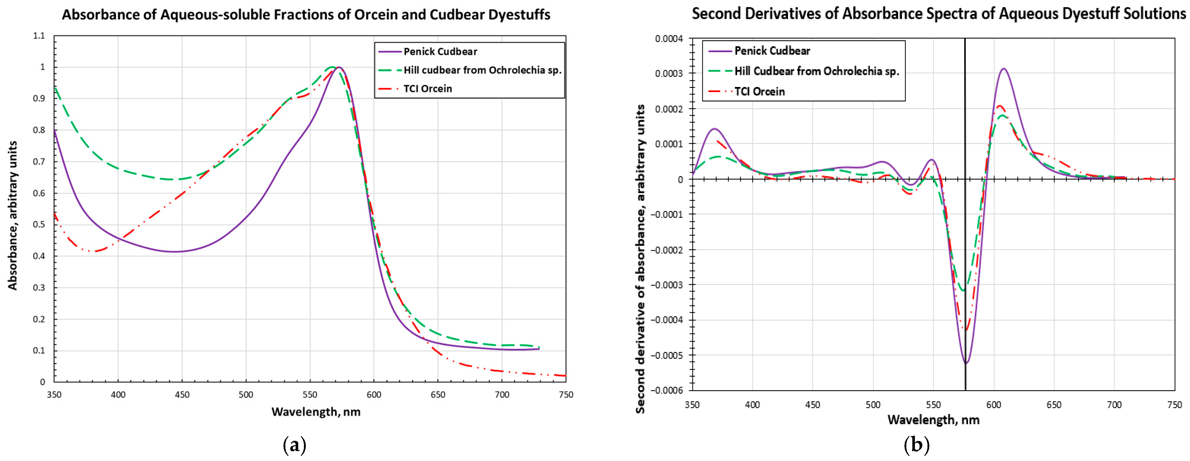
A summary of absorbance and reflectance spectroscopy results for the supernatant solutions, of dyestuff powders, and the dyed yarns is presented in Table 2, where the wavelength maxima of the three visible absorbance bands are listed. Figure 4 shows examples of the spectra for the Ochrolechia tartarea extract, and the TCI and Penick dyestuff solutions. Given the wavelength accuracy of ±3 nm for the measurements, it can be concluded that there are no significant differences in the positions of the long wavelength maximum and prominent shoulder in the spectra of the dye solutions prepared from these three different sources. When dyed onto wool with an alum mordant, with or without the addition of acidic or basic dyeing aids, both Hill and Penick dyestuffs show negligible spectroscopic differences in the visible region. The absorbance maxima for cudbear-dyed yarns are also experimentally indistinguishable from the absorbance maxima of the Schweppe yarns dyed with archil, within the accuracy of the measurements. Remarkably, the locations of the band maxima appear to be more strongly affected by dyeing conditions than by the source of the dyestuff.
Fluorescence excitation and emission data for solutions of the three lichen dyestuff samples and the dyed yarn samples are also listed in Table 2, where the wavelengths of maximum excitation and emission are also given. Figure 5 provides examples of excitation and emission spectra; these were obtained for Penick cudbear dyed onto wool without the addition of acid or base to the dye bath. Given the uncertainty of ±3 nm for the positions of the excitation and emission maxima, it can be seen from Table 2 that the spectral properties of the solutions prepared from the three different powders are indistinguishable, as are those for the corresponding lichen-dyed yarns, as exemplified in Figure 5. It should be noted that the excitation wavelength for maximum fluorescence of all the dyed yarn samples is 589 ± 3 nm. These fluorescence data are in reasonable agreement with the results of Melo et al. and Idone et al. for orchil samples [6,8].
3.2. Liquid Chromatography (LC)
3.2.1. High-Performance Liquid Chromatography Monitored by Diode Array Detector
Preliminary HPLC–DAD results for all the cudbear samples showed a similar chromatographic pattern of several peaks with absorbances in the 500 to 560 nm range. Although the same peaks were also observed in the Schweppe archil chromatograms, they appeared to be present in distinctly different relative amounts, and several additional minor peaks were detected.
3.2.2. High-Performance Liquid Chromatography Monitored by Diode Array Detector–Mass Spectrometry
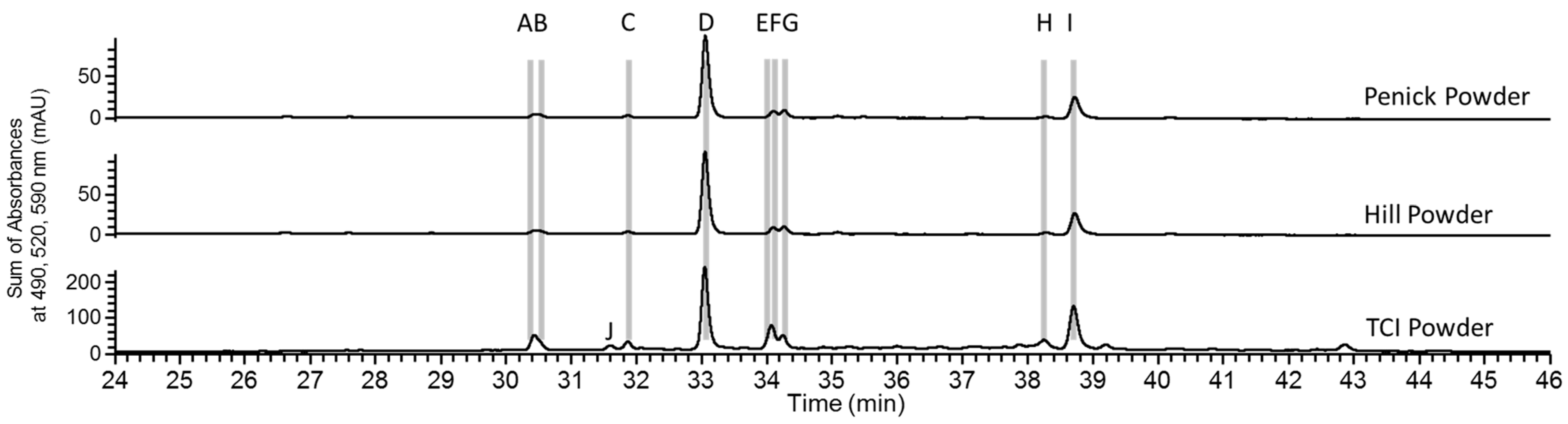
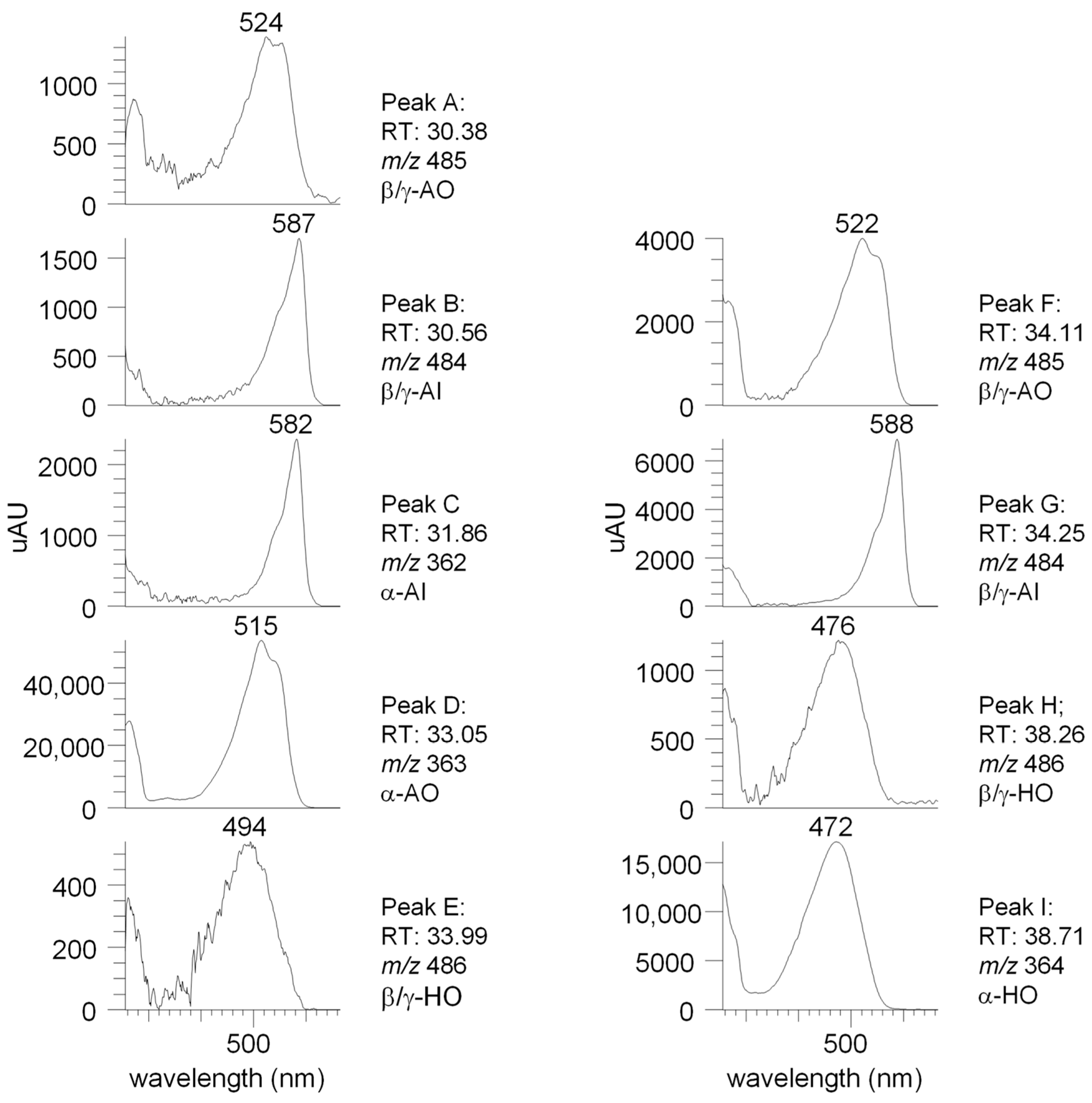
Figure 6 illustrates the chromatograms for extracts of the dye powders from the three sources. These chromatograms strongly suggest that similar dye components are present in these three dyestuffs. Based on the data presented in Table 1 and the visible spectra and mass spectrometric data shown in Figure 7, the nine analytes labeled A to I in Figure 6 have been assigned to the nine primary orceins described in the literature [21,22,25].
The visible spectra and mass data of analytes labeled with letters A to I for the Penick powder sample shown in Figure 7 agree with those reported by Pirok et al. [25] and by Lech and Fornal [22]. It should be emphasized that the samples from the other lichen dyestuffs analyzed in the current study all contained the same analytes A through I, in similar relative amounts, even though the total amount of extractable dyes varied among these samples.
The chromatographic profiles in Figure 6 indicate that the three powdered samples are likely to contain the same or very similar orceins and in similar relative amounts. Among the orceins, α-AO (analyte D) and α-HO (analyte I) are the predominant compounds, whereas the β- or γ-isomers of AO (A and F), AI (B and G), and HO (E and H) are present in lower, variable relative concentrations. Although detected only in trace amounts in the three dyestuffs, analyte C did yield sufficient data to be identified as α-AI in all three powder samples.
In Figure 6, the data show that the TCI orcein contains a minor component, J, absent from Hill and Penick dyestuffs. This component exhibits identical physiochemical properties to those of analyte D, suggesting that it may be an isomer of α-AO, possibly with the orcinol attached to a site in the molecule other than C6.
Figure 8 shows the chromatograms for extracts from woolen yarn fibers dyed with Hill or Penick cudbear powders and that for the extract of the mid-nineteenth-century cudbear-dyed yarn (WHW61M). The chromatogram for the Hill powder from Figure 6 is repeated for comparison. All these samples exhibit the same peaks for the nine orceins, with similar relative ratios for the historic and the Hill dyed yarns. However, the Penick dyed yarn sample appears to contain relatively smaller amounts of components C, D, and I when compared to the other components A, B, E, F, G, and H. The y-axes of the chromatograms also indicate that the amounts of dyes extracted from 1 mg of dyed yarn are about one-third to one-half that from 1 mg of dye powder.
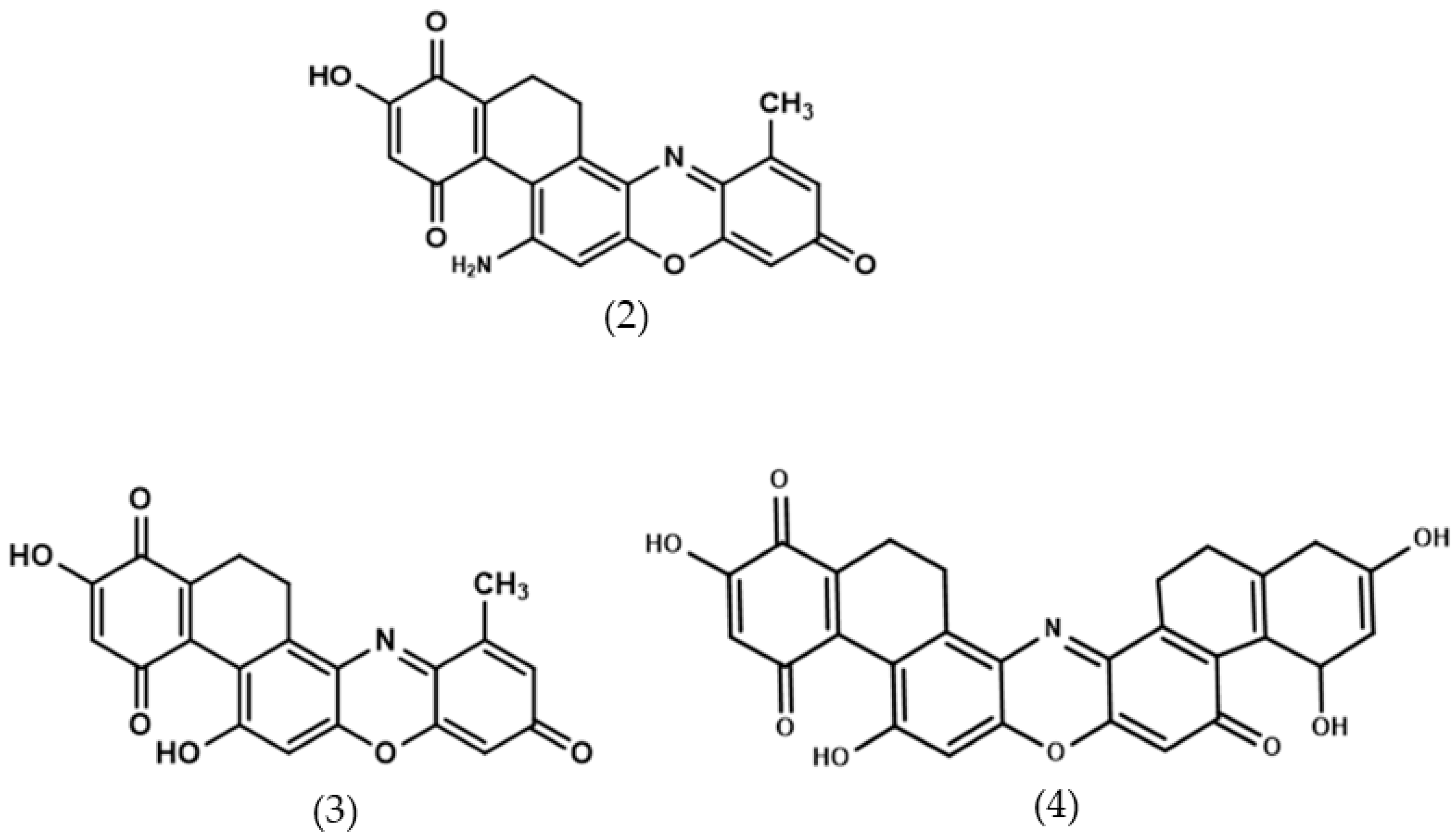
The nineteenth-century British cudbear-dyed yarn gave a chromatographic profile similar to that of the twentieth-century commercial Penick cudbear-dyed yarn, with comparable amounts of components A, B, E, F, G, and H. However, the additional analytes K and L present in the historic sample were not detected in any of the other samples. These latter two components exhibit visible spectra that when compared to the data in Table 1 and Figure 7 suggest they might be assigned to AO and HO color groups, respectively. Analyte K displays a positive ion at m/z 375, and L has two positive ions at m/z 376 and 498, which may be due to two co-migrating compounds or some unusual in-source fragmentation of m/z 498 to yield an ion with m/z 376. In Figure 9, possible structures are proposed for these three analytes.
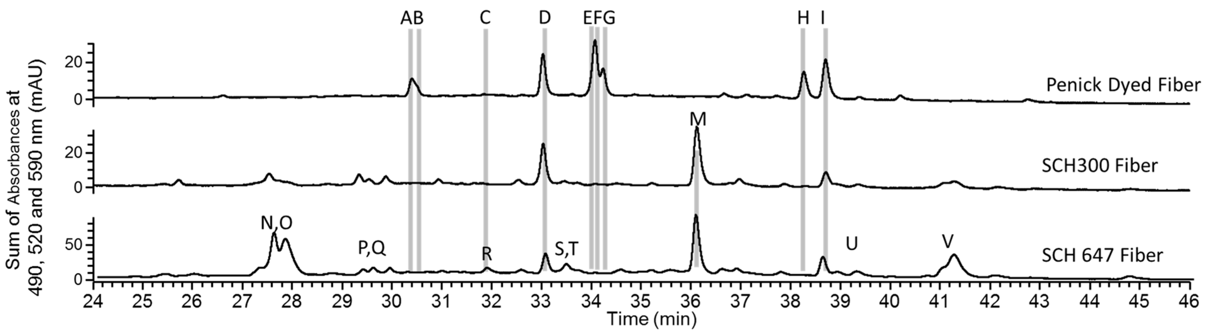
Figure 10 shows chromatograms for the extracts from Schweppe archil-dyed yarns. For comparison, the chromatogram of the extract of the Penick dyed yarns from Figure 9 has been repeated. The two Schweppe samples share similar chromatographic profiles. They contain α-AO (D) and α-HO (I), both of which are major primary orceins found in the other samples. However, these Schweppe samples have many additional peaks (Figure 10, peaks M through V). Thus, it cannot be ruled out that a low amount of A–C and E–H were present in the archil-dyed yarn but that they were obscured from detection by the relatively high amounts of M–V in this sample. Mass and absorption spectral data for these peaks are summarized in Table 3. The data suggest that none of the components are likely to belong to the primary group of orceins. Furthermore, their mass data are not consistent with the secondary orceins discussed by Beecken et al. [21], and they are not listed among orcein-like compounds characterized by Calà et al. [10]. Thus, no structures are proposed for analytes M through V.
4. Discussion
The project described in this paper was initiated to determine whether the purple powder in the S.B. Penick Company tin was indeed cudbear, as indicated by the container label. The positive answer obtained based on visible spectroscopic properties and preliminary HPLC–DAD results prompted further investigation. In this expanded study, the commercial Penick product, marketed by an American drug company in the first half of the twentieth century, was compared to cudbear and archil dyestuffs used in Britain and Europe over a span of two hundred years. The additional research was also motivated by the absence from local historical records of information suggesting that, in the southeastern United States where the Penick Company operated, locally sourced cudbear dyestuff was used to color textiles. Interestingly, aloes are the only source listed by Bemis as used historically for dyeing purple in America [26].
Although cudbear apparently was not used as a textile dye in the southwest United States during the suggested time period of the Penick powder, the dye profile of this dyestuff was found to closely resemble that of cudbears from several British sources that were used to dye yarns purple. These latter samples included the extract of a Scottish Ochrolechia lichen, a cudbear-dyed yarn from a mid-nineteenth-century English sample book, and a contemporary commercial dye from TCI marketed as “Orcein.” The similar dye profiles of all the cudbear dyestuffs and the orceins were found to differ significantly from the profiles of extracts from yarns dyed by Dr. Helmut Schweppe, with archil from Roccella tinctoria L. Serafini et al. have also reported that an extract from Ochrolechia lichens had a thin layer chromatography (TLC) profile distinctly different from those of several Roccella and Lasallia extracts [9]. The LC–DAD–MS profile of the major peaks in the Penick sample strongly suggests that this particular commercial product, belongs to the cudbear-like group of purple lichen dyestuff rather than to the archil group studied by Schweppe.
The complement of dyes in the Penick cudbear displayed a chromatographic profile very similar to those of the Worth, Hill, and TCI dyestuffs. All nine primary orceins are present, and the predominant dye compounds are α-AO and α-HO. However, analyses of the commercial TCI sample revealed additional minor components, most of which could not be unequivocally identified. The component J, for example, may be the 3-substututed isomer of α-AO. The presence of the additional orceins could be due to disparate sources of starting materials and/or to differences in the preparation of the commercial dyestuff. It should be noted that attempts to locate information from TCI about the synthetic orcein product were not successful.
In this project, the chemical assignments of many of the minor as well as the major peaks in the chromatograms of the cudbear extracts have been achieved based on their mass spectrometric and absorbance properties. Although chromatographic patterns observed for the major orcein components are consistent for all the cudbear samples, the minor components varied significantly among individual examples of the different dyestuffs. These minor color compounds could be degradation products, components present in vivo that are dependent on growing conditions, or additional compounds resulting from the extraction and analytical procedures.
The similarities between the absorption and fluorescence spectra of all samples included in the present study suggest that the same or very similar fluorophores are present in both archil and cudbear dyes. Therefore, the overall spectroscopic results for the cudbears do not provide any suggestion of significant differences in the optical properties of the components of the differently sourced lichen dyestuffs or of the archil on the Schweppe dyed reference yarns.
Two additional, non-primary, orcein-like compounds, K and L, were detected in the Worth yarn that were not present in the other samples. In Figure 9, the proposed structures (2) for K and (3) and (4) for L, if confirmed, would suggest oxidation of the corresponding primary orceins, α-AO and β/γ-HO, respectively. The oxidation of these primary orceins could be due to the mid-nineteenth-century age of the sample and/or to the dyeing conditions. Specific conclusions cannot be made in the absence of information on the dyestuff source for this historic yarn and/or to its unknown storage conditions for over a century.
The results of this investigation have highlighted some distinct differences among a group of British lichen dye samples, a yarn dyed with archil of continental European origin and an American commercial product from the early 20th century. Notably, the commercial Penick cudbear sample shares more characteristics with the British group than the European archil, although the possibility that the Penick sample was prepared from an American source of lichen cannot be ruled out.
The distinctions among the samples reported in this study were based on HPLC profiles of the primary orceins and putative orcein-like molecules. Whether the observed profiles are specific to our samples or are generally characteristic of lichen sources remain to be determined. Conceivably, factors such as dye preparation methods, handling, and/or storage conditions could lead, for example, to oxidization of the dyes to secondary or other non-primary orceins in ways that may prevent the resulting orcein profiles from being useful determinants of the biological source of lichen dyes in historical objects. Future studies should be designed to address these issues. With the growing interest in identifying and understanding the use of lichen dyestuffs as colorants in historic objects [5,6,7,8], more complete descriptions of the wide variety of dye components in orchil and cudbear from different lichen sources will become increasingly important.
Author Contributions
Conceptualization, T.T.S. and L.M.; methodology, T.T.S., L.M. and V.J.C.; investigation, T.T.S., L.M. and V.J.C.; resources, T.T.S. and L.M.; data curation, T.T.S., V.J.C. and L.M.; writing—original draft preparation, T.T.S. and L.M.; writing—review and editing, T.T.S., L.M., V.J.C. and G.D.S.; supervision, L.M. and G.D.S.; project administration, L.M. All authors have read and agreed to the published version of the manuscript.
Funding
This research received no external funding.
Data Availability Statement
The data presented in this study are available upon reasonable request from the corresponding author.
Acknowledgments
L.M. and T.T.S. thank Charlotte Eng for providing the Penick cudbear; David Hill of the University of Bristol for the gift of cudbear prepared by Robert Hill and the Roccella lichen from Tenerife; the Getty Conservation Institute for the samples of Schweppe dyed yarns; and Senior Conservation Photographer Yosi Pozeilov for the photography. The Conservation Science Laboratory at Newfields acknowledges the generous support from Sarah and John Lechleiter, The Frenzel Family Charitable Trust, Kay Koch, The Carter Family Fund, and the R.B. Annis Educational Foundation.
Conflicts of Interest
The authors declare no conflicts of interest.
References
- Cardon, D. Natural Dyes: Sources, Tradition, Technology and Science; Archetype Publications: London, UK, 2007. [Google Scholar]
- Phipps, E. The color purple in the Andes. In Dyes in History and Archaeology 37/40; Archetype Press: London, UK, 2023; pp. 1–16. [Google Scholar]
- Hofenk de Graaff, J.; Roelofs, W.G.T.; van Bommel, M.R. The Colourful Past: Origins Chemistry and Identification of Natural Dyestuffs; Abegg-Stifftung and Archetype Publications: Riggisberg, Switzerland, 2004. [Google Scholar]
- Schweppe, H. Handbuch der Naturfarbstoffe; Nikol Verlagsgesellschaft mbH & Co. KG: Hamburg, Germany, 1993. [Google Scholar]
- Aceto, M.; Arrais, A.; Marsano, F.; Agostino, A.; Fenoglio, G.; Idone, A.; Gulmini, M. A diagnostic study on folium and orchil dyes non-invasive and micro-destructive methods. Spectrochim. Acta Part A Mol. Biomol. Spectrosc. 2015, 142, 159–168. [Google Scholar] [CrossRef] [PubMed]
- Melo, M.J.; Nabais, P.; Guimarães, M.; Araújo, R.; Castro, R.; Oliveira, M.C.; Whitworth, I. Organic dyes in illuminated manuscripts: A unique cultural and historic record. Philos. Trans. R. Soc. 2016, A374, 20160050. [Google Scholar] [CrossRef] [PubMed]
- Witkowski, B.; Ganeczko, M.; Hryszko, H.; Stachurska, M.; Gierczak, T.; Biesaga, M. Identification of orcein and selected natural dyes in 14th and 15th century liturgical parameters with high-performance liquid chromatography coupled to the electrospray ionization tandem mass spectrometry (HPLC-ESI/MS/MS). Microchem. J. 2017, 133, 370–379. [Google Scholar] [CrossRef]
- Idone, A.; Miletto, I.; Davit, P.; Aceto, M.; Prenesti, E.; Gulmni, M. Direct fluorimetric characterisation of dyes in ancient purple codices. Microchem. J. 2017, 135, 122–128. [Google Scholar] [CrossRef]
- Serafini, I.; Lombardi, L.; Reverberi, M.; Ciccola, A.; Calà, E.; Sciubba, E.; Guiso, M.; Postorino, P.; Aceto, M.; Bianco, A. New advanced extraction and analytical methods applied to discrimination of different lichen species used for orcein dyed yarns: Preliminary results. Microchem. J. 2018, 138, 447–456. [Google Scholar] [CrossRef]
- Calà, E.; Benzi, M.; Gosetti, F.; Zanin, A.; Gulmini, M.; Idone, A.; Serafini, I.; Ciccola, A.; Curini, R.; Whitworth, I.; et al. Toward the identification of the lichen species in historical orchil dyes by HPLC-MS/MS. Microchem. J. 2019, 150, 104140. [Google Scholar] [CrossRef]
- Casselman, K.L. Lichen Dyes: The New Source Book, 2nd revised ed.; Dover Publications: Garden City, NY, USA, 2001. [Google Scholar]
- Schaeffer, T.T.; Maccarelli, L. Treading on Lichen: The use of cudbear by a Victorian carpet manufacturer. Dye. Hist. Archaeol. 2021, 35/36, 25–38. [Google Scholar]
- Habib, V. The Tinctorial Properties of Lichens: The Work of William Lauder Lindsay (1829–1880) and his Collection of Dyed Fabric Samples. Dye. Hist. Archaeol. 2023, 37/40, 80–92. [Google Scholar]
- Carlisle, R.D.B.; Penick Schmitz, E.v.W. Our Own Business: S. B. Penick & Company; Elizabeth Van Wie Penick Schmitz: Upper Montclair, NJ, USA, 1998. [Google Scholar]
- S.B. Penick & Company. Letter on Company Letterhead. In John H. Yardley Collection of Architectural Letterheads, Columbia University Libraries Online Exhibition; 1923; Available online: https://exhibitions.library.columbia.edu/exhibits/show/Yardley/item/8497 (accessed on 20 May 2020).
- Schaeffer, T.T.; Maccarelli, L.; Chen, V.J.; Smith, G.D. An American Purple: North Carolina Cudbear. In Dyes in History and Archaeology 38 Book of Abstracts; University of Amsterdam: Amsterdam, The Netherlands, 2019; p. 37. [Google Scholar]
- Speed, C.W. Trade-Mark 351,132, “Initial Line”. U.S. Patent 393,001, 19 October 1937. [Google Scholar]
- S.B. Penick & Company Price List. The Weehawken Time Machine. Available online: https://weehawkentimemachine.omeka.net/items/show/3521 (accessed on 7 December 2023).
- United States Pharmaceutical Association. ‘Persio (Cudbear)’ National Formulary, 8th ed.; United States Pharmaceutical Association: Washington, DC, USA, 1946. [Google Scholar]
- Brodo, I.M.; Sharnoff, S.D.; Sharnoff, S. Lichens of North America; Yale University Press: New Haven, CT, USA, 2001; pp. 81, 461–464. [Google Scholar]
- Beecken, H.; Gottschalk, E.M.; Gizycki, U.v.; Kramer, H.; Maassen, D.; Matthies, H.-G.; Musso, H.; Rathjen, C.; Zdhorszky, U. Orcein and Litmus. Biotech. Histochem. 2003, 78, 289–302. [Google Scholar] [CrossRef] [PubMed]
- Lech, K.; Fornal, E. A mass spectrometry-based approach for characterization of red, blue, and purple natural dyes. Molecules 2020, 25, 3223. [Google Scholar] [CrossRef] [PubMed]
- W.H. Worth; transcription T. Schaeffer, photographs by Y. Pozeilov. A collection of receipts for dying [sic]various colours: Begun October, 1842, Kidderminster, England. Original sample book housed in Conservation Research laboratory of the Los Angeles County Museum of Art, Accession number L.2003.29.
- Mouri, C.; Laursen, R. Identification and partial characterization of C462 glucosylflavone markers in Asian plant dyes using liquid chromatography–tandem mass spectrometry. J. Chromatogr. A 2011, 1218, 7325–7330. [Google Scholar] [CrossRef] [PubMed]
- Pirok, B.W.J.; den Uijl, M.J.; Moro, G.; Berbers, S.V.J.; Croes, C.J.M.; van Bommel, M.R.; Schoenmakers, P.J. Characterization of dye extracts from historical cultural-heritage objects using state-of-the-art comprehensive two-dimensional liquid chromatography and mass spectrometry with active modulation and optimized shifting gradients. Anal. Chem. 2019, 91, 3062–3069. [Google Scholar] [CrossRef] [PubMed]
- Adrosko, R.J. Introduction to ‘The Dyer’s Companion’; Bemis, E., Ed.; Dover Publications: New York, NY, USA, 1973. [Google Scholar]
Figure 1.
(a) Front of the S. B. Penick Co. cudbear tin; (b) side of the tin showing the Penick Company claim, “the world’s largest botanical drug supplier”.
Figure 1.
(a) Front of the S. B. Penick Co. cudbear tin; (b) side of the tin showing the Penick Company claim, “the world’s largest botanical drug supplier”.

Figure 2.
(a) Recto and (b) verso of a letter, dated 1923, on S. B. Penick Company letterhead. (c) “Cudbear” can be discerned in the faint list on the right side of the image (arrow in (b)).
Figure 2.
(a) Recto and (b) verso of a letter, dated 1923, on S. B. Penick Company letterhead. (c) “Cudbear” can be discerned in the faint list on the right side of the image (arrow in (b)).

Figure 3.
Chemical structure for the primary coloring compounds in lichen dyes. Structure 1 is the core phenoxazine of lichen dyestuff components, while (a) and (b) are two possible substituents at position R in the dye molecule.
Figure 3.
Chemical structure for the primary coloring compounds in lichen dyes. Structure 1 is the core phenoxazine of lichen dyestuff components, while (a) and (b) are two possible substituents at position R in the dye molecule.

Figure 4.
(a) Absorbance of supernatant solutions of Hill and Penick cudbears and TCI orcein; (b) Second derivatives of the absorbance spectra. Note that the position of the minimum at ~588 nm in the derivative spectrum, whose wavelength corresponds to the absorbance maximum, does not vary significantly among the different samples.

Figure 5.
Fluorescence excitation and emission spectra of a woolen yarn dyed with Penick cudbear without the addition of acid or alkali to the dye bath. The excitation spectra were recorded when fluorescence emission was detected at 615 nm (major band) and at 650 nm (long wavelength shoulder). The emission spectra were recorded when the sample was excited at 545 nm (lower intensity emission) or at 588nm (maximum emission intensity).

Figure 6.
HPL chromatograms for Penick cudbear powder extract, Ochrolechia tartarea L. extract, and for commercial TCI orcein. The prominent peaks for which the mass (m/z) of the molecular ions [M+H]+ and spectral data were obtained and are labeled with letters A through I. Note the additional small peak J in the TCI powder sample.

Figure 7.
Visible spectra and mass spectrometric data for orceins A through I obtained from the chromatogram in Figure 6 for the extract of the Penick cudbear powder.
Figure 7.
Visible spectra and mass spectrometric data for orceins A through I obtained from the chromatogram in Figure 6 for the extract of the Penick cudbear powder.

Figure 8.
HPLC chromatograms of (top to bottom) Hill cudbear powder, extracts of woolen yarn dyed with Penick cudbear, woolen yarn dyed with Hill cudbear and of a mid-nineteenth-century woolen yarn (WHW 61M) that was dyed with cudbear according to an 1840 recipe. The Penick dyed fiber and Hill dyed fiber samples were dyed in a dye bath to which acidic stannous chloride solution had been added. The chromatogram for the Hill cudbear powder sample from Figure 6 is repeated for comparison. The properties of the materials contained in the peaks labeled with letters are discussed in the text.

Figure 9.
Hypothetical Structures 2–4 giving rise to positive ions having m/z 375 (2) in peak K, and m/z 376 (3) and m/z 498 (4) in peak L.
Figure 9.
Hypothetical Structures 2–4 giving rise to positive ions having m/z 375 (2) in peak K, and m/z 376 (3) and m/z 498 (4) in peak L.

Figure 10.
HPLC chromatograms of extracts from Schweppe yarns dyed with archil extract from Roccella tinctoria Dc, both mordanted with 25% alum and 6% tartar. Sample Schweppe 647 had been subsequently treated with acetic acid according to Schweppe’s dyeing notes. For comparison, the chromatograms of the extract from Penick dyed fiber is repeated. The properties of the materials contained in the peaks labeled with letters are discussed in the text.


Table 1.
Primary orcein derivatives with various structural modifications as indicated in Figure 3.
Table 1.
Primary orcein derivatives with various structural modifications as indicated in Figure 3.
| Primary Orcein Name | X | Y | R | Nominal Mol. Wt. | Color Groups: Visible Spectral Characteristics |
|---|---|---|---|---|---|
| α-hydroxyorcein (α-HO) | O | OH | H | 363 | Hydroxyorceins: a symmetrical broad band peaking between 450 and 550 nm |
| β- or γ-hydroxyorcein (β- or γ-HO) | O | OH | (a) or (b) | 485 | |
| α-aminoorcein (α-AO) | O | NH2 | H | 362 | Aminoorceins: a set of double peaks of similar intensity with maxima between 500 and 580 nm |
| β- or γ-aminoorcein (β- or γ-AO) | O | NH2 | (a) or (b) | 484 | |
| α-aminoorceinimine (α-AI) | NH | NH2 | H | 361 | Aminoorceinimines: a sharp peak between 530 and 590 nm with low shoulder on short wavelength side |
| β- or γ-aminoorceinimine (β- or γ-AI) | NH | NH2 | (a) or (b) | 483 |
Table 2.
Absorbance and reflectance spectral data for the cudbear supernatant solutions and for all the dyed yarns. Spectral resolution was ±3 nm. Sh stands for shoulder and NA is ‘not available’.
Table 2.
Absorbance and reflectance spectral data for the cudbear supernatant solutions and for all the dyed yarns. Spectral resolution was ±3 nm. Sh stands for shoulder and NA is ‘not available’.
| Sample | Absorbance Maxima (nm) | Fluorescence Maxima (nm) | |
|---|---|---|---|
| Excitation | Emission | ||
| Solutions: | |||
| TCI orcein in water | 573, 528, 504 | 568 | 595 |
| Hill cudbear in water | 567, 531, 492 | 570 | 591 |
| Penick cudbear in water | 573, 537, 486 | 567 | 590 |
| Dyed Woolen Yarns: | |||
| Hill cudbear, alum | 587, 539, 509 (sh) | 589 | 598 |
| Hill cudbear, alum + tin (pH ~3) | 587,536,500 | 587 | 602 |
| Hill cudbear, alum + ammonia (pH ~9) | 587, 539, 494 | 589 | 606 |
| Penick cudbear, alum | NA | 589 | 618 |
| Penick cudbear, alum + tin (pH ~3) | 591, 536, 500 | 589 | 605 |
| Penick cudbear, alum + ammonia | 593, 542, 497 | 591 | 607 |
| Schweppe 300-archil, no mordants | 588, 537, 498 | 589 | 617 |
| Schweppe 302-archil, alum + ammonia | 588, 534, 492 | 588 | 620 |
| Schweppe 497-archil, alum | 588, 534, 498 | 591 | 605 |
| Schweppe 647a-archil, alum + acetic acid | 588, 537, 492 | 587 | 611 |
| Worth 60-cudbear, alum | 596, 539, 497 | 588 | 601 |
| Worth 61M, cudbear, alum + tin (acidic) | 590, 536, 497 | 590 | 617 |
Table 3.
Non-primary orceins detected in the Schweppe sample.
| Analyte | R.T. (min) | Color Group | m/z |
|---|---|---|---|
| M | 36.02 | AO | 443 |
| N | 27.55 | AO | 523 |
| O | 27.79 | HO | 524 |
| P | 29.35 | AO | 455 |
| Q | 29.54 | AO | 443 |
| R | 31.8 | HO | 444 |
| S | 33.42 | HO | 456 |
| T | 33.63 | HO | 456 |
| U | 39.24 | AO | 443 |
| V | 41.2 | HO | 444 |
Disclaimer/Publisher’s Note: The statements, opinions and data contained in all publications are solely those of the individual author(s) and contributor(s) and not of MDPI and/or the editor(s). MDPI and/or the editor(s) disclaim responsibility for any injury to people or property resulting from any ideas, methods, instructions or products referred to in the content. |
© 2024 by the authors. Licensee MDPI, Basel, Switzerland. This article is an open access article distributed under the terms and conditions of the Creative Commons Attribution (CC BY) license (https://creativecommons.org/licenses/by/4.0/).
Share and Cite
MDPI and ACS Style
Maccarelli, L.; Schaeffer, T.T.; Smith, G.D.; Chen, V.J. Components of Mid-Nineteenth- and Mid-Twentieth-Century Cudbears. Heritage 2024, 7, 1357-1371. https://doi.org/10.3390/heritage7030065
AMA Style
Maccarelli L, Schaeffer TT, Smith GD, Chen VJ. Components of Mid-Nineteenth- and Mid-Twentieth-Century Cudbears. Heritage. 2024; 7(3):1357-1371. https://doi.org/10.3390/heritage7030065
Chicago/Turabian StyleMaccarelli, Laura, Terry T. Schaeffer, Gregory D. Smith, and Victor J. Chen. 2024. "Components of Mid-Nineteenth- and Mid-Twentieth-Century Cudbears" Heritage 7, no. 3: 1357-1371. https://doi.org/10.3390/heritage7030065