Article

Identification of Degradation Products and Components in Shellfish Purple by Ultrahigh Performance Liquid Chromatography Coupled with Tandem Mass Spectrometry

by
Athina Vasileiadou
1,
Ioannis Sampsonidis
2,*,
Georgios Theodoridis
1,
Anastasia Zotou
1,
Ioannis Karapanagiotis
1 and
Stavros Kalogiannis
2,*
1
Department of Chemistry, Aristotle University of Thessaloniki, 54124 Thessaloniki, Greece
2
Department of Nutritional Sciences and Dietetics, International Hellenic University, 57400 Thessaloniki, Greece
*
Authors to whom correspondence should be addressed.
Heritage 2024, 7(4), 1935-1946; https://doi.org/10.3390/heritage7040092
Submission received: 24 January 2024 / Revised: 17 March 2024 / Accepted: 22 March 2024 / Published: 26 March 2024

Abstract

:
Ultrahigh performance liquid chromatography coupled to tandem mass spectrometry (UHPLC-MS/MS) was used to analyze a colorant and silk, which were prepared and dyed using shellfish (Hexaplex trunculus L.) purple. Solutions of colorant and silk extracts were analyzed immediately after preparation (fresh samples) and after storing them in the dark for thirty days (aged sample I). Moreover, a silk sample was subjected to artificially accelerated ageing under UV radiation (aged sample II). The application of the UHPLC-MS/MS method leads to the detection of (i) the major coloring components of shellfish purple, which are indigotin, indirubin, 6-bromoindigotin, 6′-bromoindirubin, 6-bromoindirubin, 6,6′-dibromoindigotin, 6,6′-dibromoindirubin; (ii) four minor indigoid components in shellfish purple (compounds A, B, C and D), which belong to the same structural class as indirubin, and whose identification has been reported only once in the past; and (iii) eight degradation products (isatin, degradation products DP3, DP4, DP5, DP6, DP7, DP9 and DP10). The latter were also detected in stored indigotin solution, except for DP 6, which was used as reference sample. The method development was assisted by a new solution preparation approach for investigating compound fragmentation, using a solvent system compatible with direct infusion ESI. This system replaceddimethyl sulfoxide, which inhibits detection through electrospray ionization.

1. Introduction

Shellfish (Tyrian) purple has a long history, which has been documented for 4 millennia, according to archaeometric studies [1,2,3]. For certain historical periods, purple textiles and objects were used only by the royal family and priests of high status, stressing the important role of the material in cultural heritage [1]. Shellfish purple has been used since antiquity as pigment in paintings and other heritage objects (e.g., [4,5,6,7]), as dye for textiles (e.g., [8,9,10,11,12]) and for medicinal purposes [13,14] in different areas all over the world [1]. For example, in the Mediterranean area, shellfish purple was systematically used up to the conquest of Constantinople by the Ottomans in 1453 CE [1,3], whereas in South America, it was used in the Middle Preclassic period 1000–400 BCE [15]. The production of the purple colorant and its use in textiles continued in Late Horizon period [16] and continues to exist in Mexico to this day [15,17]. However, the systematic use of shellfish purple globally has been abandoned due to the development of inexpensive synthetic purple colorants [15] and the decrease of the populations of mollusk species, which is accelerated by the climate change [18].
More than twenty species of mollusks, which can be found around the world, are sources of the purple colorant [1]. In the Mediterranean basin, however, there are only three species: Hexaplex trunculus L. (Murex trunculus), which is the most abundant, Bolinus brandaris L. (Murex brandaris) and Stramonita haemastoma L. (Thais haemastoma).
The purple color spans from dark blue to violet shades and it is highly affected by the relative composition. Several research groups reported that seven major coloring compounds are included in shellfish purple, which are indigotin (IND), indirubin (INR), 6-bromoindigotin (MBI), 6′-bromoindirubin (6′MBIR), 6-bromoindirubin (6MBIR), 6,6′-dibromoindigotin (DBI) and 6,6′-dibromoindirubin (DBIR) [19,20]. However, Nowik et al. (2011) detected four new analogues of indirubin [21], which were later characterized by Surowiec et al. [22]. After these early studies [21,22], the identification of these four compounds (named as compounds A, B, C and D) in shellfish extracts has never been confirmed.
Furthermore, only few studies have investigated the degradation products of the brominated compounds of shellfish purple [20,23,24]. It has been reported that the main degradation products of DBI are bromoisatin [20,23] and bromoisatoic anhydride [23]. Considerable attention has been drawn to the degradation of IND [25,26,27,28,29]. Mass spectrometry [25,26], high pressure liquid chromatography coupled to mass spectrometry [25,27,29] and gas chromatography coupled to mass spectrometry [28] showed that the main degradation products of IND are isatin (IS), isatoic anhydride, anthranilic acid tryptanthrin [26,27,30] and 2-benzyl-3-indolinone [28]. Witkos et al. identified ten degradation products of indigotin, indirubin and isoindigo using HPLC-MS/MS. The first six degradation products, isatin, isatoic anhydride (DP3), N-methoxycarbonyl-anthranilic acid (DP7) and DP4-6 [29] contain one nitrogen atom and are formed by the cleavage of the double bond of indol structures in indigotin. DP9-10 [29] have two nitrogen atoms and a higher mass of indigotin [29].
The goal of the present study was twofold. First, it aimed to detect and confirm the presence of the A, B, C and D compounds in Hexaplex trunculus extracts, a result which has been reported only once [22]. Second, it aimed to investigate the degradation products of the purple colorant. For this reason, two aging procedures were applied. Solution extracts of the purple colorant and silk dyed with Hexaplex trunculus were stored in ambient conditions in the dark for thirty days. In addition, the dyed silk samples were subjected to accelerated UV radiation. After these aging treatments, the samples were analyzed using ultrahigh performance liquid chromatography coupled with tandem mass spectrometry (UHPLC-MS/MS). Finally, IND solution was included in the study, as its degradation has been extensively studied in the past, and therefore a direct comparison of our results with previously published data was possible.

2. Materials and Methods

2.1. Reagents and Materials

Indigotin (IND) was obtained from TCI (TCI Chemicals, Pvt. Ltd., Tamil Nadu, India). Whereas indirubin (INR), 6-bromoindigotin (MBI), 6-bromoindirubin (6MBIR), 6′-bromoindirubin (6′MBIR), 6,6′-dibromoindigotin (DBI) and 6,6′-dibromoindirubin (DBIR) were synthesized as previously described [30]. These compounds were used as standards for determination purposes. In previous published studies, silk dyed and prepared with colorant extracted from Hexaplex trunculus was produced [30]. HPLC-grade dimethyl sulfoxide (DMSO) (>99.0%) and acetone were obtained from Sigma-Aldrich and Chem-Lab (Zedelgem, Belgium), respectively. HPLC-grade acetonitrile (ACN), methanol (MeOH) and formic acid (FA) (99%) were all of LC-MS grade and were purchased from Chem-Lab. Ultrapure water with a resistivity of 18.2 MΩ-cm was produced using a Milli-Q Direct-Q®3 UV Millipore Purification System (Millipore Corporation, Burlington, MA, USA) at 25 °C. In order to minimize the risk of contamination, all laboratory glassware was washed with Milli-Q water, acetone and methanol. During storage, they were covered with parafilm to prevent contamination from suspended particles of plasticizers.

2.2. Stock Solution Preparation for Direct Infusion

Preliminary direct infusion experiments using DMSO as the solvent for the standard compounds were unsuccessful. It was suspected that the compounds would not ionize, possibly due to DMSO’s incompatibility with the specific ion source. Therefore, it was decided to follow a different dilution approach. The stock solutions of the coloring components of shellfish purple were prepared by diluting 0.2 mg initially in 500 μL of 1.2 M NaOH aqueous solution and ultrasonicated in an ultrasonic bath at 30 °C until the deagglomeration of the solid particles. Subsequently, 4 mL of 1:1 water–methanol solution was added gradually over the course of 10 min. After 10 min, the temperature was raised to 40 °C and an additional 6 mL of water–methanol solution were added gradually within 20 min, to allow a final concentration 20 μg mL−1. After each addition of the water–methanol mixture, the solution was briefly vortexed. The pH of the final solution was 8. An aliquot from the upper clear phase was further diluted in 1:1 water–methanol to a final concentration of 10 μg mL−1. The solution was then infused in the mass spectrometer using a Hamilton 500 μL syringe pump operating with a flow of 10–20 μL min−1.

2.3. UHPLC-MS/MS Analysis

UHPLC-MS/MS analysis was carried out using an Accela UHPLC coupled to a TSQ QuantumTM Access MAX mass Spectrometer (Thermo Scientific, San Jose, CA, USA). The UHPLC was equipped with an Accela 1250 pump, an Accela autosampler and an Accela column oven. Chromatographic separation was carried out on an ACQUITY UPLC BEH C18 (1.7 μm, 2.1 × 150 mm) column (Waters, Eschborn, Germany). MS was performed using an electrospray ionization (ESI) Ion Max source with a HESI-II probe (Thermo Scientific, San Jose, CA, USA). XCalibur v. 4.1 and TSQ Tune v. 2015 (Thermo Scientific, San Jose, CA, USA) software was used for data acquisition.

2.4. Chromatographic Conditions

The mobile phase consisted of solvent A: water—0.1% (v/v) formic acid and solvent B: ACN—0.1% (v/v) formic acid. Standard solutions and colorant extracts were analyzed using a gradient elution program during a time course of 38 min, as shown in Table 1. The flow rate was 300 μL min−1. The injection volume was 10 μL, and the column temperature was set to 40 °C. An analytical run was always followed by the injection of a blank sample (DMSO), to ensure that no endogenous peaks or carry-over effects that might interfere with the indigoid compounds were detected.

2.5. MS/MS Conditions

MS detection was performed by selected reaction monitoring (SRM). The ESI was operated in positive mode (ESI+) for the coloring components of shellfish purple (IND, INR, MBI, 6MBIR, 6′MBIR, DBI, DBIR, compounds A, B, C and D [22]) and in negative mode (ESI-) for isatin (IS) and the degradation products of IND, DP3, DP4, DP5, DP6, DP7 DP9 and DP10 [29]. Table 2 shows the retention time and mass measurements of [M+H]+ and [M−H] ions for the degradation products and the minor coloring components. The source was operated at negative mode with the following parameters: spray voltage, 2500 V; capillary temperature, 380 °C; vaporizer temperature, 340 °C; sheath gas pressure, 40 arbitrary units (Arb); aux gas pressure, 10.0 Arb and collision gas pressure, argon, at 1.5 mTorr. The parameters in the source for the positive mode were as follows: spray voltage, 3000 V; capillary temperature, 380 °C; vaporizer temperature, 340 °C; sheath gas pressure, 40 arbitrary units (Arb); aux gas pressure, 10.0 Arb and collision gas pressure, argon, at 1.5 mTorr. The collision energies and tube lens voltages were optimized for each target compound using direct infusion of a 10 μg mL−1 standard solution of each compound.

2.6. Sample Preparation

Samples of dyed silk with shellfish purple and the shellfish purple colorant, which was produced from the hypobranchial glands of H. trunculus L. mollusks, collected in Tunisia and prepared as described previously [30], were treated with DMSO, following a procedure described previously [31]. In particular, weighed amounts (1.0–1.2 mg) of the samples were immersed in 200 μL of DMSO, which was heated at 80 °C for 15 min, followed by vortex-mixing for a few seconds. The clear supernatants were injected into the UHPLC system.

2.7. Aging Process

A silk textile sample dyed with shellfish purple (from Hexaplex trunculus L.) was subjected to accelerated aging conditions for 25 days. In particular, the textile sample was placed in a homemade chamber equipped with four 25 W UV lamps at 250 nm. The distance between the textile and the lamps was 30 cm. After treatment for 25 days, a fresh sample (t = 0) and the final aged sample (t = 25 days) were studied in order to reveal the effects of the UV radiation.

3. Results

Indigoids are generally difficult to analyze in ESI, mainly due to the low intensity of their signals [32]. Better ionization of indigoids and their brominated derivatives can be achieved in the positive ion mode, as reported in the literature [32,33,34,35,36,37,38,39,40]. In general, ESI conditions are individually optimized for each compound by direct infusion of low concentration standard solutions, preferably using an ESI compatible hydro-organic solvent system [41]. Initial direct infusion ESI experiments, performed by diluting each of the coloring compounds in DMSO (the most common solvent for these compounds) and infusing them into the mass spectrometer, gave no signal for any of the coloring compounds, something that we have encountered on many occasions when using DMSO as a solvent in the past. Thus, a new solution preparation approach was developed in order to enhance the individual optimization of the ESI parameters for each compound. The developed solution preparation approach, as described in Section 2.2, enables the dissolution of the coloring components in a 1:1 methanol:water solvent system, which is directly compatible with direct infusion ESI. The optimized parameters tube lens and ion breakdown curve of IS and IND, INR and their brominated derivatives are shown in Figure S1 in Supplementary Materials. IND, INR and their brominated derivatives are detectable in positive ion mode and IS in negative ion mode. The ion masses obtained for IS, IND, INR, MBI, 6MBIR, 6′MBIR, DBI and DBIR were 145.939, 262.833, 262.835, 340.848, 340.849, 340.870, 418.857 and 418.556 Da, respectively.
In Table 2, the retention times (obtained using HPLC, as detailed in Section 2.4 and also below) and mass measurements of [M+H]+ and [M − H] ions for the indigoid standard components are shown. The fragment ion of IS is at m/z 118 due to the loss of -CO [M+H−28] [29,32,39]. There are ions at m/z 235 ([M+H−28]+) and 219 ([M+H−44]+), corresponding to the subsequent loss of -CO and -NH2, respectively [25,32,33,36,39,40]. Molino et al. suggested a fragmentation mechanism for m/z 219 in IND and INR, which involves the nucleophilic attack of the hydroxyl group on the carbonyl bond, allowing for decarboxylation to take place subsequently. This process leads to the formation of an 8-membered heterocyclic ring. Proton migration and decarboxylation result in the formation of highly fused isomer cyclic structures for IND and INR with m/z 219 (C15H11N2) [25]. The product ion of protonated 6′MBIR at m/z 296 was attributed to the loss of -CO2 [22]. The selected fragment ion of protonated MBI, 6MBIR was determined experimentally by the software at m/z 262 and is probably due to the loss of a bromine radical [22,32]. As shown in Table 2, the 6′MBIR compound (Rt = 20.8 min) is detected close to the 6MBIR (Rt = 21.08 min) and differs from the MBI and 6MBIR compounds in the product ions. The product ions of protonated DBI and DBIR at m/z 341 were attributed to the loss of a bromine radical [22,32].
In view of the above, a mobile phase consisting of solvent A: water—0.1% FA (v/v) and solvent B: ACN—0.1% FA (v/v) was tested in gradient mode, and it resulted in the acceptable retention and peak shape of the seven main coloring components of shellfish purple (IND, INR, MBI, 6MBIR, 6′MBIR, DBI, DBIR) and IS; the separation is shown in Figure S2, which illustrates the chromatograms for these components at a concentration of 10 μg mL−1. The ESI positive and negative ion runs of the stock solution at 10 μg mL−1 generally showed acceptable chromatographic peaks.
The degradation products of shellfish purple were studied in three separate experiments. In the first, a silk sample dyed with shellfish purple underwent accelerated ageing under UV radiation for 25 days (aged 2), whereas in the second and third solutions of IND, shellfish purple colorant and dyed silk were left at room temperature, with no light, for 30 days (aged 1). An overall summary of the results is prepared in Table 3.

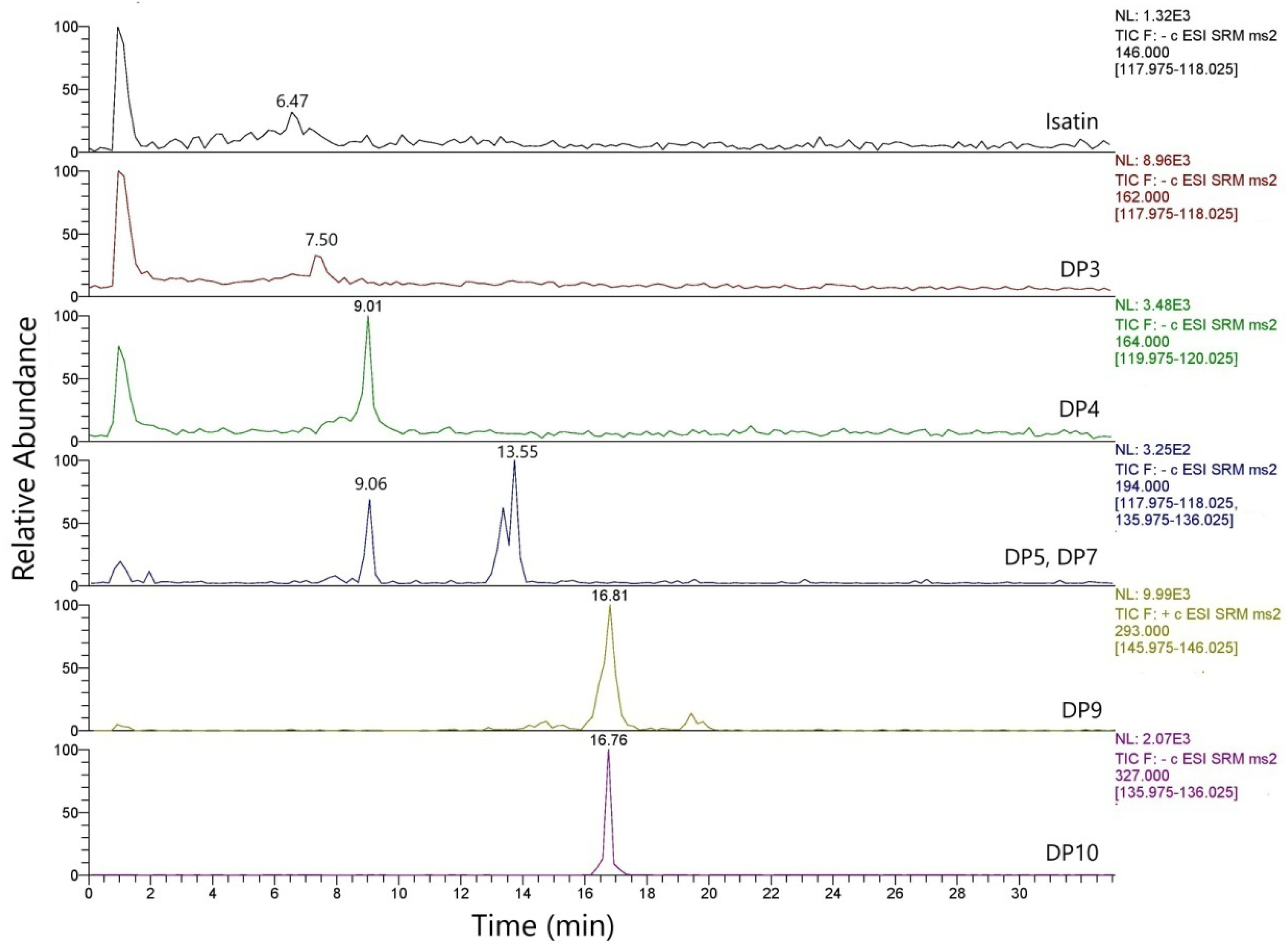
3.1. Degradation Products of Indigotin (IND) in Standard Solutions

In order to study the degradation products of shellfish purple, a stock solution of IND, used as a reference sample, at the concentration of 50 μg mL−1 was left at room temperature in the dark for 30 days (aged 1). After 30 days, the solution was analyzed using UHPLC-MS/MS. A fresh solution of IND was also analyzed for comparison.
According to the results (Table 3), the indigoid degradation products isatin (IS), isatoic anhydride (DP3), N-methoxycarbonyl-anthranilic acid (DP7), DP4, DP5, DP6, DP9 and DP10, as identified in a previous study [29], were detected in the aged sample of the standard solutions of IND. The degradation products DP3, DP4, DP5, DP6, DP7, DP10 and IS were detected in negative ion mode, and the degradation product DP9 was detected in positive ion mode. Figure 1 illustrates the chromatograms of IND solutions following 30 days at room temperature without light, in which the degradation products of the component appear.

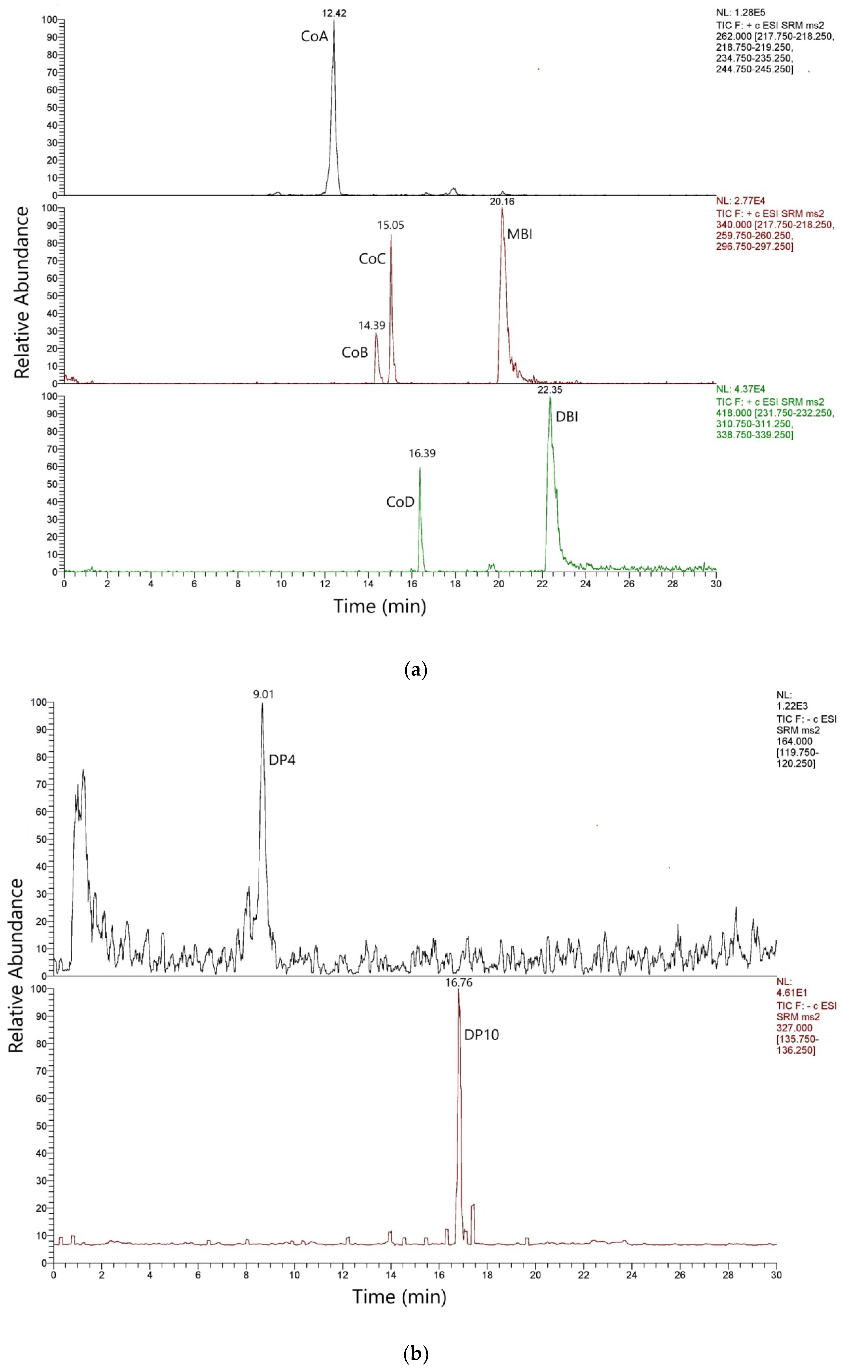
3.2. Coloring Components and Degradation Products in Hexaplex trunculus L. Pigment

Fresh solutions of Hexaplex trunculus L. extracts from Tunisia, prepared as described earlier [31], were analyzed, leading to the detection of eleven coloring components and degradation products, as shown in Table 3. The seven major coloring components (IND, INR, MBI, 6MBIR, 6′MBIR, DBI, DBIR), detected in our earlier work using HPLC-DAD [31], and minor compounds A, B, C and D [22] have been identified herein using UHPLC-MS/MS. The presence of the four minor compounds A–D, which were detected by Surowiec in Hexaplex trunculus L. extracts, is confirmed by the results presented in Table 3.
The compounds A, B, C and D belong to the same structural class as non-, mono- and di-brominated indigoids, and their relative molecular masses, which were exhibited in MS, are one unit lower than their respective calculated molecular masses. In the chemical formula of compound A, a CO group in INR is substituted by a CNH, whereas compounds B, C and D are derivatives of compound A [22]. Degradation products 4 (DP4) and 10 (DP10) [29] were also identified in traces in the fresh sample, probably as a result of the colorant preparation [30]. Figure 2 shows the chromatogram of fresh shellfish purple solution (from Hexaplex trunculus L.). The complete list of substances detected in the aged dyestuff solutions is illustrated in Figure S3 and provided in Table 3.

3.3. Degradation Products in Dyed Textiles

A silk sample dyed with the shellfish purple (Hexaplex trunculus L.) [31] was artificially aged under UV radiation, and the extract was analyzed by UHPLC (aged 2). Moreover, the extract of the fresh dyed silk sample was left for 30 days at room temperature in the dark (aged 1) and then analyzed for comparison. The results are presented in Table 3 and Figure S4.
Figure S4 illustrates the chromatograms of the degradation products of shellfish purple of dyed silk, including four coloring compounds, A, B, C and D [22], in a fresh solution and in the solution after 30 days of aging. As shown in Figure S4 and Table 3, the DP4 and DP10 [29] were identified in the freshly dyed silk sample with shellfish purple. These degradation products appear both in the fresh solution of shellfish purple colorant (Section 3.2) and in the unaged and aged silk samples dyed with the shellfish purple colorant, which can probably be attributed to the preparation of the dyestuff [31]. Degradation products DP3-6 and IS appeared in the extract of the dyed silk sample that was left at room temperature for 30 days without light. In this sample, the coloring components 6′MBIR, 6MBIR and DBIR were not detected.
The four compounds, A, B, C, D [22], and coloring components 6′MBIR, 6MBIR and DBIR were not detected after UV aging, indicating their instability towards UV irradiation and their initial small quantities in the unaged sample. In the dyed silk sample, it is observed that the coloring components found in the fresh sample in exceptionally low concentrations, such as 6′MBIR, 6MBIR and DBIR, are not detected after both aging experiments: aged 1 (left at room temperature with no light for 30 days) and aged 2 (accelerated ageing under UV radiation for 25 days). Furthermore, the four compounds A, B, C and D [22] detected in the fresh and aged solutions (aged 1) are not found in the samples subjected to UV radiation (aged 2).

4. Discussion

Artificially accelerated experiments have been carried out for several decades to study the fading of natural colorants under light aging [42,43,44,45,46]. Colorimetry has been employed to monitor the artificially accelerated fading of textiles and indigoid colorants on fibers [43,46]. The ageing process of the coloring components of the indigoids is being investigated by the use of HPLC [25,27,28,29]. IS and isatoic anhydrite (DP3) are two well-known degradation products of IND [23,25,26,27,28,29,32]. IS is produced form the acidic degradation of IND [25]. In addition, IND has been shown to be completely degraded after exposure to light or ozone, with IS and isatoic anhydride (DP3) being the reaction products [23,26]. Losses of CO and CO2 from the indol carbonyl and carboxyl group of IND are observed in the degradation products DP4, DP5, DP6 and N-methoxycarbonyl-anthranilic acid (DP7) [29]. The degradation products DP9 and DP10, which probably are indigotin’s photooxidation products, exhibit a larger relative molecular mass than IND [29]. Various of these degradation products were identified in DMSO [27,29], DMF [27] and dichloromethane [26] solutions of IND, INR, isoindigotin [29] and indigo colorant [26,27,28,29]. For the first time herein, several degradation products were detected in shellfish purple colorant and in aged textiles (silk) dyed with shellfish purple (Table 3).
Regarding the ageing experiment of the fresh shellfish purple colorant solution after 30 days at room temperature without light (aged 1), the coloring components IND, INR, MBI, DBI and coloring compounds A, B, C and D [22] were detected. However, the coloring components 6MBIR, 6′MBIR and DBIR disintegrated almost entirely below the levels of detection, while the degradation products IS, DP3, DP4, DP6 [29] were apparent (Table 3, Figure S3).
Regarding the artificial accelerated experiments in textiles under radiation, the amounts of coloring components from textiles were rapidly decreased during the first days of their exposure to light, while longer light treatment had a minor effect on their amounts [24,42,43,44]. Furthermore, the dibrominated components of shellfish purple (DBI, DBIR) appear to be less stable than IND and MBI [24]. The photodegradation of indigoids in textile fibers leads to the formation of degradation products. The main degradation products of indigo colorant identified in archaeological and historical textiles are DP4, DP5, DP7 (N-methoxycarbonyl-anthranilic acid) and DP10 [29], DP3 (isatoic anhydrite) [28,29], IS [27,28] and 2-benzyl-3-indolinone [28]. Degradation products DP3-6 [29] and IS were revealed in the extract of the dyed silk sample that was kept at room temperature for 30 days without light, while no traces of the coloring components 6′MBIR, 6MBIR and DBIR were found (Table 3, Figure S4). Following UV ageing, the four compounds A, B, C and D [22], as well as the coloring components 6′MBIR, 6MBIR and DBIR, were not detected (Table 3), indicating their instability towards UV irradiation and their initial small quantities in the unaged sample.

5. Conclusions

A UHPLC-MS/MS analytical method is presented for the detection of eleven main and minor coloring components (IND, INR, MBI, 6′MBIR, 6MBIR, DBI and DBIR, compounds A, B, C and D [22]) and eight degradation products (IS, DP 3,4,5,6,7,9 and 10 [29]) of shellfish purple, utilizing direct infusion fragment ion investigation. Dimethyl sulfoxide, a solvent widely used to dissolve low solubility substances, hindered detection in ESI; thus, a novel solution preparation approach was developed for the direct infusion analysis of the standard compounds and the optimization of detection parameters. It is the first simultaneous identification of 19 compounds that can be found in shellfish purple colorant. The four compounds A, B, C, D and the coloring components 6′MBIR, 6MBIR and DBIR were not detected after UV aging in the silk sample, indicating their instability towards UV irradiation and their initial small quantities in the unaged sample. 6′MBIR, 6MBIR, and DBIR found in the fresh sample of shellfish purple colorant solution in exceptionally low concentrations were not detected after ageing experiments. The eight indigoid degradation products were detected in at least one aged sample. The application of the proposed method in accelerated aging experiments showed that the degradation products differ depending on the initial colorant used, the textile and the aging conditions, indicating that further research on the degradation products could contribute to archaeological research regarding shellfish purple characterization.

Supplementary Materials

The following supporting information can be downloaded at: https://www.mdpi.com/article/10.3390/heritage7040092/s1. Figure S1: The optimizing tube lens and breakdown curve of ion of isatin, indigotin, indirubin and their brominated derivatives; Figure S2: The chromatograms for the components at a concentration of 10 μg mL−1; Figure S3: The degradation products and four coloring components of the solution of shellfish purple colorant that was left at room temperature for 30 days; Figure S4: Chromatograms of silk samples dyed with shellfish purple; (a) four coloring components and (b) degradation products in fresh solution and (c) degradation products in the solution after 30 days.

Author Contributions

Conceptualization, A.Z., I.K. and A.V.; methodology, S.K., I.S. and A.V.; validation, A.V. and I.S.; formal analysis, A.V. and I.S.; investigation, A.V. and I.S.; resources, S.K. and A.V.; data curation, A.V., I.S., S.K. and I.K.; writing—original draft preparation, A.V.; writing—review and editing, A.V., I.S., S.K., I.K., G.T. and A.Z.; visualization, A.V and I.S.; supervision, S.K.; project administration, A.Z.; funding acquisition, A.V. All authors have read and agreed to the published version of the manuscript.

Funding

This research was funded by the General Secretariat for Research and Technology (GSRT) and the Hellenic Foundation for Research and Innovation (HFRI), grant number: 1514 (16th) by the first scholarship for PhD students.

Data Availability Statement

The data are contained within the article and Supplementary Materials.

Conflicts of Interest

The authors declare no conflicts of interest.

References

  1. Cardon, D. Natural Dyes: Sources, Traditions, Technology and Science; Archetype: London, UK, 2007. [Google Scholar]
  2. Clark, R.J.H.; Cooksey, C.J.; Daniels, M.A.M.; Withnall, R. Indigo, woad, and Tyrian Purple: Important vat dyes from antiquity to the present. Endeavour 1993, 17, 191–199. [Google Scholar] [CrossRef]
  3. Koren, Z.C. New Chemical Insights into the Ancient Molluskan Purple Dyeing Process; Chapter 3; ACS Symposium Series; American Chemmical Society: Washigton, DC, USA, 2013. [Google Scholar]
  4. Maravelaki-Kalaitzaki, P.; Kallitrakas-Kontos, N. Pigment and terracotta of Hellenistic figurines in Crete. Anal. Chim. Acta 2003, 497, 209–225. [Google Scholar] [CrossRef]
  5. Ribechini, E.; Pérez-Arantegui, J.; Pavan, A.; Degano, I.; Zanaboni, M.; Colombini, M.P. First evidence of purple pigment production and dyeing in southern Arabia (Sumhuram, Sultanate of Oman) revealed by mass spectrometric and chromatographic techniques. J. Cult. Herit. 2016, 19, 486–491. [Google Scholar] [CrossRef]
  6. Koren, Z.C. Archaeo-chemical analysis of Royal Purple on a Darius I stone jar. Microchim. Acta 2008, 162, 381–392. [Google Scholar] [CrossRef]
  7. March, R.E.; Papanastasiou, M.; McMahon, A.W.; Allen, N.S. An investigation of paint from a mural in the church of Sainte Madeleine, Manas, France. J. Mass Spectrom. 2011, 46, 816–820. [Google Scholar] [CrossRef]
  8. Breniquet, C.; Desrosiers, S.; Nowik, W.; Rast-Eicher, A. Les textiles découverts dans les tombes de l’agedu Bronze moyen a Chagar Bazar (Syrie). In Chagar Bazar (Syrie) VIII. Les Tombes Ordinaires d’ L’¯ageduBronzeAncien et Moyen des Chantiers D-F-H-I (1999–2011); Tunca, O., Baghdo, A., Eds.; Peeters Publishers: Lueven-Liege, Belgium, 2018; pp. 11–32. [Google Scholar]
  9. Abdel-Kareem, O.; Alawi, M.A.; Mubarak, M.S. Identification of Dyestuffs in a Rare Coptic Garment Using High Performance Liquid Chromatography with Photodiode Array Detection (HPLC-PDA). JTATM 2010, 6, 1–7. [Google Scholar]
  10. James, M.A.; Reifarth, N.; Mukherjee, A.J.; Crump, M.P.; Gates, P.J.; Sandor, P.; Robertson, F.; Pfälzner, P.; Evershed, R.P. High prestige royal purple dyed textiles from the Bronze Age royal tomb at Qatna, Syria. Antiquity 2009, 83, 1109–1118. [Google Scholar] [CrossRef]
  11. Hartl, A.; van Bommel, M.R.; Joosten, I.; Hofmann-de Keijzer, R.; Grömer, K.; Rösel-Mautendorfer, H.; Reschreiter, H. Reproducing colourful woven bands from the Iron Age salt mine of Hallstatt in Austria: An interdisciplinary approach to acquire knowledge of prehistoric dyeing technology. J. Archaeol. Sci. Rep. 2015, 2, 569–595. [Google Scholar] [CrossRef]
  12. Koh, A.A.; Betancourt, P.P.; Pareja, M.N.; Brogan, T.M.; Apostolakou, V. Organic residue analysis of pottery from the dye workshop at Alatsomouri-Pefka, Crete. J. Archaeol. Sci. Rep. 2016, 7, 536–538. [Google Scholar] [CrossRef]
  13. Benkendorff, K.; Rudd, D.; Nongmaithem, B.D.; Liu, L.; Young, F.; Edwards, V.; Avila, C.; Abbott, C.A. Are the Traditional Medical Uses of Muricidae Molluscs Substantiated by Their Pharmacological Properties and Bioactive Compounds? Mar. Drugs 2015, 13, 5237–5275. [Google Scholar] [CrossRef]
  14. Katsifas, C.; Ignatiadou, D.; Zacharopoulou, A.; Kantiranis, N.; Karapanagiotis, I.; Zachariadis, G. Non-Destructive X-ray Spectrometric and Chromatographic Analysis of Metal Containers and Their Contents, from Ancient Macedonia. Separations 2018, 5, 32. [Google Scholar] [CrossRef]
  15. Vermeulen, M.; Tamburini, D.; McGeachy, A.C.; Meyers, R.D.; Walton, M.S. Multiscale characterization of shellfish purple and other organic colorants in 20th-century traditional enredos from Oaxaca, Mexico. Dyes Pigments 2022, 206, 110663. [Google Scholar] [CrossRef]
  16. Michel, R.H.; Lazar, J.; McGovern, P.E. Indigoid dyes in Peruvian and Coptic textiles of the University 632 Museum of Archaeology and Anthropology. Archeomaterials 1992, 6, 69–83. [Google Scholar]
  17. Chavez, E.A.; Michel-Morfin, J.E. Strategies for sustainable dye harvest of the purple conch Plicopurpura pansa (Gould, 1853) from west central Mexico. Am. Malacol. Bull. 2006, 21, 51–57. [Google Scholar]
  18. Rilov, G. Multi-species collapses at the warm edge of a warming sea. Sci. Rep. 2016, 6, 36897. [Google Scholar] [CrossRef] [PubMed]
  19. Koren, Z.C. High-Performance Liquid Chromatographic analysis of an ancient tyrian purple dyeing vat from Israel. Isr. J. Chem. 1995, 35, 117–124. [Google Scholar] [CrossRef]
  20. Koren, Z.C. HPLC-PDA analysis of brominated indirubinoid, indigoid, and isatinoid dyes. In Indirubin, the Red Shade of Indigo; Meijer, L., Guyard, N., Skaltsounis, A.L., Eisenbrand, G., Eds.; Life in Progress Editions; Station Biologique: Roscoff, France, 2006; Chapter 5; pp. 45–53. [Google Scholar]
  21. Nowik, W.; Marcinowska, R.; Kusyk, K.; Cardon, D.; Trojanowicz, M. High performance liquid chromatography of slightly soluble brominated indigoids from Tyrian Purple. J. Chromatogr. A 2011, 1218, 1244–1252. [Google Scholar] [CrossRef] [PubMed]
  22. Surowiec, I.; Nowik, W.; Moritz, T. Mass spectrometric identification of new minor indigoids in shellfish purple dye from Hexaplex trunculus. Dyes Pigments 2012, 94, 363–369. [Google Scholar] [CrossRef]
  23. Grosjean, D.; Whitmore, P.M.; Cass, G.R.; Druzlk, J.R. Ozone fanding of natural organic colorants: Me chanisms and products of the reaction of ozone with indigos, Environ. Sci. Technol. 1988, 22, 292–298. [Google Scholar] [CrossRef]
  24. Vasileiadou, A.; Karapanagiotis, I.; Zotou, A. UV-induced degradation of wool and silk dyed with shellfish purple. Dyes Pigments 2019, 168, 317–326. [Google Scholar] [CrossRef]
  25. Molino, R.J.E.J.; Junio, H.A. Profiling the Philippine Blue: Liquid chromatography/mass spectrometry-based metabolomics study on Philippine Indigofera. Rapid Commun. Spectrom. 2021, 35, 9037. [Google Scholar] [CrossRef]
  26. Novotna, P.; Boon, J.J.; van der Horst, J.; Paakova, V. Photodegradation of indigo in dichloromethane solution. Color. Technol. 2003, 119, 121–217. [Google Scholar] [CrossRef]
  27. Sousa, M.M.; Miguel, C.; Rodrigues, I.; Parola, A.J.; Pina, F.; de Melo, J.S.S.; Melo, M.J. A photochemical study on the blue dye indigo: From solution to ancient Andean textiles. Photochem. Photobiol. Sci. 2008, 7, 1353–1359. [Google Scholar] [CrossRef]
  28. Poulin, J. Identification of indigo and its degradation products on a silk textile fragment using gas chromatography-mass spectrometry. J. Can. Assoc. Conserv. 2007, 32, 48–56. [Google Scholar]
  29. Witkoś, K.; Lech, K.; Jarosz, M. Identification of degradation products of indigoids by tandem mass spectrometry. J. Mass Spectrom. 2015, 50, 1245–1251. [Google Scholar] [CrossRef] [PubMed]
  30. Karapanagiotis, I.; Mantzouris, D.; Cooksey, C.; Mubarak, M.; Tsiamyrtzis, P. An improved HPLC method coupled to PCA for the identification of Tyrian purple in archaeological and historical samples. Microchem. J. 2013, 110, 70–80. [Google Scholar] [CrossRef]
  31. Vasileiadou, A.; Karapanagiotis, I.; Zotou, A. Determination of Tyrian purple by high performance liquid chromatography with diode array detection. J. Chromatogr. A 2016, 1448, 67–72. [Google Scholar] [CrossRef] [PubMed]
  32. Rosenberg, E. Characterization of historical organic dyestuffs by liquid chromatography-mass spectrometry. Anal. Bioanal. Chem. 2008, 391, 33–57. [Google Scholar] [CrossRef]
  33. Lech, K.; Fornal, E. A Mass Spectrometry-Based Approach for characterization of red, blue, and purple natural dyes. Molecules 2020, 25, 3223. [Google Scholar] [CrossRef]
  34. Serrano, A.; van Bommel, M.; Hallett, J. Evaluation between ultrahigh pressure liquid chromatography and high-performance liquid chromatography analytical methods for characterizing natural dyestuffs. J. Chromatogr. A 2013, 1318, 102–111. [Google Scholar] [CrossRef]
  35. Szostek, B.; Orska-Gawrys, J.; Surowiec, I.; Trojanowicz, M. Investigation of natural dyes occurring in historical Coptic textiles by high-performance liquid chromatography with UV/Vis and mass spectrometric detection. J. Chromatogr. A 2003, 1012, 179–192. [Google Scholar] [CrossRef] [PubMed]
  36. Puchalska, M.; Polec-Pawlak, K.; Zadrozna, I.; Hryszko, H.; Jarosz, M. Identification of indigoid dyes in natural organic pigments used in historical art objects by high-performance liquid chromatography coupled to electrospay ionization mass spectrometry. J. Mass Spectrom. 2004, 39, 1441–1449. [Google Scholar] [CrossRef] [PubMed]
  37. Degano, I.; Biesaga, M.; Colombini, M.P.; Trojanowicz, M. Historical and archaeological textiles: An insight on degradation of wool and silk yarns. J. Chromatogr. A 2011, 1218, 5837–5847. [Google Scholar] [CrossRef] [PubMed]
  38. Andreotti, A.; Bonaduce, I.; Colombini, M.P.; Ribechini, E. Characterization of natural indigo and shellfish purple by mass spectrometric techniques. Rapid Commun. Mass Spectrom. 2004, 18, 1213–1220. [Google Scholar] [CrossRef] [PubMed]
  39. Zou, P.; Koh, H.L. Determination of indican, isatin, indirubin and indigotin in Isatis indigotica by liquid chromatography/electrospray ionization tandem mass spectrometry. Rapid Commun. Mass Spectrom. 2007, 21, 1239–1246. [Google Scholar] [CrossRef] [PubMed]
  40. Sanz, E.; Arteaga, A.; Garcia, M.A.; Camara, C.; Dietz, C. Chromatographic analysis of indigo from Maya Blue by LC-DAD-QTOF. J. Archaeol. Sci. 2012, 39, 3516–3523. [Google Scholar] [CrossRef]
  41. Virgiliou, C.; Sampsonidis, I.; Gika, H.G.; Raikos, N.; Theodoridis, G.A. Development and Validation of a HILIC-MS/MS multitargeted method for metabolomics applications. Electrophoresis 2015, 36, 2215–2225. [Google Scholar] [CrossRef]
  42. Giles, C.H. The fading of colouring matters. J. Appl. Chem. 1965, 15, 541–550. [Google Scholar] [CrossRef]
  43. Crews, P.C. The fading rates of some natural dyes. Stud. Conserv. 1987, 32, 65–72. [Google Scholar] [CrossRef]
  44. Cristea, D.; Vilarem, G. Improving light fastness of natural dyes on cotton yarn. Dyes Pigments 2006, 70, 238–245. [Google Scholar] [CrossRef]
  45. Ford, B.L. Monitoring colour change in textiles on display. Stud. Conserv. 1992, 37, 1–11. [Google Scholar] [CrossRef]
  46. Daniels, V. The light-fastness of textiles dyed with 6,6´-dibromoindigotin (Tyrian purple). J. Photochem. Photobiol. A 2006, 184, 73–77. [Google Scholar] [CrossRef]
Figure 1. Chromatogram of IND after 30 days at room temperature without light, in which the degradation products appear [29].
Heritage 07 00092 g001
Figure 2. Chromatograms of the fresh shellfish purple colorant solution (from Hexaplex trunculus L.). The compounds A–D [22] (a) and traces of DP4 and DP10 [29] (b) are provided above, as extracted from sample chromatograms (Table 3).
Heritage 07 00092 g002
Table 1. Gradient elution program.
Time
(min)
Flow
(mL min−1)
A (%)B (%)
00.3955
30.3955
130.37030
190.34060
220.34060
370.3595
37.010.3955
380.3955
A: H2O + FA 0.1% (v/v); B: ACN + FA 0.1% (v/v).
Table 2. Retention time and mass measurements of [M+H]+ and [M+H] ions for main and minor coloring components [22] of shellfish purple and degradation products [29].
CompoundsChemical
Formula
Rt (min)Ion ModePrecursor Ion (m/z)Fragment Ion (m/z) and CE (V)
ISC8H5NO26.47146118.03 (14) 42.45 (21) 90.10 (23)
DP3C8H5NO37.52162118
DP4C8H13NO39.01164120
DP5C9H15NO49.06194136
DP6C9H13NO39.51178134
DP7C9H13NO413.55194118
DP9C16H10N2O416.81+293146
DP10C15H12N2O6 16.76327135
Co AC16H11ON312.42+262219235 245
Co BC16H10ON3Br14.39+340218 260 297
Co CC16H10ON3Br15.05+341219 260 297
Co DC16H9ON3Br216.39+418232 311 339
INDC16H10O2N217.9+262.8218.81 (25) 234.51 (25) 189.75 (37)
INRC16H10O2N218.8+262.8218.79 (24) 189.83 (36) 234.63 (22)
MBIC16H9O2N2Br20.16+342.3261.62 (28) 204.76 (49) 340,1 (14)
6′MBIRC16H9O2N2Br20.8+341296.32 (25) 204.93 (46) 340,1 (10)
6MBIRC16H9O2N2Br21.08+341261.56 (19) 233.72 (27) 204.76 (51)
DBIC16H8O2N2Br222.35+419184.84 (22) 340.83 (6) 63.14 (36)
DBIRC16H8O2N2Br224.08+419282.6 (29) 255.1 (35) 189.2 (38)
Table 3. Overall results of the study; degradation products and coloring components.
CompoundsIND SolutionShellfish Purple PigmentSilk Dyed with Shellfish Purple
FRESHAGED 1FRESHAGED 1FRESHAGED 2AGED 1
IS + + +
DP3 + + +
DP4 ++++++
DP5 +
DP6 + +
DP7 +
DP9 + +
DP10 ++ +++
Co A ++++++ ++
Co B ++++++ ++
Co C ++++++ ++
Co D ++++++ ++
IND+++++++++++++++++++++
INR ++++++++++
MBI +++++++++++++++
6′MBIR + +
6MBIR + +
DBI +++++++++++++++
DBIR ++ +
1 Aged 1: The sample was in solution form and was left in the dark at room temperature for 30 days. 2 Aged 2: The sample was subjected to accelerated ageing under UV radiation for 25 days; +++ main compounds; ++ minor compounds; + traces.
Disclaimer/Publisher’s Note: The statements, opinions and data contained in all publications are solely those of the individual author(s) and contributor(s) and not of MDPI and/or the editor(s). MDPI and/or the editor(s) disclaim responsibility for any injury to people or property resulting from any ideas, methods, instructions or products referred to in the content.

