Article

An Evaluation of the Efficacy and Safety of TAMIXAM®, Based on Hyaluronic Acid and Tamarind Seed Extract, for Esophageal Mucosal Protection from Acid Insult

1
Department of Pharmacy, Health and Nutritional Sciences, University of Calabria, 87036 Rende, CS, Italy
2
Nutratech S.r.l., c/o Department of Pharmacy, Health and Nutritional Sciences, University of Calabria, 87036 Rende, CS, Italy
3
Neilos S.r.l., Via Bagnulo 95, 80063 Piano di Sorrento, NA, Italy
4
Biochem S.r.l., Via G.Benini 13, 40069 Zola Predosa, BO, Italy
5
Shedir Pharma Group S.p.A., Via Bagnulo 95, 80063 Piano di Sorrento, NA, Italy
6
Complife Italia S.r.l., 27028 San Martino Siccomario, PV, Italy
*
Author to whom correspondence should be addressed.
Gastrointest. Disord. 2024, 6(1), 202-213; https://doi.org/10.3390/gidisord6010015
Submission received: 6 December 2023 / Revised: 8 February 2024 / Accepted: 11 February 2024 / Published: 18 February 2024

Abstract

:
TAMIXAM® is a novel technology that combines hyaluronic acid and tamarind seed extract in its formulation. It is designed to protect the esophageal mucosa by creating a barrier through its filmogenic properties. The aim of this study is to evaluate the safety and efficacy of this technology through mucoadhesion tests, a cell viability assay, TEER measurements, and morphological analysis on reconstructed esophageal mucosa exposed to 10% hydrochloric acid before and after treatment. The mucoadhesion test highlighted the synergistic bioadhesive effect of the technology’s components. Cell viability assays revealed the substantial mucoprotective and barrier effects of the technology, preserving tissue viability when applied before exposure to acid insult. A morphological analysis illustrated TAMIXAM®’s efficacy in countering acid-induced damage, reducing erosion, necrosis, and tissue degeneration compared to the positive control, both pre- and post-acid insult. An evaluation of epithelial integrity through TEER measurements indicated a minimal reduction in tissues treated with the invention before acid exposure, demonstrating its ability to maintain epithelial integrity in the presence of an acid insult. However, this effect was less pronounced in tissues treated with the technology after the acid insult, implying a potential partial recovery of epithelial integrity. Furthermore, comprehensive in vitro and in vivo studies supported the safety profile of the invention. In conclusion, TAMIXAM® emerged as a compelling solution, providing enhanced mechanical action to maintain epithelial balance and shield the esophageal mucosa from acid-induced damage.

1. Introduction

Several digestive disorders are characterized by the backward flow of stomach acid into the esophagus, causing symptoms such as heartburn, regurgitation, and chest pain, which can lead to a condition of the esophageal lining known as gastroesophageal reflux disease (GERD). GERD is a prevalent clinical condition that affects millions of people worldwide. In Europe and the USA, approximately 10–20% of the population suffer from GERD [1] and, over time, about half of all adults will experience reflux symptoms [2]. GERD results from damage to the esophageal mucosa caused by the irritating action of hydrochloric acid and other agents, such as pepsin, found in gastric secretions [3]. Gastric juices from the stomach can rise to the esophagus throughout the day, particularly after eating, causing a burning sensation behind the breastbone and acid regurgitation. This can reduce epithelial resistance and increase visceral sensitivity, resulting in an imbalance between the aggressive action of reflux and effective defense mechanisms, such as the buffering capacity of esophageal epithelial cells [4]. The repeated exposure of the esophagus to gastric contents, primarily hydrochloric acid and pepsin, can cause tissue damage. Hydrochloric acid alters the junctions between esophageal epithelial cells, leading to dilated intercellular spaces and increased para-cellular permeability [5].
There are several means leading to a symptomatic relief in patients with GERD, including various drugs or medical devices, such as proton pump inhibitors (PPIs) or H2 receptor antagonists, characterized by different mechanisms of action involving the neutralization of gastric acid and/or suppression of its production [6]. However, relevant side effects during the long-term use of PPIs and H2 receptor antagonists have been observed, resulting in an increase in the demand for alternative approaches [7].
In this context, medical devices based on natural extracts are attracting considerable interest. These devices have been demonstrated to have the ability to enhance mucosal defenses by forming a film over the esophageal mucosa and acting as a mechanical barrier to the harmful components of both acidic and basic reflux [8,9,10,11]. The use of these novel medical devices, coupled with PPI therapy, is expected to enhance the care of individuals experiencing extraesophageal GERD symptoms.
Lately, pharmaceutical industries have shifted their attention towards the creation of novel formulations based on hyaluronic acid (HA), a key component of the extracellular matrices involved in tissue repair and regeneration processes following damage. These properties make hyaluronic acid a key active in the creation of protective films on the esophageal mucosa, acting as a shield for acid-induced damage.
In addition, HA participates in moderating the inflammatory response, inducing cell proliferation and angiogenesis, enhancing re-epithelialization through the proliferation of basal keratinocytes, and reducing collagen deposition, leading to healing [12].
In recent studies, there has been an investigation into the efficacy of xyloglucans extracted from Tamarind seeds in addressing gastroesophageal disorders that affect mucosal permeability. Tamarind seeds (Tamarindus indica, L.) are obtained from a Leguminosae tree that has found use in Eastern medicine since the 16th century [13]. Currently, tamarind seed extract (TSX) is widely used in pharmaceutical formulations. It consists of a neutral polysaccharide with thickening, binding, emulsifying, gelling, and solubilizing properties. It has been shown to be highly biocompatible and biodegradable with excellent mucoadhesive properties, making it a natural polysaccharide suitable for numerous clinical applications such as the protection of injured oral mucosa and the development of new drug delivery systems [14].
Based on this evidence, this study aims to evaluate the efficacy and safety of TAMIXAM®, a new patented technology based on the presence of HA and TSX in its formulation. The main objective of our investigation is to evaluate the potential protective and film-forming capabilities of the technology in preserving the esophageal mucosa exposed to acid insult.

2. Results

2.1. Safety Assessment

2.1.1. Cytotoxicity Test

Upon microscopic examination, cells treated with the undiluted sample extract showed deviations from the normal morphology observed in the negative control after 24 and 48 h of incubation. The undiluted extract displayed mild reactivity after 24 h and severe reactivity after 48 h. However, cells treated with the 1:5 and 1:10 diluted extract did not show any deviations from the normal morphology. Both the 1:5 and 1:10 diluted extracts exhibited no reactivity. Please refer to Table 1 for a summary of the results.

2.1.2. Oral Mucosa Irritation Test

The macroscopic evaluation results (Table S1) indicated that hamsters subjected to the tested product exhibited no signs of erythema throughout the entire treatment period of 10 days. Histologic evaluation findings (Table 2) further revealed that, under the experimental conditions, the tested product did not induce leukocyte infiltration, vascular congestion, or edema. Based on these findings, it can be inferred that the technology is non-irritating to the oral mucosa.

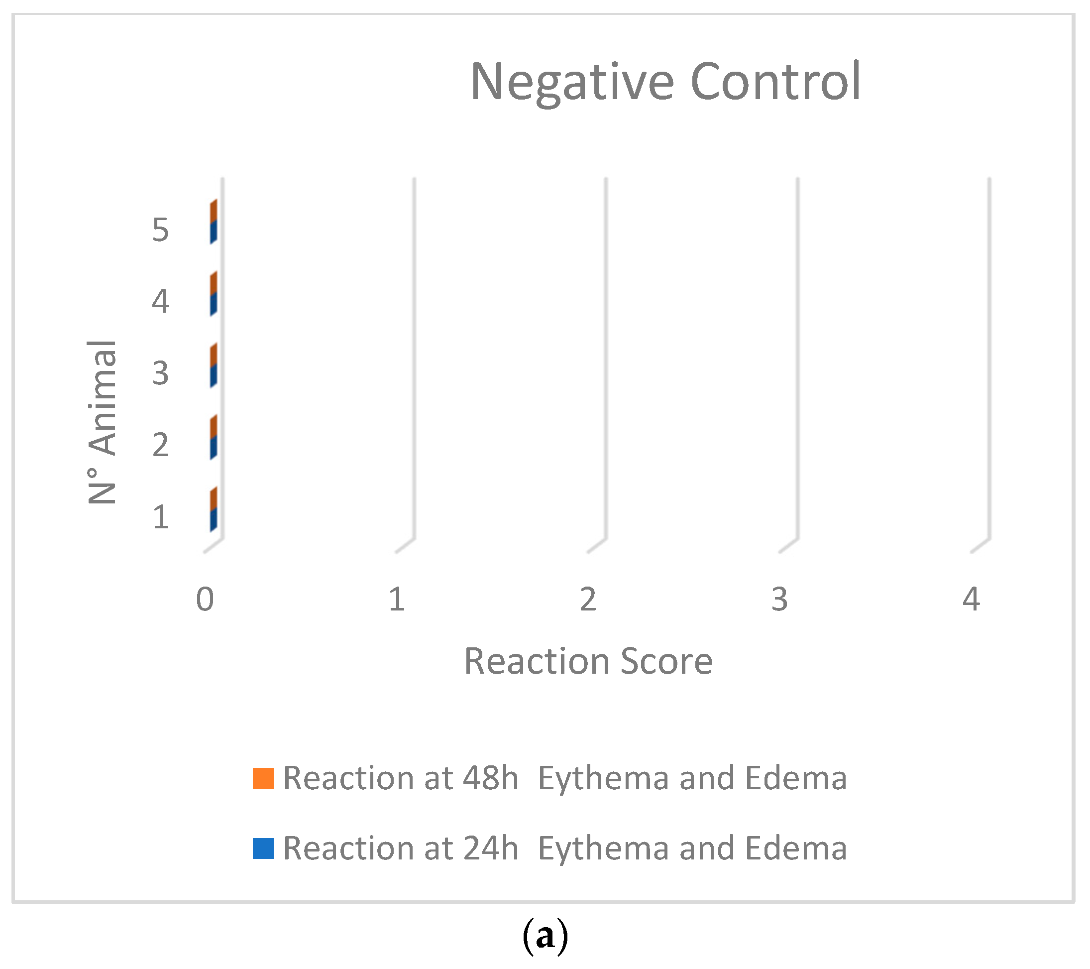
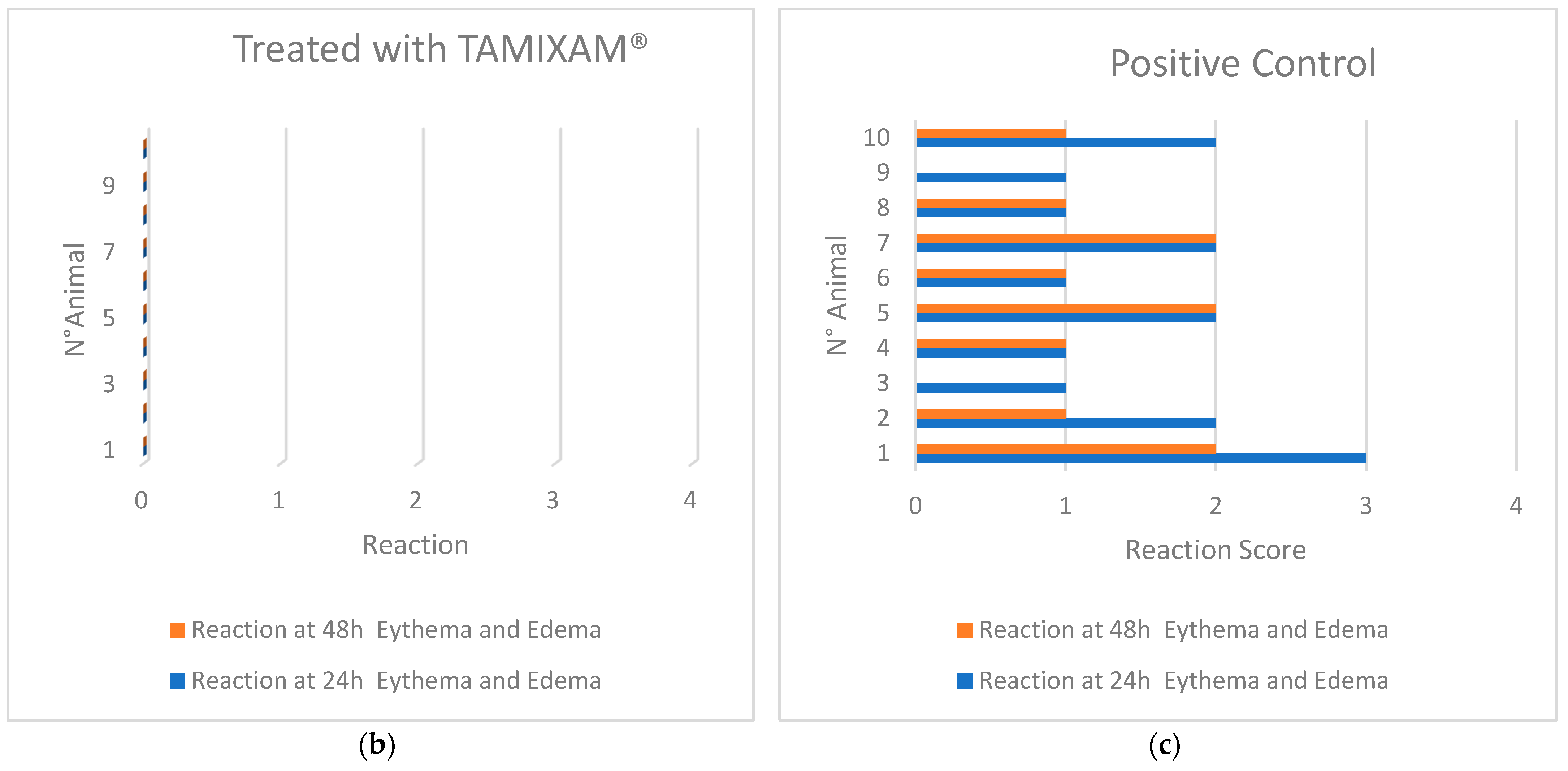
2.1.3. Skin Sensitization Test—Guinea Pig Maximization Test (GPMT)

The data depicted in Figure 1 illustrate that, within the applied experimental parameters, the sample does not exhibit any sensitizing effects.

2.2. Efficacy Assessment

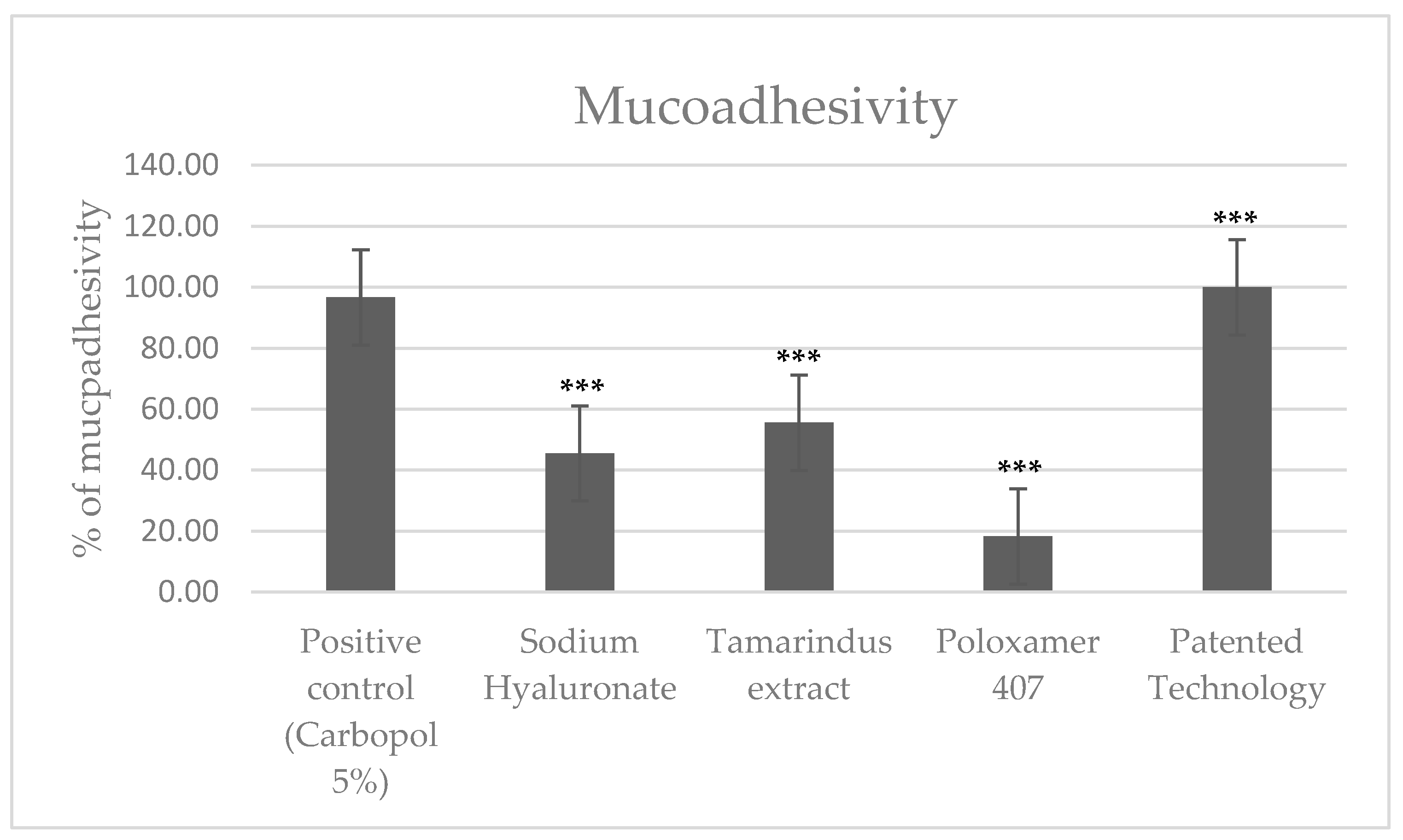
2.2.1. Mucoadhesion Test

The assessment of mucoadhesiveness was performed on both the technology and its three primary compounds. The results (refer to Table 3 and Figure 2) demonstrate the synergistic effect of the components of the technological formulation on mucoadhesive action.

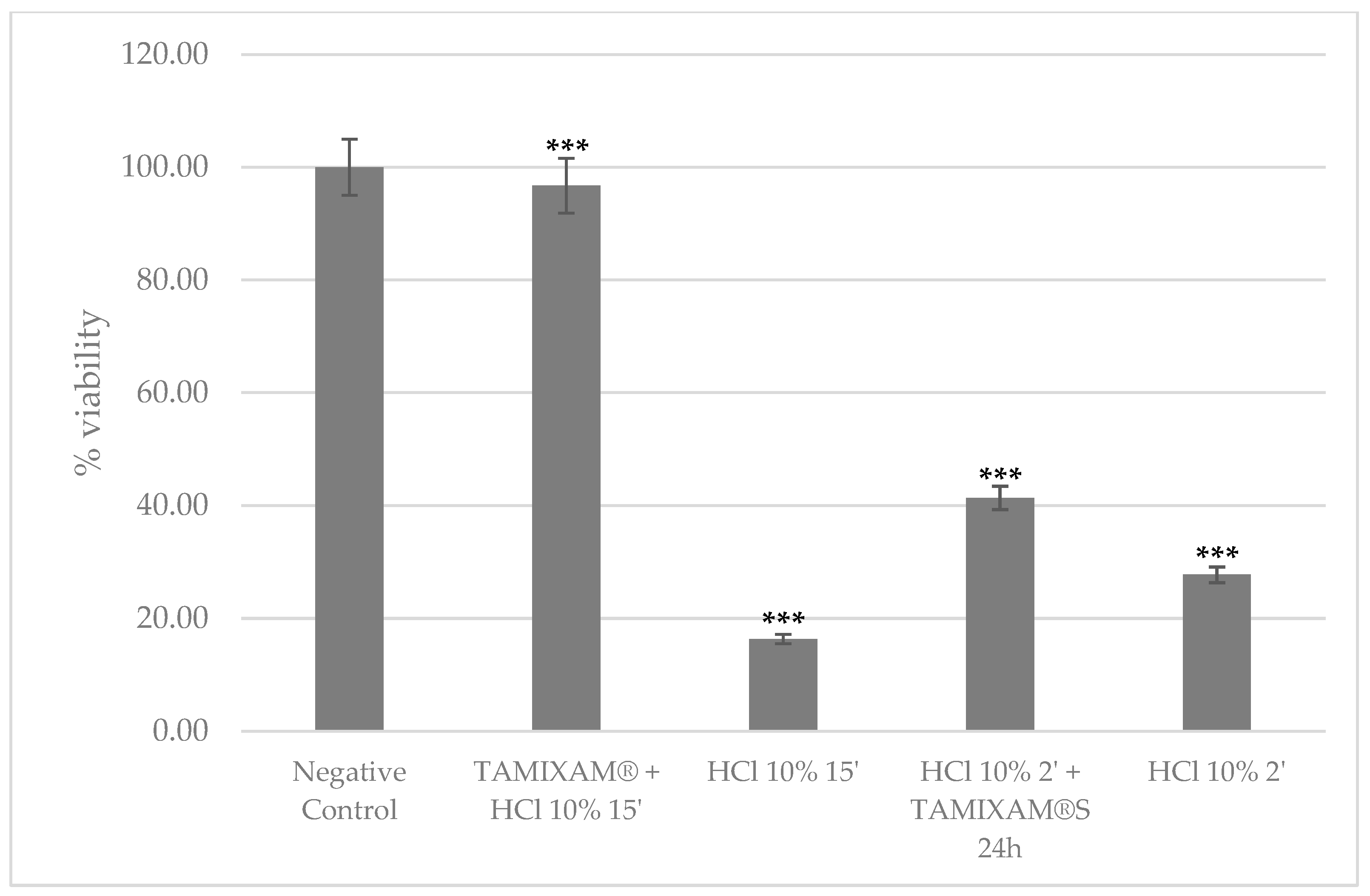
2.2.2. Viability Test

A cell viability evaluation (Figure 3) highlighted the mucoprotective and barrier effect exerted by the technology. When administered prior to the acid insult, the preservation of tissue viability reached approximately 96.73%. Tissues initially exposed to 10% HCl followed by treatment with the sample exhibited a viability of approximately 41%. This suggests that the tested sample possesses a considerable repair capacity, enabling it to partially mitigate the detrimental effects of exposure to 10% HCl.

2.2.3. TEER Measurement

The assessment of epithelial integrity showed that the TEER value was reduced by approximately 59.72% after a 15 min exposure to HCl and by 36.55% after a 2 min exposure compared to basal conditions (Table 4). The tissues treated initially with the test sample and subsequently with 10% HCl exhibited a TEER reduction of approximately 2.41%. This outcome demonstrated that TAMIXAM® has the ability to protect the esophageal mucosa and maintain its epithelial integrity when exposed to acid insults. Additionally, the tissues treated with 10% HCl and then with the test sample, evaluated 24 h post-treatment, showed a 23.53% reduction in TEER. This observation suggests that the tested technology can effectively restore epithelial integrity after damage caused by acid insult.

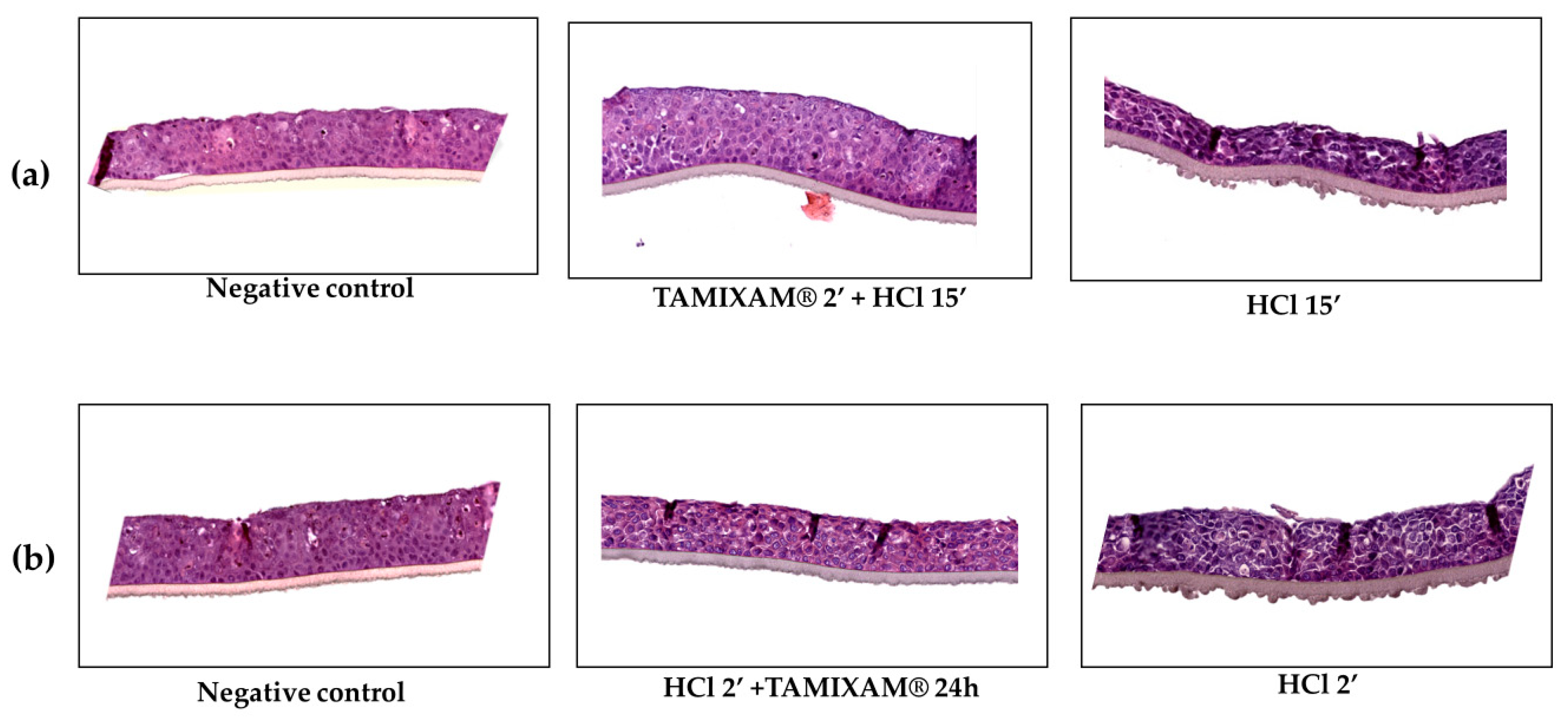
2.2.4. Morphological Analysis

A morphological analysis revealed that the technology is effective in restoring epithelial integrity after acid damage. A histological examination (Figure 4) demonstrated the technology’s strong protective capabilities compared to the positive control, particularly in reducing cellular degeneration, epithelial erosion, and necrosis after a 2 min acid insult (Table 5). Furthermore, as depicted in Figure 4b, the technology showcased its regenerative potential, albeit to a lesser extent than in the prior scenario. Morphological studies not only confirmed the product’s ability to repair damage induced by acid insult but also highlighted its significant preventive effect through the formation of a protective barrier.

3. Materials and Methods

3.1. Safety Assessment

3.1.1. Cell Cultures

The used test system was L-929 mouse fibroblast cells (ATCC CCL 1, NCTC clone 929 of strain L, Ref. ISO 10993-5: 2009 [15]; the established and preferred cell lines are listed in Note 3). Primary supplier: Istituto Zooprofilattico Sperimentale dell’Emilia-Romagna, Via A. Bianchi, 9 25,124 BRESCIA (Italy). The used culture medium was Minimum Essential Medium (MEM) with Earle’s salts and 5% fetal bovine serum, 1% L-glutamine, 0.6% penicillin/streptomycin, and 0.3% fungizone (complete MEM).

3.1.2. Animals

Syrian Hamster males and Hartley guinea pigs (nulliparous and non-pregnant females and/or males) were supplied by Charles River Laboratories Italia s.r.l. (Via Indipendenza, 11—23,885 Calco (Lecco), Italy). The housing conditions of the animals are reported in Tables S2 and S3 of the Supplementary Materials.

3.1.3. Tested Sample

TAMIXAM® is a patented technology composed of hyaluronic acid and tamarind seed extract and available in both liquid and solid form. It was supplied by Neilos s.r.l (Via Bagnulo 95—80,063 Piano di Sorrento (NA)—Italy) and it is contained within the medical device called “REXOFLUS®” supplied by Neilos s.r.l (Via Bagnulo 95—80,063 Piano di Sorrento (NA)—Italy).
REXOFLUS® is a medical device that comes in a liquid form with a high density to ensure effective and long-lasting adhesion to the oropharyngeal mucosa, allowing the product to perform its functions. Its formulation contains sodium alginate, sodium bicarbonate, calcium carbonate, tamarind seed extracts, hyaluronic acid, xanthan gum, sodium propyl paraoxybenzoate, sodium methyl paraoxybenzoate, polyoxamer 407, sucralose, flavouring, and water. It is licorice/mint-flavored and comes in 10 mL sticks. The recommended dosage is 1 sachet after each main meal and in the evening before going to bed. It is indicated in the treatment of gastro-esophageal and laryngeal pharyngeal reflux and related symptoms such as dysphagia, odynophagia, and heartburn. It is a sugar-free product. It is marketed in Italy and other European countries.

3.1.4. Cytotoxicity Test

The test was conducted following ISO 10993-5: 2009 and ISO 10993-12: 2012 [16]. Cells were grown in plates until a nearly confluent monolayer was obtained. Three cell culture plates were prepared for each sample. Additionally, three plates were prepared for negative control, three for positive control, and three for liquid control of extraction. In the plates intended to be treated with the sample, the medium was aspirated and replaced with the sample extract. The cell cultures were examined microscopically after 24 and 48 h of incubation with the extract to assess any cytotoxic effects. Cytotoxicity was evaluated by examining the general morphology, presence of vacuolization, detachments, cell lysis, and membrane integrity of the cells after 24 and 48 h of incubation with the sample extract. Deviations from normal morphology, as evidenced by the negative control, are assigned a score from 0 to 4. The scores range from 0 (none) to 4 (severe reactivity). Further details can be found in the Supplementary Materials (Table S4).

3.1.5. Oral Mucosa Irritation Test

The trial followed ISO 10993-10:2010 [17] and ISO 10993-12:2012 standards, except for histological evaluation. The sample was repeatedly administered into the right pockets of three hamsters in the Treated Group once a day, five days per week, for two weeks. The left pockets were not used for sample administration. The Control Group consisted of three other hamsters, into whose right pockets the negative control was administered using the same procedure. After the final treatment, all animals were sacrificed, and the pocket mucosa was removed for histological analysis.
A macroscopic evaluation of reactions in the treated area was conducted using a grading scale (Table S5). The scale ranges from 0, which denotes the absence of erythema, to 4, which signifies severe erythema or eschar formation. The sampled oral mucosa was compared with the control. The average score per animal was determined by totaling the scores assigned after each observation and dividing the sum by the number of observations.
Histological preparations were graded using a scale (Table S6). To obtain the mean score for the treatment, the cumulative histological scores for the sampled oral mucosa were totaled and divided by the number of observations. Similarly, the irritation index was calculated by subtracting the mean negative control score from the mean treatment score (Table S7). The index is described using adjectives based on the mean score, where 0 denotes no irritation, 1–4 indicates minimal irritation, 5–8 signifies mild irritation, 9–11 represents moderate irritation, and 12–16 signifies severe irritation.

3.1.6. Skin Sensitization Tests—Guinea Pig Maximization Test (GPMT)

After a two-stage induction using complete Freund’s adjuvant and sodium lauryl sulfate (SLS), the sample extract or the sample itself were applied to the guinea pigs’ skin using a swab. Observations were made at the application sites 24 and 48 h after the swab was removed. Each site was evaluated for erythema and edema and scored from 0 to 3 (see Table S8). Any animal that showed a reaction at 24 or 48 h, with a score of one or higher for erythema and edema, was considered sensitized, as long as the control animals exhibited evaluable reactions with lower scores. The results are presented as the frequency of observations with a grade > 1 in both the treated and control animal groups.

3.2. Efficacy Assessment

3.2.1. Materials

HO2E/S/5 Reconstructed Human Oesophageal Batch N° 23 HO2E 006 and Maintenance Medium Batch N° 23 SMM 039 were obtained from EpiSkin (Lyon Cedex, France); MTT (3-(4,5-Dimethylthiazol-2-yl)-2,5-Diphenyltetrazolium Bromide (Sigma-Aldrich s.r.l. Milan, Italy); Evolution 201 UV/Vis spectrometer (Thermo Fisher Scientific, Waltham, MA, USA), Millicell-ERS-2 instrument (Millipore, Massachusetts, United States, range 0–20 kΩ).

3.2.2. Mucoadhesion Assay

The sample was distributed homogeneously over a film of mucin on a Si-laminated gel plate (Bond Biorad gel). The plate was then placed on a 45-degree inclined plane for 60 min. Adhesion capacity was measured using the gravimetric method and expressed as a percentage of residual mass. Mucoadhesion was determined as an absolute value and a relative value compared to a standard of Carbopol 5%, which is known to have mucoadhesive properties.

3.2.3. Experimental Conditions

This study used the SkinEthicTM HO2E model. The model consisted of a human esophageal epithelium composed of the immortalized cell line Kyse 510. The cells were cultivated on an inert polycarbonate filter at the air–liquid interface in a chemically defined medium called Episkin.
Investigations were carried out to evaluate the barrier and regenerative properties of the tested sample on an in vitro reconstructed human esophageal epithelium after damage was induced by a hydrochloric acid solution (10% v/v HCl at pH 1.1). Two distinct experimental protocols were executed for this purpose.
In the first protocol, the tissues were exposed to the hydrochloric acid solution for 2 min, followed by thorough rinsing with phosphate-buffered saline (PBS). The tissues were subsequently treated with the technology for 24 h to test the regenerative potential of the product. Other tissue inserts were treated with a 0.9% (w/v) sodium chloride (NaCl) solution for 2 min as a negative control, while the remaining inserts were exposed to a hydrochloric acid solution, rinsed with PBS, and then treated with saline for another 24 h as a positive control.
The second experimental procedure aimed to assess the protective efficacy of the technology. In this approach, the tissue inserts were first treated with the test product for 2 min, followed by a 15 min exposure to the hydrochloric acid solution. Tissue inserts treated with 0.9% NaCl for 2 min and then exposed to saline or hydrochloric acid for another 15 min were used as the negative and positive control groups, respectively.

3.2.4. Viability Assessment

The MTT reduction assay was used to assess tissue viability, following the guidelines outlined in ISO 10993-23:2021. The assessment was conducted after a 42 h recovery period and post-rinsing. A dye solution was prepared with a final concentration of 1 mg/mL in phosphate-buffered saline (PBS) and filtered through a 0.22 µm filter.
Next, 300 μL of MTT solution was dispensed into 24-well plates and incubated for 3 h (± 5 min) at 37 °C, 5% CO2, and 95% relative humidity. After this period, the tissues were transferred to 2-propanol. An additional 750 μL of 2-propanol was added to each tissue, followed by a 2 h (± 5 min) incubation at room temperature with gentle agitation to facilitate formazan extraction.
After completing the 2 h (± 5 min) incubation in 2-propanol, the tissue samples were punctured with a pipette tip, and the extraction solution was homogenized through gentle pipetting to ensure complete formazan solubilization. Three 200 μL aliquots of the extraction solution were transferred into 96-well plates, and the optical density was measured at 570 nm.

3.2.5. TEER Measurement

Transepithelial electrical resistance (TEER) assessment is a method used to evaluate cellular membrane integrity [18]. TEER measurements were conducted using a Millicell-ERS instrument with a measuring range of 0 to 20 kilo-ohms (kΩ) prior to the experiment. TEER values were measured at two specific times: the initial measurement at time zero (T0), performed before any treatment with the test sample, and the final measurement at the end of each treatment regimen (T1). The T1 measurements were taken under two different conditions: (a) after treatment with the test sample followed by exposure to the acid insult, and (b) after administration of the acid insult followed by treatment with the test sample.

3.2.6. Morphological Analysis

Following each treatment, tissue samples were fixed in a 4% formaldehyde solution for histological examination. Morphological evaluations were conducted using hematoxylin–eosin staining to assess barrier efficacy and tissue permeability resulting from damage induced by the hydrochloric acid solution. The purpose of this test was to measure the effectiveness of the test product in protecting and restoring the integrity of the human esophageal epithelium that has been reconstructed in vitro and damaged by a hydrochloric acid solution (10% v/v, pH 1.1).

3.3. Data Analysis

Each datum point represents the mean ± SD of three different experiments. Data were analyzed for statistical significance (p < 0.05) using an unpaired Student’s t-test, performed using the GraphPad-Prism7 software program (GraphPad Inc., San Diego, CA, USA).

4. Discussion

Recent investigations have brought attention to the remarkable ability of xyloglucan derived from tamarind seeds to mitigate mucosal permeability [19]. Concurrently, innovative treatments for GERD, incorporating hyaluronic acid (HA), have shown promise in healing ulcers by promoting re-epithelialization in the upper gastrointestinal mucosa and contributing to epithelial cell turnover [20,21,22,23,24].
In light of these developments, our study aimed to evaluate the safety and efficacy of TAMIXAM®, a novel technology that combines HA with TSX. This research has yielded significant findings, shedding light on potential applications and paving the way for the further exploration of this cutting-edge technology.
The safety of the technology has been confirmed through a variety of assays, as previously described [15,16,17]. In vitro cytotoxicity tests have been conducted to evaluate the impact of the system on cell viability, ensuring safety at the cellular level. The Oral Mucosal Irritation tests and the GPMT have been carried out to provide insights into potential irritation, allergic reactions, and/or skin sensitization issues, which are critical aspects in evaluating the drug’s safety in practical applications.
To uphold the uniqueness of our research on efficacy assessment, it has been crucial to compare our findings with studies involving HA by itself and in combination with other compounds. Numerous investigations have illustrated the favorable impact of HA and chondroitin sulfate on mucosal protection [25]. Nonetheless, TAMIXAM® distinguishes itself through its exclusive composition, encompassing both HA and TSX.
The effectiveness of the technology has been evaluated in terms of its ability to form a film and protect the mucous membranes. Prior to evaluation, an instrumental test was performed to assess the mucoadhesive properties of the technology. The obtained data showed a higher mucoadhesion rate for the combination of HA and TSX, compared to the outcomes observed with the individual components. These results effectively showcase the synergy between the two primary components, underscoring their collective impact on bioadhesive capacity.
To determine the mucoprotective capacity, a cell viability assay, TEER measurement, and morphological analysis were performed on reconstructed esophageal mucosa exposed to a solution of hydrochloric acid (HCl 10%). The assessment of cell viability revealed that the sample exhibits a highly effective mucoprotective and barrier effect. When applied prior to the acid insult, it demonstrated the preservation of tissue viability by approximately 96.73%. The tissues initially treated with 10% HCl and then exposed to the sample exhibited a viability of approximately 41%. This suggests that the tested sample possesses a reasonable repair capacity, indicating its ability to partially mitigate the damaging effects of exposure to 10% HCl.
A morphological analysis of the tissues treated with the tested sample and then with HCl 10% for 15’ showed that the technology is able to effectively counteract the damage caused by the acid insult by protecting the tissues from erosion and necrosis and by decreasing the degree of tissue degeneration, in comparison to a positive control treated only with 10% HCl. Furthermore, the morphological analysis of the tissues initially exposed to 10% HCl and subsequently treated with the test sample demonstrated a decrease in the levels of necrosis, erosion, and tissue degeneration compared to the positive control treated exclusively with 10% HCl, although to a lesser degree than in the previous scenario.
The evaluation of epithelial integrity showed that the TEER value is reduced by approximately 59.72% after 15’ exposure to HCl and 36.55% after 2’ exposure to HCl, compared to the basal condition. The tissues treated first with the test sample and then with HCl 10% exhibited a reduction in TEER of approximately 2.41%. This result demonstrates the ability of the technology to perform a barrier effect on the esophageal mucosa, preserving its epithelial integrity when exposed to acidic insult. The tissues previously treated with 10% HCl and subsequently with the test sample, 24 h after treatment, have shown a reduction in TEER of 23.53%, revealing the invention’s considerable efficacy in restoring epithelial integrity after damage induced by acid insult.
The significance of these findings lies in the development of a technology that not only ensures safety but also provides effective in vitro mucosal protection without disrupting the natural equilibrium of the esophageal epithelium. TAMIXAM®’s ability to reduce mucosal damage and maintain tissue integrity holds promise for potential applications in GERD treatment, where enhanced mucosal protection is crucial. The study contributes valuable insights to the field by introducing a novel approach that combines natural extracts for gastroesophageal reflux disease management.
However, it is crucial to acknowledge the limitations of our study. Although the results are promising, the invention has not been tested in realistic GERD models or clinical trials yet, leaving a gap in understanding its practical implications. The lack of clinical data on symptomatic relief highlights the need for further research and comprehensive clinical trials to determine the true potential and safety of TAMIXAM® in managing GERD.

5. Conclusions

In conclusion, TAMIXAM®, due to its formulation, could represent a new tool for protecting the esophageal mucosa when exposed to acid insult. Its mode of action is unique, relying on natural extracts and bioadhesive compounds to create a mechanical barrier without disrupting the natural equilibrium of the esophageal epithelium. However, these findings are preliminary and further investigations and clinical trials are necessary to determine the true potential of TAMIXAM® in managing GERD.

6. Patents

TAMIXAM® is a technological patent approved by the Ministry of Enterprises and Made in Italy and is marketed as a medical device called REXOFLUS®.

Supplementary Materials

The following supporting information can be downloaded at https://www.mdpi.com/article/10.3390/gidisord6010015/s1. Table S1: Results of the Macroscopical Evaluation (0 = absence of erythema, 1 = slight erythema, 2 = well-defined erythema, 3 = moderate erythema, 4 = severe erythema or eschar formation).Table S2: Housing conditions for animals Oral mucosa irritation test; Table S3: Housing conditions for animals Skin sensitization tests—guinea pig maximization tests (GPMT); Table S4: Cytotoxicity test; Table S5: Oral mucosa irritation test; Table S6: Histological preparations were evaluated using the following rating scale; Table S7: Irritation Index; Table S8: Scoring System Skin sensitization tests—guinea pig maximization test (GPMT).

Author Contributions

Conceptualization, M.F.M. and R.M.; methodology, G.B. and M.F.M.; visualization, R.M.; validation, F.A. and V.N.; formal analysis, R.M.; investigation, V.N. and G.P. resources, A.B. and U.D.M.; data curation M.F.M. and G.P.; writing—original draft M.F.M. and G.P.; writing—review and editing; V.N.; project administration, A.B. and U.D.M. All authors have read and agreed to the published version of the manuscript.

Funding

This research was funded by Neilos S.r.l., Via Bagnulo 95, 80063 Piano di Sorrento, NA, Italy; NT0000655/23, 03/07/2023, NT0000958/23, 25/09/2023.

Institutional Review Board Statement

The animal study protocol was approved by the Ministry of Health (protocol code 485/2016).

Informed Consent Statement

Not applicable.

Data Availability Statement

The data presented in this study are available in article and Supplementary Data.

Conflicts of Interest

A.B. is the Neilos s.r.l C.E.O. This does not alter the author’s adherence to all the journal policies on sharing data and materials. The funder had no role in the design of the study; in the collection, analyses, or interpretation of data; in the writing of the manuscript; or in the decision to publish the results. The other authors declare no conflicts of interest.

References

  1. Badillo, R.; Francis, D. Diagnosis and Treatment of Gastroesophageal Reflux Disease. World J. Gastrointest. Pharmacol. Ther. 2014, 5, 105–112. [Google Scholar] [CrossRef]
  2. Dent, J.; Armstrong, D.; Delaney, B.; Moayyedi, P.; Talley, N.J.; Vakil, N. Symptom Evaluation in Reflux Disease: Workshop Background, Processes, Terminology, Recommendations, and Discussion Outputs. Gut 2004, 53 (Suppl. 4), iv1–iv24. [Google Scholar] [CrossRef]
  3. Gnessi, L.; Bacarea, V.; Marusteri, M.; Piqué, N. Xyloglucan for the Treatment of Acute Diarrhea: Results of a Randomized, Controlled, Open-Label, Parallel Group, Multicentre, National Clinical Trial. BMC Gastroenterol. 2015, 15, 153. [Google Scholar] [CrossRef]
  4. Nirwan, J.S.; Hasan, S.S.; Babar, Z.-U.-D.; Conway, B.R.; Ghori, M.U. Global Prevalence and Risk Factors of Gastro-Oesophageal Reflux Disease (GORD): Systematic Review with Meta-Analysis. Sci. Rep. 2020, 10, 5814. [Google Scholar] [CrossRef] [PubMed]
  5. Orlando, R.C. The Integrity of the Esophageal Mucosa. Balance between Offensive and Defensive Mechanisms. Best Pract. Res. Clin. Gastroenterol. 2010, 24, 873–882. [Google Scholar] [CrossRef] [PubMed]
  6. Zheng, Z.; Nordenstedt, H.; Pedersen, N.L.; Lagergren, J.; Ye, W. Lifestyle Factors and Risk for Symptomatic Gastroesophageal Reflux in Monozygotic Twins. Gastroenterology 2007, 132, 87–95. [Google Scholar] [CrossRef] [PubMed]
  7. Ament, P.W.; Dicola, D.B.; James, M.E. Reducing Adverse Effects of Proton Pump Inhibitors. Am. Fam. Physician 2012, 86, 66–70. [Google Scholar] [PubMed]
  8. Pellegatta, G.; Spadaccini, M.; Lamonaca, L.; Craviotto, V.; D’Amico, F.; Ceriotti, L.; Meloni, M.; Repici, A. Evaluation of Human Esophageal Epithelium Permeability in Presence of Different Formulations Containing Hyaluronic Acid and Chondroitin Sulphate. Med. Devices Evid. Res. 2020, 13, 57–66. [Google Scholar] [CrossRef] [PubMed]
  9. Hotchkiss, A.T.; Nuñez, A.; Strahan, G.D.; Chau, H.K.; White, A.K.; Marais, J.P.J.; Hom, K.; Vakkalanka, M.S.; Di, R.; Yam, K.L.; et al. Cranberry Xyloglucan Structure and Inhibition of Escherichia Coli Adhesion to Epithelial Cells. J. Agric. Food Chem. 2015, 63, 5622–5633. [Google Scholar] [CrossRef] [PubMed]
  10. Pecora, T.M.G.; Parisi, O.I.; Bertin, W.; Ragazzo, B.; Dattilo, M.; Scigliano, N.; Malivindi, R.; Amone, F.; Puoci, F. Barrier Effect and Wound Healing Activity of the Medical Device REF-FTP78 in the Treatment of Gastroesophageal Reflux Disease. Sci. Rep. 2022, 12, 6136. [Google Scholar] [CrossRef] [PubMed]
  11. Malivindi, R.; Patitucci, F.; Prete, S.; Dattilo, M.; Leonetti, A.E.; Scigliano, N.; Parisi, O.I.; Puoci, F. Efficacy and Safety Assessment of PIMIN050 Raft-Forming System as Medical Device Based on Citrus Sinensis and Crassostrea Gigas for the Management of Gastroesophageal Reflux Disease. J. Drug Deliv. Sci. Technol. 2022, 78, 103986. [Google Scholar] [CrossRef]
  12. Kapadia, C.J.; Mane, V.B. Raft-Forming Agents: Antireflux Formulations. Drug Dev. Ind. Pharm. 2007, 33, 1350–1361. [Google Scholar] [CrossRef] [PubMed]
  13. Kaur, H.; Yadav, S.; Ahuja, M.; Dilbaghi, N. Synthesis, Characterization and Evaluation of Thiolated Tamarind Seed Polysaccharide as a Mucoadhesive Polymer. Carbohydr. Polym. 2012, 90, 1543–1549. [Google Scholar] [CrossRef]
  14. Strickland, F.M.; Kuchel, J.M.; Halliday, G.M. Natural Products as Aids for Protecting the Skin’s Immune System against UV Damage. Cutis 2004, 74, 24–28. [Google Scholar]
  15. ISO 10993-5:2009; Biological Evaluation of Medical Devices—Part 5: Tests for In Vitro Cytotoxicity. ISO: Geneva, Switzerland, 2009.
  16. ISO 10993-12:2021; Biological Evaluation of Medical Devices—Part 12: Sample Preparation and Reference Materials. ISO: Geneva, Switzerland, 2021.
  17. ISO 10993-10:2010; Biological Evaluation of Medical Devices—Part 10: Tests for Irritation and Skin Sensitization. ISO: Geneva, Switzerland, 2010.
  18. Uchida, M.; Fukazawa, T.; Yamazaki, Y.; Hashimoto, H.; Miyamoto, Y. A Modified Fast (4 Day) 96-Well Plate Caco-2 Permeability Assay. J. Pharmacol. Toxicol. Methods 2009, 59, 39–43. [Google Scholar] [CrossRef] [PubMed]
  19. Alexea, O.; Bacarea, V.; Piqué, N. The Combination of Oligo- and Polysaccharides and Reticulated Protein for the Control of Symptoms in Patients with Irritable Bowel Syndrome: Results of a Randomised, Placebo-Controlled, Double-Blind, Parallel Group, Multicentre Clinical Trial. United Eur. Gastroenterol. J. 2015, 4, 455–465. [Google Scholar] [CrossRef]
  20. Volpi, N.; Schiller, J.; Stern, R.; Soltes, L. Role, Metabolism, Chemical Modifications and Applications of Hyaluronan. Curr. Med. Chem. 2009, 16, 1718–1745. [Google Scholar] [CrossRef]
  21. Nolan, A.; Baillie, C.; Badminton, J.; Rudralingham, M.; Seymour, R.A. The Efficacy of Topical Hyaluronic Acid in the Management of Recurrent Aphthous Ulceration. J. Oral Pathol. Med. 2006, 35, 461–465. [Google Scholar] [CrossRef]
  22. Kapoor, P.; Sachdeva, S.; Sachdeva, S. Topical Hyaluronic Acid in the Management of Oral Ulcers. Indian J. Dermatol. 2011, 56, 300. [Google Scholar] [CrossRef]
  23. Ialenti, A.; Rosa, M. Hyaluronic Acid Modulates Acute and Chronic Inflammation. Agents Actions 1994, 43, 44–47. [Google Scholar] [CrossRef]
  24. Lauder, R.M. Chondroitin Sulphate: A Complex Molecule with Potential Impacts on a Wide Range of Biological Systems. Complement. Ther. Med. 2009, 17, 56–62. [Google Scholar] [CrossRef] [PubMed]
  25. Pecora, T.M.G.; Ragazzo, B.; Bertin, W.; Ragonese, A.; Mascagni, M.; Maffei, P.; Pignatello, R. Rheological Behavior of a New Mucoadhesive Oral Formulation Based on Sodium Chondroitin Sulfate, Xyloglucan and Glycerol. J. Funct. Biomater. 2021, 12, 28. [Google Scholar] [CrossRef] [PubMed]
Figure 1. Results of negative control (a), positive control (b), and animals treated with TAMIXAM® (c). (Absence of erythema = 0; Discrete or irregular erythema = 1; Moderate and confluent erythema = 2; Intense erythema and/or swelling = 3).
Gastrointestdisord 06 00015 g001aGastrointestdisord 06 00015 g001b
Figure 2. Relative mucoadhesion percentage of sodium hyaluronate alone, Tamarindus extract alone, poloxamer 407 alone, and the composition subject of the present invention (sodium hyaluronate + tamarind + poloxamer 407). Values are shown as mean ± st. dev (*** p < 0.0001 t-test vs. negative control).
Gastrointestdisord 06 00015 g002
Figure 3. Evaluation of cell viability. Values are shown as mean ± st. dev (*** p < 0.0001 t-test vs. negative control).
Gastrointestdisord 06 00015 g003
Figure 4. Morphological evaluation of the tissue inserts treated (a) with HCl solution for 1 min followed by TAMIXAM® for 24 h; (b) with TAMIXAM® for 2 min followed by HCl solution for 15 min.
Gastrointestdisord 06 00015 g004
Table 1. Results after 24 h and 28 h (0 = none, 2 = light, 2 = slight, 3 = moderate, 4 = severe reactivity).
SampleMean Score after 24 hMean Score after 48 h
Positive control44
Negative control00
MEM control00
Undiluted extract24
Diluted extract 1:500
Diluted extract 1:1000
Table 2. Results of histological evaluation (0 = none, 1–4 minimum, 5–8 light, 9–11 moderate, 12–16 severe).
N° HamsterEpitheliumLeukocyte InfiltrationVascular CongestionEdemaTotal
Score
100000
200000
300000
700000
800000
900000
Mean score 0.00
Table 3. Relative mucoadhesion percentage of sodium hyaluronate alone, Tamarindus extract alone, poloxamer 407 alone, and the composition subject of the present invention (sodium hyaluronate + tamarind + poloxamer 407).
SampleAbsorbance l = 450nmMucoadhesivity (%)DEV. ST.Ttest Referred to Negative ControlTtest Referred to Positive Control
IIIIIIMEAN
Negative Control (NaCl)3.883.753.713.780.00%0.0889
Positive control (Carbopol 5%)0.110.090.180.1396.65% 3.836 × 10⁻⁷
Sodium Hyaluronate1.992.112.082.0645.50%0.062451.051 × 10−53.303 × 10⁻⁷
Tamarindus extract1.681.781.581.6855.56%0.11.089 × 10−51.668 × 10⁻⁵
Poloxamer 4073.113.043.123.0918.25%0.0435890.000271.19 × 10−7
Patented Technology0.000.000.000.00100.00%4.05 × 10⁻²⁴ 2.035 × 10−70.0071786
Table 4. Evaluation of the TEER of the inserts at T0 and at the end of treatment T1. Values are shown as mean ± st. dev. (* p < 0.05, *** p < 0.001).
Negative ControlHCl 15′“TAMIXAM®” 2′+ HCl 15′HCl 2′HCl 2′+ “TAMIXAM®” 24 h
TEER t0 (ohm * cm2) ± dev.st98.33 ± 1.5397.66 ± 2.52 96.33 ± 0.6 96.66 ± 0.696.33 ± 2.0.8
TEER t1 (ohm * cm2) ± dev.st97.66 ± 1.5339.33 ± 3.78 ***94.01 ± 1.11 *61.33 ± 4.16 ***73.66 ± 3.05***
% reduction in TEER from t0 to t10.68%59.72%2.41%36.55%23.53%
Table 5. Morphological evaluation of the treated tissue inserts. -: absent (0%); +: mild (<10%); ++: moderate (>10 to 40%); +++: serious (>40%). Results are expressed as mean of three different sections of three different experiments performed by two independent operators.
Negative Control HCl 15′ TAMIXAM® 2′ + HCl 15′ HCl 2′ + TAMIXAM® 24 h HCl 2′
Cellular Degeneration-++++++++
Necrosis-+++-+++
Erosion-++-+++
Disclaimer/Publisher’s Note: The statements, opinions and data contained in all publications are solely those of the individual author(s) and contributor(s) and not of MDPI and/or the editor(s). MDPI and/or the editor(s) disclaim responsibility for any injury to people or property resulting from any ideas, methods, instructions or products referred to in the content.

