Gastrointestinal Disease: From Molecular and Cellular Mechanisms to Novel Therapeutics

A topical collection in Cells (ISSN 2073-4409).

Viewed by 130914

Editor


E-Mail Website
Collection Editor
School of Medicine and Health Sciences, University of North Dakota, PI 58203, USA
Interests: cell signaling; cell migration; intestinal mucosa; surgery; wound healing

Topical Collection Information

Dear Colleagues,

Recent advances in cell and molecular biology have dramatically illuminated the pathophysiology of the GI tract across a wide range of diseases from injury and repair to reperfusion injury to chronic inflammatory states such as inflammatory bowel disease or pancreatitis or hepatitis to cancer. New technology and new work is helping us to identify new mechanisms of such pathophysiology and, thus, potential new targets for therapeutic intervention. In this context, it seems timely to assemble a compendium of some recent advances in our understanding of the pathobiology of GI disease. We invite you to contribute your work or nominate others who you think ought to be invited to contribute to a Topical Collection of Cells on “Gastrointestinal Disease: From Molecular and Cellular Mechanisms to Novel Therapeutics”. This peer-reviewed Topical Collection will showcase exciting new work in the field that may foreshadow the therapeutics of the future. We hope that you will consider contributing.

Prof. Marc Basson
Collection Editor

Manuscript Submission Information

Manuscripts should be submitted online at www.mdpi.com by registering and logging in to this website. Once you are registered, click here to go to the submission form. Manuscripts can be submitted until the deadline. All submissions that pass pre-check are peer-reviewed. Accepted papers will be published continuously in the journal (as soon as accepted) and will be listed together on the collection website. Research articles, review articles as well as short communications are invited. For planned papers, a title and short abstract (about 100 words) can be sent to the Editorial Office for announcement on this website.

Submitted manuscripts should not have been published previously, nor be under consideration for publication elsewhere (except conference proceedings papers). All manuscripts are thoroughly refereed through a single-blind peer-review process. A guide for authors and other relevant information for submission of manuscripts is available on the Instructions for Authors page. Cells is an international peer-reviewed open access semimonthly journal published by MDPI.

Please visit the Instructions for Authors page before submitting a manuscript. The Article Processing Charge (APC) for publication in this open access journal is 2700 CHF (Swiss Francs). Submitted papers should be well formatted and use good English. Authors may use MDPI's English editing service prior to publication or during author revisions.

Keywords

  • Stomach
  • Intestine
  • Liver
  • Pancreas
  • Mucosa
  • Cancer
  • Mucosal repair
  • Wound healing
Published Papers (27 papers)

2023

Jump to: 2022, 2021, 2020

28 pages, 2386 KiB  
Review
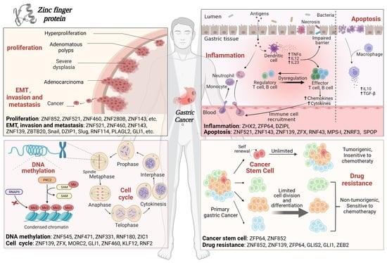
Zinc Finger Proteins in the War on Gastric Cancer: Molecular Mechanism and Clinical Potential
by Shujie Liu, Xingzhu Liu, Xin Lin and Hongping Chen
Cells 2023, 12(9), 1314; https://doi.org/10.3390/cells12091314 - 4 May 2023
Cited by 6 | Viewed by 3771
Abstract
According to the 2020 global cancer data released by the World Cancer Research Fund (WCRF) International, gastric cancer (GC) is the fifth most common cancer worldwide, with yearly increasing incidence and the second-highest fatality rate in malignancies. Despite the contemporary ambiguous molecular mechanisms [...] Read more.
According to the 2020 global cancer data released by the World Cancer Research Fund (WCRF) International, gastric cancer (GC) is the fifth most common cancer worldwide, with yearly increasing incidence and the second-highest fatality rate in malignancies. Despite the contemporary ambiguous molecular mechanisms in GC pathogenesis, numerous in-depth studies have demonstrated that zinc finger proteins (ZFPs) are essential for the development and progression of GC. ZFPs are a class of transcription factors with finger-like domains that bind to Zn2+ extensively and participate in gene replication, cell differentiation and tumor development. In this review, we briefly outline the roles, molecular mechanisms and the latest advances in ZFPs in GC, including eight principal aspects, such as cell proliferation, epithelial–mesenchymal transition (EMT), invasion and metastasis, inflammation and immune infiltration, apoptosis, cell cycle, DNA methylation, cancer stem cells (CSCs) and drug resistance. Intriguingly, the myeloid zinc finger 1 (MZF1) possesses reversely dual roles in GC by promoting tumor proliferation or impeding cancer progression via apoptosis. Therefore, a thorough understanding of the molecular mechanism of ZFPs on GC progression will pave the solid way for screening the potentially effective diagnostic indicators, prognostic biomarkers and therapeutic targets of GC. Full article
Show Figures

Graphical abstract

25 pages, 1817 KiB  
Review
Current Strategy to Treat Immunogenic Gastrointestinal Cancers: Perspectives for a New Era
by Keitaro Shimozaki, Izuma Nakayama, Toru Hirota and Kensei Yamaguchi
Cells 2023, 12(7), 1049; https://doi.org/10.3390/cells12071049 - 30 Mar 2023
Cited by 9 | Viewed by 2982
Abstract
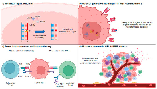
Since pembrolizumab, an anti-programmed death-1 (PD-1) antibody, showed a dramatic response to immunogenic cancers with microsatellite instability-high (MSI-H) and/or deficient mismatch repair (dMMR) in the pilot clinical trial KEYNOTE-016, subsequent studies have confirmed durable responses of anti-PD-1 inhibitors for MSI-H/dMMR solid tumors. As [...] Read more.
Since pembrolizumab, an anti-programmed death-1 (PD-1) antibody, showed a dramatic response to immunogenic cancers with microsatellite instability-high (MSI-H) and/or deficient mismatch repair (dMMR) in the pilot clinical trial KEYNOTE-016, subsequent studies have confirmed durable responses of anti-PD-1 inhibitors for MSI-H/dMMR solid tumors. As immunotherapy is described as a “game changer,” the therapeutic landscape for MSI-H/dMMR solid tumors including gastrointestinal cancers has changed considerably in the last decade. An MSI/MMR status has been established as the predictive biomarker for immune checkpoint blockades, playing an indispensable role in the clinical practice of patients with MSI-H/dMMR tumors. Immunotherapy is also now investigated for locally advanced MSI-H/dMMR gastrointestinal cancers. Despite this great success, a few populations with MSI-H/dMMR gastrointestinal cancers do not respond to immunotherapy, possibly due to the existence of intrinsic or acquired resistance mechanisms. Clarifying the underlying mechanisms of resistance remains a future task, whereas attempts to overcome resistance and improve the efficacy of immunotherapy are currently ongoing. Herein, we review recent clinical trials with special attention to MSI-H/dMMR gastrointestinal cancers together with basic/translational findings, which provide their rationale, and discuss perspectives for the further therapeutic development of treatment in this field. Full article
Show Figures

Figure 1

2022

Jump to: 2023, 2021, 2020

12 pages, 2803 KiB  
Article
Hydrophobic Bile Salts Induce Pro-Fibrogenic Proliferation of Hepatic Stellate Cells through PI3K p110 Alpha Signaling
by Sebastian Zimny, Dennis Koob, Jingguo Li, Ralf Wimmer, Tobias Schiergens, Jutta Nagel, Florian Paul Reiter, Gerald Denk and Simon Hohenester
Cells 2022, 11(15), 2344; https://doi.org/10.3390/cells11152344 - 29 Jul 2022
Cited by 5 | Viewed by 2023
Abstract
Bile salts accumulating during cholestatic liver disease are believed to promote liver fibrosis. We have recently shown that chenodeoxycholate (CDC) induces expansion of hepatic stellate cells (HSCs) in vivo, thereby promoting liver fibrosis. Mechanisms underlying bile salt-induced fibrogenesis remain elusive. We aimed to [...] Read more.
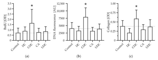
Bile salts accumulating during cholestatic liver disease are believed to promote liver fibrosis. We have recently shown that chenodeoxycholate (CDC) induces expansion of hepatic stellate cells (HSCs) in vivo, thereby promoting liver fibrosis. Mechanisms underlying bile salt-induced fibrogenesis remain elusive. We aimed to characterize the effects of different bile salts on HSC biology and investigated underlying signaling pathways. Murine HSCs (mHSCs) were stimulated with hydrophilic and hydrophobic bile salts. Proliferation, cell mass, collagen deposition, and activation of signaling pathways were determined. Activation of the human HSC cell line LX 2 was assessed by quantification of α-smooth muscle actin (αSMA) expression. Phosphatidyl-inositol-3-kinase (PI3K)-dependent signaling was inhibited both pharmacologically and by siRNA. CDC, the most abundant bile salt accumulating in human cholestasis, but no other bile salt tested, induced Protein kinase B (PKB) phosphorylation and promoted HSC proliferation and subsequent collagen deposition. Pharmacological inhibition of the upstream target PI3K-inhibited activation of PKB and pro-fibrogenic proliferation of HSCs. The PI3K p110α-specific inhibitor Alpelisib and siRNA-mediated knockdown of p110α ameliorated pro-fibrogenic activation of mHSC and LX 2 cells, respectively. In summary, pro-fibrogenic signaling in mHSCs is selectively induced by CDC. PI3K p110α may be a potential therapeutic target for the inhibition of bile salt-induced fibrogenesis in cholestasis. Full article
Show Figures

Figure 1

22 pages, 5236 KiB  
Article
GLP-1 Mediates Regulation of Colonic ACE2 Expression by the Bile Acid Receptor GPBAR1 in Inflammation
by Michele Biagioli, Silvia Marchianò, Rosalinda Roselli, Cristina Di Giorgio, Rachele Bellini, Martina Bordoni, Eleonora Distrutti, Bruno Catalanotti, Angela Zampella, Luigina Graziosi, Annibale Donini and Stefano Fiorucci
Cells 2022, 11(7), 1187; https://doi.org/10.3390/cells11071187 - 1 Apr 2022
Cited by 13 | Viewed by 3691
Abstract
Background & Aims: ACE2, a carboxypeptidase that generates Ang-(1-7) from Ang II, is highly expressed in the lung, small intestine and colon. GPBAR1, is a G protein bile acid receptor that promotes the release of the insulinotropic factor glucagon-like peptide (GLP)-1 and attenuates [...] Read more.
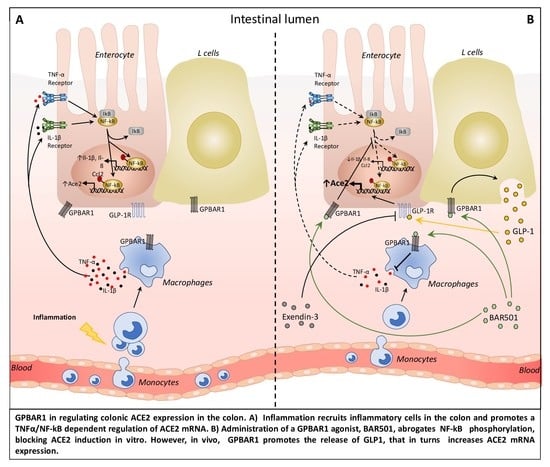
Background & Aims: ACE2, a carboxypeptidase that generates Ang-(1-7) from Ang II, is highly expressed in the lung, small intestine and colon. GPBAR1, is a G protein bile acid receptor that promotes the release of the insulinotropic factor glucagon-like peptide (GLP)-1 and attenuates intestinal inflammation. Methods: We investigated the expression of ACE2, GLP-1 and GPBAR1 in two cohorts of Crohn’s disease (CD) patients and three mouse models of colitis and Gpbar1−/− mice. Activation of GPBAR1 in these models and in vitro was achieved by BAR501, a selective GPBAR1 agonist. Results: In IBD patients, ACE2 mRNA expression was regulated in a site-specific manner in response to inflammation. While expression of ileal ACE2 mRNA was reduced, the colon expression was induced. Colon expression of ACE2 mRNA in IBD correlated with expression of TNF-α and GPBAR1. A positive correlation occurred between GCG and GPBAR1 in human samples and animal models of colitis. In these models, ACE2 mRNA expression was further upregulated by GPABR1 agonism and reversed by exendin-3, a GLP-1 receptor antagonist. In in vitro studies, liraglutide, a GLP-1 analogue, increased the expression of ACE2 in colon epithelial cells/macrophages co-cultures. Conclusions: ACE2 mRNA expression in the colon of IBD patients and rodent models of colitis is regulated in a TNF-α- and GLP-1-dependent manner. We have identified a GPBAR1/GLP-1 mechanism as a positive modulator of ACE2. Full article
Show Figures

Graphical abstract

2021

Jump to: 2023, 2022, 2020

18 pages, 8287 KiB  
Article
Activating Transcription Factor 3 Protects against Restraint Stress-Induced Gastrointestinal Injury in Mice
by Dun-Jie Chuang, Subhashree Pethaperumal, Bijaya Siwakoti, Hung-Jen Chien, Ching-Feng Cheng, Shih-Che Hung, Te-Sheng Lien, Der-Shan Sun and Hsin-Hou Chang
Cells 2021, 10(12), 3530; https://doi.org/10.3390/cells10123530 - 14 Dec 2021
Cited by 12 | Viewed by 4038
Abstract
Psychological stress increases the risk of gastrointestinal (GI) tract diseases, which involve bidirectional communication of the GI and nerves systems. Acute stress leads to GI ulcers; however, the mechanism of the native cellular protection pathway, which safeguards tissue integrality and maintains GI homeostasis, [...] Read more.
Psychological stress increases the risk of gastrointestinal (GI) tract diseases, which involve bidirectional communication of the GI and nerves systems. Acute stress leads to GI ulcers; however, the mechanism of the native cellular protection pathway, which safeguards tissue integrality and maintains GI homeostasis, remains to be investigated. In a mouse model of this study, restraint stress induced GI leakage, abnormal tight junction protein expression, and cell death of gut epithelial cells. The expression of activating transcription factor 3 (ATF3), a stress-responsive transcription factor, is upregulated in the GI tissues of stressed animals. ATF3-deficient mice displayed an exacerbated phenotype of GI injuries. These results suggested that, in response to stress, ATF3 is part of the native cellular protective pathway in the GI system, which could be a molecular target for managing psychological stress-induced GI tract diseases. Full article
Show Figures

Figure 1

20 pages, 2368 KiB  
Article
Alterations in SLC4A2, SLC26A7 and SLC26A9 Drive Acid–Base Imbalance in Gastric Neuroendocrine Tumors and Uncover a Novel Mechanism for a Co-Occurring Polyautoimmune Scenario
by Oriol Calvete, José Reyes, Hernán Valdés-Socin, Paloma Martin, Mónica Marazuela, Alicia Barroso, Javier Escalada, Antoni Castells, Raúl Torres-Ruiz, Sandra Rodríguez-Perales, María Currás-Freixes and Javier Benítez
Cells 2021, 10(12), 3500; https://doi.org/10.3390/cells10123500 - 10 Dec 2021
Cited by 8 | Viewed by 3137
Abstract
Autoimmune polyendocrine syndrome (APS) is assumed to involve an immune system malfunction and entails several autoimmune diseases co-occurring in different tissues of the same patient; however, they are orphans of its accurate diagnosis, as its genetic basis and pathogenic mechanism are not understood. [...] Read more.
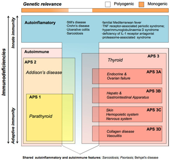
Autoimmune polyendocrine syndrome (APS) is assumed to involve an immune system malfunction and entails several autoimmune diseases co-occurring in different tissues of the same patient; however, they are orphans of its accurate diagnosis, as its genetic basis and pathogenic mechanism are not understood. Our previous studies uncovered alterations in the ATPase H+/K+ Transporting Subunit Alpha (ATP4A) proton pump that triggered an internal cell acid–base imbalance, offering an autoimmune scenario for atrophic gastritis and gastric neuroendocrine tumors with secondary autoimmune pathologies. Here, we propose the genetic exploration of APS involving gastric disease to understand the underlying pathogenic mechanism of the polyautoimmune scenario. The whole exome sequencing (WES) study of five autoimmune thyrogastric families uncovered different pathogenic variants in SLC4A2, SLC26A7 and SLC26A9, which cotransport together with ATP4A. Exploratory in vitro studies suggested that the uncovered genes were involved in a pathogenic mechanism based on the alteration of the acid–base balance. Thus, we built a custom gene panel with 12 genes based on the suggested mechanism to evaluate a new series of 69 APS patients. In total, 64 filtered putatively damaging variants in the 12 genes of the panel were found in 54.17% of the studied patients and none of the healthy controls. Our studies reveal a constellation of solute carriers that co-express in the tissues affected with different autoimmune diseases, proposing a unique genetic origin for co-occurring pathologies. These results settle a new-fangled genetics-based mechanism for polyautoimmunity that explains not only gastric disease, but also thyrogastric pathology and disease co-occurrence in APS that are different from clinical incidental findings. This opens a new window leading to the prediction and diagnosis of co-occurring autoimmune diseases and clinical management of patients. Full article
Show Figures

Figure 1

11 pages, 26181 KiB  
Article
T-Bet Deficiency Attenuates Bile Duct Injury in Experimental Biliary Atresia
by Sujit K. Mohanty, Bryan Donnelly, Haley Temple, Alexander Bondoc, Monica McNeal and Greg Tiao
Cells 2021, 10(12), 3461; https://doi.org/10.3390/cells10123461 - 8 Dec 2021
Cited by 4 | Viewed by 2961
Abstract
Biliary atresia (BA) is an obstructive neonatal cholangiopathy leading to liver cirrhosis and end stage liver disease. A Kasai portoenterostomy may restore biliary drainage, but most patients ultimately require liver transplantation for survival. At diagnosis, immune cells within the liver of patients with [...] Read more.
Biliary atresia (BA) is an obstructive neonatal cholangiopathy leading to liver cirrhosis and end stage liver disease. A Kasai portoenterostomy may restore biliary drainage, but most patients ultimately require liver transplantation for survival. At diagnosis, immune cells within the liver of patients with BA demonstrate a T-helper 1 (Th1) inflammatory profile similar to rhesus rotavirus (RRV)-infected mice livers developing BA. The transcription factor Tbx21 (T-bet) is essential for induction of a Th1 immune response in both the adaptive and innate immune system. Here we used animals with targeted deletion of the T-bet gene to determine its role in the progression of BA. Infection of newborn T-bet knockout (KO) pups with RRV resulted in a decreased Th1 inflammatory chemokine/cytokine profile when compared to infected wild-type mice. Analysis of the mononuclear cells profile from T-bet KO mice revealed both a significant decrease in the total number of CD3, CD4, and CD8 T cells and their effector molecules granzyme A, perforin, and FasL. Even though the percentage of T-bet KO mice displaying symptoms of an obstructive cholangiopathy and overall mortality rate was not different compared to wild-type mice, the extrahepatic bile ducts of T-bet KO mice remained patent. Full article
Show Figures

Figure 1

15 pages, 1478 KiB  
Review
The Pathophysiology of Farnesoid X Receptor (FXR) in the GI Tract: Inflammation, Barrier Function and Innate Immunity
by Kemp M. Anderson and Christopher P. Gayer
Cells 2021, 10(11), 3206; https://doi.org/10.3390/cells10113206 - 17 Nov 2021
Cited by 30 | Viewed by 5771
Abstract
The Farnesoid-X Receptor, FXR, is a nuclear bile acid receptor. Its originally described function is in bile acid synthesis and regulation within the liver. More recently, however, FXR has been increasingly appreciated for its breadth of function and expression across multiple organ systems, [...] Read more.
The Farnesoid-X Receptor, FXR, is a nuclear bile acid receptor. Its originally described function is in bile acid synthesis and regulation within the liver. More recently, however, FXR has been increasingly appreciated for its breadth of function and expression across multiple organ systems, including the intestine. While FXR’s role within the liver continues to be investigated, increasing literature indicates that FXR has important roles in responding to inflammation, maintaining intestinal epithelial barrier function, and regulating immunity within the gastrointestinal (GI) tract. Given the complicated and multi-factorial nature of intestinal barrier dysfunction, it is not surprising that FXR’s role appears equally complicated and not without conflicting data in different model systems. Recent work has suggested translational applications of FXR modulation in GI pathology; however, a better understanding of FXR physiology is necessary for these treatments to gain widespread use in human disease. This review aims to discuss current scientific work on the role of FXR within the GI tract, specifically in its role in intestinal inflammation, barrier function, and immune response, while also exploring areas of controversy. Full article
Show Figures

Figure 1

20 pages, 1455 KiB  
Review
Treatment for Viral Hepatitis as Secondary Prevention for Hepatocellular Carcinoma
by Saleh A. Alqahtani and Massimo Colombo
Cells 2021, 10(11), 3091; https://doi.org/10.3390/cells10113091 - 9 Nov 2021
Cited by 15 | Viewed by 3074
Abstract
Chronic infections with either hepatitis B or C virus (HBV or HCV) are among the most common risk factors for developing hepatocellular carcinoma (HCC). The hepatocarcinogenic potential of these viruses is mediated through a wide range of mechanisms, including the induction of chronic [...] Read more.
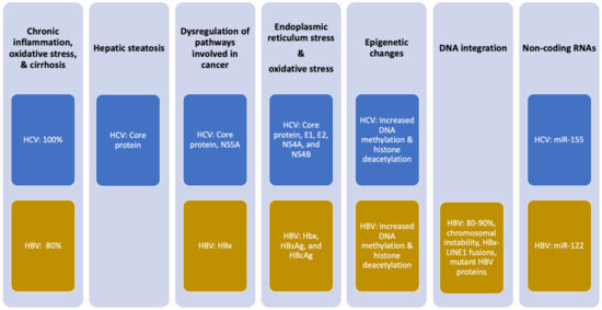
Chronic infections with either hepatitis B or C virus (HBV or HCV) are among the most common risk factors for developing hepatocellular carcinoma (HCC). The hepatocarcinogenic potential of these viruses is mediated through a wide range of mechanisms, including the induction of chronic inflammation and oxidative stress and the deregulation of cellular pathways by viral proteins. Over the last decade, effective anti-viral agents have made sustained viral suppression or cure a feasible treatment objective for most chronic HBV/HCV patients. Given the tumorigenic potential of HBV/HCV, it is no surprise that obtaining sustained viral suppression or eradication proves to be effective in preventing HCC. This review summarizes the mechanisms by which HCV and HBV exert their hepatocarcinogenic activity and describes in detail the efficacy of anti-HBV and anti-HCV therapies in terms of HCC prevention. Although these treatments significantly reduce the risk for HCC in patients with chronic viral hepatitis, this risk is not eliminated. Therefore, we evaluate potential strategies to improve these outcomes further and address some of the remaining controversies. Full article
Show Figures

Figure 1

18 pages, 6793 KiB  
Review
Bile Acid Receptors and the Gut–Liver Axis in Nonalcoholic Fatty Liver Disease
by Rui Xue, Lianyong Su, Shengyi Lai, Yanyan Wang, Derrick Zhao, Jiangao Fan, Weidong Chen, Phillip B. Hylemon and Huiping Zhou
Cells 2021, 10(11), 2806; https://doi.org/10.3390/cells10112806 - 20 Oct 2021
Cited by 49 | Viewed by 6110
Abstract
The prevalence of nonalcoholic fatty liver disease (NAFLD) has been significantly increased due to the global epidemic of obesity. The disease progression from simple steatosis (NAFL) to nonalcoholic steatohepatitis (NASH) is closely linked to inflammation, insulin resistance, and dysbiosis. Although extensive efforts have [...] Read more.
The prevalence of nonalcoholic fatty liver disease (NAFLD) has been significantly increased due to the global epidemic of obesity. The disease progression from simple steatosis (NAFL) to nonalcoholic steatohepatitis (NASH) is closely linked to inflammation, insulin resistance, and dysbiosis. Although extensive efforts have been aimed at elucidating the pathological mechanisms of NAFLD disease progression, current understanding remains incomplete, and no effective therapy is available. Bile acids (BAs) are not only important physiological detergents for the absorption of lipid-soluble nutrients in the intestine but also metabolic regulators. During the last two decades, BAs have been identified as important signaling molecules involved in lipid, glucose, and energy metabolism. Dysregulation of BA homeostasis has been associated with NAFLD disease severity. Identification of nuclear receptors and G-protein-coupled receptors activated by different BAs not only significantly expanded the current understanding of NAFLD/NASH disease progression but also provided the opportunity to develop potential therapeutics for NAFLD/NASH. In this review, we will summarize the recent studies with a focus on BA-mediated signaling pathways in NAFLD/NASH. Furthermore, the therapeutic implications of targeting BA-mediated signaling pathways for NAFLD will also be discussed. Full article
Show Figures

Figure 1

31 pages, 7118 KiB  
Article
Combination Treatment with the Vimentin-Targeting Antibody hzVSF and Tenofovir Suppresses Woodchuck Hepatitis Virus Infection in Woodchucks
by Kyle E. Korolowicz, Manasa Suresh, Bin Li, Xu Huang, Changsuek Yon, Bhaskar V. Kallakury, Kyoung-pil Lee, Sungman Park, Yoon-Won Kim and Stephan Menne
Cells 2021, 10(9), 2321; https://doi.org/10.3390/cells10092321 - 5 Sep 2021
Cited by 5 | Viewed by 3464
Abstract
Current treatment options for patients infected with hepatitis B virus (HBV) are suboptimal, because the approved drugs rarely induce cure due to the persistence of the viral DNA genome in the nucleus of infected hepatocytes, and are associated with either severe side effects [...] Read more.
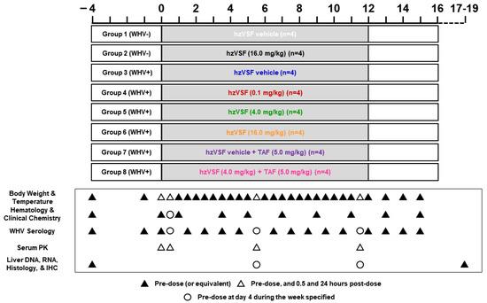
Current treatment options for patients infected with hepatitis B virus (HBV) are suboptimal, because the approved drugs rarely induce cure due to the persistence of the viral DNA genome in the nucleus of infected hepatocytes, and are associated with either severe side effects (pegylated interferon-alpha) or require life-long administration (nucleos(t)ide analogs). We report here the evaluation of the safety and therapeutic efficacy of a novel, humanized antibody (hzVSF) in the woodchuck model of HBV infection. hzVSF has been shown to act as a viral entry inhibitor, most likely by suppressing vimentin-mediated endocytosis of virions. Targeting the increased vimentin expression on liver cells by hzVSF after infection with HBV or woodchuck hepatitis virus (WHV) was demonstrated initially. Thereafter, hzVSF safety was assessed in eight woodchucks naïve for WHV infection. Antiviral efficacy of hzVSF was evaluated subsequently in 24 chronic WHV carrier woodchucks by monotreatment with three ascending doses and in combination with tenofovir alafenamide fumarate (TAF). Consistent with the proposed blocking of WHV reinfection, intravenous hzVSF administration for 12 weeks resulted in a modest but transient reduction of viral replication and associated liver inflammation. In combination with oral TAF dosing, the antiviral effect of hzVSF was enhanced and sustained in half of the woodchucks with an antibody response to viral proteins. Thus, hzVSF safely but modestly alters chronic WHV infection in woodchucks; however, as a combination partner to TAF, its antiviral efficacy is markedly increased. The results of this preclinical study support future evaluation of this novel anti-HBV drug in patients. Full article
Show Figures

Figure 1

17 pages, 979 KiB  
Review
Regulation of Paneth Cell Function by RNA-Binding Proteins and Noncoding RNAs
by Hee K. Chung, Lan Xiao, Krishna C. Jaladanki and Jian-Ying Wang
Cells 2021, 10(8), 2107; https://doi.org/10.3390/cells10082107 - 17 Aug 2021
Cited by 18 | Viewed by 4130
Abstract
Paneth cells are specialized intestinal epithelial cells that are located at the base of small intestinal crypts and play a vital role in preserving the gut epithelium homeostasis. Paneth cells act as a safeguard from bacterial translocation across the epithelium and constitute the [...] Read more.
Paneth cells are specialized intestinal epithelial cells that are located at the base of small intestinal crypts and play a vital role in preserving the gut epithelium homeostasis. Paneth cells act as a safeguard from bacterial translocation across the epithelium and constitute the niche for intestinal stem cells in the small intestine by providing multiple niche signals. Recently, Paneth cells have become the focal point of investigations defining the mechanisms underlying the epithelium-microbiome interactions and pathogenesis of chronic gut mucosal inflammation and bacterial infection. Function of Paneth cells is tightly regulated by numerous factors at different levels, while Paneth cell defects have been widely documented in various gut mucosal diseases in humans. The post-transcription events, specific change in mRNA stability and translation by RNA-binding proteins (RBPs) and noncoding RNAs (ncRNAs) are implicated in many aspects of gut mucosal physiology by modulating Paneth cell function. Deregulation of RBPs and ncRNAs and subsequent Paneth cell defects are identified as crucial elements of gut mucosal pathologies. Here, we overview the posttranscriptional regulation of Paneth cells by RBPs and ncRNAs, with a particular focus on the increasing evidence of RBP HuR and long ncRNA H19 in this process. We also discuss the involvement of Paneth cell dysfunction in altered susceptibility of the intestinal epithelium to chronic inflammation and bacterial infection following disrupted expression of HuR and H19. Full article
Show Figures

Graphical abstract

24 pages, 1548 KiB  
Review
The State of Immunotherapy in Hepatobiliary Cancers
by Farhan Z. Ilyas, Joal D. Beane and Timothy M. Pawlik
Cells 2021, 10(8), 2096; https://doi.org/10.3390/cells10082096 - 15 Aug 2021
Cited by 24 | Viewed by 5254
Abstract
Hepatobiliary cancers, including hepatocellular carcinoma (HCC), cholangiocarcinoma (CCA), and gallbladder carcinoma (GBC), are lethal cancers with limited therapeutic options. Curative-intent treatment typically involves surgery, yet recurrence is common and many patients present with advanced disease not amenable to an operation. Immunotherapy represents a [...] Read more.
Hepatobiliary cancers, including hepatocellular carcinoma (HCC), cholangiocarcinoma (CCA), and gallbladder carcinoma (GBC), are lethal cancers with limited therapeutic options. Curative-intent treatment typically involves surgery, yet recurrence is common and many patients present with advanced disease not amenable to an operation. Immunotherapy represents a promising approach to improve outcomes, but the immunosuppressive tumor microenvironment of the liver characteristic of hepatobiliary cancers has hampered the development and implementation of this therapeutic approach. Current immunotherapies under investigation include immune checkpoint inhibitors (ICI), the adoptive transfer of immune cells, bispecific antibodies, vaccines, and oncolytic viruses. Programmed cell death protein 1 (PD-1) and cytotoxic T-lymphocyte-associated protein 4 (CTLA-4) are two ICIs that have demonstrated utility in HCC, and newer immune checkpoint targets are being tested in clinical trials. In advanced CCA and GBC, PD-1 ICIs have resulted in antitumor responses, but only in a minority of select patients. Other ICIs are being investigated for patients with CCA and GBC. Adoptive transfer may hold promise, with reports of complete durable regression in metastatic CCA, yet this therapeutic approach may not be generalizable. Alternative approaches have been developed and promising results have been observed, but clinical trials are needed to validate their utility. While the treatment of hepatobiliary cancers involves unique challenges that these cancers present, the progress seen with ICIs and adoptive transfer has solidified immunotherapy as an important approach in these challenging patients with few other effective treatment options. Full article
Show Figures

Figure 1

14 pages, 2475 KiB  
Review
Renalase: A Multi-Functional Signaling Molecule with Roles in Gastrointestinal Disease
by Thomas C. Pointer, Fred S. Gorelick and Gary V. Desir
Cells 2021, 10(8), 2006; https://doi.org/10.3390/cells10082006 - 6 Aug 2021
Cited by 15 | Viewed by 3652
Abstract
The survival factor renalase (RNLS) is a recently discovered secretory protein with potent prosurvival and anti-inflammatory effects. Several evolutionarily conserved RNLS domains are critical to its function. These include a 20 aa site that encodes for its prosurvival effects. Its prosurvival effects are [...] Read more.
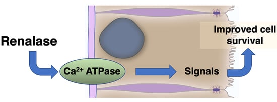
The survival factor renalase (RNLS) is a recently discovered secretory protein with potent prosurvival and anti-inflammatory effects. Several evolutionarily conserved RNLS domains are critical to its function. These include a 20 aa site that encodes for its prosurvival effects. Its prosurvival effects are shown in GI disease models including acute cerulein pancreatitis. In rodent models of pancreatic cancer and human cancer tissues, increased RNLS expression promotes cancer cell survival but shortens life expectancy. This 37 kD protein can regulate cell signaling as an extracellular molecule and probably also at intracellular sites. Extracellular RNLS signals through a specific plasma membrane calcium export transporter; this interaction appears most relevant to acute injury and cancer. Preliminary studies using RNLS agonists and antagonists, as well as various preclinical disease models, suggest that the immunologic and prosurvival effects of RNLS will be relevant to diverse pathologies that include acute organ injuries and select cancers. Future studies should define the roles of RNLS in intestinal diseases, characterizing the RNLS-activated pathways linked to cell survival and developing therapeutic agents that can increase or decrease RNLS in relevant clinical settings. Full article
Show Figures

Graphical abstract

17 pages, 2390 KiB  
Article
The Genes Encoding Small Leucine-Rich Proteoglycans Undergo Differential Expression Alterations in Colorectal Cancer, Depending on Tumor Location
by Maria Pilar Solis-Hernandez, Carla Martín, Beatriz García, Natalia Pérez-López, Yolanda García-Mesa, Sara González-Fernández, Olivia García-Suárez, Jesús Merayo, Iván Fernández-Vega and Luis M. Quirós
Cells 2021, 10(8), 2002; https://doi.org/10.3390/cells10082002 - 6 Aug 2021
Cited by 3 | Viewed by 2936
Abstract
Small leucine-rich proteoglycans (SLRPs) regulate different processes and undergo significant alterations in various diseases. Colon carcinomas (CCs) are heterogeneous pathologies with important clinical and molecular differences depending on their location, which makes it interesting to analyze the alterations in SLRPs in right- and [...] Read more.
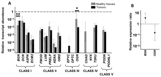
Small leucine-rich proteoglycans (SLRPs) regulate different processes and undergo significant alterations in various diseases. Colon carcinomas (CCs) are heterogeneous pathologies with important clinical and molecular differences depending on their location, which makes it interesting to analyze the alterations in SLRPs in right- and left-sided tumors (RS- and LSCCs). SLRP transcription levels were studied in 32 CCs using qPCR compared to healthy colon mucosae samples from the same patients, 20 of them from LSCCs and the remaining 12 from RSCCs. Protein expression of genes with significant differences in their transcriptions was analyzed by immunohistochemistry. The alterations observed were related to survival data. The arrangement of transcription of SLRPs was quite similar in ascending and descending colon, but RS- and LSCCs displayed different patterns of alteration, with a greater number of deregulations occurring in the latter. The analysis of protein expression also indicated changes in the location of these molecules, largely moving to the cell interior. While podocan underexpression showed a trend toward better outcomes, no differences were observed in terms of overall survival. In vitro studies using the HT29 tumor cell line suggest that deregulation of SLRPs could affect cell proliferation. SLRPs constitute new differential markers of RS- and LSCCs, showing differences dependent on the anatomical location of the tumor. Full article
Show Figures

Figure 1

19 pages, 7604 KiB  
Review
The Critical Role of Growth Factors in Gastric Ulcer Healing: The Cellular and Molecular Mechanisms and Potential Clinical Implications
by Andrzej S. Tarnawski and Amrita Ahluwalia
Cells 2021, 10(8), 1964; https://doi.org/10.3390/cells10081964 - 2 Aug 2021
Cited by 64 | Viewed by 10041
Abstract
In this article we review the cellular and molecular mechanisms of gastric ulcer healing. A gastric ulcer (GU) is a deep defect in the gastric wall penetrating through the entire mucosa and the muscularis mucosae. GU healing is a regeneration process that encompasses [...] Read more.
In this article we review the cellular and molecular mechanisms of gastric ulcer healing. A gastric ulcer (GU) is a deep defect in the gastric wall penetrating through the entire mucosa and the muscularis mucosae. GU healing is a regeneration process that encompasses cell dedifferentiation, proliferation, migration, re-epithelialization, formation of granulation tissue, angiogenesis, vasculogenesis, interactions between various cells and the matrix, and tissue remodeling, all resulting in scar formation. All these events are controlled by cytokines and growth factors (e.g., EGF, TGFα, IGF-1, HGF, bFGF, TGFβ, NGF, VEGF, angiopoietins) and transcription factors activated by tissue injury. These growth factors bind to their receptors and trigger cell proliferation, migration, and survival pathways through Ras, MAPK, PI3K/Akt, PLC-γ, and Rho/Rac/actin signaling. The triggers for the activation of these growth factors are tissue injury and hypoxia. EGF, its receptor, IGF-1, HGF, and COX-2 are important for epithelial cell proliferation, migration, re-epithelialization, and gastric gland reconstruction. VEGF, angiopoietins, bFGF, and NGF are crucial for blood vessel regeneration in GU scars. The serum response factor (SRF) is essential for VEGF-induced angiogenesis, re-epithelialization, and blood vessel and muscle restoration. Local therapy with cDNA of human recombinant VEGF165 in combination with angiopoietin1, or with the NGF protein, dramatically accelerates GU healing and improves the quality of mucosal restoration within ulcer scars. The future directions for accelerating and improving healing include local gene and protein therapies with growth factors, their combinations, and the use of stem cells and tissue engineering. Full article
Show Figures

Graphical abstract

19 pages, 901 KiB  
Review
Tissue-Resident T Cells in Chronic Relapsing–Remitting Intestinal Disorders
by Juliana Barreto de Albuquerque, Christoph Mueller and Bilgi Gungor
Cells 2021, 10(8), 1882; https://doi.org/10.3390/cells10081882 - 25 Jul 2021
Cited by 3 | Viewed by 3625
Abstract
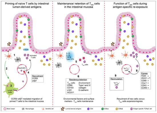
Tissue-resident memory T (TRM) cells critically contribute to the rapid immunoprotection and efficient immunosurveillance against pathogens, particularly in barrier tissues, but also during anti-tumor responses. However, the involvement of TRM cells also in the induction and exacerbation of immunopathologies, notably [...] Read more.
Tissue-resident memory T (TRM) cells critically contribute to the rapid immunoprotection and efficient immunosurveillance against pathogens, particularly in barrier tissues, but also during anti-tumor responses. However, the involvement of TRM cells also in the induction and exacerbation of immunopathologies, notably in chronically relapsing auto-inflammatory disorders, is becoming increasingly recognized as a critical factor. Thus, TRM cells may also represent an attractive target in the management of chronic (auto-) inflammatory disorders, including multiple sclerosis, rheumatoid arthritis, celiac disease and inflammatory bowel diseases. In this review, we focus on current concepts of TRM cell biology, particularly in the intestine, and discuss recent findings on their involvement in chronic relapsing–remitting inflammatory disorders. Potential therapeutic strategies to interfere with these TRM cell-mediated immunopathologies are discussed. Full article
Show Figures

Figure 1

16 pages, 2518 KiB  
Review
Farnesoid X Receptor as Target for Therapies to Treat Cholestasis-Induced Liver Injury
by Anca D. Petrescu and Sharon DeMorrow
Cells 2021, 10(8), 1846; https://doi.org/10.3390/cells10081846 - 21 Jul 2021
Cited by 22 | Viewed by 5428
Abstract
Recent studies on liver disease burden worldwide estimated that cirrhosis is the 11th most common cause of death globally, and there is a great need for new therapies to limit the progression of liver injuries in the early stages. Cholestasis is caused by [...] Read more.
Recent studies on liver disease burden worldwide estimated that cirrhosis is the 11th most common cause of death globally, and there is a great need for new therapies to limit the progression of liver injuries in the early stages. Cholestasis is caused by accumulation of hydrophobic bile acids (BA) in the liver due to dysfunctional BA efflux or bile flow into the gall bladder. Therefore, strategies to increase detoxification of hydrophobic BA and downregulate genes involved in BA production are largely investigated. Farnesoid X receptor (FXR) has a central role in BA homeostasis and recent publications revealed that changes in autophagy due to BA-induced reactive oxygen species and increased anti-oxidant response via nuclear factor E2-related factor 2 (NRF2), result in dysregulation of FXR signaling. Several mechanistic studies have identified new dysfunctions of the cholestatic liver at cellular and molecular level, opening new venues for developing more performant therapies. Full article
Show Figures

Figure 1

13 pages, 1017 KiB  
Review
Butyrate and the Intestinal Epithelium: Modulation of Proliferation and Inflammation in Homeostasis and Disease
by Pooja S. Salvi and Robert A. Cowles
Cells 2021, 10(7), 1775; https://doi.org/10.3390/cells10071775 - 14 Jul 2021
Cited by 195 | Viewed by 12169
Abstract
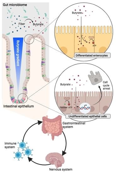
The microbial metabolite butyrate serves as a link between the intestinal microbiome and epithelium. The monocarboxylate transporters MCT1 and SMCT1 are the predominant means of butyrate transport from the intestinal lumen to epithelial cytoplasm, where the molecule undergoes rapid β-oxidation to generate cellular [...] Read more.
The microbial metabolite butyrate serves as a link between the intestinal microbiome and epithelium. The monocarboxylate transporters MCT1 and SMCT1 are the predominant means of butyrate transport from the intestinal lumen to epithelial cytoplasm, where the molecule undergoes rapid β-oxidation to generate cellular fuel. However, not all epithelial cells metabolize butyrate equally. Undifferentiated colonocytes, including neoplastic cells and intestinal stem cells at the epithelial crypt base preferentially utilize glucose over butyrate for cellular fuel. This divergent metabolic conditioning is central to the phenomenon known as “butyrate paradox”, in which butyrate induces contradictory effects on epithelial proliferation in undifferentiated and differentiated colonocytes. There is evidence that accumulation of butyrate in epithelial cells results in histone modification and altered transcriptional activation that halts cell cycle progression. This manifests in the apparent protective effect of butyrate against colonic neoplasia. A corollary to this process is butyrate-induced inhibition of intestinal stem cells. Yet, emerging research has illustrated that the evolution of the crypt, along with butyrate-producing bacteria in the intestine, serve to protect crypt base stem cells from butyrate’s anti-proliferative effects. Butyrate also regulates epithelial inflammation and tolerance to antigens, through production of anti-inflammatory cytokines and induction of tolerogenic dendritic cells. The role of butyrate in the pathogenesis and treatment of intestinal neoplasia, inflammatory bowel disease and malabsorptive states is evolving, and holds promise for the potential translation of butyrate’s cellular function into clinical therapies. Full article
Show Figures

Graphical abstract

18 pages, 2110 KiB  
Article
Interference of LPS H. pylori with IL-33-Driven Regeneration of Caviae porcellus Primary Gastric Epithelial Cells and Fibroblasts
by Weronika Gonciarz, Agnieszka Krupa, Anthony P. Moran, Agata Tomaszewska and Magdalena Chmiela
Cells 2021, 10(6), 1385; https://doi.org/10.3390/cells10061385 - 4 Jun 2021
Cited by 13 | Viewed by 2930
Abstract
Background: Lipopolysaccharide (LPS) of Helicobacter pylori (Hp) bacteria causes disintegration of gastric tissue cells in vitro. It has been suggested that interleukin (IL)-33 is involved in healing gastric injury. Aim: To elucidate whether Hp LPS affects regeneration of gastric barrier initiated by IL-33. [...] Read more.
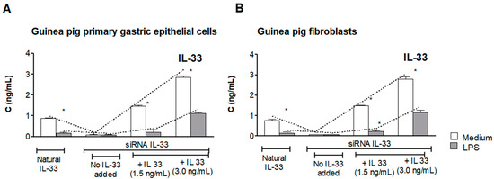
Background: Lipopolysaccharide (LPS) of Helicobacter pylori (Hp) bacteria causes disintegration of gastric tissue cells in vitro. It has been suggested that interleukin (IL)-33 is involved in healing gastric injury. Aim: To elucidate whether Hp LPS affects regeneration of gastric barrier initiated by IL-33. Methods: Primary gastric epithelial cells or fibroblasts from Caviae porcellus were transfected with siRNA IL-33. Such cells, not exposed or treated with LPS Hp, were sub-cultured in the medium with or without exogenous IL-33. Then cell migration was assessed in conjunction with oxidative stress and apoptosis, activation of extracellular signal-regulated kinase (Erk), production of collagen I and soluble ST2 (IL-33 decoy). Results: Control cells not treated with LPS Hp migrated in the presence of IL-33. The pro-regenerative activity of IL-33 was related to stimulation of cells to collagen I production. Wound healing by cells exposed to LPS Hp was inhibited even in the presence of IL-33. This could be due to increased oxidative stress and apoptosis in conjunction with Erk activation, sST2 elevation and modulation of collagen I production. Conclusions: The recovery of gastric barrier cells during Hp infection potentially can be affected due to downregulation of pro-regenerative activity of IL-33 by LPS Hp. Full article
Show Figures

Figure 1

10 pages, 4481 KiB  
Communication
Positive Correlation between nNOS and Stress-Activated Bowel Motility Is Confirmed by In Vivo HiBiT System
by Jeong Pil Han, Jeong Hyeon Lee, Geon Seong Lee, Ok Jae Koo and Su Cheong Yeom
Cells 2021, 10(5), 1028; https://doi.org/10.3390/cells10051028 - 27 Apr 2021
Cited by 6 | Viewed by 2997
Abstract
Neuronal nitric oxide synthase (nNOS) has various roles as a neurotransmitter. However, studies to date have produced insufficient data to fully support the correlation between nNOS and bowel motility. This study aimed to investigate the correlation between nNOS expression and gastrointestinal (GI) tract [...] Read more.
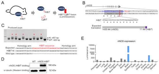
Neuronal nitric oxide synthase (nNOS) has various roles as a neurotransmitter. However, studies to date have produced insufficient data to fully support the correlation between nNOS and bowel motility. This study aimed to investigate the correlation between nNOS expression and gastrointestinal (GI) tract motility using a stress-induced neonatal maternal separation (NMS) mouse model. In this study, we generated a genetically modified mouse with the HiBiT sequence knock-in into the nNOS gene using CRISPR/Cas9 for analyzing accurate nNOS expression. nNOS expression was measured in the stomach, small intestine, large intestine, adrenal gland, and hypothalamus tissues after establishing the NMS model. The NMS model exhibited a significant increase in nNOS expression in large intestine, adrenal gland, and hypothalamus. Moreover, a significant positive correlation was observed between whole gastrointestinal transit time and the expression level of nNOS. We reasoned that NMS induced chronic stress and consequent nNOS activation in the hypothalamic-pituitary-adrenal (HPA) axis, and led to an excessive increase in intestinal motility in the lower GI tract. These results demonstrated that HiBiT is a sensitive and valuable tool for analyzing in vivo gene activation, and nNOS could be a biomarker of the HPA axis-linked lower intestinal tract dysfunction. Full article
Show Figures

Figure 1

17 pages, 2879 KiB  
Article
ZINC40099027 Promotes Gastric Mucosal Repair in Ongoing Aspirin-Associated Gastric Injury by Activating Focal Adhesion Kinase
by Sema Oncel, Rashmi Gupta, Qinggang Wang and Marc D. Basson
Cells 2021, 10(4), 908; https://doi.org/10.3390/cells10040908 - 15 Apr 2021
Cited by 15 | Viewed by 3159
Abstract
Nonsteroidal anti-inflammatory drugs cause gastric ulcers and gastritis. No drug that treats GI injury directly stimulates mucosal healing. ZINC40099027 (ZN27) activates focal adhesion kinase (FAK) and heals acute indomethacin-induced small bowel injury. We investigated the efficacy of ZN27 in rat and human gastric [...] Read more.
Nonsteroidal anti-inflammatory drugs cause gastric ulcers and gastritis. No drug that treats GI injury directly stimulates mucosal healing. ZINC40099027 (ZN27) activates focal adhesion kinase (FAK) and heals acute indomethacin-induced small bowel injury. We investigated the efficacy of ZN27 in rat and human gastric epithelial cells and ongoing aspirin-associated gastric injury. ZN27 (10 nM) stimulated FAK activation and wound closure in rat and human gastric cell lines. C57BL/6J mice were treated with 300 mg/kg/day aspirin for five days to induce ongoing gastric injury. One day after the initial injury, mice received 900 µg/kg/6 h ZN27, 10 mg/kg/day omeprazole, or 900 µg/kg/6 h ZN27 plus 10 mg/kg/day omeprazole. Like omeprazole, ZN27 reduced gastric injury vs. vehicle controls. ZN27-treated mice displayed better gastric architecture, with thicker mucosa and less hyperemia, inflammation, and submucosal edema, and lost less weight than vehicle controls. Gastric pH, serum creatinine, serum alanine aminotransferase (ALT), and renal and hepatic histology were unaffected by ZN27. Blinded scoring of pFAK-Y-397 immunoreactivity at the edge of ZN27-treated lesions demonstrated increased FAK activation, compared to vehicle-treated lesions, confirming target activation in vivo. These results suggest that ZN27 ameliorates ongoing aspirin-associated gastric mucosal injury by a pathway involving FAK activation. ZN27-derivatives may be useful to promote gastric mucosal repair. Full article
Show Figures

Figure 1

16 pages, 3743 KiB  
Article
Mechanism of Na-K-ATPase Inhibition by PGE2 in Intestinal Epithelial Cells
by Niraj Nepal, Subha Arthur, Jennifer Haynes, Balasubramanian Palaniappan and Uma Sundaram
Cells 2021, 10(4), 752; https://doi.org/10.3390/cells10040752 - 29 Mar 2021
Cited by 11 | Viewed by 3211
Abstract
The primary means of intestinal absorption of nutrients by villus cells is via Na-dependent nutrient co-transporters located in the brush border membrane (BBM). These secondary active co-transport processes require a favorable transcellular Na gradient that is provided by Na-K-ATPase. In chronic enteritis, malabsorption [...] Read more.
The primary means of intestinal absorption of nutrients by villus cells is via Na-dependent nutrient co-transporters located in the brush border membrane (BBM). These secondary active co-transport processes require a favorable transcellular Na gradient that is provided by Na-K-ATPase. In chronic enteritis, malabsorption of essential nutrients is partially due to inhibition of villus Na-K-ATPase activity mediated by specific immune inflammatory mediators that are known to be elevated in the inflamed mucosa. However, how Prostaglandin E2 (PGE2), a specific mediator of nutrient malabsorption in the villus BBM, may mediate the inhibition of Na-K-ATPase is not known. Therefore, this study aimed to determine the effect of PGE2 on Na-K-ATPase in villus cells and define its mechanism of action. In vitro, in IEC-18 cells, PGE2 treatment significantly reduced Na-K-ATPase activity, accompanied by a significant increase in the intracellular levels of cyclic Adenosine Monophosphate (cAMP). The treatment with cAMP analog 8-Bromo-cAMP mimicked the PGE2-mediated effect on Na-K-ATPase activity, while Rp-cAMP (PKA inhibitor) pretreatment reversed the same. The mechanism of inhibition of PGE2 was secondary to a transcriptional reduction in the Na-K-ATPase α1 and β1 subunit genes, which was reversed by the Rp-cAMP pretreatment. Thus, the PGE2-mediated activation of the PKA pathway mediates the transcriptional inhibition of Na-K-ATPase activity in vitro. Full article
Show Figures

Figure 1

12 pages, 25308 KiB  
Article
MFG-E8 Plays an Important Role in Attenuating Cerulein-Induced Acute Pancreatitis in Mice
by Heng-Fu Bu, Saravanan Subramanian, Hua Geng, Xiao Wang, Fangyi Liu, Pauline M. Chou, Chao Du, Isabelle G. De Plaen and Xiao-Di Tan
Cells 2021, 10(4), 728; https://doi.org/10.3390/cells10040728 - 25 Mar 2021
Cited by 5 | Viewed by 3183
Abstract
Milk fat globule-EGF factor 8 (MFG-E8) is a secreted glycoprotein that regulates tissue homeostasis, possesses potent anti-inflammatory properties, and protects against tissue injury. The human pancreas expresses MFG-E8; however, the role of MFG-E8 in the pancreas remains unclear. We examined the expression of [...] Read more.
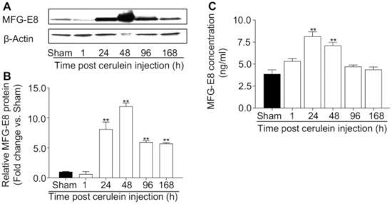
Milk fat globule-EGF factor 8 (MFG-E8) is a secreted glycoprotein that regulates tissue homeostasis, possesses potent anti-inflammatory properties, and protects against tissue injury. The human pancreas expresses MFG-E8; however, the role of MFG-E8 in the pancreas remains unclear. We examined the expression of MFG-E8 in the pancreas at baseline and during cerulein-induced acute pancreatitis in mice and determined whether MFG-E8 attenuates the progression of pancreatitis, a serious inflammatory condition that can be life-threatening. We administered cerulein to wild-type (WT) and Mfge8 knockout (KO) mice to induce pancreatitis. Immunoblot analysis showed that MFG-E8 is constitutively expressed in the murine pancreas and is increased in mice with cerulein-induced acute pancreatitis. In situ hybridization revealed that ductal epithelial cells in the mouse pancreas express Mfge8 transcripts at baseline. During pancreatitis, Mfge8 transcripts were abundantly expressed in acinar cells and endothelial cells in addition to ductal epithelial cells. Knocking out Mfge8 in mice exacerbated the severity of cerulein-induced acute pancreatitis and delayed its resolution. In contrast, administration of recombinant MFG-E8 attenuated cerulein-induced acute pancreatitis and promoted repair of pancreatic injury in Mfge8 KO mice. Taken together, our study suggests that MFG-E8 protects the pancreas against inflammatory injury and promotes pancreatic tissue repair. MFG-E8 may represent a novel therapeutic target in acute pancreatitis. Full article
Show Figures

Figure 1

10 pages, 1012 KiB  
Review
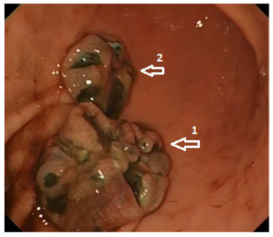
Malignant Melanoma of the Gastrointestinal Tract: Symptoms, Diagnosis, and Current Treatment Options
by Darina Kohoutova, Dominic Worku, Hala Aziz, Julian Teare, Justin Weir and James Larkin
Cells 2021, 10(2), 327; https://doi.org/10.3390/cells10020327 - 5 Feb 2021
Cited by 43 | Viewed by 5938
Abstract
Malignant melanoma (MM) has become the fifth most frequent cancer in the UK. It is the most common carcinoma to metastasize to the gastrointestinal (GI) tract. MM particularly has an affinity to spread to the small bowel, which is followed by the involvement [...] Read more.
Malignant melanoma (MM) has become the fifth most frequent cancer in the UK. It is the most common carcinoma to metastasize to the gastrointestinal (GI) tract. MM particularly has an affinity to spread to the small bowel, which is followed by the involvement of the stomach and large intestine. Excellent endoscopic options including video capsule endoscopy and enteroscopy are available for a precise diagnosis of GI involvement by a metastatic MM. The complete surgical resection of GI metastatic MM in carefully selected patients not only provides symptom control, but has also been associated with an increase in overall survival. The approval of BRAF-targeted therapies and immune checkpoint inhibitors has transformed therapeutic approaches for patients with metastatic MM over the past decade. Currently, the overall survival of patients with advanced metastatic MM who have been treated with a combination of immunotherapeutic agents reaches 52% at five years. The role of surgery for patients with the metastatic involvement of the GI tract with MM is evolving in the era of effective systemic treatments. Full article
Show Figures

Figure 1

16 pages, 4507 KiB  
Article
Role of Chitinase 3-Like 1 Protein in the Pathogenesis of Hepatic Insulin Resistance in Nonalcoholic Fatty Liver Disease
by Songhua Zhang, Aryanna Sousa, Mengqui Lin, Ayako Iwano, Rishubh Jain, Bing Ma, Chang Min Lee, Jin Wook Park, Suchitra Kamle, Rolf Carlson, Ghun Geun Lee, Jack A. Elias and Jack R. Wands
Cells 2021, 10(2), 201; https://doi.org/10.3390/cells10020201 - 20 Jan 2021
Cited by 12 | Viewed by 4144
Abstract
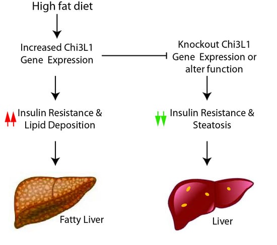
A recently discovered human glycoprotein, chitinase 3-like 1 (Chi3L1), may play a role in inflammation, tissue remodeling, and visceral fat accumulation. We hypothesize that Chi3L1 gene expression is important in the development of hepatic insulin resistance characterized by the generation of pAKT, pGSK, [...] Read more.
A recently discovered human glycoprotein, chitinase 3-like 1 (Chi3L1), may play a role in inflammation, tissue remodeling, and visceral fat accumulation. We hypothesize that Chi3L1 gene expression is important in the development of hepatic insulin resistance characterized by the generation of pAKT, pGSK, and pERK in wild type and Chi3L1 knockout (KO) murine liver following insulin stimulation. The Chi3L1 gene and protein expression was evaluated by Real Time PCR and ELISA; lipid accumulation in hepatocytes was also assessed. To alter Chi3L1 function, three different anti-Chi3L1 monoclonal antibodies (mAbs) were administered in vivo and effects on the insulin signaling cascade and hepatic lipid deposition were determined. Transmission of the hepatic insulin signal was substantially improved following KO of the CHi3L1 gene and there was reduced lipid deposition produced by a HFD. The HFD-fed mice exhibited increased Chi3L1 expression in the liver and there was impaired insulin signal transduction. All three anti-Chi3L1 mAbs partially restored hepatic insulin sensitivity which was associated with reduced lipid accumulation in hepatocytes as well. A KO of the Chi3L1 gene reduced lipid accumulation and improved insulin signaling. Therefore, Chi3L1 gene upregulation may be an important factor in the generation of NAFLD/NASH phenotype. Full article
Show Figures

Graphical abstract

2020

Jump to: 2023, 2022, 2021

37 pages, 627 KiB  
Review
Helicobacter pylori Virulence Factors—Mechanisms of Bacterial Pathogenicity in the Gastric Microenvironment
by Jacek Baj, Alicja Forma, Monika Sitarz, Piero Portincasa, Gabriella Garruti, Danuta Krasowska and Ryszard Maciejewski
Cells 2021, 10(1), 27; https://doi.org/10.3390/cells10010027 - 25 Dec 2020
Cited by 191 | Viewed by 14542
Abstract
Gastric cancer constitutes one of the most prevalent malignancies in both sexes; it is currently the fourth major cause of cancer-related deaths worldwide. The pathogenesis of gastric cancer is associated with the interaction between genetic and environmental factors, among which infection by Helicobacter [...] Read more.
Gastric cancer constitutes one of the most prevalent malignancies in both sexes; it is currently the fourth major cause of cancer-related deaths worldwide. The pathogenesis of gastric cancer is associated with the interaction between genetic and environmental factors, among which infection by Helicobacter pylori (H. pylori) is of major importance. The invasion, survival, colonization, and stimulation of further inflammation within the gastric mucosa are possible due to several evasive mechanisms induced by the virulence factors that are expressed by the bacterium. The knowledge concerning the mechanisms of H. pylori pathogenicity is crucial to ameliorate eradication strategies preventing the possible induction of carcinogenesis. This review highlights the current state of knowledge and the most recent findings regarding H. pylori virulence factors and their relationship with gastric premalignant lesions and further carcinogenesis. Full article
Back to TopTop