Molecular Mechanisms and Current Treatment Strategy of Sarcopenia and Cachexia

A topical collection in Cells (ISSN 2073-4409). This collection belongs to the section "Cellular Pathology".

Viewed by 82079

Editor


E-Mail Website
Collection Editor
Institute for Liberal Arts, Tokyo Institute of Technology, Tokyo, Japan
Interests: skeletal muscle; molecular mechanism of congenital muscular dystrophy; normal muscle regeneration; molecular mechanism of sarcopenia

Topical Collection Information

Dear Colleagues,

Skeletal muscle is the most abundant tissue in the body, comprising 40–50% of body mass and playing vital roles in locomotion, heat production during periods of cold stress, and overall metabolism. That skeletal muscle consists of the largest pool of proteins in the whole organism, which highlights why this specific tissue is highly sensitive under conditions that act to alter the balance between protein synthesis and degradation.

Two common but distinct conditions characterized by a loss of skeletal muscle mass are sarcopenia and cachexia. Muscle wasting is an inevitable part of aging (sarcopenia). Cachexia is associated not only with chronic diseases, most commonly cancer, but also with other inflammatory conditions, such as chronic obstructive pulmonary disease (COPD), heart failure, chronic kidney disease, AIDS, and sepsis.

Sarcopenia and cachexia occur due to a multifactorial process that involves physical activity, nutritional intake, metabolic homeostasis, oxidative stress, hormonal changes, and life span. The specific contribution of each of these factors is unknown, but more recent studies have indicated an apparent functional defect in autophagy-dependent signaling in both sarcopenic and cachectic muscles. In contrast, many investigators have failed to demonstrate age-related enhancement in the ubiquitin-proteasome system in the case of sarcopenia. Since many researchers try to elucidate the molecular mechanism of sarcopenia and cachexia, recent understanding is very broad.

A recent review indicated the effectiveness of exercise training and some hormonal, nutritional, and pharmacological approaches for these two conditions. In addition, sarcopenia has been most attenuated by mild caloric restriction (CR) in all mammals. In contrast, treatment with ghrelin is well known to exhibit positive effects on cancer cachexia.

This Topical Collection aims to provides recent research advances dealing with molecular mediators modulating muscle mass in both sarcopenia and cachexia. In addition, this topic includes recent attenuating strategies (hormone, pharmacology, nutrition, etc.) for this wasting.

We look forward to your contributions.

Prof. Dr. Kunihiro Sakuma
Collection Editor

Manuscript Submission Information

Manuscripts should be submitted online at www.mdpi.com by registering and logging in to this website. Once you are registered, click here to go to the submission form. Manuscripts can be submitted until the deadline. All submissions that pass pre-check are peer-reviewed. Accepted papers will be published continuously in the journal (as soon as accepted) and will be listed together on the collection website. Research articles, review articles as well as short communications are invited. For planned papers, a title and short abstract (about 100 words) can be sent to the Editorial Office for announcement on this website.

Submitted manuscripts should not have been published previously, nor be under consideration for publication elsewhere (except conference proceedings papers). All manuscripts are thoroughly refereed through a single-blind peer-review process. A guide for authors and other relevant information for submission of manuscripts is available on the Instructions for Authors page. Cells is an international peer-reviewed open access semimonthly journal published by MDPI.

Please visit the Instructions for Authors page before submitting a manuscript. The Article Processing Charge (APC) for publication in this open access journal is 2700 CHF (Swiss Francs). Submitted papers should be well formatted and use good English. Authors may use MDPI's English editing service prior to publication or during author revisions.

Keywords

  • skeletal muscle
  • muscle fiber
  • sarcopenia
  • cachexia
  • autophagy
  • mitophagy
  • apoptosis
  • inflammation
  • ubiquitin-proteasome system
  • myostatin
  • ribosome
  • mitochondria
  • Akt
  • mTOR
  • FOXO
  • amino acids
  • supplementation
  • hormonal treatment
  • caloric restriction
  • satellite cell

Published Papers (17 papers)

2023

Jump to: 2022, 2021

17 pages, 3697 KiB  
Article
Chemokine/ITGA4 Interaction Directs iPSC-Derived Myogenic Progenitor Migration to Injury Sites in Aging Muscle for Regeneration
by Muhammad Ashraf, Srinivas M. Tipparaju, Joung Woul Kim and Wanling Xuan
Cells 2023, 12(14), 1837; https://doi.org/10.3390/cells12141837 - 12 Jul 2023
Viewed by 1801
Abstract
The failure of muscle to repair after injury during aging may be a major contributor to muscle mass loss. We recently generated muscle progenitor cells (MPCs) from human-induced pluripotent stem-cell (iPSC) cell lines using small molecules, CHIR99021 and Givinostat (Givi-MPCs) sequentially. Here, we [...] Read more.
The failure of muscle to repair after injury during aging may be a major contributor to muscle mass loss. We recently generated muscle progenitor cells (MPCs) from human-induced pluripotent stem-cell (iPSC) cell lines using small molecules, CHIR99021 and Givinostat (Givi-MPCs) sequentially. Here, we test whether the chemokines overexpressed in injured endothelial cells direct MPC migration to the site by binding to their receptor, ITGA4. ITGA4 was heavily expressed in Givi-MPCs. To study the effects on the mobilization of Givi-MPCs, ITGA4 was knocked down by an ITGA4 shRNA lentiviral vector. With and without ITGA4 knocked down, cell migration in vitro and cell mobilization in vivo using aged NOD scid gamma (NSG) mice and mdx/scid mice were analyzed. The migration of shITGA4-Givi-MPCs was significantly impaired, as shown in a wound-healing assay. The knockdown of ITGA4 impaired the migration of Givi-MPCs towards human aortic endothelial cells (HAECs), in which CX3CL1 and VCAM-1 were up-regulated by the treatment of TNF-α compared with scramble ones using a transwell system. MPCs expressing ITGA4 sensed chemokines secreted by endothelial cells at the injury site as a chemoattracting signal to migrate to the injured muscle. The mobilization of Givi-MPCs was mediated by the ligand–receptor interaction, which facilitated their engraftment for repairing the sarcopenic muscle with injury. Full article
Show Figures

Figure 1

21 pages, 2617 KiB  
Review
Emerging Mechanisms of Skeletal Muscle Homeostasis and Cachexia: The SUMO Perspective
by Bushra Khan, Luis Vincens Gand, Mamta Amrute-Nayak and Arnab Nayak
Cells 2023, 12(4), 644; https://doi.org/10.3390/cells12040644 - 17 Feb 2023
Cited by 3 | Viewed by 3838
Abstract
Mobility is an intrinsic feature of the animal kingdom that stimulates evolutionary processes and determines the biological success of animals. Skeletal muscle is the primary driver of voluntary movements. Besides, skeletal muscles have an immense impact on regulating glucose, amino acid, and lipid [...] Read more.
Mobility is an intrinsic feature of the animal kingdom that stimulates evolutionary processes and determines the biological success of animals. Skeletal muscle is the primary driver of voluntary movements. Besides, skeletal muscles have an immense impact on regulating glucose, amino acid, and lipid homeostasis. Muscle atrophy/wasting conditions are accompanied by a drastic effect on muscle function and disrupt steady-state muscle physiology. Cachexia is a complex multifactorial muscle wasting syndrome characterized by extreme loss of skeletal muscle mass, resulting in a dramatic decrease in life quality and reported mortality in more than 30% of patients with advanced cancers. The lack of directed treatments to prevent or relieve muscle loss indicates our inadequate knowledge of molecular mechanisms involved in muscle cell organization and the molecular etiology of cancer-induced cachexia (CIC). This review highlights the latest knowledge of regulatory mechanisms involved in maintaining muscle function and their deregulation in wasting syndromes, particularly in cachexia. Recently, protein posttranslational modification by the small ubiquitin-like modifier (SUMO) has emerged as a key regulatory mechanism of protein function with implications for different aspects of cell physiology and diseases. We also review an atypical association of SUMO-mediated pathways in this context and deliberate on potential treatment strategies to alleviate muscle atrophy. Full article
Show Figures

Figure 1

19 pages, 1902 KiB  
Review
The Contribution of Tumor Derived Exosomes to Cancer Cachexia
by Christopher R. Pitzer, Hector G. Paez and Stephen E. Alway
Cells 2023, 12(2), 292; https://doi.org/10.3390/cells12020292 - 12 Jan 2023
Cited by 11 | Viewed by 4245
Abstract
Cancer cachexia is defined as unintentional weight loss secondary to neoplasia and is associated with poor prognosis and outcomes. Cancer cachexia associated weight loss affects both lean tissue (i.e., skeletal muscle) and adipose tissue. Exosomes are extracellular vesicles that originate from multivesicular bodies [...] Read more.
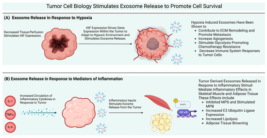
Cancer cachexia is defined as unintentional weight loss secondary to neoplasia and is associated with poor prognosis and outcomes. Cancer cachexia associated weight loss affects both lean tissue (i.e., skeletal muscle) and adipose tissue. Exosomes are extracellular vesicles that originate from multivesicular bodies that contain intentionally loaded biomolecular cargo. Exosome cargo includes proteins, lipids, mitochondrial components, and nucleic acids. The cargo carried in exosomes is thought to alter cell signaling when it enters into recipient cells. Virtually every cell type secretes exosomes and exosomes are known to be present in nearly every biofluid. Exosomes alter muscle and adipose tissue metabolism and biological processes, including macrophage polarization and apoptosis which contribute to the development of the cachexia phenotype. This has led to an interest in the role of tumor cell derived exosomes and their potential role as biomarkers of cancer cell development as well as their contribution to cachexia and disease progression. In this review, we highlight published findings that have studied the effects of tumor derived exosomes (and extracellular vesicles) and their cargo on the progression of cancer cachexia. We will focus on the direct effects of tumor derived exosomes and their cellular cross talk on skeletal muscle and adipose tissue, the primary sites of weight loss due to cancer cachexia. Full article
Show Figures

Figure 1

30 pages, 2240 KiB  
Review
Age-Related Dysfunction in Proteostasis and Cellular Quality Control in the Development of Sarcopenia
by Hector G. Paez, Christopher R. Pitzer and Stephen E. Alway
Cells 2023, 12(2), 249; https://doi.org/10.3390/cells12020249 - 7 Jan 2023
Cited by 19 | Viewed by 4765
Abstract
Sarcopenia is a debilitating skeletal muscle disease that accelerates in the last decades of life and is characterized by marked deficits in muscle strength, mass, quality, and metabolic health. The multifactorial causes of sarcopenia have proven difficult to treat and involve a complex [...] Read more.
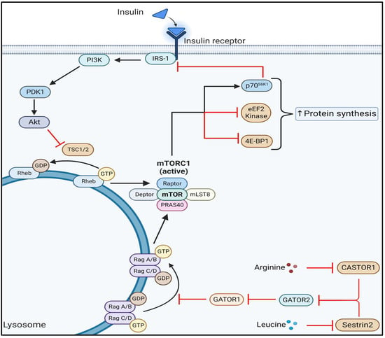
Sarcopenia is a debilitating skeletal muscle disease that accelerates in the last decades of life and is characterized by marked deficits in muscle strength, mass, quality, and metabolic health. The multifactorial causes of sarcopenia have proven difficult to treat and involve a complex interplay between environmental factors and intrinsic age-associated changes. It is generally accepted that sarcopenia results in a progressive loss of skeletal muscle function that exceeds the loss of mass, indicating that while loss of muscle mass is important, loss of muscle quality is the primary defect with advanced age. Furthermore, preclinical models have suggested that aged skeletal muscle exhibits defects in cellular quality control such as the degradation of damaged mitochondria. Recent evidence suggests that a dysregulation of proteostasis, an important regulator of cellular quality control, is a significant contributor to the aging-associated declines in muscle quality, function, and mass. Although skeletal muscle mammalian target of rapamycin complex 1 (mTORC1) plays a critical role in cellular control, including skeletal muscle hypertrophy, paradoxically, sustained activation of mTORC1 recapitulates several characteristics of sarcopenia. Pharmaceutical inhibition of mTORC1 as well as caloric restriction significantly improves muscle quality in aged animals, however, the mechanisms controlling cellular proteostasis are not fully known. This information is important for developing effective therapeutic strategies that mitigate or prevent sarcopenia and associated disability. This review identifies recent and historical understanding of the molecular mechanisms of proteostasis driving age-associated muscle loss and suggests potential therapeutic interventions to slow or prevent sarcopenia. Full article
Show Figures

Figure 1

2022

Jump to: 2023, 2021

19 pages, 1386 KiB  
Review
The Role of Crosstalk between Adipose Cells and Myocytes in the Pathogenesis of Sarcopenic Obesity in the Elderly
by Mauro Zamboni, Gloria Mazzali, Anna Brunelli, Tanaz Saatchi, Silvia Urbani, Anna Giani, Andrea P. Rossi, Elena Zoico and Francesco Fantin
Cells 2022, 11(21), 3361; https://doi.org/10.3390/cells11213361 - 25 Oct 2022
Cited by 10 | Viewed by 3785
Abstract
As a result of aging, body composition changes, with a decline in muscle mass and an increase in adipose tissue (AT), which reallocates from subcutaneous to visceral depots and stores ectopically in the liver, heart and muscles. Furthermore, with aging, muscle and AT, [...] Read more.
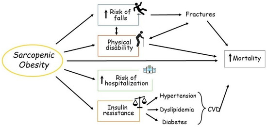
As a result of aging, body composition changes, with a decline in muscle mass and an increase in adipose tissue (AT), which reallocates from subcutaneous to visceral depots and stores ectopically in the liver, heart and muscles. Furthermore, with aging, muscle and AT, both of which have recognized endocrine activity, become dysfunctional and contribute, in the case of positive energy balance, to the development of sarcopenic obesity (SO). SO is defined as the co-existence of excess adiposity and low muscle mass and function, and its prevalence increases with age. SO is strongly associated with greater morbidity and mortality. The pathogenesis of SO is complex and multifactorial. This review focuses mainly on the role of crosstalk between age-related dysfunctional adipose and muscle cells as one of the mechanisms leading to SO. A better understanding of this mechanisms may be useful for development of prevention strategies and treatments aimed at reducing the occurrence of SO. Full article
Show Figures

Figure 1

15 pages, 4863 KiB  
Article
Metabolic Advantage of 25(OH)D3 versus 1,25(OH)2D3 Supplementation in Infantile Nephropathic Cystinosis-Associated Adipose Tissue Browning and Muscle Wasting
by Ping Zhou, Wai W. Cheung, Alex Gonzalez, Venya Vaddi, Eduardo A. Oliveira and Robert H. Mak
Cells 2022, 11(20), 3264; https://doi.org/10.3390/cells11203264 - 17 Oct 2022
Cited by 4 | Viewed by 1777
Abstract
Manifestations of infantile nephropathic cystinosis (INC) often include cachexia and deficiency of circulating vitamin D metabolites. We examined the impact of 25(OH)D3 versus 1,25(OH)2D3 repletion in Ctns null mice, a mouse model of INC. Six weeks of intraperitoneal administration [...] Read more.
Manifestations of infantile nephropathic cystinosis (INC) often include cachexia and deficiency of circulating vitamin D metabolites. We examined the impact of 25(OH)D3 versus 1,25(OH)2D3 repletion in Ctns null mice, a mouse model of INC. Six weeks of intraperitoneal administration of 25(OH)D3 (75 μg/kg/day) or 1,25(OH)2D3 (60 ng/kg/day) resulted in Ctns−/− mice corrected low circulating 25(OH)D3 or 1,25(OH)2D3 concentrations. While 25(OH)D3 administration in Ctns−/− mice normalized several metabolic parameters characteristic of cachexia as well as muscle function in vivo, 1,25(OH)2D3 did not. Administration of 25(OH)D3 in Ctns−/− mice increased muscle fiber size and decreased fat infiltration of skeletal muscle, which was accompanied by a reduction of abnormal muscle signaling pathways. 1,25(OH)2D3 administration was not as effective. In conclusion, 25(OH)D3 supplementation exerts metabolic advantages over 1,25(OH)2D3 supplementation by amelioration of muscle atrophy and fat browning in Ctns−/− mice. Full article
Show Figures

Figure 1

9 pages, 1974 KiB  
Article
Modulation of Neuroendocrine and Immunological Biomarkers Following Rehabilitation in Sarcopenic Patients
by Federica Piancone, Francesca La Rosa, Ivana Marventano, Ambra Hernis, Rossella Miglioli, Fabio Trecate, Marina Saresella and Mario Clerici
Cells 2022, 11(16), 2477; https://doi.org/10.3390/cells11162477 - 10 Aug 2022
Cited by 1 | Viewed by 1843
Abstract
This study aimed to investigate if rehabilitation could down-regulated sarcopenia-associated inflammation by modulating the crosstalk between the neuroendocrine and immune systems, with the aim of ameliorating quality of life of sarcopenic subjects. A total of 60 sarcopenic patients (49 females and 11 males; [...] Read more.
This study aimed to investigate if rehabilitation could down-regulated sarcopenia-associated inflammation by modulating the crosstalk between the neuroendocrine and immune systems, with the aim of ameliorating quality of life of sarcopenic subjects. A total of 60 sarcopenic patients (49 females and 11 males; median age 74.5, interquartile range 71–79), undergoing a personalized rehabilitation program, have been recruited and subjected to: (1) functional and physical evaluation (Short Physical Performance Battery (SPPB), Barthel Index and Tinetti Test); (2) pro-inflammatory IL-1β, TNF-α, IL-6, IL-18, and anti-inflammatory IL-10 cytokines plasmatic level measures; and (3) norepinephrine, epinephrine, dopamine, and serotonin neurotransmitter level evaluation at time of enrollment (T0) and once rehabilitation was concluded (1 month, T1). Rehabilitation combined a balance and strength training program with two daily sessions that were fine-tuned and personalized according to the ability of the patient. The results showed a significant increase at T1 in the plasmatic levels of IL-10 (p = 0.018) and of norepinephrine (p = 0.016)), whereas the concentration of IL-18 was significantly reduced (p = 0.012). Notably, changes in norepinephrine were positively correlated with clinical improvements (Tinetti and Barthel scores, p ≤ 0.0001; SPPB scores, p = 0.0002). These results show that efficient rehabilitation induces a reduction of inflammation, suggesting that this effect could be mediated by a modulation of the neuro-immune axis that results in an increase of norepinephrine. Full article
Show Figures

Figure 1

26 pages, 1554 KiB  
Review
Shared and Divergent Epigenetic Mechanisms in Cachexia and Sarcopenia
by Laura Yedigaryan, Martina Gatti, Vittoria Marini, Tullia Maraldi and Maurilio Sampaolesi
Cells 2022, 11(15), 2293; https://doi.org/10.3390/cells11152293 - 25 Jul 2022
Cited by 10 | Viewed by 4665
Abstract
Significant loss of muscle mass may occur in cachexia and sarcopenia, which are major causes of mortality and disability. Cachexia represents a complex multi-organ syndrome associated with cancer and chronic diseases. It is often characterized by body weight loss, inflammation, and muscle and [...] Read more.
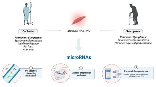
Significant loss of muscle mass may occur in cachexia and sarcopenia, which are major causes of mortality and disability. Cachexia represents a complex multi-organ syndrome associated with cancer and chronic diseases. It is often characterized by body weight loss, inflammation, and muscle and adipose wasting. Progressive muscle loss is also a hallmark of healthy aging, which is emerging worldwide as a main demographic trend. A great challenge for the health care systems is the age-related decline in functionality which threatens the independence and quality of life of elderly people. This biological decline can also be associated with functional muscle loss, known as sarcopenia. Previous studies have shown that microRNAs (miRNAs) play pivotal roles in the development and progression of muscle wasting in both cachexia and sarcopenia. These small non-coding RNAs, often carried in extracellular vesicles, inhibit translation by targeting messenger RNAs, therefore representing potent epigenetic modulators. The molecular mechanisms behind cachexia and sarcopenia, including the expression of specific miRNAs, share common and distinctive trends. The aim of the present review is to compile recent evidence about shared and divergent epigenetic mechanisms, particularly focusing on miRNAs, between cachexia and sarcopenia to understand a facet in the underlying muscle wasting associated with these morbidities and disclose potential therapeutic interventions. Full article
Show Figures

Graphical abstract

24 pages, 788 KiB  
Review
The Functional Role of Long Non-Coding RNA in Myogenesis and Skeletal Muscle Atrophy
by Keisuke Hitachi, Masahiko Honda and Kunihiro Tsuchida
Cells 2022, 11(15), 2291; https://doi.org/10.3390/cells11152291 - 25 Jul 2022
Cited by 13 | Viewed by 4558
Abstract
Skeletal muscle is a pivotal organ in humans that maintains locomotion and homeostasis. Muscle atrophy caused by sarcopenia and cachexia, which results in reduced muscle mass and impaired skeletal muscle function, is a serious health condition that decreases life longevity in humans. Recent [...] Read more.
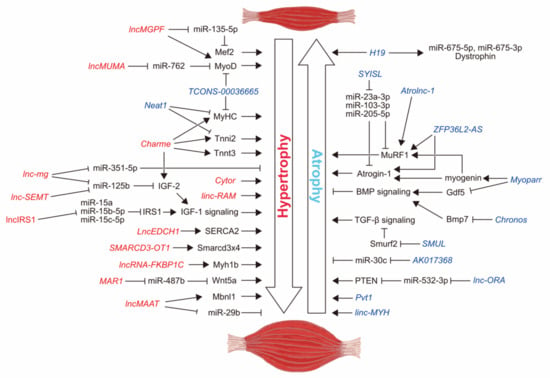
Skeletal muscle is a pivotal organ in humans that maintains locomotion and homeostasis. Muscle atrophy caused by sarcopenia and cachexia, which results in reduced muscle mass and impaired skeletal muscle function, is a serious health condition that decreases life longevity in humans. Recent studies have revealed the molecular mechanisms by which long non-coding RNAs (lncRNAs) regulate skeletal muscle mass and function through transcriptional regulation, fiber-type switching, and skeletal muscle cell proliferation. In addition, lncRNAs function as natural inhibitors of microRNAs and induce muscle hypertrophy or atrophy. Intriguingly, muscle atrophy modifies the expression of thousands of lncRNAs. Therefore, although their exact functions have not yet been fully elucidated, various novel lncRNAs associated with muscle atrophy have been identified. Here, we comprehensively review recent knowledge on the regulatory roles of lncRNAs in skeletal muscle atrophy. In addition, we discuss the issues and possibilities of targeting lncRNAs as a treatment for skeletal muscle atrophy and muscle wasting disorders in humans. Full article
Show Figures

Figure 1

19 pages, 2263 KiB  
Systematic Review
Association of Oral Function and Dysphagia with Frailty and Sarcopenia in Community-Dwelling Older Adults: A Systematic Review and Meta-Analysis
by Kotomi Sakai, Enri Nakayama, Daisuke Yoneoka, Nobuo Sakata, Katsuya Iijima, Tomoki Tanaka, Kuniyoshi Hayashi, Kunihiro Sakuma and Eri Hoshino
Cells 2022, 11(14), 2199; https://doi.org/10.3390/cells11142199 - 14 Jul 2022
Cited by 21 | Viewed by 5593
Abstract
Studies investigating the associations of oral function and dysphagia with frailty and sarcopenia in community-dwelling older adults are increasing; however, they have not been systematically summarized. We conducted a systematic review to investigate these associations. We searched electronic databases and synthesized relevant data [...] Read more.
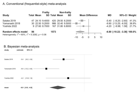
Studies investigating the associations of oral function and dysphagia with frailty and sarcopenia in community-dwelling older adults are increasing; however, they have not been systematically summarized. We conducted a systematic review to investigate these associations. We searched electronic databases and synthesized relevant data using conventional (frequentist-style) and Bayesian meta-analyses. Twenty-four studies were found to be eligible for our review, including 20 cross-sectional and four prospective cohort studies. Older adults with frailty or sarcopenia had lower tongue pressure, according to the results of conventional meta-analysis (mean difference [95% confidence interval or credible interval]: −6.80 kPa [−10.22 to −3.38] for frailty and −5.40 kPa [−6.62 to −4.17] for sarcopenia) and Bayesian meta-analysis (−6.90 kPa [−9.0 to −4.8] for frailty, −5.35 kPa [−6.78 to −3.89] for sarcopenia). People with frailty had a higher odds ratio (OR) for dysphagia according to the results of conventional meta-analysis (3.99 [2.17 to 7.32]) and Bayesian meta-analysis (1.38 [0.77 to 1.98]). However, the results were inconclusive for people with sarcopenia. A prospective association could not be determined because of the lack of information and the limited number of studies. Decreased oral function and dysphagia can be important characteristics of frailty and sarcopenia in community-dwelling older adults. Full article
Show Figures

Figure 1

12 pages, 625 KiB  
Review
Selected Methods of Resistance Training for Prevention and Treatment of Sarcopenia
by Tomohiro Yasuda
Cells 2022, 11(9), 1389; https://doi.org/10.3390/cells11091389 - 20 Apr 2022
Cited by 19 | Viewed by 7416
Abstract
Resistance training is an extremely beneficial intervention to prevent and treat sarcopenia. In general, traditional high-load resistance training improves skeletal muscle morphology and strength, but this method is impractical and may even reduce arterial compliance by about 20% in aged adults. Thus, the [...] Read more.
Resistance training is an extremely beneficial intervention to prevent and treat sarcopenia. In general, traditional high-load resistance training improves skeletal muscle morphology and strength, but this method is impractical and may even reduce arterial compliance by about 20% in aged adults. Thus, the progression of resistance training methods for improving the strength and morphology of muscles without applying a high load is essential. Over the past two decades, various resistance training methods that can improve skeletal muscle mass and muscle function without using high loads have attracted attention, and their training effects, molecular mechanisms, and safety have been reported. The present study focuses on the relationship between exercise load/intensity, training effects, and physiological mechanisms as well as the safety of various types of resistance training that have attracted attention as a measure against sarcopenia. At present, there is much research evidence that blood-flow-restricted low-load resistance training (20–30% of one repetition maximum (1RM)) has been reported as a sarcopenia countermeasure in older adults. Therefore, this training method may be particularly effective in preventing sarcopenia. Full article
Show Figures

Figure 1

10 pages, 1109 KiB  
Article
Skeletal Muscle Pathological Fat Infiltration (Myosteatosis) Is Associated with Higher Mortality in Patients with Cirrhosis
by Maryam Ebadi, Cynthia Tsien, Rahima A. Bhanji, Abha R. Dunichand-Hoedl, Elora Rider, Maryam Motamedrad, Vera C. Mazurak, Vickie Baracos and Aldo J. Montano-Loza
Cells 2022, 11(8), 1345; https://doi.org/10.3390/cells11081345 - 14 Apr 2022
Cited by 33 | Viewed by 3762
Abstract
Myosteatosis (pathological fat accumulation in muscle) is defined by lower mean skeletal muscle radiodensity in CT. We aimed to determine the optimal cut-offs for myosteatosis in a cohort of 855 patients with cirrhosis. CT images were used to determine the skeletal muscle radiodensity [...] Read more.
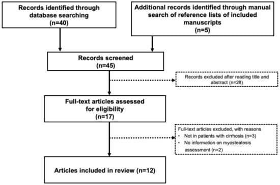
Myosteatosis (pathological fat accumulation in muscle) is defined by lower mean skeletal muscle radiodensity in CT. We aimed to determine the optimal cut-offs for myosteatosis in a cohort of 855 patients with cirrhosis. CT images were used to determine the skeletal muscle radiodensity expressed as Hounsfield Unit (HU). Patients with muscle radiodensity values below the lowest tertile were considered to have myosteatosis. Competing-risk analysis was performed to determine associations between muscle radiodensity and pre-transplant mortality. Muscle radiodensity less than 33 and 28 HU in males and females, respectively, were used as cut-offs to identify myosteatosis. In the univariate analysis, cirrhosis etiology, MELD score, refractory ascites, variceal bleeding, hepatic encephalopathy, sarcopenia and myosteatosis were predictors of mortality. Myosteatosis association with mortality remained significant after adjusting for confounding factors (sHR 1.47, 95% CI 1.17–1.84, p = 0.001). Patients with concurrent presence of myosteatosis and sarcopenia constituted 17% of the patient population. The cumulative incidence of mortality was the highest in patients with concomitant sarcopenia and myosteatosis (sHR 2.22, 95% CI 1.64–3.00, p < 0.001). In conclusion, myosteatosis is common in patients with cirrhosis and is associated with increased mortality. The concomitant presence of myosteatosis and sarcopenia is associated with worse outcomes. Full article
Show Figures

Figure 1

24 pages, 638 KiB  
Review
Sarcopenia in Children with Solid Organ Tumors: An Instrumental Era
by Annika Ritz, Eberhard Lurz and Michael Berger
Cells 2022, 11(8), 1278; https://doi.org/10.3390/cells11081278 - 9 Apr 2022
Cited by 10 | Viewed by 3756
Abstract
Sarcopenia has recently been studied in both adults and children and was found to be a prognostic marker for adverse outcome in a variety of patient groups. Our research showed that sarcopenia is a relevant marker in predicting outcome in children with solid [...] Read more.
Sarcopenia has recently been studied in both adults and children and was found to be a prognostic marker for adverse outcome in a variety of patient groups. Our research showed that sarcopenia is a relevant marker in predicting outcome in children with solid organ tumors, such as hepatoblastoma and neuroblastoma. This was especially true in very ill, high-risk groups. Children with cancer have a higher likelihood of ongoing loss of skeletal muscle mass due to a mismatch in energy intake and expenditure. Additionally, the effects of cancer therapy, hormonal alterations, chronic inflammation, multi-organ dysfunction, and a hypermetabolic state all contribute to a loss of skeletal muscle mass. Sarcopenia seems to be able to pinpoint this waste to a high degree in a new and objective way, making it an additional tool in predicting and improving outcome in children. This article focuses on the current state of sarcopenia in children with solid organ tumors. It details the pathophysiological mechanisms behind sarcopenia, highlighting the technical features of the available methods for measuring muscle mass, strength, and function, including artificial intelligence (AI)-based techniques. It also reviews the latest research on sarcopenia in children, focusing on children with solid organ tumors. Full article
Show Figures

Figure 1

15 pages, 1858 KiB  
Review
Myosteatosis in Cirrhosis: A Review of Diagnosis, Pathophysiological Mechanisms and Potential Interventions
by Maryam Ebadi, Cynthia Tsien, Rahima A. Bhanji, Abha R. Dunichand-Hoedl, Elora Rider, Maryam Motamedrad, Vera C. Mazurak, Vickie Baracos and Aldo J. Montano-Loza
Cells 2022, 11(7), 1216; https://doi.org/10.3390/cells11071216 - 4 Apr 2022
Cited by 37 | Viewed by 5944
Abstract
Myosteatosis, or pathological excess fat accumulation in muscle, has been widely defined as a lower mean skeletal muscle radiodensity on computed tomography (CT). It is reported in more than half of patients with cirrhosis, and preliminary studies have shown a possible association with [...] Read more.
Myosteatosis, or pathological excess fat accumulation in muscle, has been widely defined as a lower mean skeletal muscle radiodensity on computed tomography (CT). It is reported in more than half of patients with cirrhosis, and preliminary studies have shown a possible association with reduced survival and increased risk of portal hypertension complications. Despite the clinical implications in cirrhosis, a standardized definition for myosteatosis has not yet been established. Currently, little data exist on the mechanisms by which excess lipid accumulates within the muscle in individuals with cirrhosis. Hyperammonemia may play an important role in the pathophysiology of myosteatosis in this setting. Insulin resistance, impaired mitochondrial oxidative phosphorylation, diminished lipid oxidation in muscle and age-related differentiation of muscle stem cells into adipocytes have been also been suggested as potential mechanisms contributing to myosteatosis. The metabolic consequence of ammonia-lowering treatments and omega-3 polyunsaturated fatty acids in reversing myosteatosis in cirrhosis remains uncertain. Factors including the population of interest, design and sample size, single/combined treatment, dosing and duration of treatment are important considerations for future trials aiming to prevent or treat myosteatosis in individuals with cirrhosis. Full article
Show Figures

Figure 1

19 pages, 2490 KiB  
Review
Adaptive Remodeling of the Neuromuscular Junction with Aging
by Michael R. Deschenes, Rachel Flannery, Alexis Hawbaker, Leah Patek and Mia Mifsud
Cells 2022, 11(7), 1150; https://doi.org/10.3390/cells11071150 - 29 Mar 2022
Cited by 18 | Viewed by 7976
Abstract
Aging is associated with gradual degeneration, in mass and function, of the neuromuscular system. This process, referred to as “sarcopenia”, is considered a disease by itself, and it has been linked to a number of other serious maladies such as type II diabetes, [...] Read more.
Aging is associated with gradual degeneration, in mass and function, of the neuromuscular system. This process, referred to as “sarcopenia”, is considered a disease by itself, and it has been linked to a number of other serious maladies such as type II diabetes, osteoporosis, arthritis, cardiovascular disease, and even dementia. While the molecular causes of sarcopenia remain to be fully elucidated, recent findings have implicated the neuromuscular junction (NMJ) as being an important locus in the development and progression of that malady. This synapse, which connects motor neurons to the muscle fibers that they innervate, has been found to degenerate with age, contributing both to senescent-related declines in muscle mass and function. The NMJ also shows plasticity in response to a number of neuromuscular diseases such as amyotrophic lateral sclerosis (ALS) and Lambert-Eaton myasthenic syndrome (LEMS). Here, the structural and functional degradation of the NMJ associated with aging and disease is described, along with the measures that might be taken to effectively mitigate, if not fully prevent, that degeneration. Full article
Show Figures

Graphical abstract

17 pages, 937 KiB  
Review
Sarcopenia Is a Cause and Consequence of Metabolic Dysregulation in Aging Humans: Effects of Gut Dysbiosis, Glucose Dysregulation, Diet and Lifestyle
by James W. Daily and Sunmin Park
Cells 2022, 11(3), 338; https://doi.org/10.3390/cells11030338 - 20 Jan 2022
Cited by 39 | Viewed by 9308
Abstract
Skeletal muscle mass plays a critical role in a healthy lifespan by helping to regulate glucose homeostasis. As seen in sarcopenia, decreased skeletal muscle mass impairs glucose homeostasis, but it may also be caused by glucose dysregulation. Gut microbiota modulates lipopolysaccharide (LPS) production, [...] Read more.
Skeletal muscle mass plays a critical role in a healthy lifespan by helping to regulate glucose homeostasis. As seen in sarcopenia, decreased skeletal muscle mass impairs glucose homeostasis, but it may also be caused by glucose dysregulation. Gut microbiota modulates lipopolysaccharide (LPS) production, short-chain fatty acids (SCFA), and various metabolites that affect the host metabolism, including skeletal muscle tissues, and may have a role in the sarcopenia etiology. Here, we aimed to review the relationship between skeletal muscle mass, glucose homeostasis, and gut microbiota, and the effect of consuming probiotics and prebiotics on the development and pathological consequences of sarcopenia in the aging human population. This review includes discussions about the effects of glucose metabolism and gut microbiota on skeletal muscle mass and sarcopenia and the interaction of dietary intake, physical activity, and gut microbiome to influence sarcopenia through modulating the gut–muscle axis. Emerging evidence suggests that the microbiome can regulate both skeletal muscle mass and function, in part through modulating the metabolisms of short-chain fatty acids and branch-chain amino acids that might act directly on muscle in humans or indirectly through the brain and liver. Dietary factors such as fats, proteins, and indigestible carbohydrates and lifestyle interventions such as exercise, smoking, and alcohol intake can both help and hinder the putative gut–muscle axis. The evidence presented in this review suggests that loss of muscle mass and function are not an inevitable consequence of the aging process, and that dietary and lifestyle interventions may prevent or delay sarcopenia. Full article
Show Figures

Figure 1

2021

Jump to: 2023, 2022

20 pages, 6490 KiB  
Article
Decreased miR-497-5p Suppresses IL-6 Induced Atrophy in Muscle Cells
by Paula P. Freire, Sarah S. Cury, Letícia O. Lopes, Geysson J. Fernandez, Jianming Liu, Leonardo Nazario de Moraes, Grasieli de Oliveira, Jakeline S. Oliveira, Diogo de Moraes, Otavio Cabral-Marques, Maeli Dal-Pai-Silva, Xiaoyun Hu, Da-Zhi Wang and Robson F. Carvalho
Cells 2021, 10(12), 3527; https://doi.org/10.3390/cells10123527 - 14 Dec 2021
Cited by 11 | Viewed by 4062
Abstract
Interleukin-6 (IL-6) is a pro-inflammatory cytokine associated with skeletal muscle wasting in cancer cachexia. The control of gene expression by microRNAs (miRNAs) in muscle wasting involves the regulation of thousands of target transcripts. However, the miRNA-target networks associated with IL6-induced muscle atrophy remain [...] Read more.
Interleukin-6 (IL-6) is a pro-inflammatory cytokine associated with skeletal muscle wasting in cancer cachexia. The control of gene expression by microRNAs (miRNAs) in muscle wasting involves the regulation of thousands of target transcripts. However, the miRNA-target networks associated with IL6-induced muscle atrophy remain to be characterized. Here, we show that IL-6 promotes the atrophy of C2C12 myotubes and changes the expression of 20 miRNAs (5 up-regulated and 15 down-regulated). Gene Ontology analysis of predicted miRNAs targets revealed post-transcriptional regulation of genes involved in cell differentiation, apoptosis, migration, and catabolic processes. Next, we performed a meta-analysis of miRNA-published data that identified miR-497-5p, a down-regulated miRNAs induced by IL-6, also down-regulated in other muscle-wasting conditions. We used miR-497-5p mimics and inhibitors to explore the function of miR-497-5p in C2C12 myoblasts and myotubes. We found that miR-497-5p can regulate the expression of the cell cycle genes CcnD2 and CcnE1 without affecting the rate of myoblast cellular proliferation. Notably, miR-497-5p mimics induced myotube atrophy and reduced Insr expression. Treatment with miR-497-5p inhibitors did not change the diameter of the myotubes but increased the expression of its target genes Insr and Igf1r. These genes are known to regulate skeletal muscle regeneration and hypertrophy via insulin-like growth factor pathway and were up-regulated in cachectic muscle samples. Our miRNA-regulated network analysis revealed a potential role for miR-497-5p during IL6-induced muscle cell atrophy and suggests that miR-497-5p is likely involved in a compensatory mechanism of muscle atrophy in response to IL-6. Full article
Show Figures

Figure 1

Planned Papers

The below list represents only planned manuscripts. Some of these manuscripts have not been received by the Editorial Office yet. Papers submitted to MDPI journals are subject to peer-review.

Title: Dysfuntional macrophages: Impact on disappearing lung tissue in COPD and bone tissue in osteoporosis
Authors: Stephan van Eeden
Affiliation: University of British Columbia, Canada

Back to TopTop