Article

Combined Toxicity of Xenobiotics Bisphenol A and Heavy Metals on Zebrafish Embryos (Danio rerio)

by
Davide Di Paola
1,†,
Fabiano Capparucci
1,†,
Giovanni Lanteri
1,
Marika Cordaro
2,
Rosalia Crupi
3,
Rosalba Siracusa
1,
Ramona D’Amico
1,
Roberta Fusco
1,
Daniela Impellizzeri
1,
Salvatore Cuzzocrea
1,4,*,
Nunziacarla Spanò
2,*,
Enrico Gugliandolo
3,‡ and
Alessio Filippo Peritore
1,‡
1
Department of Chemical, Biological, Pharmaceutical, and Environmental Science, University of Messina, 98166 Messina, Italy
2
Department of Biomedical and Dental Sciences and Morphofunctional Imaging, University of Messina, 98100 Messina, Italy
3
Department of Veterinary Science, University of Messina, 98100 Messina, Italy
4
Department of Pharmacological and Physiological Science, Saint Louis University School of Medicine, Saint Louis, MO 63108, USA
*
Authors to whom correspondence should be addressed.
The first two authors contributed equally to this study.
These authors shared senior authorship.
Toxics 2021, 9(12), 344; https://doi.org/10.3390/toxics9120344
Submission received: 19 October 2021 / Revised: 5 November 2021 / Accepted: 26 November 2021 / Published: 9 December 2021
(This article belongs to the Special Issue Environmental and Health Effects of Heavy Metal)

Abstract

:
Environmental pollutants may cause adverse effects on the immune system of aquatic organisms. This study revealed that combination of environmental pollutants and Bisphenol A(BPA) could cause an acute inflammatory response in zebrafish larvae as shown by body alterations, which may imply a common immunotoxicity mechanism for most environmental pollutants. In the present study we evaluated the toxicity after co-exposure of BPA and Cd or Cr (III) in zebrafish embryos and larvae, and the oxidative stress pathway involved. Evaluation of lethal and developmental endpoints such as hatching, edema, malformations, abnormal heart rate and survival rate were evaluated after 96 h of exposure. Combination of BPA at 10 μM with Cd or Cr at 0.5 μM exposure induce malformations at 96 hpf in zebrafish larvae, as well as significantly increases oxidative stress and induce apoptosis on larvae. Our study suggested how environmental pollutant showed a synergistic effect at common not-effective doses, promoting decrease of antioxidant defense and contrasted fish development.

1. Introduction

Endocrine-disrupting chemicals (EDCs) are molecules with a structural similarity with endogenous hormones, that can alter hormone biosynthesis, excretion or biodegradation. Environmental exposures to high levels of EDCs can result in alterations to biological homeostasis. In recent years, numerous classes of chemical compounds have been identified as endocrine disruptors, and their widespread presence in the environment has increased concerns for wildlife and consequently for humans.
Bisphenol A (BPA) is a building block of polycarbonate plastics and is used heavly in the production of consumer products including food containers (toys, water pipes, or medical tubing. In addition, BPA can be found in epoxy resins and store receipts, as it is an additive in thermal paper [1,2,3]. Since BPA is heavily present in consumer products, it is one of the highest produced chemicals in the world with approximately comsumption reaching 7.7 million metric tons in 2015, with a projection for 2022 of 10.6 million metric tons [4]. In the early 2000s, Asian markets saw an increase in demand for BPA, polycarbonate and epoxy resin, which has led to an increase in BPA contamination over the years [5].
Many industrial and household chemicals are disposed of in aquatic environments, including bisphenols, leading to health impacts for a range of aquatic animal species. Several studies have focused on BPA exposure to fish, with particular attention to development and sexual function [6,7]. However, the effects of toxic exposure to BPA, on fish development, have only been reported at concentrations greater than those found in natural environments [8,9].
Natural environments can be filled, not only with chemical compounds such as EDCs, but also with other types of contaminants such as heavy metals. Heavy metals in different environments arise from natural sources such as erosion, volcanic activity and forest fires, raising great concern to aquatic environments, reaching through contaminated sediments, wastewater or oil spills [10]. Their persistence and bioaccumulation may cause adverse effects in the development, growth, and reproduction of various aquatic organisms [11,12]. Several branches industries frequently use both Cadmium (Cd) and Chromium (Cr), for example batteries, cast iron, metal finishes and others, resulting in massive releases of them into the environment by different pathways. [13,14]. Indeed, high levels of Cd and Cr were observed in the waterborne and sediment especially in developing countries. Elevated levels of dissolved Cd and Cr have been detected in freshwater, ranging from 10 to 500 ng/L, and in some industrialized areas can reach as 1 mg/L [15,16]. Previous studies have shown that Cd and Cr can cause hepatotoxicity and nephrotoxicity, as well as disrupt the endocrine systems of many aquatic organisms. Underlying the multiple toxic effects of Cd and Cr there is the involvement of the oxidative stress pathway and the consequent decrease of the antioxidant defenses [17,18]
Prolonged exposure to heavy metals can induce oxidative stress resulting in increased production of reactive oxygen species (ROS). An increase in ROS production, which have different targets (such as proteins, lipids or DNA) can lead to several diseases, as well as cell death and aging. Previous studies have shown that exposure to BPA can result in increased oxidative stress in the liver of male rats [19]. In addition, BPA-induced oxidative stress has also been seen in zebrafish models, through biochemical analyses with antioxidant markers and phosphatase [20]. In oxygen-consuming organisms, antioxidant enzymes play a key role in the first line of defense. Among the various antioxidant enzymes, an important role is played by the superoxide dismutase (SOD). ISOD converts the superoxide anion (O2ࢤ) into hydrogen peroxide (H2O2), which is then converted into H2O and O2 by another important antioxidant enzyme the catalase (CAT). In several aquatic organisms, such as mollusk, fish and polychaete, the effects of xenobiotics on SOD and CAT enzymes expression have been ascertained [21,22,23,24,25,26]. Furthermore, it has been seen that heavy metals can cross the blood-brain membrane, accumulating at the brain level where they can cause damage induction and apoptosis [27,28,29]. In this regard, these enzymes can be used as biomarkers of oxidative stress induced by environmental contaminants [27].
As a model of drug screening in recent years is being used a lot the zebrafish (Danio rerio). Zebrafish model is useful not only for their short generation time and low costs, but also due to the high size of the clutch in the embryonic and larval stages, and their optical transparency that allows the visualization of pathogens and lesions in real time [30]. In the present study, we investigated the acute toxicity of two heavy metals (Cd and Cr) and an EDCs (BPA) in zebrafish larvae. Furthermore, we analyzed the transcriptional modulation of stress oxidative and apoptotic pathway after exposure the xenobiotic BPA alone and in combination with heavy metals contaminants at various concentration.

2. Materials and Methods

2.1. Zebrafish Maintenance and Embryo Collection

Zebrafish Maintenance and Embryo Collection of fertilized eggs were provided from the Center of Experimental Fish Patology (Centro di ittiopatologia Sperimentale della Sicilia, Messina, CISS), University of Messina, Italy. Eggs were collected and selected within 4 h post fertilization (hpf) under a stereomicroscope (Leica M0205C, Multifocus, Wetzlar, Germany). All embryos were derived from the same spawns of eggs.

2.2. Dose Preparation and Exposure Protocols

The Cadmium chloride (CdCl2) (Cas Number: 654054-66-7) and potassium dichromate (K2Cr2O7) (CAS Number: 7778-50-9) (Sigma-Aldrich, St. Louis, MO, USA) was dissolved in distilled and deionized water to produce a 10 mM stock solution. The 100 mM stock solution of BPA (CAS 80-05-7) (Sigma-Aldrich, St. Louis, MO, USA) was obtained by dissolving BPA powder in dimethyl sulfoxide (DMSO) at 0.01% (v/v). (Sigma-Aldrich, St. Louis, MO, USA). BPA in the stock solution was diluted with 0.01M phosphate buffered saline (PBS, pH 7.0) and further diluted with Embryo medium (15 mM NaCl, 0.5 mM KCl, 1 mM CaCl2, 1 mM MgSO4, 0.15 mM KH2PO4, 0.05 mM Na2HPO4, 0.7 mM NaHCO3 a pH 7.3) to the tested concentrations (10 and 50 μM). Before application, both stock solutions were diluted in fresh embryo culture medium to generate the final concentrations needed (0.5 μM Cd and Cr, 10 and 50 μM BPA).

2.3. Fish Embryo Toxicity (FET) Test

Fish Embryo Toxicity (FET) test was performed according to OECD [31] and ISO 15088. Zebrafish embryos exposed to BPA concentrations (10 and 50 μM), and after BPA 10 μM in combination with 0.5 μM of Cd and Cr in 1mL of freshwater for 4–96 h post-fertilization hpf were measured for toxic effects of a continuing observation period. All the solutions were renewed, and embryonic/larval mortality and hatching rate were evaluated every 24 h, while the heart rate was measured at 96 hpf. As we described in previous paper [32], healthy embryos were placed in 24-well culture plates (1 embryo in 1ml solution/well). Each group had 20 embryos and each experiment was replicated three times. During the exposure period, photographs of the embryos were made under a stereomicroscope (Leica M0205C, Multifocus, Wetzlar, Germany) and the percentage of abnormal embryos was counted every 24 h.

2.4. Viability, Morphology and Hatching Rate

The health of zebrafish larva was assessed as previously described [33]. To examine the survival rate and morphology of embryos/larvae, BPA in the stock solution was diluted with 0.01 M phosphate buffered saline (PBS, pH7.0) and further diluted with Embryo medium (15 mM NaCl, 0.5 mM KCl, 1 mM CaCl2, 1 mM MgSO4, 0.15 mM KH2PO4, 0.05 mM Na2HPO4, 0.7 mM NaHCO3 a pH 7.3) to the tested concentrations (10 and 50 μM). Healthy and normally developing WT embryos at 4 hpf were collected and exposed to vehicle or various concentrations of BPA, Cd and Cr in egg water. During the exposure time, it was observed the embryonic development and various parameters including mortality, hatching rate, heartbeat rate, as well as abnormalities like pericardial edema, pigmentation, and axial spinal curvature in hatched larvae were documented [34]. Morphology scores were determined at 96 hpf as previously described. Nine endpoints, including body shape, somites, notochord, tail, fins, heart, face, brain, and pharyngeal arches/jaws, were examined to evaluate the phenotypes of the zebrafish, and eight larval specimens per group were used for scoring [35].
To measure heart rate, embryos at 48, 72 and 96 hpf were moved to room temperature and allowed to stabilize for 30 min prior to manual counting. For each treatment condition, ten embryos were selected at random, and their heart rates were measured for four intervals of 20 s under a stereomicroscope.

2.5. Total RNA Extraction and RT-PCR

Total RNA was extracted from zebrafish larvae tissue using the NanoMag Animal and Fish RNA Isolation Kit (Shannuo Scientific Company, Shanghai, China). One microgram of total RNA was subjected to cDNA synthesis using PrimeScript@ RT Master Mix. Real-time PCR was performed in a total volume of 20 μL containing 10 μL of SYBR Green supermix, 1 μL of cDNA, 7 μL of RNase/DNase-free water and 500 nM of each primer. Gene expression was analyzed using the 2ΔΔCT method, the b-actin (gene encoding beta-actin) was used as housekeeping gene for the normalization of RT-qPCR. The sequences of primers for real-time PCR are shown in Table 1.

2.6. Detection of ROS and Antioxidative Enzyme Assay

The larvae from each group were defrosted and homogenized on ice with 180 μL ice-cold physiological saline. The homogenate was centrifuged at 12,000× g at 4 °C for 10min to obtain the supernatant. ROS concentrations were assessed as previously described [36]. The content of SOD and CAT of the supernatant was analyzed as described previously [37,38,39,40,41].

2.7. Materials

All compounds used in this study were purchased from Sigma-Aldrich Company Ltd.

2.8. Statistical Evaluation

All values in the figures and text are expressed as the mean ± standard error of the mean (SEM) of N number of animals. The results were analyzed by one-way ANOVA followed by a Bonferroni post-hoc test for multiple comparisons.

3. Results

3.1. Effect of Bisphenol A on Morphology, Viability, and Hatching Rate

In order to identify the suitable concentrations, time points were used in the following experiments: BPA in the range of 10 and 50 μM doses was applied to observe morphology of larvae until 96 hpf. Embryo development in the CTRL was normal: hatching began at 48 to 72 hpf while no mortality was showed. As presented in Figure 1 BPA 10 μM did not alter the zebrafish morphology until 96 hpf compared to CTRL group. BPA 50 μM groups induced a suite of abnormalities in zebrafish embryos. The effect of BPA 50 μM on the embryo development was observed from 48 hpf. Furthermore, when embryos were treated with BPA 50 μM, no hatching rate was observed after 96 hpf. For embryos exposed to BPA 10 μM, no hatching influence was found compared with CTRL.

3.2. Malformation Scores, Body Length, and Heart Rate

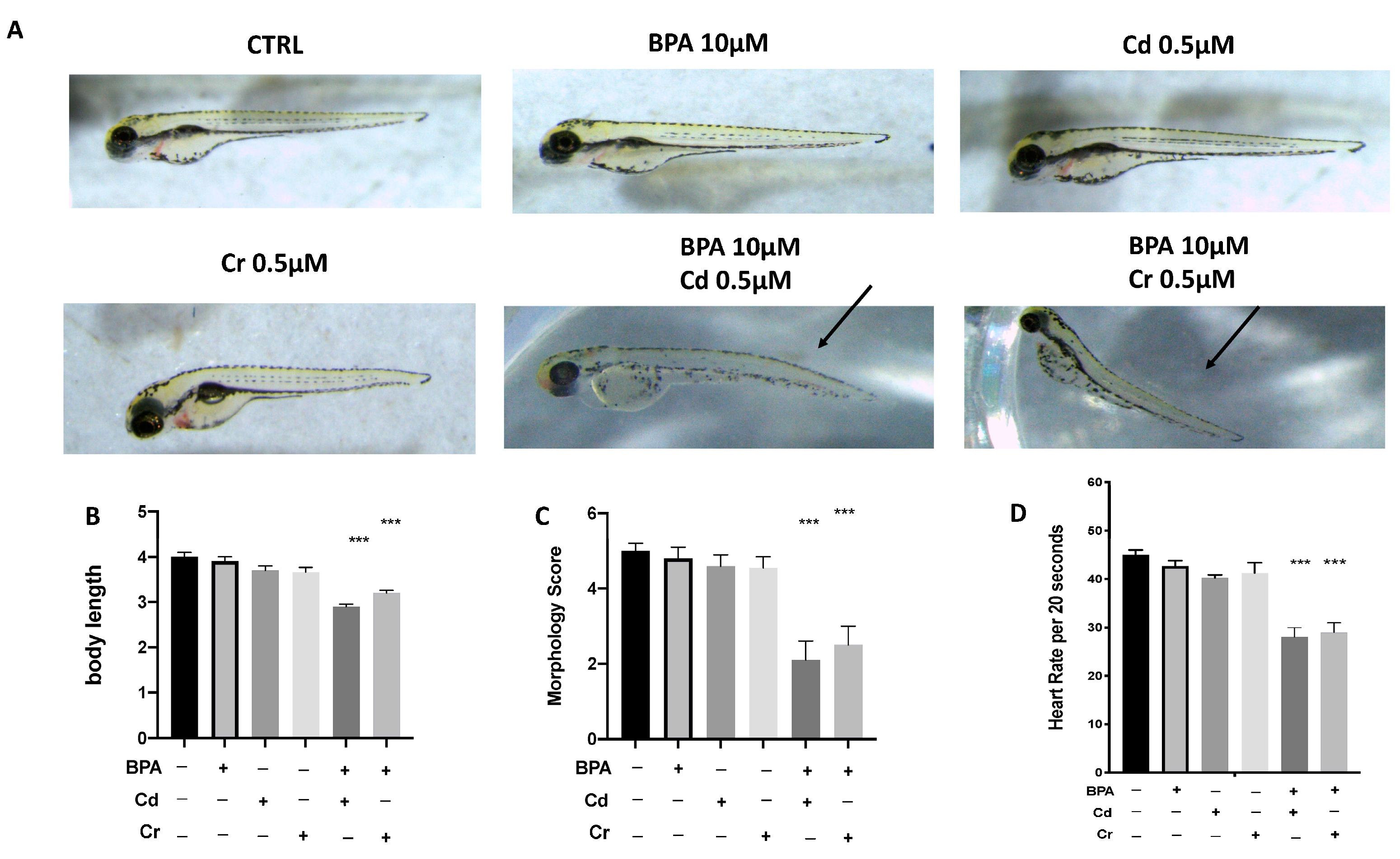
Phenotypic defections at time points up to 96 hpf were noted. Compared with the control group, the malformation rate of the BPA 10 μM group showed no significant change. Abnormalities, primarily modest yolk retention, and pericardial edema were found in the combined groups, with BPA 10 μM and Cd 0.5 μM (BPA + Cd) or BPA 10 μM and Cr 0.5 μM (BPA + Cr). The body lengths of the larvae were measured at 96 hpf to assess the degree of development (Figure 2C). The body lengths of the larvae at 96 hpf were significantly reduced in both the BPA + Cd and the BPA + Cr group, which indicated that the combination of low dose of BPA and heavy metals significantly inhibited larval growth. Heart rates were recorded to determine the effect of combination of BPA and Cd and Cr on cardiac function. In the BPA 10 μM group embryos, as well as in the CTRL, heart rate did not show differences at 96 hpf. However, significant bradycardia was observed in embryos treated both BPA + Cd and BPA + Cr compared to the CTRL (Figure 2D). No differences on morphology, body length, and heart rate were found in the groups with single Cd and Cr exposure (Figure 2A–D).

3.3. Effect of BPA, Cd, and Cr on Gene Expression of Antioxidant Pathway

The changes in the expression of antioxidant-related genes in the zebrafish larvae after exposure to BPA alone and in combination with Cd and Cr are shown in Figure 3. Both BPA + Cd and BPA + Cr significantly downregulated the expression levels of cat, sod1 and gstp2, compared to CTRL group. However, there was no significant change in the expression level of antioxidant-gene in the BPA alone group compared to the CTRL group (Figure 3). Moreover, the expression of mRNA of Nrf2, a common transcription factor of antioxidant defenses, was decreased after both BPA + Cd and BPA + Cr compared to CTRL, while no effect was showed in the single BPA group. No differences on oxidative stress related mRNA expression were found in the groups with single Cd and Cr exposure. No differences on oxidative stress related mRNA expression was found in the groups with single Cd and Cr exposure. At the same time, we analyzed the content of SOD and CAT and determined the percentage of ROS, following exposure to BPA, Cd and Cr individually and in synergy. We showed a decrease in both CAT and SOD content following co-exposure to BPA and Cd or Cr, as well as an increase in ROS production. In contrast, the groups with single exposure of the various contaminants (BPA, Cd and Cr), there were no differences in CAT and SOD content and ROS production compared to the control group.

3.4. Effect of BPA, Cd, and Cr on Gene Expression of Apoptotic Pathway

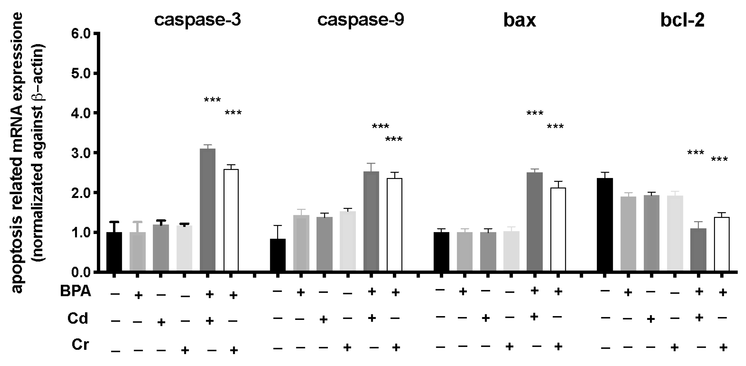
To investigate the possible mechanisms of the toxic effects induced by combination of BPA and Cd or Cr, we used RT-PCR to examine the mRNA expression levels of larvae exposed from 6 to 120 hpf. The mRNA expression levels of apoptosis-related genes (caspase-3, caspase-9, and bax) increased in BPA + CD more than in the BPA + Cr group, compared to CTRL (Figure 4). The mRNA expression level of bcl-2 was downregulated both in BPA + Cd and BPA + Cr group. In addition, no significant differences in apoptotic and anti-apoptotic related genes were observed between the BPA group and CTRL alone (Figure 4). No differences on apoptosis related mRNA expression were found in the groups with single Cd and Cr exposure.

4. Discussion

BPA, Cd and Cr applied widely in consumers [42,43]. To date, several studies regarding the toxicity of BPA, Cd and Cr have been conducted to investigate their adverse effects on human and animal health, but they have been exposed individually and never together. Furthermore, given the likelihood of the simultaneous presence of several EDCs, increasing attention is being paid to the potential toxicity of their co-exposure. In the present study, we evaluated the possibility that BPA, Cd, and Cr together increase oxidative stress damage, in normally not effective single doses, which means that there is an additive or synergistic effect. The first objective of the study was to identify an appropriate concentration range for further validation studies on the BPA dose to be exposed in combination with Cd and Cr. We showed that BPA induced developmental toxicity in zebrafish embryos, particularly delayed hatching and morphological abnormalities. The 10 and 50 μM doses chosen for the toxicity studies ranges from no injury to noticeable toxic effect on development. BPA exposure at 10 μM did not show clear signs of toxicity, while doses of 50μM strongly reduced survival and hatching rate at 96 and 72 hpf respectively. Hatching is a critical moment of zebrafish embryogenesis; consequently, the decreased hatching rate was induced by functional and structural disturbances during embryonic development [44,45]. In addition, the suppression of embryogenesis or the inhibition of mitosis [46] or the incapability of the embryonic larvae to open the eggshell [47] also likely caused the developmental delay. Our data showed a significant dose-dependent reduction of the hatching rate, with the 100 % of no hatched egg for the dose 50 μM at 96 hpf, which is a critical indicator of developmental toxicity. Furthermore, BPA exposure at 50 μM resulted in embryonic teratogenesis, characterized by spinal curvature, pericardial edema, bent tails and uninflated swim bladders.
It have been reported that mean dissolved Cd concentrations in rivers strongly impacted by mining activities in the Riou Mort, in France is 26 μg/L [48], and the average concentrations of BPA exposure of adult is no more than 0.4–1.5 μg/kg body weight (BW) per day [49]. Because the metabolic rate and elimination ofheavy metals are very slow infishes, the outcomes induced by Cd andCr may be permanent for zebrafish [50]. Although the doses of BPA, Cd and Cr used in our present study are lower than the environmental exposure levels, the aim of this study employed so low doses of BPA, Cd and Cr is to mimic the occupational exposure and to explore their combined genotoxic effects. In our study BPA exposure alone did not show genotoxicity and cytotoxicity at 10 μM. Our results are consistent with previous data showing that BPA exposure at 25 μM does not cause oxidative stress-associated DNA damage in INS-1 cells according to the comet assay [51]. Moreover, no significant changes in cell viability or DNA damage levels have been observed in GM00637 or HeLa S3 cells when exposed to 5 μM or 10 μM Cd [52]. The combination of BPA and heavy metals such as Cd or Cr, at doses that are themselves non-toxic, showed alteration in early zebrafish development. In fact, co-exposure of 10 μM BPA and 0.5 μM Cd or 0.5 μM Cr(III) increasing the incidence of malformations such as spinal lordosis, pericardial edemas and heart rate decrease among others. Moreover, the body length reduction indicated that BPA in combination both Cd than Cr exposure could affect the growth of the larvae, which could be explained by delayed deciduation. All these data agree in proving that developmental toxicity induced by combination of BPA with both Cd or Cr exposure. Critical pathways involved in the developmental toxicity are inflammation and oxidative stress [53]. About the EDCs oxidative stress two main mechanisms are involved: the reduction of the cellular antioxidant defenses and the ROS overproduction [54,55]. ROS are the main promotors of oxidative stress [56] because they excessively combined with CAT, GSH and SOD unbalancing the antioxidant protection mechanism. SOD is an important enzyme in the endogenous antioxidant system thanks to its ability to prevent lipid peroxidation and remove ROS [41]. Another key antioxidant factor is gstp2, member of the GST Pi family, which is involved with glutathione in removing ROS [57]. The mRNA levels of sod1, cat, and gstp2 have been decreased in larvae exposure with both BPA 10 μM + Cd 0.5 μM and BPA 10 μM +Cr 0.5 μM, but not in BPA 10 μM group. Furthermore, the combination of BPA and Cd and Cr also caused an increase in CAT and SOD content, as well as the production of ROS, suggested that the the antioxidant defenses were impaired in the combinated-exposure groups. Thus, these data displayed that the decreased oxidative stress has a key role in the developmental sinergistic toxicity of EDCs and heavy metals. Recent studies have suggested Nrf2 as a potent key factor, responsible for the regulation of cellular oxidative stress response [58]. A large number of genes are reported to be influenced by Nrf2 in zebrafish [37]. Moreover, Nrf2 is an adaptive measure of cells against oxidative stress induced by Cd [59] and also Cr(VI), a precursor of Cr(III) before entering the intracellular level [60]. In the present study, a decrease in mRNA levels of Nrf2 was confirmed both BPA + Cd and BPA + Cr groups, as opposed to the BPA 10 μM group alone, which showed no effect on Nrf2 mRNA. A decrease in Nrf2 expression in the nucleus can be translated in an antioxidant protein levels decrease, confirming what was seen with the sod1, cat, and gstp2 datas. In fact increased ROS production associated with decreased of antioxidant defense expression are not the only involved in EDCs toxicity of zebrafish embryonic development, but also apoptosis and the caspase pathway play an important role [61]. Apoptosis is a regulated mechanism of cell death and with a key role in several physiological and pathological processes [62]. ROS over-production has been shown to be an important apoptotic signal [63,64,65]. The induction of apoptosis in zebrafish embryos was detected by the mRNA levels analysis of apoptosis-related genes Caspase 3 and 9, Bax and Bcl-2. Previous study suggested that metal ion-induced ROS production acts directly on mitochondria to cause cytochrome c release from mitochondria into the cytosol, which leads to caspase-9 activation and apoptosis [66]. Caspases are a large family of proteinases that play crucial roles in the process of apoptosis and are considered markers of oxidative stress-induced apoptosis in zebrafish embryos [67]. Our data showed that the exposure of BPA with Cd or Cr increased expression of the apoptosis-inducing target genes and reduced the expression of the anti-apoptotic factor, while the only BPA. These results suggested that apoptosis induction has a key role in the developmental toxicity of EDCs and heavy metals in early stage of fish development.

5. Conclusions

In conclusion, we demonstrated that BPA in combination with Cd and Cr, at a single ineffective dose, induced genotoxicity and cytotoxicity on early life-stage zebrafish. These results may support the idea that BPA, which have been shown to be non-toxic or safe at low levels, can become highly toxic when combined with other marine pollutants such as heavy metals. Our results suggested stress oxidative involvement in the molecular mechanisms involved in BPA-heavy metals combinate toxicity, with a decrease of antioxidant enzyme expression and an augment of ROS production. Further investigation into the combined effects of environmental pollutants and future work may illuminate the mechanisms underlying this synergistic toxicity.

Author Contributions

Conceptualization, S.C.; methodology D.I., R.C. and D.D.P.; validation, M.C., R.S. and, E.G., formal analysis and investigation, A.F.P. and F.C.; resources, R.F.; data curation G.L. and R.D.; writing—original draft preparation, A.F.P.; writing—review and editing, A.F.P.; visualization, E.G.; supervision S.C.; project administration, S.C.; funding acquisition, S.C. and N.S. All authors have read and agreed to the published version of the manuscript.

Funding

This research received no external funding.

Institutional Review Board Statement

Not applicable.

Informed Consent Statement

Not applicable.

Data Availability Statement

Not applicable.

Conflicts of Interest

The authors declare no conflict of interest.

References

  1. Liao, C.; Kannan, K. High levels of bisphenol A in paper currencies from several countries, and implications for dermal exposure. Environ. Sci. Technol. 2011, 45, 6761–6768. [Google Scholar] [CrossRef] [PubMed]
  2. Geens, T.; Aerts, D.; Berthot, C.; Bourguignon, J.-P.; Goeyens, L.; Lecomte, P.; Maghuin-Rogister, G.; Pironnet, A.-M.; Pussemier, L.; Scippo, M.-L. A review of dietary and non-dietary exposure to bisphenol-A. Food Chem. Toxicol. 2012, 50, 3725–3740. [Google Scholar] [CrossRef]
  3. Li, X.; Yin, P.; Zhao, L. Effects of individual and combined toxicity of bisphenol A, dibutyl phthalate and cadmium on oxidative stress and genotoxicity in HepG 2 cells. Food Chem. Toxicol. 2017, 105, 73–81. [Google Scholar] [CrossRef]
  4. Experts, I. Bisphenol-A-A Global Market Overview; Hexion Inc.: Columbus, OH, USA, 2016. [Google Scholar]
  5. Huang, Y.; Wong, C.; Zheng, J.; Bouwman, H.; Barra, R.; Wahlström, B.; Neretin, L.; Wong, M.H. Bisphenol A (BPA) in China: A review of sources, environmental levels, and potential human health impacts. Environ. Int. 2012, 42, 91–99. [Google Scholar] [CrossRef]
  6. Oehlmann, J.; Schulte-Oehlmann, U.; Kloas, W.; Jagnytsch, O.; Lutz, I.; Kusk, K.O.; Wollenberger, L.; Santos, E.M.; Paull, G.C.; Van Look, K.J. A critical analysis of the biological impacts of plasticizers on wildlife. Philos. Trans. R. Soc. B Biol. Sci. 2009, 364, 2047–2062. [Google Scholar] [CrossRef] [Green Version]
  7. Flint, S.; Markle, T.; Thompson, S.; Wallace, E. Bisphenol A exposure, effects, and policy: A wildlife perspective. J. Environ. Manag. 2012, 104, 19–34. [Google Scholar] [CrossRef]
  8. Lam, S.H.; Hlaing, M.M.; Zhang, X.; Yan, C.; Duan, Z.; Zhu, L.; Ung, C.Y.; Mathavan, S.; Ong, C.N.; Gong, Z. Toxicogenomic and phenotypic analyses of bisphenol-A early-life exposure toxicity in zebrafish. PLoS ONE 2011, 6, e28273. [Google Scholar] [CrossRef] [PubMed] [Green Version]
  9. Chow, W.S.; Chan, W.K.L.; Chan, K.M. Toxicity assessment and vitellogenin expression in zebrafish (Danio rerio) embryos and larvae acutely exposed to bisphenol A, endosulfan, heptachlor, methoxychlor and tetrabromobisphenol A. J. Appl. Toxicol. 2013, 33, 670–678. [Google Scholar] [CrossRef]
  10. Nriagu, J.O. A global assessment of natural sources of atmospheric trace metals. Nature 1989, 338, 47–49. [Google Scholar] [CrossRef]
  11. Juárez-Franco, M.F.; Sarma, S.; Nandini, S. Effect of cadmium and zinc on the population growth of Brachionus havanaensis (Rotifera: Brachionidae). J. Environ. Sci. Health Part A 2007, 42, 1489–1493. [Google Scholar] [CrossRef] [PubMed]
  12. Das, S.; Khangarot, B. Bioaccumulation of copper and toxic effects on feeding, growth, fecundity and development of pond snail Lymnaea luteola L. J. Hazard. Mater. 2011, 185, 295–305. [Google Scholar] [CrossRef]
  13. Bagchi, D.; Joshi, S.S.; Bagchi, M.; Balmoori, J.; Benner, E.; Kuszynski, C.; Stohs, S. Cadmium-and chromium-induced oxidative stress, DNA damage, and apoptotic cell death in cultured human chronic myelogenous leukemic K562 cells, promyelocytic leukemic HL-60 cells, and normal human peripheral blood mononuclear cells. J. Biochem. Mol. Toxicol. 2000, 14, 33–41. [Google Scholar] [CrossRef]
  14. Alghasham, A.; Salem, T.A.; Meki, A.-R.M. Effect of cadmium-polluted water on plasma levels of tumor necrosis factor-α, interleukin-6 and oxidative status biomarkers in rats: Protective effect of curcumin. Food Chem. Toxicol. 2013, 59, 160–164. [Google Scholar] [CrossRef] [PubMed]
  15. Jones, I.; Kille, P.; Sweeney, G. Cadmium delays growth hormone expression during rainbow trout development. J. Fish Biol. 2001, 59, 1015–1022. [Google Scholar] [CrossRef]
  16. Ma, W.; Wang, L.; He, Y.; Yan, Y. Tissue-specific cadmium and metallothionein levels in freshwater crab Sinopotamon henanense during acute exposure to waterborne cadmium. Environ. Toxicol. Int. J. 2008, 23, 393–400. [Google Scholar] [CrossRef]
  17. Woo, S.; Yum, S.; Park, H.-S.; Lee, T.-K.; Ryu, J.-C. Effects of heavy metals on antioxidants and stress-responsive gene expression in Javanese medaka (Oryzias javanicus). Comp. Biochem. Physiol. Part C Toxicol. Pharmacol. 2009, 149, 289–299. [Google Scholar] [CrossRef] [PubMed]
  18. Li, Z.-H.; Chen, L.; Wu, Y.-H.; Li, P.; Li, Y.-F.; Ni, Z.-H. Effects of waterborne cadmium on thyroid hormone levels and related gene expression in Chinese rare minnow larvae. Comp. Biochem. Physiol. Part C Toxicol. Pharmacol. 2014, 161, 53–57. [Google Scholar] [CrossRef]
  19. Bindhumol, V.; Chitra, K.; Mathur, P. Bisphenol A induces reactive oxygen species generation in the liver of male rats. Toxicology 2003, 188, 117–124. [Google Scholar] [CrossRef]
  20. Wu, M.; Xu, H.; Shen, Y.; Qiu, W.; Yang, M. Oxidative stress in zebrafish embryos induced by short-term exposure to bisphenol A, nonylphenol, and their mixture. Environ. Toxicol. Chem. 2011, 30, 2335–2341. [Google Scholar] [CrossRef]
  21. Isani, G.; Monari, M.; Andreani, G.; Fabbri, M.; Carpenè, E. Effect of copper exposure on the antioxidant enzymes in bivalve mollusc Scapharca inaequivalvis. Vet. Res. Commun. 2003, 27, 691–693. [Google Scholar] [CrossRef]
  22. Vale, G.; Franco, C.; Diniz, M.S.; dos Santos, M.M.; Domingos, R.F. Bioavailability of cadmium and biochemical responses on the freshwater bivalve Corbicula fluminea–the role of TiO2 nanoparticles. Ecotoxicol. Environ. Saf. 2014, 109, 161–168. [Google Scholar] [CrossRef] [PubMed]
  23. Ken, C.-F.; Lin, C.-T.; Shaw, J.-F.; Wu, J.-L. Characterization of fish Cu/Zn–superoxide dismutase and its protection from oxidative stress. Mar. Biotechnol. 2003, 5, 167–173. [Google Scholar] [CrossRef] [PubMed]
  24. Cao, L.; Huang, W.; Liu, J.; Yin, X.; Dou, S. Accumulation and oxidative stress biomarkers in Japanese flounder larvae and juveniles under chronic cadmium exposure. Comp. Biochem. Physiol. Part C Toxicol. Pharmacol. 2010, 151, 386–392. [Google Scholar] [CrossRef] [PubMed]
  25. Won, E.-J.; Lee, J.-S.; Lee, Y.-M. Combined effects of cadmium and copper on the expression of antioxidant enzyme—Coding genes in the polychaete, Perinereis nuntia. Toxicol. Environ. Health Sci. 2013, 5, 26–33. [Google Scholar] [CrossRef]
  26. Won, E.-J.; Ra, K.; Kim, K.-T.; Lee, J.-S.; Lee, Y.-M. Three novel superoxide dismutase genes identified in the marine polychaete Perinereis nuntia and their differential responses to single and combined metal exposures. Ecotoxicol. Environ. Saf. 2014, 107, 36–45. [Google Scholar] [CrossRef]
  27. Matés, J.M.; Segura, J.A.; Alonso, F.J.; Márquez, J. Roles of dioxins and heavy metals in cancer and neurological diseases using ROS-mediated mechanisms. Free Radic. Biol. Med. 2010, 49, 1328–1341. [Google Scholar] [CrossRef] [PubMed]
  28. Richetti, S.K.; Rosemberg, D.B.; Ventura-Lima, J.; Monserrat, J.M.; Bogo, M.R.; Bonan, C.D. Acetylcholinesterase activity and antioxidant capacity of zebrafish brain is altered by heavy metal exposure. Neurotoxicology 2011, 32, 116–122. [Google Scholar] [CrossRef] [Green Version]
  29. Jaishankar, M.; Tseten, T.; Anbalagan, N.; Mathew, B.B.; Beeregowda, K.N. Toxicity, mechanism and health effects of some heavy metals. Interdiscip. Toxicol. 2014, 7, 60. [Google Scholar] [CrossRef] [PubMed] [Green Version]
  30. Lesley, R.; Ramakrishnan, L. Insights into early mycobacterial pathogenesis from the zebrafish. Curr. Opin. Microbiol. 2008, 11, 277–283. [Google Scholar] [CrossRef] [Green Version]
  31. Chemicals, D. OECD Guideline for Testing of Chemicals; The Organisation for Economic Co-operation and Development: Paris, France, 2015; pp. 1–13. [Google Scholar]
  32. Brundo, M.V.; Pecoraro, R.; Marino, F.; Salvaggio, A.; Tibullo, D.; Saccone, S.; Bramanti, V.; Buccheri, M.A.; Impellizzeri, G.; Scuderi, V. Toxicity evaluation of new engineered nanomaterials in zebrafish. Front. Physiol. 2016, 7, 130. [Google Scholar] [CrossRef]
  33. Di Paola, D.; Iaria, C.; Capparucci, F.; Cordaro, M.; Crupi, R.; Siracusa, R.; D’Amico, R.; Fusco, R.; Impellizzeri, D.; Cuzzocrea, S. Aflatoxin B1 Toxicity in Zebrafish Larva (Danio rerio): Protective Role of Hericium erinaceus. Toxins 2021, 13, 710. [Google Scholar] [CrossRef]
  34. Kuder, R.S.; Gundala, H.P. Developmental toxicity of deltamethrin and 3-phenoxybenzoic acid in embryo-larval stages of zebrafish (Danio rerio). Toxicol. Mech. Methods 2018, 28, 415–422. [Google Scholar] [CrossRef] [PubMed]
  35. Li, J.; Zhang, Y.; Liu, K.; He, Q.; Sun, C.; Han, J.; Han, L.; Tian, Q. Xiaoaiping induces developmental toxicity in zebrafish embryos through activation of ER stress, apoptosis and the Wnt pathway. Front. Pharmacol. 2018, 9, 1250. [Google Scholar] [CrossRef] [PubMed] [Green Version]
  36. Deng, J.; Yu, L.; Liu, C.; Yu, K.; Shi, X.; Yeung, L.W.; Lam, P.K.; Wu, R.S.; Zhou, B. Hexabromocyclododecane-induced developmental toxicity and apoptosis in zebrafish embryos. Aquat. Toxicol. 2009, 93, 29–36. [Google Scholar] [CrossRef] [PubMed]
  37. Liu, Y.; Wang, J.; Wei, Y.; Zhang, H.; Xu, M.; Dai, J. Induction of time-dependent oxidative stress and related transcriptional effects of perfluorododecanoic acid in zebrafish liver. Aquat. Toxicol. 2008, 89, 242–250. [Google Scholar] [CrossRef] [PubMed]
  38. Jin, Y.; Zhang, X.; Shu, L.; Chen, L.; Sun, L.; Qian, H.; Liu, W.; Fu, Z. Oxidative stress response and gene expression with atrazine exposure in adult female zebrafish (Danio rerio). Chemosphere 2010, 78, 846–852. [Google Scholar] [CrossRef]
  39. Jin, Y.; Wang, L.; Ruan, M.; Liu, J.; Yang, Y.; Zhou, C.; Xu, B.; Fu, Z. Cypermethrin exposure during puberty induces oxidative stress and endocrine disruption in male mice. Chemosphere 2011, 84, 124–130. [Google Scholar] [CrossRef] [PubMed]
  40. Stucki, G.; Alexander, M. Role of dissolution rate and solubility in biodegradation of aromatic compounds. Appl. Environ. Microbiol. 1987, 53, 2603. [Google Scholar] [CrossRef] [Green Version]
  41. Jin, Y.; Liu, Z.; Liu, F.; Ye, Y.; Peng, T.; Fu, Z. Embryonic exposure to cadmium (II) and chromium (VI) induce behavioral alterations, oxidative stress and immunotoxicity in zebrafish (Danio rerio). Neurotoxicology Teratol. 2015, 48, 9–17. [Google Scholar] [CrossRef]
  42. Kortenkamp, A. Ten years of mixing cocktails: A review of combination effects of endocrine-disrupting chemicals. Environ. Health Perspect. 2007, 115, 98–105. [Google Scholar] [CrossRef] [Green Version]
  43. Kocaoba, S.; Akcin, G. Removal of chromium (III) and cadmium (II) from aqueous solutions. Desalination 2005, 180, 151–156. [Google Scholar] [CrossRef]
  44. Samaee, S.M.; Rabbani, S.; Jovanovic, B.; Mohajeri-Tehrani, M.R.; Haghpanah, V. Efficacy of the hatching event in assessing the embryo toxicity of the nano-sized TiO(2) particles in zebrafish: A comparison between two different classes of hatching-derived variables. Ecotoxicol. Environ. Saf. 2015, 116, 121–128. [Google Scholar] [CrossRef]
  45. Liu, L.; Li, Y.; Coelhan, M.; Chan, H.M.; Ma, W.; Liu, L. Relative developmental toxicity of short-chain chlorinated paraffins in Zebrafish (Danio rerio) embryos. Environ. Pollut. 2016, 219, 1122–1130. [Google Scholar] [CrossRef] [PubMed]
  46. Ismail, A.; Yusof, S. Effect of mercury and cadmium on early life stages of Java medaka (Oryzias javanicus): A potential tropical test fish. Mar. Pollut. Bull. 2011, 63, 347–349. [Google Scholar] [CrossRef] [PubMed]
  47. Papiya, S.; Kanamadi, R. Effect of mercurial fungicide Emisan®-6 on the embryonic developmental stages of zebrafish, Brachydanio (Danio) rerio. J. Adv. Zool. 2000, 21, 12–18. [Google Scholar]
  48. Morin, S.; Duong, T.; Dabrin, A.; Coynel, A.; Herlory, O.; Baudrimont, M.; Delmas, F.; Durrieu, G.; Schäfer, J.; Winterton, P. Long-term survey of heavy-metal pollution, biofilm contamination and diatom community structure in the Riou Mort watershed, South-West France. Environ. Pollut. 2008, 151, 532–542. [Google Scholar] [CrossRef] [PubMed]
  49. Lakind, J.S.; Naiman, D.Q. Bisphenol A (BPA) daily intakes in the United States: Estimates from the 2003–2004 NHANES urinary BPA data. J. Expo. Sci. Environ. Epidemiol. 2008, 18, 608–615. [Google Scholar] [CrossRef] [Green Version]
  50. Kusch, R.C.; Krone, P.H.; Chivers, D.P. Chronic exposure to low concentrations of waterborne cadmium during embryonic and larval development results in the long-term hindrance of antipredator behavior in zebrafish. Environ. Toxicol. Chem. Int. J. 2008, 27, 705–710. [Google Scholar] [CrossRef] [PubMed]
  51. Xin, F.; Jiang, L.; Liu, X.; Geng, C.; Wang, W.; Zhong, L.; Yang, G.; Chen, M. Bisphenol A induces oxidative stress-associated DNA damage in INS-1 cells. Mutat. Res. Genet. Toxicol. Environ. Mutagenesis 2014, 769, 29–33. [Google Scholar] [CrossRef]
  52. Youn, C.-K.; Kim, S.-H.; Song, S.H.; Chang, I.-Y.; Hyun, J.-W.; Chung, M.-H.; You, H.J. Cadmium down-regulates human OGG1 through suppression of Sp1 activity. J. Biol. Chem. 2005, 280, 25185–25195. [Google Scholar] [CrossRef] [Green Version]
  53. Nazıroğlu, M. Role of selenium on calcium signaling and oxidative stress-induced molecular pathways in epilepsy. Neurochem. Res. 2009, 34, 2181–2191. [Google Scholar] [CrossRef] [PubMed]
  54. Baillie, T.A.; Rettie, A.E. Role of biotransformation in drug-induced toxicity: Influence of intra- and inter-species differences in drug metabolism. Drug Metab. Pharm. 2011, 26, 15–29. [Google Scholar] [CrossRef] [PubMed] [Green Version]
  55. Leung, L.; Kalgutkar, A.S.; Obach, R.S. Metabolic activation in drug-induced liver injury. Drug Metab. Rev. 2012, 44, 18–33. [Google Scholar] [CrossRef] [PubMed]
  56. Wijesinghe, W.A.; Kim, E.A.; Kang, M.C.; Lee, W.W.; Lee, H.S.; Vairappan, C.S.; Jeon, Y.J. Assessment of anti-inflammatory effect of 5beta-hydroxypalisadin B isolated from red seaweed Laurencia snackeyi in zebrafish embryo in vivo model. Environ. Toxicol. Pharmacol. 2014, 37, 110–117. [Google Scholar] [CrossRef] [PubMed]
  57. Dong, M.; Zhu, L.; Shao, B.; Zhu, S.; Wang, J.; Xie, H.; Wang, J.; Wang, F. The effects of endosulfan on cytochrome P450 enzymes and glutathione S-transferases in zebrafish (Danio rerio) livers. Ecotoxicol. Environ. Saf. 2013, 92, 1–9. [Google Scholar] [CrossRef]
  58. Kobayashi, A.; Kang, M.-I.; Okawa, H.; Ohtsuji, M.; Zenke, Y.; Chiba, T.; Igarashi, K.; Yamamoto, M. Oxidative stress sensor Keap1 functions as an adaptor for Cul3-based E3 ligase to regulate proteasomal degradation of Nrf2. Mol. Cell. Biol. 2004, 24, 7130–7139. [Google Scholar] [CrossRef] [PubMed] [Green Version]
  59. Zheng, J.-L.; Yuan, S.-S.; Wu, C.-W.; Lv, Z.-M. Acute exposure to waterborne cadmium induced oxidative stress and immunotoxicity in the brain, ovary and liver of zebrafish (Danio rerio). Aquat. Toxicol. 2016, 180, 36–44. [Google Scholar] [CrossRef]
  60. Shaw, P.; Mondal, P.; Bandyopadhyay, A.; Chattopadhyay, A. Environmentally relevant concentration of chromium activates Nrf2 and alters transcription of related XME genes in liver of zebrafish. Chemosphere 2019, 214, 35–46. [Google Scholar] [CrossRef]
  61. Cai, G.; Zhu, J.; Shen, C.; Cui, Y.; Du, J.; Chen, X. The effects of cobalt on the development, oxidative stress, and apoptosis in zebrafish embryos. Biol. Trace Elem. Res. 2012, 150, 200–207. [Google Scholar] [CrossRef]
  62. Cole, L.; Ross, L. Apoptosis in the developing zebrafish embryo. Dev. Biol. 2001, 240, 123–142. [Google Scholar] [CrossRef] [Green Version]
  63. Corda, S.; Laplace, C.; Vicaut, E.; Duranteau, J. Rapid reactive oxygen species production by mitochondria in endothelial cells exposed to tumor necrosis factor-α is mediated by ceramide. Am. J. Respir. Cell Mol. Biol. 2001, 24, 762–768. [Google Scholar] [CrossRef] [PubMed]
  64. Meier, B.; Radeke, H.; Selle, S.; Younes, M.; Sies, H.; Resch, K.; Habermehl, G. Human fibroblasts release reactive oxygen species in response to interleukin-1 or tumour necrosis factor-α. Biochem. J. 1989, 263, 539–545. [Google Scholar] [CrossRef]
  65. Shoji, Y.; Uedono, Y.; Ishikura, H.; Takeyama, N.; Tanaka, T. DNA damage induced by tumour necrosis factor-alpha in L929 cells is mediated by mitochondrial oxygen radical formation. Immunology 1995, 84, 543. [Google Scholar]
  66. Pulido, M.D.; Parrish, A.R. Metal-induced apoptosis: Mechanisms. Mutat. Res./Fundam. Mol. Mech. Mutagenesis 2003, 533, 227–241. [Google Scholar] [CrossRef] [PubMed]
  67. Wolf, B.B.; Green, D.R. Suicidal tendencies: Apoptotic cell death by caspase family proteinases. J. Biol. Chem. 1999, 274, 20049–20052. [Google Scholar] [CrossRef] [PubMed] [Green Version]
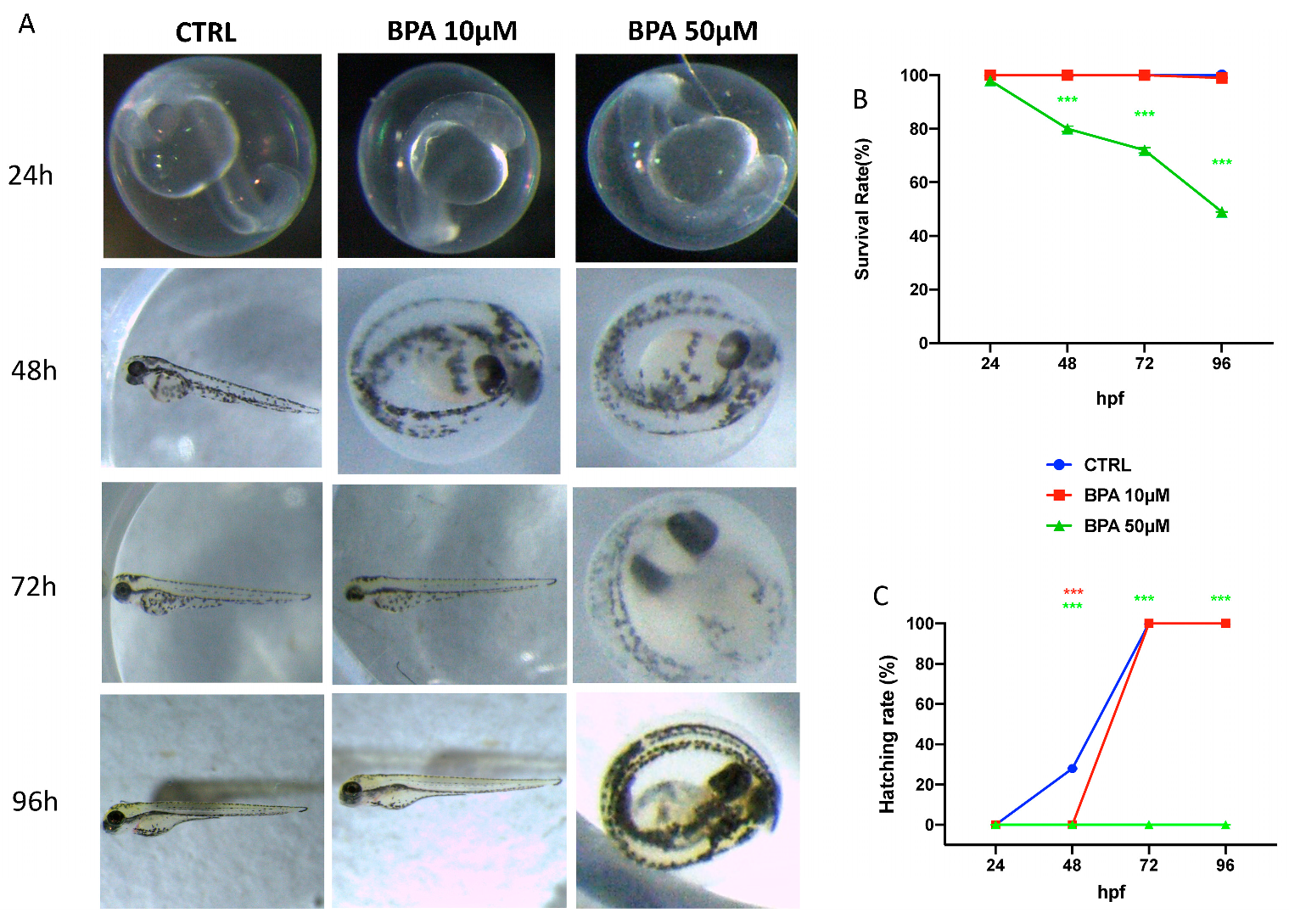
Figure 1. The morphological abnormalities in zebrafish caused by BPA different dose exposure (A), survival rate (B), and hatching rate (C). Images were taken from the lateral view under a dissecting microscope (magnification 25). Scale bar, 500 mm. *** p < 0.001 versus CTRL.
Toxics 09 00344 g001
Figure 2. Effects of BPA, Cd and Cr single and co-exposure on morphological changes in zebrafish larvae at 96 hpf. Malformation like the curvature of body axis are indicated by the arrows (A) Representative lateral views (A), body length (B), and morphological scoring (C) and heart rate (D) of zebrafish larvae treated. *** p < 0.001 versus CTRL.
Toxics 09 00344 g002
Figure 3. Effects of BPA, Cd and Cr single and co-exposure on the mRNA levels of stress oxidative pathway (cat, sod, gstp2, and nrf2) (A), CAT and SOD content (B,C) and ROS determination (D) in larval zebrafish. Values = means ± SEM of three independent experiment datas; *** at p < 0.001 against CTRL.
Toxics 09 00344 g003
Figure 4. Effects of BPA, Cd, and Cr single and co-exposure on the mRNA levels of apoptotic pathway (caspase3 and 9, bax, and bcl-2) in larval zebrafish. Values = means ± SEM of three independent experiment data; *** at p < 0.001 CTRL.
Toxics 09 00344 g004
Table 1. Primers for real-time PCR.
GenePrimer OrientationNucleotide Sequence
b-actinforward 5′-AGAGCTATGAGCTGCCTGACG-3′
reverse 5′-CCGCAAGATTCCATACCCA-3′
Oxidative stress
pathway genes
sod1forward 5′-GGCCAACCGATAGTGTTAGA-3′
reverse 5′-CCAGCGTTGCCAGTTTTTAG-3′
catforward 5′-AGGGCAACTGGGATCTTACA-3′
reverse 5′-TTTATGGGACCAGACCTTGG-3′
gstp2forward 5′-CACAGACCTCGCTTTTCACAC-3′
reverse 5′-GAGAGAAGCCTCACAGTCGT-3′
Nrf2forward 5′-TCGGGTTTGTCCCTAGATG-3′
reverse 5′-AGGTTTGGAGTGTCCGCTA-3′
Apoptosis pathway genes
casp-3forward 5′-CCGCTGCCCATCACTA-3′
reverse 5′-ATCCTTTCACGACCATCT-3′
Baxforward 5′-GGCTATTTCAACCAGGGTTCC-3′
reverse 5′-TGCGAATCACCAATGCTGT-3′
bcl-2forward 5′-TCACTCGTTCAGACCCTCAT-3′
reverse 5′-ACGCTTTCCACGCACAT-3′
casp-9forward5′-AAATACATAGCAAGGCAACC-3′
reverse5′-CACAGGGAATCAAGAAAGG-3′
Publisher’s Note: MDPI stays neutral with regard to jurisdictional claims in published maps and institutional affiliations.