Article

X-ray Fluorescence Techniques in Determining the Habitat Preferences of Species—Ulva pilifera (Ulvales, Chlorophyta) from Montenegro Case Study

by
Joanna Czerwik-Marcinkowska
1,*,
Renata Piwowarczyk
1,
Bohuslav Uher
2,
Ewa Tomal
3 and
Anna Wojciechowska
4
1
Institute of Biology, Jan Kochanowski University, 25-406 Kielce, Poland
2
Department of Microbiology, University of Vienna, 1010 Vienna, Austria
3
Provincial Sanitary and Epidemiological Station, 25-734 Kielce, Poland
4
Department of Geobotany and Landscape Planning, Nicolaus Copernicus University, 87-100 Toruń, Poland
*
Author to whom correspondence should be addressed.
Molecules 2020, 25(21), 5022; https://doi.org/10.3390/molecules25215022
Submission received: 4 September 2020 / Revised: 23 October 2020 / Accepted: 27 October 2020 / Published: 29 October 2020

Abstract

:
The paper presents four new sites where bright green Ulva thalli were found inhabiting freshwater (a river, a ditch, the Milet Canal) and marine (on the rocky shore of the Adriatic Sea) habitats in Montenegro. The aims of this study were to determine, for the first time, whether specimens of Ulva pilifera collected in Montenegro are phylogenetically and morphologically the same species as the one occurring in Europe. Using total reflection X-ray fluorescence (TXRF) and wavelength dispersive X-ray fluorescence (WDXRF) techniques it assessed the elemental composition of their thalli and its influence to colonise new habitats. Elements: Al, As, Ba, Br, Ca, Cl, Cr, Cu, Fe, Hf, I, K, Mg, Mn, Na, Ni, P, Pb, Rb, S, Si, Sr, Ti, V, and Zn were determined. The highest elemental concentrations were found for Ca = 16.3% (using WDXRF) and for Sr = 292 ppm (using TXRF) in the Ulva thalli. Ulva pilifera analysed from Montenegro, based on classical morphological methods and molecular techniques, are closely related to the same species from inland and coastal waters throughout Europe. The analysis of trace elements showed that the metal content in Ulva thalli is correlated with the trace elements in water and sediments. Ulva pilifera fits numerous features that make it one of the bioindicators of marine pollution, thanks to its worldwide distribution and capacity to accumulate trace elements.

1. Introduction

Ulva species grow attached to different substrates (such as soil, rocks, tree bark, leaves, and others macroalgae), but mature thalli are easily detached and become free living algae [1,2]. Due to the fact that Ulva species belong to the cosmopolitan genus, occurring in marine (with salinities ranging from 0.5–34%) and freshwater ecosystems [2,3,4] the seasonal massive growth (green tides worldwide) of the free living species is usually a result of high waters eutrophication [5,6]. The genus Ulva is one of the first marine genera described by Linneaus (previously known as Enteromorpha). Initially Ulva and Enteromorpha were considered as separate genera, but based on the internal transcribed spacer (ITS) region of nuclear ribosomal DNA (nrDNA) analysis reported by Hayden et al. [7], it was concluded that the genus Enteromorpha is synonymous with the genus Ulva [8]. It is generally known that the Ulva species are difficult to identify morphologically because of its high intraspecific variation and phenotypic plasticity [9,10]. In some habitats (e.g., littoral zone, coral reefs) some species of Ulva (e.g., U. flexuosa, U. reticulata) can be regarded as invasive with fouling potential [11]. However, Mareš et al. [12] showed that marine Ulva cannot be recognised as alien or invasive species in inland European waters, but they can be recognised as the primary algae colonisers and indicators of coastal waters eutrophication. Lougheed and Stevenson [13] suggested that Ulva species colonise habitats where ecological disturbances are present or conditions of water quality change because of various kinds of contamination.
Ulva pilifera (Kützing) Škaloud and Leliaert [14] is currently regarded as a synonym of Enteromorpha flexuosa subsp. pilifera (Kütz.) Bliding [15]; basionym Enteromorpha pilifera Kütz. [16]; the synonym of Ulva flexuosa subsp. pilifera (Kütz.) M. J. Wynne [17] is regularly encountered in freshwaters, but is equally well represented in brackish waters. In Europe, this species was reported from two distinct areas: a salt marsh in the Czech Republic [12] and from ponds and pools with increased salinity in Poland [18,19], Sweden [15], Slovakia [20], Germany [21], Hungary [12], and the United Kingdom [1]. U. pilifera has not until now been observed in Montenegro, although, Kapetanović et al. [22] and Kosanić et al. [23] found two marine ulvalean species U. lactuca and U. intestinalis in the Adriatic Sea.
This paper presents the study of freshwater and marine Ulva pilifera specimens from Montenegro, using morphological, molecular and X-ray fluorescence techniques (total reflection X-ray fluorescence (TXRF) and wavelength dispersive X-ray fluorescence (WDXRF)). It was assumed that Ulva marine species adapt effectively and rapidly to develop in freshwater habitats and along the coast of the Adriatic Sea, and also that the Ulva pilifera could be used as bioindicator of trace elemental composition in water and sediments in future.

2. Results and Discussion

Ulva specimens grow in the form of abundant tubular monostromatic thalli in waters (mesotrophic to eutrophic), which are characterised by high levels of nutrients, conductivity (400–1000 μS m−1), pH (5.5–9.5), and low oxygen concentration (2.1 mg L−1). Ulva has been collected in sites rich in N-NH4 (0.55 mg L−1), P-PO4 (0.28 mg L−1), and displayed high value conductivity (857.4 μS m−1). High concentrations of total iron (0.32 mg L−1) and chloride (92.3 mg L−1) were defined, in well-oxygenated sites. NaCl formed the major dissolved salt with sulphate and Mg/Ca bicarbonate. High concentrations of sulphate (38.7 mg L−1) were noted in habitats where the Ulva specimens were found.
Based on morphology, molecular, and elemental analysis (the X-ray fluorescence techniques: TXRF and WDXRF), all of the specimens found at the four sites in Montenegro (Figure 1 and Figure 2) were identified as U. pilifera.

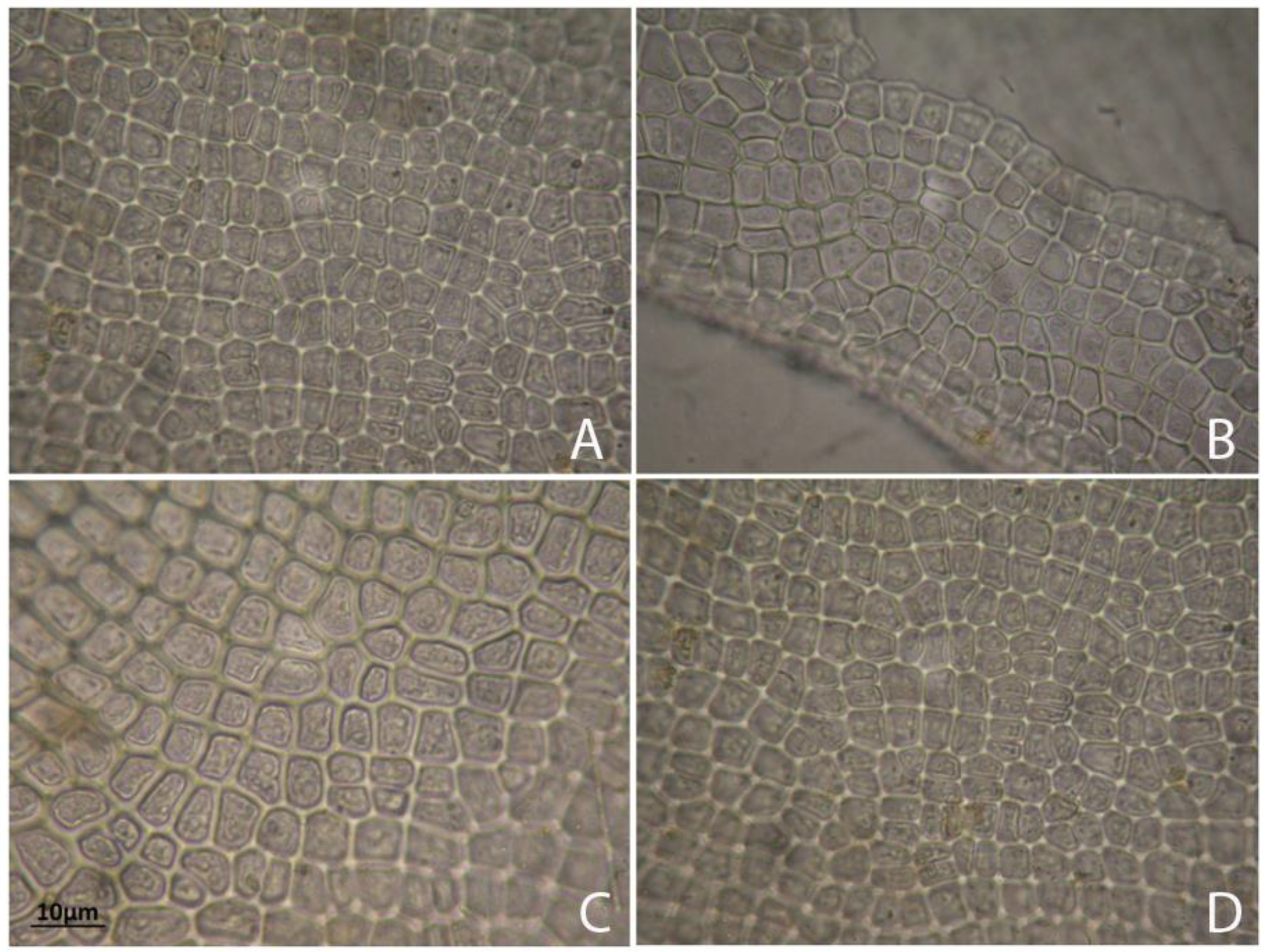
2.1. Habitat and Morphological Observations

The mature thalli of the studied Ulva were irregular in shape, bright to dark green in colour, forming masses of perforated blades, branched and with numerous uniseriate proliferations, and a typical globular cell on the apex (Figure 3). In site B, at a rocky cliff on large rocks washed by the waters of the Adriatic Sea (marine site), square or rectangular apex cells with rounded angles were found. In the apical and central parts of the thalli, in the marine site (B), irregular cell arrangements were observed, whereas in the freshwater sites (A, C, and D) the cells were arranged regularly in longitudinal rows (Figure 4). Considering cells measurements, all material was divided into young and mature thalli collected directly during the studies and analysed separately (there was no culturing in the lab). The free floating thalli were always measured within the stipe region. Thalli of the submerged specimens (young thalli) were from 10.3 cm to 63.6 cm long and 0.3 cm to 1.8 cm wide. Free floating thalli (mature and dying) were from 15.7 cm to 52.3 cm long and 0.6 cm to 2.1 cm wide. Vegetative cells contained one parietal, perforated chloroplast (usually covering the majority of the cell) with 2 or 4 spherical pyrenoids (in the upper basal region). In young thalli, rectangular cells were ranging from 32–56 μm in width to 20–35 μm in length and they formed distinct longitudinal rows. In mature thalli, the vegetative cells were less regular in shape and measured from 24–46 μm in width to 18–32 μm in length, and the rows were not as distinct as those in young thalli. Single row proliferation, culminating in a conical or rounded apex cell of 6.20–7.40 µm in length and 5.80–7.00 µm in width was observed. The thalli of U. pilifera created mats with the largest surface (among all the studied samples) in stagnant water (i.e., in the river, in the ditch, and in the Milet Canal—all from Montenegro). There were no zoospores or gametes observed within any of the specimens.

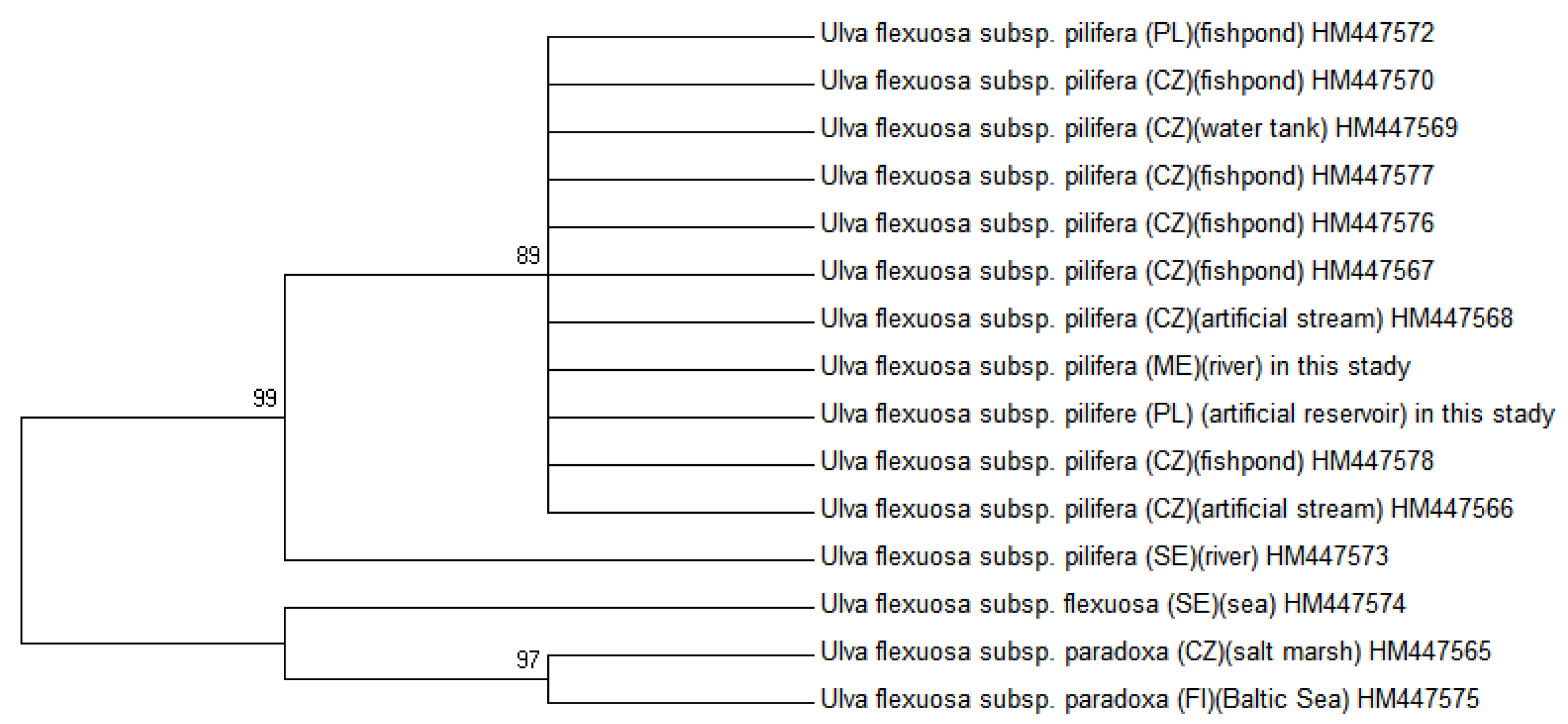
2.2. Molecular Phylogenetic Analysis

Molecular analysis confirmed that all of the freshwater and marine specimens occurring for the four sites from Montenegro were indeed U. pilifera. The specimens of the same species Ulva from freshwater habitats in Poland [18] were compared with the specimens from the similar habitats in Montenegro. The neighbour joining (NJ) and parsimony analysis (MP), as presented in Figure 5 and Figure 6, were congruent with maximum likelihood (ML), considering bootstrap proportions (BP) from ML, NJ, and MP demonstrated at the nodes. The maximum likelihood tree was presented due to the fact that topologies of obtained maximum likelihood, neighbour joining, and parsimony trees were compatible. All new sequences of U. pilifera collected from different habitats in Montenegro constituted a monophyletic group, supported by bootstrap analysis. In a Bayesian tree with mapped branches from the MP and ML were considered all phylogenetic analysis, which created trees with very similar clustering at lower levels, but with differences in higher topology. The SH test and robustness of topology, comparing the ML tree with a tree in monophyletic of Ulva flexuosa were performed. The results of the SH test stated that the ML tree was significantly preferable than the tree with monophyletic U. pilifera, as well as the, rbcL data set was combined with related sequences from GenBank (BLAST). There were no differences in the coded polypeptide amino acid sequences. The cells of Ulva thalli (Poland) are not morphologically similar to those from Montenegro (Table 1), but the monophyletic clusters are characterised by unique morphological traits representing synapomorphy, which further allow them to be distinguished from their sister clades.

2.3. Elemental Analysis

In Ulva thalli 25 elements: Al, As, Ba, Br, Ca, Cl, Cr, Cu, Fe, Hf, I, K, Mg, Mn, Na, Ni, P, Pb, Rb, S, Si, Sr, Ti, V, and Zn, using WDXRF and TXRF techniques, were identified (Table 2 and Table 3). Moreover, 14 elements were classified by WDXRF and 11 elements by TXRF. At site A of the Ulva pilifera thalli the calcium (Ca) levels showed the highest concentration, relatively large in site D, while at site B there were only trace amounts using WDXRF technique (see Table 2). At site B, using TXRF technique, the strontium (Sr) reached the highest value, while in sites A and D the amounts were 50% lower (Table 3). The elements concentration using WDXRF technique at the site A for barium (Ba) and phosphorus (P), similarly to the site B for iodine (I) and bromine (Br), and at the site D for bromine (Br) and sodium (Na) were the lowest (see Table 2). While using TXRF technique (Table 3) at sites A and D, the elements concentration for arsenic (As) and chromium (Cr), and at site B for vanadium (V) and lead (Pb) were the lowest.

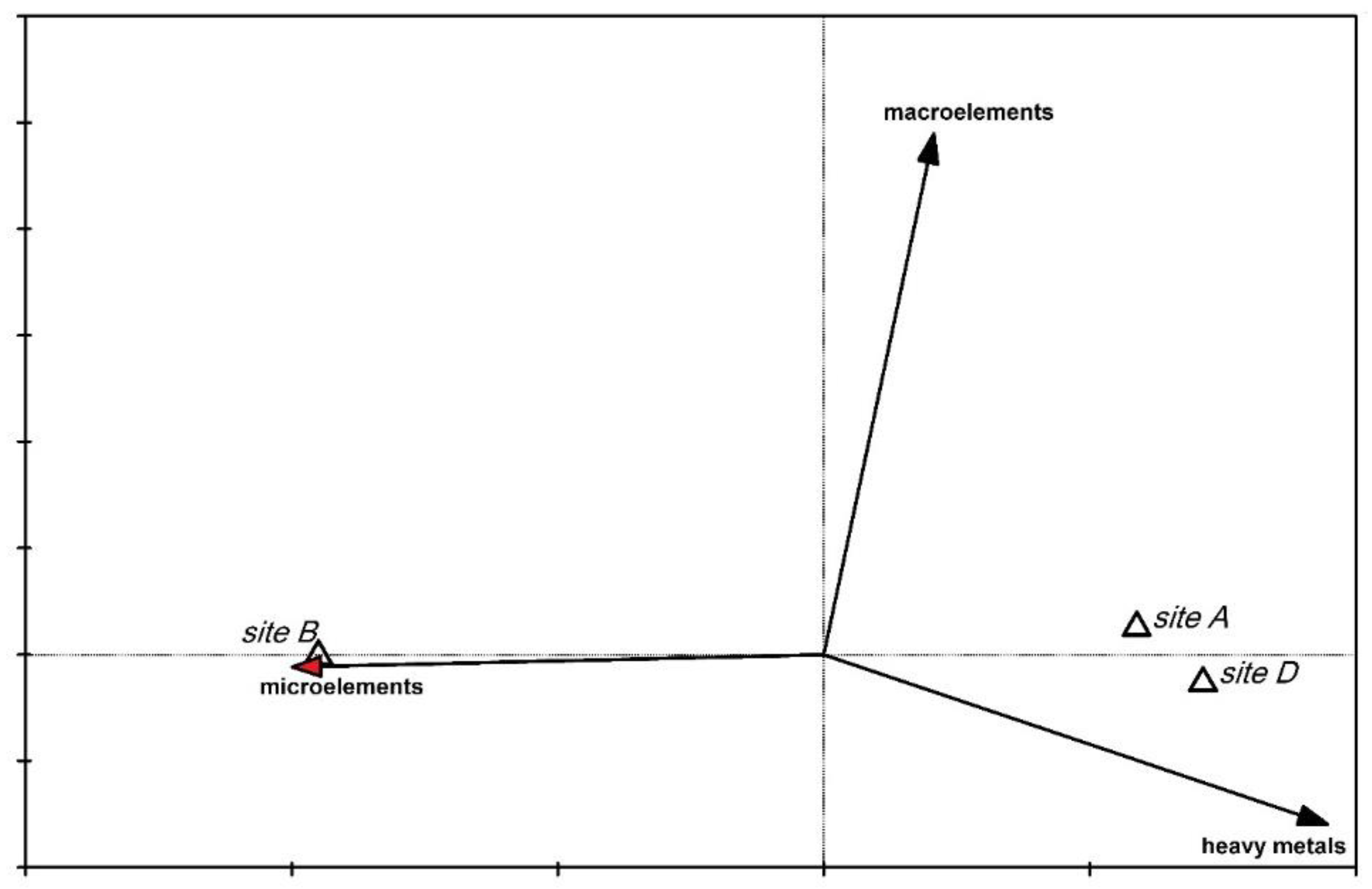
2.4. Comparison of Data Analysis

The Principal Component Analysis (PCA) indicated that the Ulva elemental composition from site B (from the Adriatic Sea) differs from that of sites A and D. The analysis was done only from sites A, B and D, while site C was below the limit of significance (and therefore was not included in the analysis). The Ulva thalli from site B were characterised by more strontium and zinc, whereas those from A and D had more copper and titanium (Figure 7). The first axis explains 93% of the variation, because the variation in the data was small and the differences between these sites were not too large. The whole group of chemical elements visible on the left side of the graph were no different among the sites. The Canonical Variate Analysis (CVA) showed that Ulva from site B (marine) contained more microelements than those from the sites A and D (Figure 8). The microelements variable explains 50% of the data variability. The CVA confirmed the results obtained from the PCA. Apaydin et al. [24] investigated the contents of trace elements by the energy-dispersive X-ray fluorescence spectrometry (EDXRF) in Ulva lactuca from eight different regions of Istanbul (Turkey). Their results demonstrated that Ulva thalli contain trace element contamination (but no toxic element), and that the mineral content of Ulva lactuca varies according to the composition of sediment, sea water, sand, and rock.

2.5. Distribution and Habitat Preferences of the Ulva Species

U. pilifera is considered a cosmopolitan species that has a worldwide distribution [13]. The distribution of U. pilifera was based on all the references available. Table 4 presents the occurrence and habitats preference of U. pilifera in most continents, excluding Antarctica.
The taxa of the genus Ulva were difficult in terms of morphological identification, due to the plasticity of the thallus and cellular details [1,41]. Ulva pilifera is commonly encountered in both freshwaters, and slightly saline waters [42,43]. Mareš et al. [12] stated that Ulva flexuosa subsp. paradoxa and Ulva pilifera are freshwater algae which dominate in Europe habitats, and this is confirmed by the morphological and molecular results. Kapetanović et al. [22] and Kosanić et al. [23] found Ulva lactuca and Enteromorpha intestinalis in Montenegro on the Adriatic Coast (in waters with a high salt content). In studies Ulva pilifera was observed in one marine and in three freshwater sites. Variable environmental conditions are responsible for the plasticity of Ulvales morphology characters [44]. On the one hand, pyrenoids number is believed by some authors to be of taxonomic values but it is questioned by others [45]. Ulva pilifera found in the four sites from Montenegro formed longer, wider thalli and thicker cell walls than the same species observed in Poland and the Czech Republic. Morphological analysis of this species from 21 freshwater sites in Poland [41] displayed tubular, curled and bubbled thalli with strongly corrugated surfaces and uniseriate branches (4.7–40.4 cm in length, 0.10–23.0 mm in width) containing from 1 to 4 pyrenoids. Whereas the Ulva pilifera found by Mareš et al. [12] in enriched freshwaters habitat formed tube-like or leaf-like thalli, free floating masses, abundant or absent branching from 7 to 18 (25) µm in length, with from 2 to 4 pyrenoids.
Poole and Raven [46] and Rybak et al. [41] stated that the subspecies of Ulva flexuosa is characterised by wide tolerances to factors such as water salinity, temperature and nutrients. However, Ulva’s are known to grow in a wide range of tolerance to salinity, but optimal salinity is different for adult thalli or spores [47]. The local conditions of Ulva habitats depend also on the tolerance range of salinity for individual species. Rybak et al. [41] found that Ulva pilifera in different freshwater ecosystems (natural and anthropogenic) dominated most often lakes, water basins, and rivers, where the salinity did not exceed (i.e., 270 mg L−1 Cl).
Both eutrophication and climatic factors, such as light and temperature, promotes the proliferation and growth of the green tide forming algae known as Ulva species. Factors, such as temperature, pH, salinity, and nutrient availability, have limited spatial and temporal distribution of Ulva pilifera in different habitats. Holzinger et al. [48] observed that algae respond rapidly to changes in water quality due to different environmental factors and that Ulva compressa, characterised by rbcL phylogeny, is a common species in the Mediterranean Sea. Ulva as an intertidal species tolerates repeated desiccation-rehydration cycles in nature, with drastic variation in salinity requiring a high osmotic potential and water-holding capacity [49]. West and Pitman [50] studied ionic relations in Ulva, and they found an accumulation mechanism for potassium, while sodium accumulation was excluded. However, Holzinger et al. [48] concluded that structural changes in the cell walls of Ulva compressa during desiccation-rehydration cycles play a key role in surviving high and low tides. Ulva pilifera existing both in the freshwater and marine environment, implies high ecological plasticity. Typically, Ulva species are regarded as good indicators of heavy metal concentration in sea water [51]. The concentration of many elements (including heavy metals) in algae fluctuates with the seasons due to changes in their bioaccumulation during development and independent of ion concentration in the environment [52]. According to Rybak et al. [41], the highest metal concentrations were for calcium (Ca) and magnesium (Mg) in the thalli of freshwater Ulva, both in lake and the river populations. In the Montenegro specimens, the highest elemental concentrations were for calcium (Ca) (freshwater site A) and strontium (Sr) (sea water site B), which has physical and chemical properties similar to calcium (Ca). Calcium and barium have physical and chemical properties similar to strontium, and all three of them are vertical neighbours in the periodic table.
Montenegro’s diversity of geology, landscapes, climate types and soils, also different water habitats and geographical location on the Balkan Peninsula and Adriatic Sea, result in remarkable biological diversity. Due to these specific qualities, Montenegro ranks among Europe’s and the world’s most biologically diverse hot-spots. Since 2000, in Montenegro, the National Environmental Monitoring Programme discovered 1500 species of algae (some 1000 taxa are known from Skadar Lake), 8000 species of vascular plants and 372 Balkan endemic taxa, therefore, the number of taxa per area is 0.837 which constitutes the highest index for all known European countries [22]. Future work should extend the search for other new localities of Ulva species in the Balkans.

3. Materials and Methods
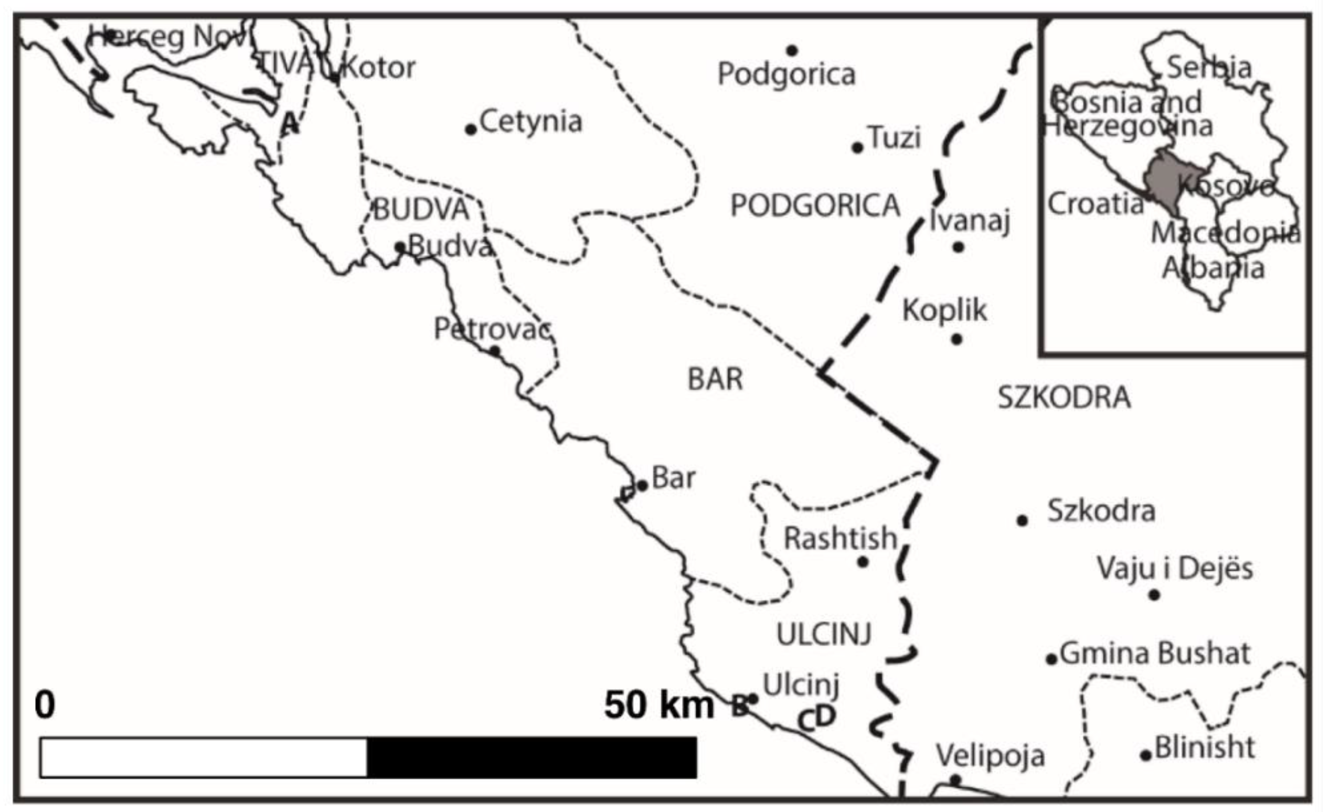
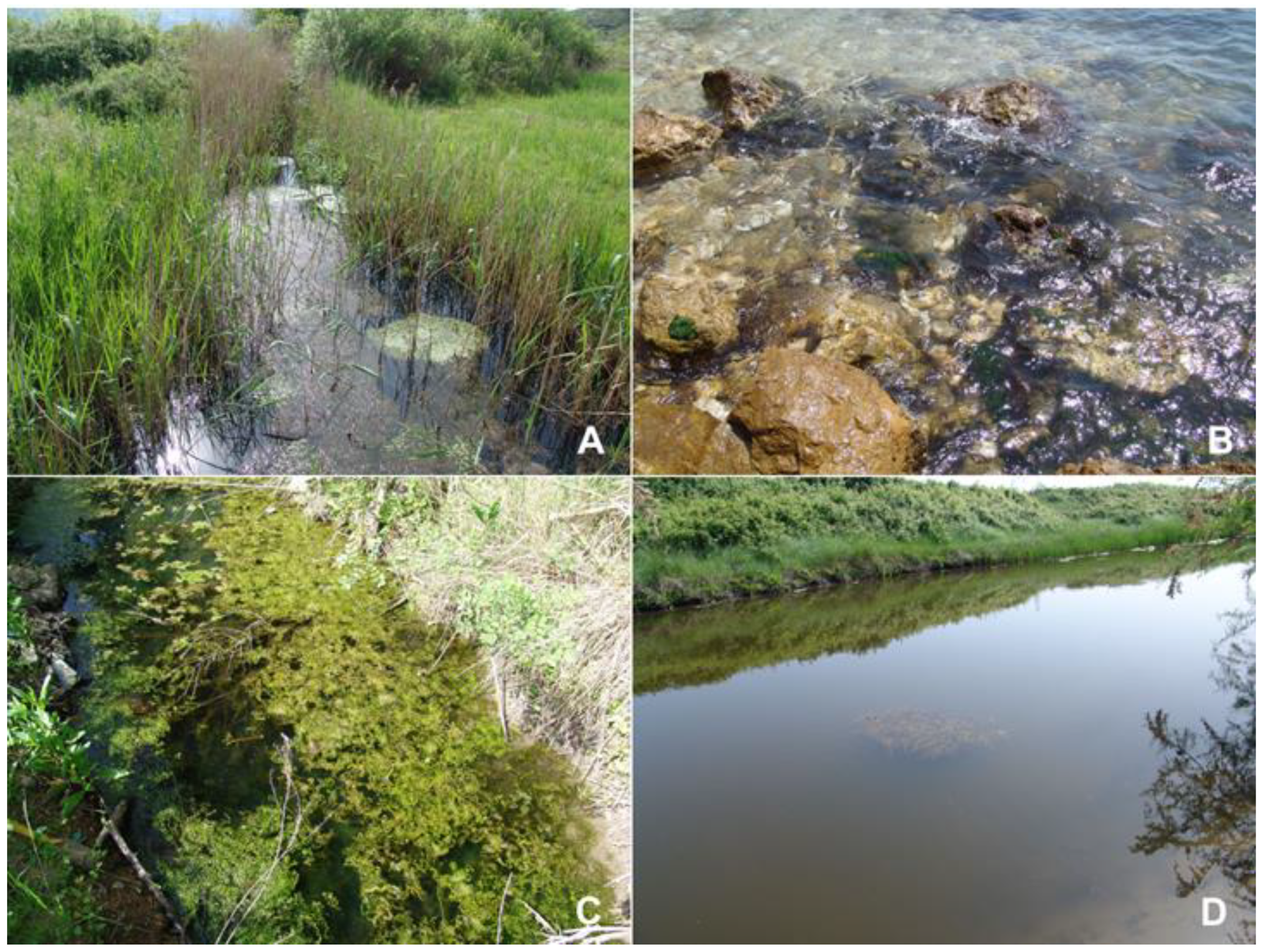
3.1. Study Site

Specimens of Ulva pilifera were collected (in 50 mL sterile clean bottles) on May 2015 from four sites in Montenegro: (A) 42°23′24″ N, 18°42′51″ E, 5 May 2015, the outskirts of Peninsula Luštica, in the river 2 km SE from Durasevici (Figure 1). The river is overgrown with Phragmites, Callitriche, and Zannichellia communities. There are numerous and scattered salt springs and saline wetlands in the immediate vicinity of the river. (B) 41°55′31″ N, 19°11′58″ E, 6 May 2015, NW of Old Town of Ulcinj city; the site is located at the base of a rocky cliff on large rocks washed by the waters of the Adriatic Sea. (C) 41°54′52″ N, 19°16′34″ E, 6 May 2015, a ditch near Ulcinjska Solana (the vast space in the area of Adriatic Coast, where salt is produced by the evaporation; this is one of the largest salt basins in the Balkans. (D) 41°54′52″ N; 19°16′36″ E, 6 May 2015, the Milet Canal in Donji Štoj near Ulcinjska Solana (Figure 2). Besides Ulva pilifera, there were also large quantities of Potamogeton pectinatus (L.) Börner. Each sampling sites was recorded by Garmin Differential GPS, and all specimens were photographed fresh to show cell arrangement, cell size, and branching patterns.

3.2. Samples Preparation

A total of 30 Ulva samples were collected, subsequently, each sample was washed clean with water and thoroughly dried with absorbent paper. Fresh material was pressed and dried in herbarium sheets, whilst part of the material from the Ulva specimens was cleaned well using distilled water to remove epiphytes and preserved in silica gel (Sigma Aldrich, St. Louis, MO, USA) for molecular analysis. Each sterile clean bottles was labelled with the date of collected, the habitats number, and sample number. For morphological studies (including measurement of Ulva thalli, presence or absence of indentation, cell wall thickness, cell dimensions and number of pyrenoids within the chloroplast), the specimens were examined using a JENAMED 2 microscope. Morphometric diagnostic features were measured and photographed using an inverted Nikon Eclipse Ti microscope, and confocal equipment a Nikon A1 (Nikon, Tokyo, Japan). The 405.488.561 nm laser heads and detection system was a PMT-DU4 detector in the range of 400–820 nm, Plan Apo VC 100XOil inverse lens, Nikon C-HgFiE mercury illumination, and a DS0Fi1C-U3 digital camera. Data acquisition in Nikon NIS-Elements obtained digital images, processed with Adobe Photoshop CS5 (Adobe, San Jose, CA, USA). The elemental analysis of Ulva thalli was carried out using wavelength dispersive X-ray fluorescence (WDXRF) and total reflection X-ray fluorescence (TXRF) in the Laboratory of X-ray methods (Institute of Physics, Jan Kochanowski University in Kielce, Poland). The Ulva specimens were morphologically identified using Bliding (1963) and following [14,17].
Ulva preparation for the WDXRF technique. For the wavelength dispersive X-ray fluorescence analysis, the samples were prepared as a pellet. Prior to pellet formation, the Ulva samples were dried and ground with the compact mill MiniMill2 at the rotation speed 200 rpm and grinding time t = 1 min. Next, the powdered Ulva (about 5 g) was mixed with wax binder (C18H36O2N2), whose mass was about 5% of the algae mass, and afterwards it was formed into the pellet using pressure equal to 20 tons for 30 s. WDXRF measurement was carried out for 40 min.
Ulva preparation for the TXRF technique. The TXRF analysis requires a liquid form of the sample. For this purpose the powdered Ulva samples weighing (~0.03 g) was mixed in an Eppendorf tube with 500 µL of high purity 65% HNO3 (Merck). Due to the violent reaction of acid with the Ulva sample, the acid was added very carefully in the following stages: 100 µL + 100 µL + 100 µL + 200 µL. Finally, 40 µL of 100 ppm of SeO2 (in HNO3 2–3%; Merck) was added to the mixture as internal standard. The solution was next mineralised in a microwave oven. Next, the solution was allowed to stand for 24 h in order for total mineralisation. Then, the 5 µL of sample solution was pipetted into a silicon sample carrier and dried on at hot plate at 60 °C. Finally, each Ulva sample was analysed using TXRF technique (the measurement time was 60 min). The concentration of elements obtained in the solution (Ulva powder + acid + Se as internal standard) was converted into the dry weight of Ulva. Eppendorf tubes, pipette tips and sample carriers were previously washed in 10% nitric acid, rinsed with deionised water and then dried by centrifugation.

3.3. Physicochemical Water Quality

Water samples from all the sites were collected using clean hard rubber bottles. Water temperature (°C), pH, specific conductivity (µS cm−1) and total dissolved solids (mg L−1) were measured in situ using the Bernant 30 pH meter, while dissolved oxygen (DO) was determined using YSI 51B DO meter. Detailed hydrochemical characteristics of each study sites, including irons, nutrients, trace elements and metals, were determined following [53]. The ions: Ca2+, Cl, HCO3, Mg2+, Na+ and SO42− were measured using ion-exchange chromatography (ICS 3000 Dionex). The nutrients: NO2, NO3, NH4+, TP, and SRP were determined by molecular absorption spectroscopy (UniCam 969 AA Spectrometer).

3.4. Molecular and Phylogenetic Analysis

Total genomic DNA was extracted from 100 mg sample of fresh tissue (Ulva specimens from Montenegro and Poland, each one separately) and grounded with liquid nitrogen using the DNeasy Plant Mini Kit (Qiagen GmbH, Hilden, Germany). HotStartTaq DNA Polymerase (Qiagen) was used to amplify the selected genomic regions using newly designed ITS primers (ITS-F 5′-CTG CGG AGG GAT CAT TGA AA-3′; ITS-R 5′-GTA GCG AGC TAC CTA CCT AGT-3′) and rbcL primers (rbcL-F 5′-CAA GGT CCA CCT CAT GGT ATT C-3′; rbcL-R 5′-GTC ACC TGA CAT ACG TAA A-3′). Thermal cycling parameters were as follows: initial denaturation at 95 °C for 15 min followed by 40 cycles at 94 °C for 60 s, 58 °C (ITS) or 62 °C (rbcL) for 30 s, and 72 °C for 40 s. Direct sequencing of PCR products were done by GenoMed S.A. (Warsaw, Poland), and manually inspected using Chromas Lite 2.1.1 (Technelysium Pty Ltd., South Brisbane, Australia). The divergence (in%), distance in the sequence, have a Montenegro species in ITS and rbcL sequences. All others sequences for phylogenetic trees were taken from GenBank and they were highly similar to our sequences. The phylogenetic trees were constructed by the maximum-likelihood algorithm of the MEGA 6 program Tamura et al. [54]. The obtained sequences were deposited in GenBank under the following accession numbers: KX398148 for rbcL, KX398149 for ITS (Ulva from Montenegro), KX398150 for rbcL, KX398151 for ITS (Ulva from Poland).

3.5. Wavelength Dispersive X-ray Fluorescence (WDXRF) Technique

The spectrometer Axios (PANalytical) with X-ray tube with Rh anode was used to take WDXRF measurements. The X-ray tube has 2.4 kW maximum power and 60 kV maximum voltage, as well as 100 mA maximum current. The primary beam, its intensity, and spectrum was modified using different filters. The excitation of the X-rays sample, early generated in an X-ray tube, is a source of information about elemental composition. Due to the analysis of crystal, according to Bragg’s law, characteristic polychromatic X-ray was dispersed into monochromatic constituents using a beam appearing from a sample surface and collimated by a collimator. Five crystals: LiF200, Ge111, PE 002, PX1, and LiF220 are present in the Axios spectrometer and their choice is necessary to cover the wavelength. Two detectors: a flow counter is used to optimise elements detection up to iron (Fe) or a scintillation counter used for heavier elements served the characteristic X-rays. Elemental analysis from oxygen (O) to uranium (U) was carried out by Axios spectrometer. The measurements of elemental analysis were performed in vacuum.

3.6. Total Reflection X-ray Fluorescence (TXRF) Technique

TXRF measurements were performed with the S2 PICOFOX spectrometer (Bruker AXS Microanalysis GmbH). On the basis of [55,56] the characteristic X-rays were excited in the samples with the 30 W Mo-anode X-ray tube operated at 50 kV with an electron current of 0.6 mA. Fluorescence X-rays from the samples were detected by Peltier-cooled XFlash silicon drift detector (with an area 10 mm2) having an energy resolution of approx. 160 eV at 100 kcps for Mn-Kα line [56]. The measurements were performed in the air. The PICOFOX spectrometer allows measurement of characteristic X-rays of elements from Al (aluminium) to U (uranium), with the exception of zirconium (Zr) to ruthenium (Ru). A good detection value (from 10 ppb in the case of real multielemental sample) is achieved as a result of optimisation of the setup geometry and use of a multilayer monochromator.

3.7. Statistical Analysis

Principal Component Analysis (PCA) was used to show similarities and differences in the Ulva elemental composition among the studied sites. The ordination diagram presents the arrangement of variables: elements (geometric symbols) and sites (vectors) in the ordination space. The closer vectors location indicates larger chemical elemental similarity on the sites. For the purpose of the performed analysis, the assigned elements were divided into chemical groups. Ulva thalli elemental composition in particular sites by Canonical Variate Analysis (CVA) was determined. Three chemical groups of elements were distinguished: heavy metals (Ti, Cr, Ni, As, Hf, and Pb), microelements (Ba, Cl, Fe, Mn, I, V, Cu, Zn, Rb, and Sr), and macroelements (Al, Br, Ca, K, Mg, Na, P, S, and Si), and all of these were encoded accordingly in the system 0–1. The data were correlated with the elemental Ulva composition. Monte Carlo permutation test was also performed to determine the statistical significance of the used variables. On the ordination diagram, the sites were marked with triangles and the elemental chemical groups were marked with vectors. Both ordination analyses were carried out in Canoco 5.0 [57].

4. Conclusions

A green macroalgae was collected in Montenegro from four sites (freshwater and marine): a river, a ditch, the Milet Canal, and on the rocky shore of the Adriatic Sea. A detailed morphology, autecology, and molecular studies allowed to identify the Ulva population as Ulva pilifera. WDXRF and TXRF techniques were applied for the analysis of elemental composition of Ulva thalli. Molecular studies demonstrated a close phylogenetic relationship between Ulva species isolated from Poland, the Czech Republic and Montenegro. It was focused on the correlation between different kinds techniques applied to taxonomic identification of the Ulva species and its elemental properties. In elemental analysis, 25 elements were determined, i.e., Al, As, Ba, Br, Ca, Cl, Cr, Cu, Fe, Hf, I, K, Mg, Mn, Na, Ni, P, Pb, Rb, S, Si, Sr, Ti, V, and Zn. The concentrations of the metals Cr, Cu, Ni, PB, and Zn in the marine site were higher than in freshwater sites and significantly correlated with the content of trace elements in water and sediments. CVA and PCA statistical analysis confirming the obtained results were performed. The application of WDXRF and TXRF techniques as chemotaxonomic marker might be useful, in the future for identifying freshwater and marine Ulva species and also determining their bioindicative properties.

Author Contributions

Conceptualisation, J.C.-M. and R.P.; investigation, R.P.; resources, J.C.-M.; methodology, J.C.-M. and E.T.; writing—original draft preparation, J.C.-M. and B.U.; writing—review and editing, J.C.-M.; visualisation, A.W. All authors have read and agreed to the published version of the manuscript.

Funding

This research was funded by SMGR.RN 20.107 (J.C.-M).

Acknowledgments

The authors are grateful to Karolina Fučíková-Ryerson from the University of Connecticut for critically reading the manuscript. Thanks also to Kinga Półchłopek from Jan Kochanowski University for her technical assistance. Many thanks also to Ilona Stabrawa from Jan Kochanowski University for her assistance with X-ray based techniques.

Conflicts of Interest

The authors in the present study have declared that there are no conflicts of interest.

References

  1. John, D.M.; Whitton, B.A.; Brook, A.J. The Freshwater Algal Flora of the British Isles: An Identification Guide to Freshwater and Terrestrial Algae, 2nd ed.; Cambridge University Press: Cambridge, UK, 2011; pp. 5–15. [Google Scholar]
  2. Wolf, M.A.; Sciuto, K.; Andreoli, C.; Moro, I. Ulva (Chlorophyta, Ulvales) biodiversity in the North Adriatic Sea (Mediterranean, Italy): Cryptic species and new introductions. J. Phycol. 2012, 48, 1510–1521. [Google Scholar] [CrossRef] [PubMed]
  3. Ogawa, T.; Ohki, K.; Kamiya, M. Difference of spatial distribution and seasonal succession among Ulva species (Ulvophyceae) across salinity gradients. J. Phycol. 2013, 52, 637–651. [Google Scholar] [CrossRef]
  4. Saber, A.A.; Mareš, J.; Guella, G.; Anesi, A.; Štenclová, L.; Cantonati, M. Polyphasic approach to a characteristic Ulva population from a limno-rheocrenic, mineral (chloride, sodium, sulphate) in the Siwa Oasis (Western Desert of Egypt). Fottea 2018, 18, 227–242. [Google Scholar] [CrossRef] [Green Version]
  5. Merceron, M.; Antoine, V.; Auby, I.; Morand, P. In situ growth potential of the subtidal part of green tide forming Ulva spp. stocks. Sci. Total Environ. 2007, 384, 293–305. [Google Scholar] [CrossRef] [Green Version]
  6. Ye, N.; Zhang, X.; Mao, Y.; Liang, C.; Xu, D.; Zou, J.; Zhuang, Z.; Qing, W. Green tides are overwhelming the coastline of our blue planet: Taking the world’s largest example. Ecol. Res. 2011, 26, 477–485. [Google Scholar] [CrossRef]
  7. Hayden, H.S.; Blomster, J.; Maggs, C.A.; Silva, P.C.; Stanhope, M.J.; Waaland, J.R. Linnaeus was right all along: Ulva and Enteromorpha are not distinct genera. Eur. J. Phycol. 2003, 38, 277–294. [Google Scholar] [CrossRef]
  8. Shimada, S.; Hiraoka, M.; Nabata, S.; Iima, M.; Masuda, M. Molecular phylogenetic analyses of the Japanese Ulva and Enteromorpha (Ulvales, Ulvophyceae), with special reference to the free-floating Ulva. Phycol. Res. 2003, 51, 99–108. [Google Scholar] [CrossRef]
  9. Leskinen, E.; Alström-Rapaport, C.; Pamilo, P. Phytogeographical structure, distribution and genetic variation of the green algae Ulva intestinalis and U. compressa (Chlorophyta) in the Baltic Sea area. Mol. Ecol. 2014, 13, 2257–2265. [Google Scholar] [CrossRef]
  10. Rybak, A.S. Species of Ulva (Ulvophyceae, Chlorophyta) as indicators of salinity. Ecol. Ind. 2018, 85, 253–261. [Google Scholar] [CrossRef]
  11. Beach, K.S.; Smith, C.M.; Michael, T.; Shin, H.W. Photosynthesis in reproductive unicells of Ulva fasciata and Enteromorpha flexuosa: Implications for ecological success. Mar. Ecol. Prog. Ser. 1995, 125, 229–237. [Google Scholar] [CrossRef]
  12. Mareš, J.; Leskinen, E.; Sitkowska, M.; Skácelová, O.; Blomster, J. True identity of the European freshwater Ulva (Chlorophyta, Ulvophyceae) revealed by a combined molecular and morphological approach. J. Phycol. 2011, 47, 1177–1192. [Google Scholar] [CrossRef]
  13. Lougheed, V.L.; Stevenson, R.J. Exotic marine macroalga (Enteromorpha flexuosa) reaches bloom proportions in a coastal lake of Lake Michigan. J. G. Lak. Res. 2004, 30, 538–544. [Google Scholar] [CrossRef]
  14. Škaloud, P.; Rindi, F.; Boedeker, C.; Leliaert, F. Freshwater Flora of Central Europe, Chlorophyta: Ulvophyceae (Süßwasserflora von Mitteleuropa, Bd. 13: Chlorophyta: Ulvophyceae); Springer Spektrum: Berlin, Germany, 2018; Volume 13, 289p. [Google Scholar]
  15. Bliding, C.A. A critical survey of European taxa in Ulvales, Part Ulva, Ulvaria, Monostroma, Kornmannia. Bot. Not. 1968, 121, 535–629. [Google Scholar]
  16. Enteromorpha pilifera Kütz., 1856 in Gargominy 0. TAXREF; Version 4; UMS PatriNAT (OFB-CNRS-MNHN): Paris, France, 2020.
  17. Wynne, M.J. A checklist of benthic marine algae of the tropical and subtropical western Atlantic: Third revision. Nova Hedwig. Beih. 2011, 140, 7–166. [Google Scholar]
  18. Messyasz, B.; Czerwik-Marcinkowska, J.; Massalski, A.; Uher, B.; Rybak, A.; Szendzina, L.; Pikosz, M. Morphological and ultrastructural studies on Ulva flexuosa subsp. pilifera (Chlorophyta) from Poland. Acta Soc. Bot. Pol. 2013, 82, 157–163. [Google Scholar] [CrossRef] [Green Version]
  19. Richter, D.; Pietryka, M. New localities of Ulva flexuosa subsp. pilifera (Chlorophyta, Ulvophyceae) in Lower Silesia—Morphometric and habitat characteristics. Frag. Flor. Geobot. Pol. 2016, 23, 305–319. [Google Scholar]
  20. Kopp, R. Phytoplankton of the Zámecký Pond. Fottea 2006, 6, 111–125. [Google Scholar]
  21. Skácelová, O. Flóra Sinic a Řas Tůní v Inundačních Pásmech Řek; Faculty of Biological Science: Českeé Budějovice, Czech Republic, 2004; pp. 1–143. [Google Scholar]
  22. Kapetanović, R.; Sladić, D.; Popov, S.; Zlatović, M.; Kljajić, Z.; Gašić, M.J. Sterol composition of the Adriatic sea algae Ulva lactuca, Codium dichotonium, Cystoseira adriatica and Fucus virsoides. J. Serb. Chem. Soc. 2005, 70, 1395–1400. [Google Scholar] [CrossRef]
  23. Kosanić, M.; Ranković, B.; Stanjković, T. Biological activities of two macroalgae from Adriatic coast of Montenegro. Saudi J. Biol. Sci. 2016, 22, 390–397. [Google Scholar] [CrossRef] [Green Version]
  24. Apaydm, G.; Ayhkcr, V.; Cengiz, E.; Saydam, M.; Küp, N.; Tirasoglu, E. Analysis of metal contents of seaweed (Ulva lactuca) from Istanbul, Turkey by EDXRF. Turk. J. Fish. Aquat. Sci. 2010, 10, 215–220. [Google Scholar]
  25. Shaaban, A.E.S.; Mansour, H.A.; Saber, A.A. Soil algae of El-Farfara Oasis (The Western Desert, Egypt), and N2–fixation efficiency of five heterocytous cyanopytes. Egypt J. Bot. 2013, 57, 517–524. [Google Scholar]
  26. Wang, Y.; Qu, T.; Zhao, X.; Tang, X.; Xiao, H.; Tang, X. A comparative study of the photosynthetic capacity in two green tide macroalgae using chlorophyll fluorescence. Spring 2016, 5, 775. [Google Scholar] [CrossRef] [PubMed] [Green Version]
  27. Phang, S.M.; Yeong, H.Y.; Ganzon-Fortes, E.T.; Lewmanomont, K.; Prathep, A.; Hau, L.N.; Gerung, G.S.; Tan, K.S. Marine algae of the South China Sea bordered by Indonesia, Malaysia, Philippines, Singapore, Thailand and Vietnam. Raffles Bull. Zool. 2016, 40, 13–59. [Google Scholar]
  28. Shimada, S.; Yokoyama, N.; Arai, S.; Hiraoka, M. Phylogeography of the genus Ulva (Ulvophyceae, CHlorophyta), with special reference to the Japanese freshwater and brackish taxa. J. Appl. Phycol. 2008, 20, 979–989. [Google Scholar] [CrossRef]
  29. Lee, S.H.; Kang, P.J.; Nam, K.W. New record of two marine ulvalean species (Chlorophyta) in Korea. J. Ecol. Environ. 2014, 37, 375–389. [Google Scholar] [CrossRef] [Green Version]
  30. Taskin, E.; Öztürk, M.; Kurt, O.; Öztürk, M. Checklist of marine flora of Turkey. J. Phycol. 2018, 40, 71–72. [Google Scholar]
  31. Nguyen, T.V.; Le, N.H.; Lin, S.M.; Steen, F.; De Clerck, O. Checklist of the marine macroalgae of Vietnam. Bot. Mar. 2013, 56, 207–320. [Google Scholar] [CrossRef]
  32. Kraft, L.; Kraft, G.T.; Waller, R. Investigations into southern Australian Ulva (Ulvophyceae, Chlorophyta) taxonomy and molecular phylogeny indicate both cosmopolitanism and endemic cryptic species. J. Phycol. 2010, 46, 1257–1277. [Google Scholar] [CrossRef]
  33. Kirkendale-Saunders, L.; Winberg, P. A molecular survey of Ulva (Chlorophyta) in Temperate Australia reveals enhanced levels of cosmopolitanism. J. Phycol. 2012, 49, 324–328. [Google Scholar] [CrossRef]
  34. Messyasz, B.; Rybak, A. Abiotic factors affecting the development of Ulva sp. (Ulvophyceae; Chlorophyta) in freshwater ecosystems. Aquat Ecol. 2011, 45, 75–87. [Google Scholar] [CrossRef] [Green Version]
  35. Brodie, J.; Maggs, C.A.; John, D.M. Green Seaweeds of Britain and Ireland; British Phycological Society: London, UK, 2007; 250p. [Google Scholar]
  36. Tsiamis, K.; Panayotidis, P.; Economou-Amilli, A.; Katsaros, C. Seaweeds of the Greek coasts. II. Ulvophyceae. Med. Mar. Sci. 2014, 15, 449–461. [Google Scholar] [CrossRef] [Green Version]
  37. Anon, A. Inventaire National du Patrimoine Naturel; Muséum National d’Histoire Naturelle: Paris, France, 2017; pp. 45–51. [Google Scholar]
  38. Sfriso, A. Chlorophyta Multicellulari e Fanerogame Acquatiche. Ambiente di Transizione Italiani e Litorali Adiacenti. I Quaderni di ARPA; ARPA Emilia-Romagna: Bologna, Italy, 2011; 320p. [Google Scholar]
  39. Favot, G.; Engelen, A.H.; Cunha, M.E.; Serräo, M.E. Identification of Ulva sp. grown in multitropic aquaculture systems. J. Aquac Fish. 2019, 3, 24–31. [Google Scholar]
  40. De Zaixso, A.L.B. Elementos para el Estudio de las Macroalgas de Argentina; De Zaixso, C.C.J.M., Ed.; Editorial Universitaria de la Patagonia: Comodoro Rivadavia, Argentina, 2013; 204p. [Google Scholar]
  41. Rybak, S.A.; Czerwoniec, A.; Gąbka, M.; Messyasz, B. Ulva flexuosa (Ulvaceae, Chlorophyta) inhabiting inland aquatic ecosystems: Molecular, morphological and ecological discrimination of subspecies. Eur. J. Phycol. 2014, 49, 471–485. [Google Scholar] [CrossRef] [Green Version]
  42. Ichihara, K.; Arai, S.; Uchimura, M.; Fay, J.E.; Ebata, H.; Hiraoka, M.; Shimada, S. New species of freshwater Ulva, Ulva limnetica (Ulvales, Ulvophyceae) from the Ryukyu Islands, Japan. Phycol. Res. 2009, 57, 94–103. [Google Scholar] [CrossRef]
  43. Rybak, S.A. Revision of herbarium specimens of freshwater Enteromorpha-like Ulva (Ulvaceae, Chlorophyta) collected from Central Europe during the years 1849. Phytotaxa 2015, 1, 1–29. [Google Scholar] [CrossRef]
  44. Malta, E.J.; Draisma, S.G.A.; Kamermans, P. Free-floating Ulva in the southwest Netherlands; species or morphotypes? A morphological, molecular and ecological comparison. Eur. J. Phycol. 1999, 34, 443–454. [Google Scholar] [CrossRef]
  45. Loughnane, C.J.; McIvor, L.M.; Rindi, F.; Stengel, B.; Guiry, M. Morphology, rbcL phylogeny and distribution of distromatic Ulva (Ulvophyceae, Chlorophyta) in Ireland and southern Britan. J. Phycol. 2008, 47, 416–429. [Google Scholar] [CrossRef]
  46. Poole, L.J.J.; Raven, A. The biology of Enteromorpha. Prog. Phycol. Res. 1997, 12, 1–148. [Google Scholar]
  47. Imchen, T. Recruitment potential of a green alga Ulva flexuosa Wulfen dark preserved zoospore and its development. PLoS ONE 2012, 7, e32651. [Google Scholar] [CrossRef] [Green Version]
  48. Holzinger, A.; Herburger, K.; Kaplan, F.; Lewis, L.A. Desiccation tolerance the chlorophyte green alga Ulva compressa: Does cell wall architecture contribute to ecological success? Planta 2015, 242, 477–492. [Google Scholar] [CrossRef] [Green Version]
  49. Kirst, G.O. Salinity tolerance of eukaryotic marine algae. Annu. Rev. Plant Physiol. Plant Mol. Biol. 1999, 41, 21–53. [Google Scholar] [CrossRef]
  50. West, K.R.; Pitman, M.G. Ionic relations and ultrastructure in Ulva lactuca. Aust. J. Biol. Sci. 1967, 20, 901–914. [Google Scholar] [CrossRef] [Green Version]
  51. Ustunada, M.; Erdugan, H.; Yilmaz, S.; Akgul, R.; Aysel, V. Seasonal concentrations of some heavy metals (Cd, Pb, Zn, and Cu) in Ulva rigida J. Agardh (Chlorophyta) from Dardanelles (Canakkale, Turkey). Environ. Monit. Assess. 2011, 177, 337–342. [Google Scholar] [CrossRef]
  52. Villares, R.; Puente, X.; Carballeira, A. Ulva and Enteromorpha as indicators of heavy metal pollution. Hydrobiology 2001, 462, 221–232. [Google Scholar] [CrossRef]
  53. Clesceri, L.S.; Eaton, A.D.; Baird, R.B. Standard Methods for the Examination of Water and Waste Water; American Public Health Association: Washington, DC, USA, 2020; pp. 1–19. [Google Scholar]
  54. Tamura, K.; Stecher, G.; Peterson, D.; Filipski, A.; Kumar, S. Molecular evolutionary genetics analyses version 6. Mol. Biol. Evol. 2013, 30, 2725–2729. [Google Scholar] [CrossRef] [Green Version]
  55. Kubala-Kukuś, A.; Arabski, M.; Stabrawa, I.; Banaś, D.; Różański, W.; Lipiński, M.; Majewska, U.; Wudarczyk-Moćko, J.; Braziewicz, J.; Pajek, M.; et al. Application of TXRF and XRPD techniques for analysis of elemental and chemical composition of human kidney stones. X-Ray Spec. 2017, 46, 412–420. [Google Scholar] [CrossRef] [Green Version]
  56. Arabski, M.; Stabrawa, I.; Kubala-Kukuś, A.; Gałczyńska, K.; Banaś, D.; Piskorz, Ł.; Forma, E.; Bryś, M.; Różański, W.; Lipiński, M. The correlation of crystalline and elemental composition of urinary stones with a history of bacterial infections: TXRF, XRPD and PCR-DGGE studies. Eur. Biophys. J. 2018, 48, 111–118. [Google Scholar] [CrossRef] [Green Version]
  57. Braak, C.J.F.T.; Šmilauer, P. Canoco Reference Manual and User’s Guide: Software for Ordination (Version 5.0); Microcomputer Power: Ithaca, NY, USA, 2012; pp. 1–496. [Google Scholar]
Figure 1. Location of Ulva pilifera sampling sites in Montenegro: A, the River Ulcinj 2 km SE from Durasevici; B, NW of the Ulcinj City, on the Adriatic Coast; C, the ditch near Ulcinjska Solana; D, the Milet Canal in Donji Štoj near Ulcinjska Solana.
Molecules 25 05022 g001
Figure 2. Landscape views showing of the sampling sites of Ulva pilifera: (A) the river (freshwater), mass growth of the green thallus of Ulva; (B) a rocky cliff on large rocks of the Adriatic Sea (marine); (C) the ditch (freshwater); (D) the Milet Canal (freshwater).
Molecules 25 05022 g002
Figure 3. Thalli of Ulva pilifera from the river (A), and from the Adriatic Sea (B,C). Thallus corrugated, laminar, light green filiform, and intensive ramification.
Molecules 25 05022 g003
Figure 4. Ulva pilifera (A,D) close-up view on the slightly rounded and/or polygonal vegetative cells well-ordered in long rows in the mid region of the thallus with parietal chloroplast and 2–3 pyrenoids; (B) rectangular cells in a branch showing parietal chloroplasts covering the majority of the cell walls; (C) irregularly arranged vegetative cells in the thallus with 2–3 pyrenoids. Scale bars: 10 µm.
Molecules 25 05022 g004
Figure 5. Maximum likelihood of ITS DNA sequences of U. pilifera samples obtained from Montenegro and Poland. Numbers on branches bootstrap support analysis 1000/50. The phylogenetic tree was constructed by the ML algorithm of the MEGA 6 program. Location and habitat of Ulva species taken from GenBank [3,12].
Molecules 25 05022 g005
Figure 6. Maximum likelihood tree of rbcL DNA sequences U. pilifera samples obtained from Montenegro and Poland. Numbers on branches represent bootstrap support analysis 1000/50. The phylogenetic tree was constructed by ML algorithm of the MEGA 6 program. Location and habitat of Ulva species taken from Mareš et al. [12].
Molecules 25 05022 g006
Figure 7. Principal Component Analysis (PCA) showed a composition data of the Ulva elements from the studied sites. The first axis explained 93% of the data variation. The vectors indicate the sites and the symbols elements divided into chemical groups.
Molecules 25 05022 g007
Figure 8. Canonical Variate Analysis (CVA) for Ulva elemental composition from study sites with the chemical groups of elements. The sites were marked with triangles and the elemental chemical groups were marked with vectors. The red tip of the vector signifies a statistically significant variable and 50% of the variance of the data.
Molecules 25 05022 g008
Table 1. Morphological characteristics of Ulva pilifera from Montenegro and Poland [18].
CharactersMontenegroPoland
morphologytube-liketubular, curled, and bubbled
thalli colour bright to dark green (mature yellow-green)bright green
branchingfrequentabundant to almost absent
uniseriate branchesfrequent to rare, obtusely rounded endspresent
cell shaperounded polygonal to quadrangular rectangular, polygonal
structure of branch tipsglobular cell on the apexrectangular or square
number of pyrenoids2–3–42–4
chloroplast shapeparietalparietal, girdle-shaped
cell size:
length of cells (μm)
width of cells (μm)

42.25–66.56
7.58–19.80

32.05–55.20
10.50–35.50
thallus size:
length of thalli (cm)
width of thalli (mm)

6.50–56.20
0.20–1.50

10.1–65.1
0.20–2.50
cell arrangementdisordered, in small groups or short rowsdisordered, in small groups or short rows
habitatbrackish and marine waters (sea coast)enriched freshwaters (ponds, pools, small streams)
mode of lifeattached or free floatingattached or free floating, occasionally in masses
Table 2. Element concentrations (±measurement expanded uncertainty, expanded factor k = 2) determined in Ulva pilifera thalli using wavelength dispersive X-ray fluorescence (WDXRF) technique. Concentrations are given in %. The analysis were done only from sites A, B, and D while site C was below the limit of significance.
ElementConcentration (%) ± Expanded Uncertainty
Site ASite BSite D
Al0.186 ± 0.0260.243 ± 0.0300.310 ± 0.034
Ba0.020 ± 0.008----------------
Br--------0.039 ± 0.0120.013 ± 0.006
Ca16.300 ± 0.2000.854 ± 0.05610.300 ± 0.200
Cl0.560 ± 0.0447.630 ± 0.1700.498 ± 0.042
Fe0.120 ± 0.0200.129 ± 0.0220.366 ± 0.036
K0.698 ± 0.0503.330 ± 0.1101.450 ± 0.070
Mg0.999 ± 0.0601.390 ± 0.0701.550 ± 0.060
Mn0.299 ± 0.032--------1.020 ± 0.060
Na0.454 ± 0.0404.990 ± 0.1300.149 ± 0.024
P0.070 ± 0.0160.242 ± 0.0300.372 ± 0.036
S1.860 ± 0.0801.860 ± 0.0801.640 ± 0.080
Si1.670 ± 0.0801.250 ± 0.0701.640 ± 0.080
I-----------0.009 ± 0.006-------
The uncertainty values are the expanded uncertainty with an expansion of level about 95% with factor of k = 2. The value of extended uncertainty is given as a range (±U95%).
Table 3. Element concentrations (±measurement expanded uncertainty, expanded factor k = 2) determined in Ulva pilifera using total reflection X-ray fluorescence (TXRF) technique. Concentrations are given μg/g. The analysis were done only from sites A, B, and D while site C was below the limit of significance.
ElementConcentration (μg/g) ± Expanded Uncertainty
Site ASite BSite D
Ti148.000 ± 2.00082.600 ± 1.000135.000 ± 2.000
V13.800 ± 0.8007.140 ± 0.5408.090 ± 1.080
Cr1.800 ± 0.6409.190 ± 0.4204.530 ± 0.900
Ni22.200 ± 0.20015.700 ± 0.20039.200 ± 0.400
Cu10.700 ± 0.2009.030 ± 0.1807.390 ± 0.240
Zn11.300 ± 0.20019.400 ± 0.20020.100 ± 0.200
As0.531 ± 0.13811.100 ± 0.2001.900 ± 0.140
Rb5.680 ± 0.14022.200 ± 0.2006.920 ± 0.140
Sr292.000 ± 2.000541.000 ± 2.000238.000 ± 2.000
Hf58.400 ± 0.40015.700 ± 0.20027.200 ± 0.400
Pb9.510 ± 0.1806.030 ± 0.14013.300 ± 0.200
The uncertainty values are the expanded uncertainty with an expansion of level about 95% with factor of k = 2. The value of extended uncertainty is given as a range (±U95%).
Table 4. Occurrence (in freshwater and marine ecosystems) and habitat preference of Ulva pilifera.
RegionHabitatsReferencesNotes
Africa
Egyptlimno-rheocrenic, thermal, mineral (chloride, sodium, sulphate) spring known as “Ain Abu Sherouf” in the Siwa Oasis; Red Sea coasts; brackish estuaries of the Bile RiverShaaban et al. [25] Saber et al. [4]the Western Desert of Egypt
Asia
ChinaYellow Sea, South China Sea Wang et al. [26] Phang et al. [27]the Subei Shoal coastal waters; bordered by Philippines
Japanfreshwater and brackish Shimada et al. [28]
KoreamarineLee et al. [29]
TurkeyAegean SeaTaskin et al. [30]
VietnammarineNguyen et al. [31]mats
Australia
marineKraft et al. [32] Kirkendale-Saunders and Winberg [33]algal blooms, southern Australia
Europe
Czech Republiclakes, ponds, streams, rivers in Central EuropeMareš et al. [12] Messyasz and Rybak [34]
GermanypondKopp [20]
Mareš et al. [12]
Great Britainnear the top of the shore, on rocks or other algae, on open coasts or in estuaries and harboursBrodie et al. [35] John et al. [1]
GreeceIonian SeaTsiamis et al. [36]the Greek coasts
HungaryfreshwaterMareš et al. [12]
FrancefreshwaterAnon [37]
ItalymarineSfriso [38]Veneto, Mediterranean Sea
Montenegrofreshwater (a river, a ditch, the Milet Canal) and marine (on the rocky shore of the Adriatic Sea)Czerwik-Marcinkowska et al.
Polandfrom freshwater to hyperhaline, and brine habitatsMareš et al. [12] Messyasz et al. [18] Richter and Pietryka [19]mats
PortugalpondsFavot et al. [39]the Ria Formosa Lagoon
SlovakiafreshwaterMareš et al. [12]
Sweden Mareš et al. [12]
North America
USAfreshwaterLougheed and Stevenson [13]Lake Michigan, Muskegon Lake
South America
ArgentinafreshwaterBoraso and Zaixso [40]
Publisher’s Note: MDPI stays neutral with regard to jurisdictional claims in published maps and institutional affiliations.

