Article

Probing Molybdenum Active Sites during In Situ Photoreduction of the Mo6+/SiO2 Catalyst

Department of Chemistry and NIS Centre, University of Torino, Via Giuria 7, 10125 Torino, Italy
*
Author to whom correspondence should be addressed.
Molecules 2021, 26(6), 1700; https://doi.org/10.3390/molecules26061700
Submission received: 28 February 2021 / Revised: 14 March 2021 / Accepted: 16 March 2021 / Published: 18 March 2021
(This article belongs to the Special Issue Design, Characterization and Applications of Functional Nanomaterials)

Abstract

:
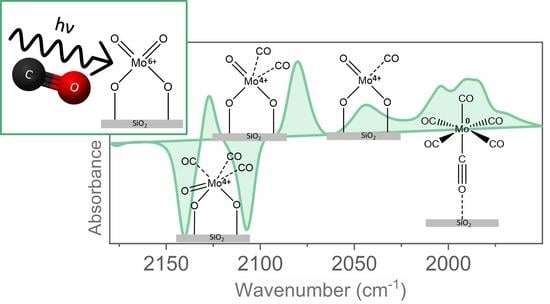
The photoreduction of the Mo6+/SiO2 system with CO was investigated in situ, employing a recently developed experimental setup allowing for the acquisition of transmission FT-IR spectra under simultaneous UV irradiation. Carbon monoxide, besides acting as a reducing agent in such processes, is also a useful probe molecule able to detect coordinatively unsaturated sites exposed on the surface. The unprecedented quality of the spectroscopic data, obtained as a function of the reduction time, allowed us to better rationalize the different mechanisms previously proposed for the photoreduction process. These results, coupled with UV-Vis spectroscopic data, shed light on the oxidation state and surface structure of supported molybdenum species, which are key active sites for several important reactions, such as selective oxidation, polymerization, hydrodesulfurization, epoxidation and olefin metathesis.

Graphical Abstract

1. Introduction

Heterogeneous systems based on molybdenum species supported on the surface of pure or mixed oxides are still extensively investigated because of their importance as catalysts for a wide number of significant industrial reactions such as oxidation, polymerization, hydrogenation, isomerization, hydrodesulfurization, epoxidation and olefin metathesis [1,2,3].
In this respect, molybdena-silica catalysts are often considered suitable reference systems for the investigation of the nature of the active sites and of the reaction mechanisms since the use of SiO2 as support ensures excellent dispersion of the metal phase at low loadings, which remains mainly in a monomeric form [3,4,5]. Indeed, accurate control of the dispersion of the metal on the support surface is crucial to establish clear structure-activity relationships.
In addition to nuclearity (monomer, dimer/oligomer), the coordination environment and the oxidation state of molybdenum sites are key factors influencing catalytic activity [6]. Although such aspects have been extensively studied employing both theoretical and experimental methods, to date they are still subject of debate [7,8,9] and several studies have tried to identify the best synthesis conditions to improve the catalytic activity [10,11,12]. In this respect, several preparation methods have been employed, including wet impregnation, the adsorption equilibrium deposition method, the oxoperoxo route and the use of different metal precursors, such as molybdates, organo-molybdenum compounds and Mo-oxoperoxo species. Moreover, the conditions of precalcination, the molybdenum concentration and the reduction conditions of Mo6+/SiO2 ions are important factors in governing the fraction of Mo exposed sites active for catalysis [13].
Focusing on the reduction conditions, Kazansky and coworkers investigated in detail the oxidation state and nuclearity of the Mo species obtained by the reduction of Mo6+/SiO2 precursors following different routes, namely thermal reduction at a high temperature (673–873 K) and photoreduction in mild conditions (300 K) in the presence of CO or H2 [14,15]. In particular, the thermal approach leads to the formation of highly heterogeneous systems with molybdenum ions in various coordination and valence states. On the contrary, photoreduction in a CO atmosphere by UV irradiation at room or low temperature resulted more selective, producing mainly coordinatively unsaturated, grafted Mo4+ ions with a small fraction of Mo5+ species [16]. Unlike the thermally reduced samples, the photoreduced Mo4+ species showed remarkable activity in metathesis reactions [17,18]. This difference could be explained by the fact that oxygen vacancies near Mo ions with lower coordination states are produced by the low-temperature photoreduction process, whereas during thermal reduction, they are easily eliminated by rearrangements of the first coordination sphere of Mo ions [14].
Interestingly, during the photoreduction process CO adsorbs on the reduced Mo sites forming carbonyls whose spectroscopic properties in the mid-IR region depend on the metal oxidation and coordination state. Therefore, the spectroscopic study of the adsorbed molecules represents a useful way to probe the nature of the different surface species that are progressively formed [19,20,21]. Many researchers have investigated these carbonyl species, giving sometimes contradictory interpretations of the spectroscopic data. For instance, some authors assigned the main IR signals to molybdenum dicarbonyls in different configurations [22,23,24,25,26], while others postulated the initial presence of molybdenum tricarbonyls [27]. In this paper, we try to contribute to this still lively debate investigating the photoreduction of the Mo6+/SiO2 system in CO, employing an experimental setup recently developed by Mino et al. [28,29,30], allowing for the acquisition of FT-IR spectra in transmission while simultaneously irradiating the sample with a UV-Vis beam. This in situ setup allowed us to follow in greater detail each step of the reduction process, monitoring the temporal evolution of each intermediate of the photoinduced reaction.

2. Results and Discussion

All the experiments were performed on a Mo6+/SiO2 catalyst prepared by the classical wet impregnation method with a Mo concentration of 0.5 wt % (i.e., below the maximum dispersion limit of Mo). The SiO2 support employed was a high-surface-area commercial fumed silica, which is a synthetic amorphous silicon dioxide produced by burning silicon tetrachloride in an oxygen-hydrogen flame. Before starting the in situ investigation of the reduction step, the Mo/SiO2 samples were dehydrated by vacuum treatment at a high temperature and fully oxidized with O2 (see Section 3 for details). Based on previous literature studies combining Raman, UV-Vis, XAS and DFT [18] this procedure leads to isolated Mo6+ ions in a Td environment (dioxo species, see Figure 1).
Spectral series such as those reported in Figure 2 were obtained, after dosing CO at room temperature, acquiring the spectrum of the system in the dark (without UV-Vis illumination) and then recording FT-IR spectra at fixed time intervals while illuminating with UV-Vis light directly inside the instrument sample compartment (black, gray and red curves in Figure 2).
As soon as the UV-Vis lamp is switched on (black curve in Figure 2A) we can note the immediate appearance of two bands at 2140 and 2107 cm−1. During the first 30 min of irradiation the intensity of such bands increases and a weak signal also appears at 2177 cm−1. Carbonylic bands in the 2180–2100 cm−1 spectral region have been already reported in previous studies with ex situ UV irradiation [22,23,24,25]. They were ascribed to the formation of Mo4+ species [31], via an unstable Mo5+ intermediate [16,32], which can adsorb CO by removal of one ligand from the first coordination sphere of the Mo ion forming a surface-distorted tetrahedral structure with concomitant CO2 evolution [14]. In particular, Williams et al. [27], by combining FT-IR, temperature-programmed decomposition and oxygen isotopic exchange, assigned the carbonyl signals to a meridionally-coordinated molybdenum tricarbonyl (T in Figure 1). They suggested that the mer-Mo4+(CO)3 should give rise to three IR signals: (i) a weak (A1)1 symmetric trans C-O stretch at 2181 cm−1; (ii) a strong B2 antisymmetric trans C-O stretch at 2141 cm−1; (iii) a strong (A1)2 cis-C-O stretch at 2108 cm−1, with relative intensities of 0.07:0.92:1.0, respectively. However, other assignments have been proposed and some authors ascribed the bands at ca. 2140 and ca. 2107 cm−1 to molybdenum dicarbonyls [22,23].
From a detailed analysis of the accurate spectral series (in terms of both number of spectra and quality) obtained in our experiments in the first 30 min (black to dark gray spectra in Figure 2) it is inferred that the intensity ratio of the 2177, 2140 and 2107 cm−1 bands is nearly constant all over the series and is ca. 0.07:1.04:1.00. These data strongly support the hypothesis that the first products formed by mild photoreduction with CO of the isolated Mo6+ species grafted at the SiO2 surface are highly coordinatively unsaturated reduced Mo4+ species capable of adsorbing up to 3 CO ligands to give surface Mo4+(CO)3 carbonyls.
Upon increasing the irradiation time from 30 to 150 min (dark gray to red spectra in Figure 2A), the first formed triplet at 2177, 2140, 2107 cm−1 gradually decreases in favor of two new absorptions centered at 2127 and 2080 cm−1 (see also the difference spectrum reported in Figure 2A’), which grow in intensity in a strictly parallel way. It is most noticeable that the transformation of the triplet at 2177, 2140 and 2107 cm−1 in the doublet at 2127 and 2080 cm−1 is accompanied by the appearance of two clear isosbestic points at 2132 and 2097 cm−1. This is a clear indication that the species responsible for the triplet are transformed into those responsible for the doublet in a 1:1 ratio. Based on the previous consideration and on the number of observed bands and their intensity ratio, the assignment of the bands at 2127 and 2080 cm−1 respectively to the in-phase and out-of-phase vibrations of a dicarbonylic Mo4+(CO)2 surface complex, formed in agreement with the reaction scheme of Figure 1, is straightforward [22,23,24,27,33].
As it is possible to see in Figure 2, the triplet and the doublet discussed above are not the only manifestations observed during the irradiation of the sample in the presence of CO. Indeed, a complex system of superimposed absorptions also gradually develops at wavenumbers below 2060 cm−1. Among them, an absorption at 2044 cm−1 and an envelope of bands in the 2010–1960 cm−1 region with a main maximum at ca. 1990 cm−1 becomes more and more evident. In the literature, a band at 2045 cm−1 was attributed to a linear molybdenum monocarbonyl, Mo(CO) (M in Figure 1). As far as the manifestations at lower wavenumbers are concerned, in ex situ experiments photoreduction in CO of Mo6+/SiO2 is supposed to also lead to the formation of Mo metal centers able to form physisorbed and chemisorbed hexacarbonyls Mo0(CO)6 (H in Figure 1), which show a stretching C-O mode at 1990 cm−1 [27,33]. We think that similar species are responsible for the complex spectrum we observe in the 2010–1960 cm−1 region (Figure 2).
In Figure 2(B,B’), we show the effect of switching off the UV lamp after 150 min. As it is possible to see, stopping the irradiation results in the disappearance of the manifestations of the residual Mo4+(CO)3 and of the Mo4+(CO) complexes, leaving unchanged those of the Mo4+(CO)2 dicarbonyls. It is also interesting to notice that switching off the irradiation dramatically changes the complex absorptions in the 2010–1960 cm−1 interval, where the various components appear to collapse in the main band at 1990 cm−1 related to CO adsorbed on Mo0 sites, which consequently gains in intensity. Similar effects were not reported in previous ex situ studies and deserve in our opinion further investigation.
Figure 3(A,A’) shows the effect of CO evacuation at room temperature. In agreement with previous studies [23,27], the room temperature evacuation of the gas-phase CO in equilibrium with molybdenum tri- and dicarbonyl species progressively removes CO ligands, resulting in the growth of the IR signal at 2044 cm−1 due to linear Mo4+(CO) (M species in Figure 1). After the complete removal of CO from Mo(CO)2 or Mo(CO) by outgassing at room temperature, the readmission of CO on the reduced Mo/SiO2 system mainly leads to repopulation of the D and H species (Figure 3(B,B’)). This results in the decrease of the monocarbonyl species (M) at 2044 cm−1 and the simultaneous growth of the bands at 2127 and 2080 cm−1. Furthermore, the band at 1990 cm−1, related to the hexacarbonyl Mo0 species (H), is restored together with some weak manifestations related to tricarbonyls. This behavior indicates the reversibility of the carbonylation-decarbonylation mechanism of the different surface Mo centers.
The electronic structure and the surface coordination geometry of the Mo species formed during the photoreduction process were investigated also by diffuse reflectance UV-Vis spectroscopy. The relationships between UV-Vis spectral features and molybdenum surface state have been widely debated in the literature, trying to relate the band position not only to the local symmetry but also to the molybdenum environment (e.g., structure and dimension of surface aggregates, interaction with the support) [34,35,36]. From the analysis of the spectra reported in Figure 4, we can note that the Mo6+/SiO2 system in the initial fully oxidized conditions shows two absorption bands centered at 237 and 276 nm, which are only slightly perturbed upon CO adsorption in dark conditions (red curve in Figure 4A). On the basis of previous investigations of different molybdenum complexes [3,14,15,36,37], we can conclude that these spectral features can be associated with ligand-to-metal charge-transfers (LMCT) of isolated Mo6+/SiO2 (Td) species (isolated dioxo species in Figure 1). After short UV irradiation, the surface state of the Mo sites is gradually modified, as described in the previous paragraphs, and new signals at about 340, 430 and 595 nm appear, which can be ascribed to d-d transitions in Mo4+ ions (in a d2 electronic configuration) coordinated by CO molecules [14]. Finally, after prolonged irradiation and subsequent spectra acquisition in dark conditions, the shoulder at about 600 nm disappears and two main signals grow at 320 and 415 nm (Figure 4B). These treatment conditions, according to the FT-IR results, lead to the disappearance of meridionally-coordinated molybdenum tricarbonyls (T) in favor of molybdenum dicarbonyl (D) species. The two main UV-Vis absorptions undergo only a small shift upon CO outgassing, likely due to the increase of molybdenum monocarbonyl (M) species.

3. Materials and Methods

3.1. Preparation of the Mo/SiO2 Catalyst

Mo/SiO2 (0.5 wt % molybdenum) was prepared by the impregnation method using an aqueous solution of ammonium molybdate (NH4)2MoO4 (Aldrich) as the metal precursor and silica Aerosil 300 (SSA = 300 m2/g) by Evonik as support. After impregnation, the sample was dried in an oven at 323 K and calcined in air at 773 K.

3.2. Pretreatment Conditions and Spectroscopic Measurements

Before photoreduction and spectroscopic measurements, the sample was pressed in the form of a thin self-supporting pellet and pretreated in an IR cell under a controlled atmosphere [38]. The pretreatment procedure consisted of degassing at 973 K, oxidation in an O2 atmosphere at 773 K and cooling down to room temperature in O2. After evacuation of the gas phase CO (equilibrium pressure 50 mbar) was dosed on the sample before photoreduction.
The infrared spectra were recorded on a Bruker Equinox 55 FT-IR spectrometer, equipped with an MCT cryogenic detector; 64 interferograms (recorded at a 2 cm−1 resolution) were typically averaged for each spectrum.
In situ UV-Vis irradiation (at increasing times up to 150 min) was performed using a Newport 500 W Hg(Xe) arc lamp, equipped with a water filter to eliminate the infrared portion of the spectrum. The radiation emitted by the lamp was focused on the Mo/SiO2 sample inside the quartz cell in a controlled atmosphere using an aspherical fiber bundle focusing assembly and a large core Newport liquid light guide [28,39].
Diffuse reflectance (DR) UV-Vis measurements were carried out on the Mo/SiO2 sample in the form of a thick self-supporting pellet, pretreated in the same conditions used for the FT-IR experiment. After each step of the photoreduction process in CO at room temperature (which, in this case, was performed ex situ), the UV-Vis spectra were recorded on a Cary 5000 Varian spectrophotometer equipped with a reflectance sphere.

4. Conclusions

In this study, we combined in situ FT-IR spectroscopy and diffuse reflectance UV–Vis spectroscopy to follow, with unprecedented detail, the photoreduction of Mo6+/SiO2 samples at room temperature in the presence of CO, accurately monitoring in the time the changes in coordination and oxidation state of Mo sites under UV–Vis irradiation.
We propose that UV irradiation of the initially fully oxidized, tetrahedrally coordinated, Mo6+/SiO2 surface complexes promotes electron transfer from the O2- anion to the Mo6+ cation. Then, this excited Mo5+ state interacts with CO to eliminate one oxygen ligand from its first coordination sphere, generating CO2. This change in oxidation state is testified by the complete disappearance of the electronic absorptions centered at 237 and 276 nm, related to the ligand-to-metal charge-transfers of isolated Mo6+/SiO2 (Td) species.
After this first reaction step, tricarbonylic Mo4+(CO)3 complexes are formed (possibly with the meridional structure proposed by Williams et al. [27]), which gradually convert into dicarbonyl species, Mo4+(CO)2. This transformation is clearly proven by the appearance of two isosbestic points at 2132 and 2097 cm−1, which were not highlighted in previous studies with ex situ UV irradiation. Prolonged photoreduction in CO leads then to the appearance of molybdenum metal hexacarbonyls, Mo0(CO)6.
Finally, CO evacuation at room temperature mainly promotes the formation of monocarbonyl species, Mo4+(CO).
Our results contribute to shed light on the unique redox properties of supported Mo6+ species, which were found to play a significant role in several catalytic and photocatalytic reactions. For instance, catalysts containing Mo6+ were successfully employed in the preferential photocatalytic oxidation of CO with O2 in the presence of excess H2 (photo-PROX) to obtain CO-free hydrogen for fuel cells [40,41].

Author Contributions

Conceptualization and supervision, G.S.; methodology, G.S. and L.M.; investigation, R.S.; writing and original draft preparation, R.S. and L.M.; review and editing, R.S., L.M. and G.S. All authors have read and agreed to the published version of the manuscript.

Funding

This research received no external funding.

Institutional Review Board Statement

Not applicable.

Informed Consent Statement

Not applicable.

Data Availability Statement

Not applicable.

Conflicts of Interest

The authors declare no conflict of interest.

Sample Availability

Mo/SiO2 samples are available from the authors.

References

  1. Haber, J. Chapter 10—Molybdenum Compounds in Heterogeneous Catalysis. In Studies in Inorganic Chemistry; Braithwaite, E.R., Haber, J., Eds.; Elsevier: Amsterdam, The Netherlands, 1994; Volume 19, pp. 477–617. [Google Scholar]
  2. Ohler, N.; Bell, A.T. Selective oxidation of methane over MoOx/SiO2: Isolation of the kinetics of reactions occurring in the gas phase and on the surfaces of SiO2 and MoOx. J. Catal. 2005, 231, 115–130. [Google Scholar] [CrossRef]
  3. Lee, E.L.; Wachs, I.E. In situ spectroscopic investigation of the molecular and electronic structures of SiO2 supported surface metal oxides. J. Phys. Chem. C 2007, 111, 14410–14425. [Google Scholar] [CrossRef]
  4. Banares, M.A.; Hu, H.C.; Wachs, I.E. Molybdena on silica catalysts: Role of preparation methods on the structure-selectivity properties for the oxidation of methanol. J. Catal. 1994, 150, 407–420. [Google Scholar] [CrossRef]
  5. Amakawa, K.; Sun, L.; Guo, C.; Havecker, M.; Kube, P.; Wachs, I.E.; Lwin, S.; Frenkel, A.I.; Patlolla, A.; Hermann, K.; et al. How strain affects the reactivity of surface metal oxide catalysts. Angew. Chem. Int. Ed Engl. 2013, 52, 13553–13557. [Google Scholar] [CrossRef] [PubMed]
  6. Anpo, M.; Kondo, M.; Coluccia, S.; Louis, C.; Che, M. Application of dynamic photoluminescence spectroscopy to the study of the active surface sites on supported Mo/SiO2 catalysts: Features of anchored and impregnated catalysts. J. Am. Chem. Soc. 1989, 111, 8791–8799. [Google Scholar] [CrossRef]
  7. Chempath, S.; Zhang, Y.; Bell, A.T. DFT Studies of the structure and vibrational spectra of isolated molybdena species supported on silica. J. Phys. Chem. C 2007, 111, 1291–1298. [Google Scholar] [CrossRef]
  8. Kurleto, K.; Tielens, F.; Handzlik, J. Isolated molybdenum(VI) and tungsten(VI) oxide species on partly dehydroxylated silica: A computational perspective. J. Phys. Chem. C 2020, 124, 3002–3013. [Google Scholar] [CrossRef]
  9. Amakawa, K.; Wang, Y.Q.; Krohnert, J.; Schlogl, R.; Trunschke, A. Acid sites on silica-supported molybdenum oxides probed by ammonia adsorbtion: Experiment and theory. Mol. Catal. 2019, 478, 9. [Google Scholar] [CrossRef]
  10. Mestl, G.; Srinivasan, T.K.K. Raman spectroscopy of monolayer-type catalysts: Supported molybdenum oxides. Catal. Rev. 1998, 40, 451–570. [Google Scholar] [CrossRef]
  11. Guesmi, H.; Grybos, R.; Handzlik, J.; Tielens, F. Characterization of molybdenum monomeric oxide species supported on hydroxylated silica: A DFT study. Phys. Chem. Chem. Phys. 2014, 16, 18253–18260. [Google Scholar] [CrossRef]
  12. Gholampour, N.; Yusubov, M.; Verpoort, F. Investigation of the preparation and catalytic activity of supported Mo, W, and Re oxides as heterogeneous catalysts in olefin metathesis. Catal. Rev. 2016, 58, 113–156. [Google Scholar] [CrossRef]
  13. Wachs, I.E.; Segawa, K. Supported metal oxide. In Characterization of Catalytic Materials, 2nd ed.; Wachs, I.E., Ed.; Momentum Press LLC: New York, NY, USA, 2010; pp. 69–88. [Google Scholar]
  14. Shelimov, B.N.; Pershin, A.N.; Kazansky, V.B. Selective photoreduction of molybdenum ions supported on silica. J. Catal. 1980, 64, 426–436. [Google Scholar] [CrossRef]
  15. Shelimov, B.N.; Elev, L.V.; Kazansky, V.B. Use of photoreduction for activation of silica-molybdena catalysts for propylene metathesis: Comparison with thermal reduction. J. Catal. 1986, 98, 70–81. [Google Scholar] [CrossRef]
  16. Kazansky, V.B.; Shelimov, B.N. Supported silica-molybdena catalysts for olefin metathesis activated by photoreduction. Res. Chem. Intermed. 1991, 15, 1–16. [Google Scholar] [CrossRef]
  17. Aigler, J.M.; Houalla, M.; Hercules, D.M. Surface structure and metathesis activity of photoreduced allyl-based Mo/SiO2 catalysts. Top. Catal. 2000, 10, 123–126. [Google Scholar] [CrossRef]
  18. Lwin, S.; Wachs, I.E. Olefin metathesis by supported metal oxide catalysts. ACS Catal. 2014, 4, 2505–2520. [Google Scholar] [CrossRef]
  19. Mino, L.; Spoto, G.; Bordiga, S.; Zecchina, A. Particles morphology and surface properties as investigated by HRTEM, FTIR, and Periodic DFT calculations: From pyrogenic TiO2 (P25) to nanoanatase. J. Phys. Chem. C 2012, 116, 17008–17018. [Google Scholar] [CrossRef]
  20. Zaera, F. New advances in the use of infrared absorption spectroscopy for the characterization of heterogeneous catalytic reactions. Chem. Soc. Rev. 2014, 43, 7624–7663. [Google Scholar] [CrossRef]
  21. Mino, L. IR spectroscopy as a tool to investigate photocatalytic reactions at oxide surfaces. Rend. Lincei. Sci. Fis. Nat. 2017, 28, 143–149. [Google Scholar] [CrossRef]
  22. Guglielminotti, E.; Giamello, E. Spectroscopic characterization of a molybdena silica system photoreduced in a carbon-monoxide atmosphere. J. Chem. Soc. Faraday Trans. I 1985, 81, 2307–2322. [Google Scholar] [CrossRef]
  23. Gerasimov, S.F. CO and NO adsorption on photoreduced Mo/SiO2 catalysts. React. Kinet. Catal. Lett. 1986, 32, 275–280. [Google Scholar] [CrossRef]
  24. Rodrigo, L.; Marcinkowska, K.; Roberge, P.C.; Kaliaguine, S. Photoreducibility of Mo/SiO2 catalysts with CO. J. Catal. 1987, 107, 8–22. [Google Scholar] [CrossRef]
  25. Kamegawa, T.; Takeuchi, R.; Matsuoka, M.; Anpo, M. Photocatalytic oxidation of CO with various oxidants by Mo oxide species highly dispersed on SiO2 at 293K. Catal. Today 2006, 111, 248–253. [Google Scholar] [CrossRef]
  26. Toyao, T.; Morishima, J.; Saito, M.; Horiuchi, Y.; Kamegawa, T.; Martra, G.; Coluccia, S.; Matsuoka, M.; Anpo, M. FT-IR study of the reaction mechanisms for photocatalytic reduction of NO with CO promoted by various single-site photocatalysts. J. Catal. 2013, 299, 232–239. [Google Scholar] [CrossRef]
  27. Williams, C.C.; Ekerdt, J.G. Infrared spectroscopic characterization of molybdenum carbonyl species formed by ultraviolet photoreduction of silica-supported Mo(VI) in carbon-monoxide. J. Phys. Chem. 1993, 97, 6843–6852. [Google Scholar] [CrossRef]
  28. Mino, L.; Zecchina, A.; Martra, G.; Rossi, A.M.; Spoto, G. A surface science approach to TiO2 P25 photocatalysis: An in situ FTIR study of phenol photodegradation at controlled water coverages from sub-monolayer to multilayer. Appl. Catal. B Environ. 2016, 196, 135–141. [Google Scholar] [CrossRef]
  29. Barzan, C.; Mino, L.; Morra, E.; Groppo, E.; Chiesa, M.; Spoto, G. Photoinduced ethylene polymerization on titania nanoparticles. ChemCatChem 2017, 9, 4324–4327. [Google Scholar] [CrossRef]
  30. Mino, L.; Barzan, C.; Martino, G.A.; Piovano, A.; Spoto, G.; Zecchina, A.; Groppo, E. Photoinduced ethylene polymerization on the CrVI/SiO2 Phillips catalyst. J. Phys. Chem. C 2019, 123, 8145–8152. [Google Scholar] [CrossRef]
  31. Aigler, J.M.; Kazansky, V.B.; Houalla, M.; Proctor, A.; Hercules, D.M. Photoreduction study of Mo allyl-based Mo/SiO2 catalysts. J. Phys. Chem. 1995, 99, 11489–11493. [Google Scholar] [CrossRef]
  32. Louis, C.; Che, M.; Anpo, M. Activation and properties of Mo=O bonds in Mo/SiO2 catalysts. Res. Chem. Intermed. 1991, 15, 81–98. [Google Scholar] [CrossRef]
  33. Ogata, A.; Kazusaka, A.; Enyo, M.; Toyoshima, I. Active-sites on the MoO3/SiO2 catalyst photoreduced with CO for CO2 reduction to CO. Chem. Phys. Lett. 1986, 127, 283–285. [Google Scholar] [CrossRef]
  34. Praliaud, H. Diffuse reflectance spectra of molybdenum ions supported by magnesia, γ-alumina or silica. J. Less Common Met. 1977, 54, 387–399. [Google Scholar] [CrossRef]
  35. Fournier, M.; Louis, C.; Che, M.; Chaquin, P.; Masure, D. Polyoxometallates as models for oxide catalysts: Part I. An UV-visible reflectance study of polyoxomolybdates: Influence of polyhedra arrangement on the electronic transitions and comparison with supported molybdenum catalysts. J. Catal. 1989, 119, 400–414. [Google Scholar] [CrossRef]
  36. Vikulov, K.A.; Shelimov, B.N.; Kazansky, V.B. IR and UV-Vis spectroscopic studies of the surface Mo=CH2 and Mo=CH-CH3 carbene complexes produced by methylcyclopropane chemisorption over photoreduced silica molybdena catalysts. J. Mol. Catal. 1991, 65, 393–402. [Google Scholar] [CrossRef]
  37. Williams, C.C.; Ekerdt, J.G.; Jehng, J.M.; Hardcastle, F.D.; Turek, A.M.; Wachs, I.E. A Raman and ultraviolet diffuse reflectance spectroscopic investigation of silica-supported molybdenum oxide. J. Phys. Chem. 1991, 95, 8781–8791. [Google Scholar] [CrossRef]
  38. Mino, L.; Mandrile, L.; Iannarelli, L.; Portesi, C.; Martra, G.; Rossi, A.M. Vibrational spectroscopy. In Characterization of Nanoparticles: Measurement Processes for Nanoparticles; Hodoroaba, V., Unger, W., Shard, A., Eds.; Elsevier: Amsterdam, The Netherlands, 2019; pp. 457–480. [Google Scholar] [CrossRef]
  39. Mino, L.; Negri, C.; Zecchina, A.; Spoto, G. Photodegradation of organic pollutants on TiO2 P25 surfaces investigated by transmission FTIR spectroscopy under in situ UV-Vis irradiation. Z. Phys. Chem. Int. J. Res. Phys. Chem. Chem. Phys. 2016, 230, 1441–1451. [Google Scholar] [CrossRef]
  40. Matsuoka, M.; Kamegawa, T.; Takeuchi, R.; Anpo, M. In situ characterization of the highly dispersed Mo6+-oxide species supported onto various oxides and their photocatalytic reactivities. Catal. Today 2007, 122, 39–45. [Google Scholar] [CrossRef]
  41. Yoshida, Y.; Izumi, Y. Recent advances in the preferential thermal-/photo-oxidation of carbon monoxide: Noble versus inexpensive metals and their reaction mechanisms. Catal. Surv. Asia 2016, 20, 141–166. [Google Scholar] [CrossRef]
Figure 1. Proposed photoreduction pathways of silica-supported Mo in CO at room temperature. T, D, M and H indicate tricarbonyl, dicarbonyl, monocarbonyl and hexacarbonyl species, respectively. The vibrational frequencies associated with the different Mo-CO surface complexes are also reported.
Molecules 26 01700 g001
Figure 2. FT-IR spectra during photoreduction of Mo/SiO2 in the presence of CO at 50 mbar. (A) Effect of photoreduction time up to 150 min of UV irradiation. (B) Effect of 15 h in dark conditions after photoreduction. The spectrum of the oxidized material in the presence of CO gas, before UV irradiation, has been subtracted from all spectra. (A’) Difference spectrum (c−b) to underline the spectral evolution from 30 min to 150 min. (B’) Difference spectrum (d−c) showing the effect of switching off the UV lamp. The letters T, D, M and H refer to the molybdenum species shown in Figure 1.
Molecules 26 01700 g002
Figure 3. FT-IR spectra of Mo/SiO2 after photoreduction in CO. (A) Comparison of the spectra after stopping the UV irradiation (blue curve) and after 200 min of CO evacuation in dark conditions (purple curve). (B) Comparison of the spectra of the outgassed sample and after readmission of CO at 50 mbar (black curve). The spectrum of the oxidized material in the presence of CO gas, before UV irradiation, has been subtracted from all spectra. (A’) Difference spectrum (b−a) to underline the effect of CO evacuation. (B’) Difference spectrum (d−c) showing the effect of CO readsorption. The letters T, D, M and H refer to the molybdenum species shown in Figure 1.
Molecules 26 01700 g003
Figure 4. Diffuse reflectance spectra of the Mo/SiO2 system. (A) In the initial fully oxidized conditions (black curve), after CO admission (red curve) and after UV irradiation for 30 min. (B) Effect of UV irradiation up to 60 min (blue curves) and of CO outgassing (purple curve).
Molecules 26 01700 g004
Publisher’s Note: MDPI stays neutral with regard to jurisdictional claims in published maps and institutional affiliations.