Article

In Vitro Antileishmanial and Antitrypanosomal Activities of Plicataloside Isolated from the Leaf Latex of Aloe rugosifolia Gilbert & Sebsebe (Asphodelaceae)

1
Department of Pharmaceutical Chemistry and Pharmacognosy, School of Pharmacy, College of Health Sciences, Addis Ababa University, Addis Ababa P.O. Box 1176, Ethiopia
2
School of Pharmacy, Faculty of Health Sciences Institute of Health, Jimma University, Jimma P.O. Box 378, Ethiopia
*
Author to whom correspondence should be addressed.
Molecules 2022, 27(4), 1400; https://doi.org/10.3390/molecules27041400
Submission received: 21 January 2022 / Revised: 16 February 2022 / Accepted: 16 February 2022 / Published: 18 February 2022
(This article belongs to the Section Natural Products Chemistry)

Abstract

:
Trypanosomiasis and leishmaniasis are among the major neglected diseases that affect poor people, mainly in developing countries. In Ethiopia, the latex of Aloe rugosifolia Gilbert & Sebsebe is traditionally used for the treatment of protozoal diseases, among others. In this study, the in vitro antitrypanosomal activity of the leaf latex of A. rugosifolia was evaluated against Trypanosoma congolense field isolate using in vitro motility and in vivo infectivity tests. The latex was also tested against the promastigotes of Leishmania aethiopica and L. donovani clinical isolates using alamar blue assay. Preparative thin-layer chromatography of the latex afforded a naphthalene derivative identified as plicataloside (2,8-O,O-di-(β-D-glucopyranosyl)-1,2,8-trihydroxy-3-methyl-naphthalene) by means of spectroscopic techniques (HRESI-MS, 1H, 13C-NMR). Results of the study demonstrated that at 4.0 mg/mL concentration plicataloside arrested mobility of trypanosomes within 30 min of incubation period. Furthermore, plicataloside completely eliminated subsequent infectivity in mice for 30 days at concentrations of 4.0 and 2.0 mg/mL. Plicataloside also displayed antileishmanial activity against the promastigotes of L. aethopica and L. donovani with IC50 values 14.22 ± 0.41 µg/mL (27.66 ± 0.80 µM) and 18.86 ± 0.03 µg/mL (36.69 ± 0.06 µM), respectively. Thus, plicataloside may be used as a scaffold for the development of novel drugs effective against trypanosomiasis and leishmaniasis.

1. Introduction

Neglected tropical diseases (NTDs) consist of a group of diseases which currently account about 12% of the total global health burden in 149 tropical and subtropical countries [1,2]. Among these, trypanosomiasis and leishmaniasis are the two major NTDs in Africa [3]. Trypanosomiasis is a disease caused by the parasitic protozoa of the genus Trypanosoma, which is potentially fatal to both human and animal [4]. This disease mainly occurs in tropical Africa and South America [5]. Human African Trypanosomiasis (HAT), also known as sleeping sickness, is caused by two subspecies of Trypanosoma parasites, Trypanosoma brucei gambiense and Trypanosoma brucei rhodesiense [6], of which the former accounts for 98% of the cases [7]. There are also reports that indicate that other trypanosome species such as T. congolense and T. evansi cause human trypanosomiasis [8] in regions ranging from the Sahara to the Kalahari Desert [9].
Leishmaniasis is a parasitic disease caused by the protozoan parasite Leishmania [10]. The disease is prevalent in Africa, the Mediterranean basin, Asia, Latin America, and the Middle East, and has recently been identified in East Timor, Thailand and in Kangaroos in Australia [11]. In Ethiopia, visceral leishmaniasis occurs mainly in arid and semiarid areas [12], whereas cutaneous leishmaniasis is prevalent in highland areas [13]. Leishmaniasis is associated with malnutrition, population displacement, poor housing, a weak immune system, and a lack of financial resources. Depending upon the forms of the disease, it leads to various health problems. Cutaneous leishmaniasis causes skin lesions that leave life-long scars and serious disability, while mucocutaneous leishmaniasis leads to partial or total destruction of mucous membranes of the nose, mouth, and throat. Visceral leishmaniasis is fetal if left untreated. Similarly, the major symptoms of African trypanosomiasis include fever, skin lesions, and swollen lymph nodes on the back of the neck. The infection may become meningoencephalitis. If the patient fails to receive medical treatment, death will occur, usually within months.
Recently, there have been some attempts to control protozoal diseases, partly because they have become more difficult to control due to a number of factors that limit the utility of current drugs in resource-poor settings, such as high cost, poor compliance, drug resistance, low efficacy, and poor safety [14,15,16]. Therefore, safe, non-toxic, and cost-effective drugs are urgently required to eliminate this problem from every corner of world [7,17]. The latex of Aloe rugosifolia Gilbert & Sebsebe is traditionally used for treatments of malaria, wounds, worms, and internal parasites in human and livestock in Hamer district of southern Ethiopia [18]. Latexes of several Aloe species have been reported to possess antiprotozoal activity [19,20,21]. Thus, in this study, the antileishmanial and antitrypanosomal activities of Aloe rugosifolia and its major constituent plicataloside have been investigated.

2. Results

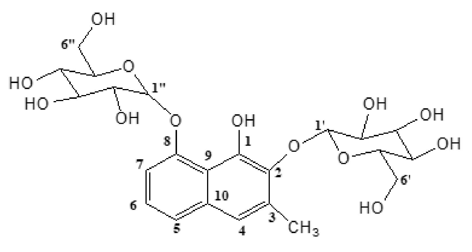
2.1. Structural Elucidation of Plicataloside

The leaf latex of A. rugosifolia showed only one peak when analyzed by HPLC (Figure S1). Following this, application of the leaf latex of A. rugosifolia on silica gel- PTLC using chloroform: methanol (2:1) as a solvent system led to the isolation of a light-brown-coloured amorphous substance with Rf value of 0.27. A pseudomolecular ion at m/z 513.1622 was observed in the negative-mode HRESI-MS as the deprotonated molecular ion [M−H] (Figure S2), which confirmed the molecular formula to be C23H30O13 (calcd. m/z = 513.1608 [M−H]). Two main fragment ions at m/z 351.1095 and 189.0565 were also observed in the negative-mode HRESI-MS, corresponding to the loss of a glucose moiety and a subsequent loss of another glucose moiety from the first fragment ion, respectively.
The 13C-NMR (Figure S3) and DEPT-135 (Figure S4) spectral data of the compound indicated the presence of 23 carbon atoms. Of these, 17 carbon atoms appeared in the DEPT-135 spectrum, including one sp3 methyl (CH3), eight sp3 oxymethines (OCH), two sp3 dioxymethines (OCHO), two sp3 oxymethylenes (OCH2), and four aromatic sp2 methines (=CH). Differential profiling of 13C-NMR and DEPT-135 spectral data allowed the identification of six quaternary aromatic carbons (δ 115.21, 133.25, 134.00, 140.04, 144.49, and 154.37) in the compound. Diglucosylation was evident from the 13C-NMR spectral data (two anomeric carbons (δ 103.05, 104.58), two sp3 oxymethylene carbons (δ 61.18, 61.32) and eight sp3 oxymethine carbons (δ 70.29, 70.29, 74.05, 74.66, 76.59, 76.75, 77.47, 77.92).
The 1H-NMR spectrum (Figure S5) of the compound (see Materials and Methods section) revealed the presence of four aromatic protons assigned as H-4 (δ 7.19, 1H, brs), H-5 (δ 7.40, 1H, brd, J = 8 Hz), H-6 (δ 7.26, 1H, t, J = 8 Hz), and H-7 (δ 7.19, 1H, brd, J = 8 Hz). Diglucosylation was also confirmed by the 1H-NMR spectrum as indicated by twelve proton signals between δ 2.51 and 3.44, along with other two anomeric protons as H-1′ (δ 4.82, 1H, d, J = 8 Hz) and H-1 (δ 4.99, 1H, d, J = 8 Hz). The positions of attachment of the two O-glucosyl units at C-2 and C-8 were confirmed by HMBC experiments (Figure S6), which revealed correlations between the anomeric proton H-1′ (δ 4.82, d) and C-2 (δ 140.04) and also between the anomeric proton H-1 (δ 4.99, d) and C-8 (δ 154.37). Two singlets were detected at δ 2.38 and δ 9.30 due to the presence of a methyl group and a hydroxyl group attached to a naphthalene ring, respectively. From the above spectroscopic data and comparison with those reported in the literature [22,23], the compound was identified as 2,8-O,O-di-(β-D-glucopyranosyl)-1,2,8-trihydroxy-3-methylnaphthalene (plicataloside) (Figure 1).

2.2. Oral Acute Toxicity

Oral administration of plicataloside (2000 mg/kg) led to minor signs of toxicity such as temporary shivering and diarrhea. However, these signs disappeared after 24 h. No mortality was also observed following the administration of plicataloside (2000 mg/kg). This implies that plicataloside is safe up to the dose levels used. As per the OECD guideline, its LD50 is above 2 g/kg [24].

2.3. In Vitro Antitrypanosomal Activity

The current study demonstrated that the incubation of infected blood with 4 and 2 mg/mL concentrations of both the latex and plicataloside arrested the motility of T. congolense within the first hour of incubation period. Similarly, at concentrations of 4.0, 2.0, and 0.4 mg/mL, diminazene diaceturate completely eliminated trypanosomal motility within the first hour (Table 1). The results suggested that that A. rugosifolia has promising in vitro antitrypanosomal activity.

2.4. In Vivo Infectivity Test

As shown in Table 2, the leaf latex of A. rugosifolia and plicataloside inhibited healthy mice from developing infection for more than 30 days or significantly prolonged the pre-patent period especially at a concentration of 4.0 mg/mL unlike the animals in the negative control group, which developed infection within 11 days after inoculation.

2.5. In Vitro Antipromastigote Activity

IC50 (effective concentration required to achieve 50% growth inhibition) values of the latex and plicataloside were determined against promastigotes of L. aethiopica and L. donovani in vitro (Table 3). The current study revealed that the leaf latex of A. rugosifolia possesses antileishmanial activity with IC50 values of 24.5±0.24 and 31.21±0.01 μg/mL on the promastigotes stage of L. aethiopica and L. donovani, respectively. On the other hand, plicataloside showed stronger activity when compared to that of the latex against both promastigotes of L. aethiopica and L. donovani with IC50 values of 14.22±0.41 μg/mL (27.66 ± 0.80 µM) and 18.86±0.03 μg/mL (36.69 ± 0.06 µM), respectively.

3. Discussion

Trypanosomiasis and leishmaniasis are the two major neglected tropical diseases (NTDs), which currently affect poor people mainly in developing countries [1,2]. Several plants from the genus Aloe are endowed with antiprotozoal activities [19,20,21,25,26,27,28]. As a continuation of our search for antiprotozoal compounds from the genus Aloe, the leaf latex of A. rugosifolia and its major compound were investigated for their in vitro antitrypanosomal and antileishmanial activities.
As shown in Table 1, both the leaf latex of A. rugosifolia and its major compound plicataloside showed in vitro activity against T. congolense parasites. Plicataloside further eliminated subsequent infectivity in mice for 30 days. It has been reported that the motility of parasites constitutes a relatively reliable indicator of viability among most zoo flagellate parasites [29]. In the in vitro system when no extract is present, it has been proved that trypanosomes can survive for about 4 h [30]. Trypanocidal activity was measured based on comparison of drop or cessation in motility between the parasites in test substances treated blood- and parasite-loaded control blood without test substances [30,31]. Generally, the shorter the time of cessation of motility of the parasite, the more active the extract is considered to be [30].
A previous study on the leaf latex of Aloe gilbertii, its major compound aloin, and its derivatives demonstrated promising antitrypanosomal effects [25]. The report indicates that at a concentration of 4.0 mg/mL, rhein, aloe-emodin, the latex, and aloin completely immobilized or killed the parasites within 15, 20, 25, and 40 min, respectively. Another study by Tadesse et al. [32] demonstrated that the dichloromethane and methanol leaf extracts of Dovyalis abyssinica ceased the motility of T. congolense within 1 h at concentrations of 20.0, 10.0, and 2.0 mg/mL. Furthermore, both extracts eliminated subsequent infectivity in mice. Several plant extracts have also been reported to have antitrypanosomal activities against T. congolense [30,33,34]. Previous reports [29,30,35] indicate that the mean minimum inhibitory concentration (MIC) value of the common trypanocidal drugs is 10.7 mg/mL, and that agents with MIC values of 5–20 mg/mL could be regarded as very active. Therefore, the leaf latex of A. rugosifolia and plicataloside can be considered as promising antitrypanosomal agents, although their activity was not as strong as the positive control diminazene diaceturate.
It should be noted that in the in vitro model, complete immobility of parasites does not necessarily mean that the parasites are dead, but rather the parasites may have lost their infectivity. This was confirmed through the infectivity test, which showed that the leaf latex of A. rugosifolia and plicataloside inhibited healthy mice from developing infection for more than 30 days or significantly prolonged the pre-patent period, especially at a concentration of 4.0 mg/mL (Table 2), unlike the animals in the negative control group, which developed infection within 11 days after inoculation.
Even though the lowest concentration of both plicataloside and diminazene diaceturate (0.4 mg/mL) highly reduced the motility of the parasite, mice administered with these doses contracted infection. Thus, it appears reasonable to speculate that these doses may belong to the group that acts by static action affecting the growth and multiplication of trypanosomes rather than eliminating them completely [30]. These results are consistent with other findings reported elsewhere [25,32].
The latex of A. rugosifolia also showed good antileishmanial activity against the promastigotes of L. aethiopica (IC50 = 24.5 ± 0.24 µg/mL) and L. donovani (IC50 = 31.21 ± 0.01 µg/mL), although its effect was inferior to that of plicataloside, which showed IC50 values of 14.22 ± 0.41 and 18.86 ± 0.03 µg/mL, respectively. These activities are much lower than those reported for the latex of Aloe macrocarpa (IC50 = 1.90 and 1.92 μg/mL, respectively) [36], but higher than those reported for Aloe vera leaf exudate against L. donovani strains (IC50 = 70–115 μg/mL) [19]).
As shown in Table 3, although the antipromastigote activity of plicataloside was less than that of the positive-control amphotericin B (IC50 = 8.1 ± 0.11 µg/mL (8.77 ± 0.12 µM) and 7.20 ± 0.15 µg/mL (7.79 ± 0.16 µM) against L. aethopica and L. donovani, respectively), it was more potent than the antileishmanial drug pentostam, which showed an IC50 value of 28.41 μg/mL against L. donovani promastigotes [37]. Plicataloside is a naphthalene derivative first isolated from the leaf latex of South African Aloe plicatilis [22] and then from the leaf latex of Ethiopian Aloe otallensis [23]. Several biological activities including antimalarial and antioxidant [22] and antityrosinase [38] have been associated with it.
It is difficult to speculate the mechanism by which these test substances exhibit their trypanocidal action. However, accumulated evidence suggests that many natural products show trypanocidal activity by interfering with the redox balance of the parasites [39]. A number of natural products possess structures which are capable of generating radicals that can cause peroxidative damage to trypanothione reductase which is very sensitive to alterations in redox balance. It is also known that some compounds act by binding with the kinetoplast DNA of the parasite [39].
Perusal of literature unveils that a large number of test substances show relatively higher activities against the promastigotes of L. aethopica than those of L. donovani [36,40,41]. These differences in drug susceptibility between the two species could be associated with slight differences in their membrane composition and/or metabolic characteristics. The extent to which the two parasites are exposed to different chemical substances and culture conditions may also affect their susceptibility to drugs [42,43]. To the best of our knowledge, this is the first report on antitrypanosomal and antilishmanial activities of the leaf latex of A. rugosifolia and its major compound plicataloside. Future optimization of plicataloside through structural alteration may lead to synthesis of molecules with improved trypanocidal and leishmanicidal activities.

4. Materials and Methods

4.1. Plant Material

Fresh leaves of A. rugosifolia were collected in September 2019 from Dida Chena plain between Yabelo and Mega, Oromia region, southern Ethiopia. The plant was authenticated by Mr. Anteneh Desta, the National Herbarium, Department of Plant Biology and Biodiversity Management, College of Natural and Computational Sciences, Addis Ababa University (AAU), where a voucher specimen was deposited (collection number AR010) for future reference.

4.2. Chemicals and Drugs

Analytical TLC was performed on a pre-coated aluminium backed silica gel 60 F254 plates (Merck KGaA, Darmstadt, Germany). Chloroform and methanol (HPLC grade, Carlo Erba, Milan, Italy). Dimethylsulfoxide (DMSO) (Sigma-Aldrich Co., St. Louis, MO, USA), amphotericin B (Laborchemikalien GmbH, Seelze, Germany) and Diminasan® (1.05 g diminazene diaceturate × 1.31 gantipyrine (Woerden, the Netherlands) were used in the biological assay.

4.3. Instruments

A Rotary evaporator R-200 (Buchi, Switzerland) was used to remove organic solvents. TLC chromatograms were viewed under a UV cabinet (CAMAG, Muttenz, Switzerland), 1H-NMR and 13C-NMR spectra were recorded at a room temperature on a Bruker Avance DMX400 FT-NMR spectrometer (Bruker, Billerica, MA, USA). MS was run on an ultra-high-performance liquid chromatography-mass spectrometer (UHPLC-MS) Waters® Acquity UHPLC system (Waters, Milford, MA, USA) equipped with electrospray ionization (ESI) in the negative mode.

4.4. Experimental Animals

Swiss albino mice (20–35 g) of either sex were obtained from the School of Pharmacy breeding colony, AAU. The animals were kept at room temperature and 12 h light and dark cycle in polypropylene cages, and provided with commercial pelleted ration and clean water ad libitum. The animals were acclimatized for one week under controlled conditions before conducting the experiments. All the experiments were conducted in accordance with internationally accepted laboratory animal use and care guideline [44] and were approved by the Institutional Review Board of School of Pharmacy, AAU (approval code: ERB/SoP/028/01/21).

4.5. Leaf Latex Preparation

Latex was collected from the leaves of A. rugosifolia by cutting the leaves at the bottom and allowing the latex to drain on a plastic sheet. The latex was then left to dry in air for 3 days, which yielded a light powder, then the dried material was weighed and stored in a refrigerator at 4 °C until use.

4.6. Isolation of Plicataloside

The latex was dissolved in methanol, then filtered and dried under reduced pressure using A Rotary Evaporator R-200 (Buchi, Switzerland). The dried extract was subjected to PTLC over silica gel plates using a mixture of chloroform and methanol (2:1) as a mobile phase. Chromatographic zones were visualized under UV light of 254 nm and 366 nm.

4.7. Spectral Data Plicataloside

Plicataloside was isolated as a light-brown-coloured amorphous substance; Rf = 0.27 (CHCl3/MeOH; 2:1); HRESI-MS (-ve mode, Figure S2) m/z: 513.1622 [M−H] (exact calcd. 513.1608 for [M−H]) indicating a molecular formula of C23H30O13; m/z: 351.1095 [M−Glc−H]; 189.0565 [M−Glc−Glc−H]; 1H-NMR (400 MHz, DMSO-d6, Figure S5) δ: 2.38 (3H, s, Me), 2.51-3.44 (12H, m, H-2′-H-6′ & H-2″-H-6″), 4.82 (1H, d, J = 8 Hz, H-1′), 4.99 (1H, d, J = 8 Hz, H-1″), 7.19 (1H, brs, H-4), 7.19 (1H, brd, J = 8 Hz, H-7), 7.26 (1H, t, J = 8 Hz, H-6), 7.40 (1H, brd, J = 8 Hz, H-5), 9.30 (1H, s, OH-1); 13C-NMR & DEPT (100 MHz, DMSO-d6, Figure S3 and Figure S4) δ: 18.2 (C-11), 61.18 (C-6′), 61.32 (C-6″), 70.29 (C-4′ & C-4″), 74.05 (C-2″), 74.66 (C-2′), 76.59 (C-3″), 76.75 (C-3′), 77.47 (C-5′), 77.92 (C-5″), 103.05 (C-1″), 104.58 (C-1′), 110.25 (C-7), 115.21 (C-9), 119.45 (C-4), 122.50 (C-5), 125.85 (C-6), 133.25 (C-10), 134.00 (C-3), 140.04 (C-2) 144.49 (C-1) 154.37 (C-8).

4.8. Acute Toxicity Study

Acute oral toxicity of plicataloside was carried out according to the OECD guidelines for testing of chemicals on Swiss albino mice [24]. Ten healthy Swiss female mice weighing 23–25 g was randomly divided into 2 groups of 5 mice per group. After fasting for 3 h, mice in the first group were given 2 g/kg of plicataloside and the second group received 0.2 mL distilled water (control group) orally and observed for any signs of toxicity for 14 days to assess safety of the test substances. The mice were observed for any gross behavioral changes (loss of appetite, hair erection, lacrimation, mortality) and other signs of toxicity manifestation.

4.9. Isolation of Trypanosoma congolense

Trypanosoma congolense field isolate was collected from Oromia region, Buno Bedele Zone, Dabu Woreda, Sebategna kebele, southwestern Ethiopia. The Murray method was used to check the presence of T. congolense in cattle [45]. The presence of T. congolense parasites was identified based on their type of motility and further confirmed by the National Tsetse and Trypanosomiasis Control Centre (NTTCC) in Bedelle town by preparing thin blood smear. The slides were read using an oil immersion 100x objective after fixing with methanol and staining with Giemsa stain [46,47]. The stained field was then compared with that of the reference species. This was followed by collection of blood from the positive cattle jugular vein using heparinized vacutainer tube. Then, 0.2 mL of blood containing the parasites was injected intraperitoneally to 10 healthy laboratory mice [32], and transported to the Department of Pharmaceutical Chemistry and Pharmacognosy, College of Health Sciences, AAU, Addis Ababa for serial passage to other mice used in the experiments.

4.10. Leishmania Test Strains

In vitro antileishmanial activity tests were performed on the promastigote stage of L. aethiopica and L. donovani clinical isolates obtained from Leishmania Diagnostic and Research Laboratory collection, Faculty of Medicine, AAU. They were grown in tissue culture flasks containing RPMI 1640 medium supplemented with 10% heat-inactivated fetal calf serum, 100 IU penicillin/mL, and 100 μg/mL streptomycin solution at 24 °C for L. donovani and 22 °C for L. aethiopica. In vitro cell-free medium was used to set up the test system for the determination of IC50 values of the test substances and to grow the parasites [36,48].

4.11. In Vitro Antitrypanosomal Activity Tests

Assessment of in vitro antitrypanosomal activity of the test substances was performed in triplicates in 96-well microtiter plates. Infected mice with a high parasitemic state were sacrificed and blood was collected in an ethylenediaminetetraacetic acid (EDTA; Thermo Fisher Scientific, Waltham, MA, USA) tube prepared with phosphate buffer saline glucose (PBSG; Gibco, Waltham, MA, USA). A measure of 20 μL of blood containing about 20–25 parasites per field was mixed with 5 μL of each of the test substances dissolved in 10% DMSO at concentrations of 20.0, 10.0, 2.0, and 0.5 mg/mL to produce 4.0, 2.0, 0.4, and 0.1 mg/mL of final concentrations, respectively [49]. A set of negative controls containing the parasite (20 μL of infected blood) suspended in 10% DMSO was introduced to ensure that the effect monitored was that of the test substances alone. Tests were also performed with similar concentrations of Diminasan® to serve as a positive control [30,50].
After 5 min of incubation in closed microtiter plates maintained at 37 °C, the parasites were observed every 5 min for death/motility using 400× objective for a total of 60 min by placing separately 2 μL of test mixtures on microscope slide and covered with 22 mm × 22 mm cover slips [51].

4.12. In Vivo Infectivity Test

An in vivo infectivity test was performed to establish whether there were any remaining infective parasites after the in vitro test. In this test, the inoculum contained in the remaining incubation mixture from each well of the micro titer plate where the in vitro test was carried out, i.e., 4, 2, 0.4, and 0.1 mg/mL of test substance separately in 0.02 mL of infected blood as described elsewhere [25]. A total of 65 healthy mice (5 animals per dose) received each mixture intraperitoneally (after 2 h of incubation period) and were observed for 30 days for the development of infection.

4.13. In Vitro Antileishmanial Activity Assay

To a separate 96-well microtiter plate containing 100 µL of complete culture medium, each of the test substances was added to triplicate wells to achieve a final concentration of 100 µg/mL. Then, 100 µL suspension of the parasites containing 3.5 × 10⁶ promastigotes of L. aethiopica or L. donovani were added to each well, which was obtained from the previous culture. After 68 h of incubation, 20 µL (10% of the total volume of each well) resazurin (0.125 mg/mL) was added and covered with aluminum foil, and left at room temperature. After a total incubation period of 72 h, fluorescence intensity was measured using Victor3 Multilabel Counter (PerkinElmer, Waltham, MA, USA) at an emission wavelength of 590 nm and excitation wavelength of 544 nm. During the assay, cell viability was monitored by measuring the fluorescent signal. The number of viable cells was proportional to the fluorescence intensities produced [36,48].

4.14. Data Analysis

Antileishmanial activity (IC50) values were expressed as mean ± SD of triplicate experiments, and they were evaluated from sigmoidal dose-response curves of percent inhibition using GraphPad Prism 8 (GraphPad Software, CA, USA). Values of antitrypanosomal activity were expressed as mean ± SEM using Microsoft Excel 2013.

5. Conclusions

The results of the current study revealed that the leaf latex of A. rugosifolia possesses genuine in vitro antitrypanosomal activity against field isolate of T. congolense and antileishmanial effect against L. aethopica and L. donovani promastigotes. Phytochemical investigation of the latex afforded a rare naphthalene derivative identified as plicataloside. Plicataloside showed superior activity against these parasites when compared to the effect of the latex. It can therefore be concluded that the antitrypanosomal and antileishmanial effects of the latex of A. rugosifolia are attributed in full or in part to the presence of plicataloside. Furthermore, plicataloside has the potential to be used as a lead compound for the development of cost-effective and more potent alternative drugs for the treatment of trypanosomiasis and leishmaniasis.

Supplementary Materials

The following supporting information can be downloaded online. Figure S1: HPLC chromatogram of the leaf latex of Aloe rugosifolia (plicataloside (1)). Figure S2: Negative-mode HR-ESI-mass spectrum of plicataloside; Figure S3: 13C-NMR spectrum of plicataloside in DMSO-d6; Figure S4: DEPT-135 spectrum of plicataloside in DMSO-d6; Figure S5: 1H-NMR spectral data of plicataloside in DMSO-d6; Figure S6: HMBC spectrum of plicataloside in DMSO-d6.

Author Contributions

Conceptualization, G.C., D.B., and K.A.; methodology, G.C., D.B., and K.A.; writing—original draft preparation, G.C.; writing—review and editing, K.A., D.B., and M.Y.Y.; supervision, K.A. and D.B.; project administration, K.A., D.B., and M.Y.Y.; funding acquisition, K.A. and M.Y.Y. All authors have read and agreed to the published version of the manuscript.

Funding

This study was supported by the School of Graduate Studies of Addis Ababa University research grant and was partially funded by International Science Program (ISP), Uppsala University (ETH:02).

Institutional Review Board Statement

All the animal study procedures followed were reviewed and approved by the Institutional Review Board of the SoP, CHS, AAU. The mice were handled in accordance with the Guide for the Care and Use of Laboratory Animals [44].

Informed Consent Statement

Not applicable.

Data Availability Statement

The authors declare that all data supporting the finding of this study are included in this article.

Acknowledgments

One of the authors (G.C.) would like to acknowledge Addis Ababa University, School of Graduate Studies, for the research grant as well as Jimma University for sponsoring. We are grateful to Anteneh Desta, the National Herbarium, Department of Biology, College of Natural and Computational Sciences, AAU for identification and authentication of the plant material. The authors would like to express their gratitude to ISP, Uppsala University for partly funding the research work.

Conflicts of Interest

The authors declare no conflict of interest.

Sample Availability

Samples of the compound are available from the authors.

References

  1. Mello, J.; Gomes, R.A.; Vital-Fujii, D.G.; Ferreira, G.M.; Trossini, G.H.G. Fragment-based drug discovery as alternative strategy to the drug development for neglected diseases. Chem. Biol. Drug Des. 2017, 90, 1067–1107. [Google Scholar] [CrossRef]
  2. Ready, P.D. Epidemiology of visceral leishmaniasis. Clin. Epidemiol. 2014, 6, 147–154. [Google Scholar] [CrossRef] [PubMed] [Green Version]
  3. Mckerrow, J.H. Recognition of the role of natural products as drugs to treat neglected tropical diseases by the 2015 Nobel Prize in physiology or medicine. Nat. Prod. Reports 2015, 32, 1610–1611. [Google Scholar] [CrossRef] [PubMed]
  4. Peacock, L.; Cook, S.; Ferris, V.; Bailey, M.; Gibson, W. The life cycle of Trypanosoma (Nannomonas) congolense in the tsetse fly. Parasit. Vectors 2012, 5, 109. [Google Scholar] [CrossRef] [Green Version]
  5. Enyanw, O.M.; Igwe, O.H.; Emmanue, E. In vivo antitrypanosomal activity of aqueous extract of Azadirachta indica leaves on Trypanosoma brucei brucei infected mice. Int. J. Infect. Dis. Ther. 2018, 3, 13–17. [Google Scholar]
  6. Flohé, L. In search of trypanocoidal drugs. In Antiparasitic and Antibacterial Drug Discovery: From Molecular Targets to Drug Candidates; Selzer, P.M., Ed.; Wiley-Blackwell: Hoboken, NJ, USA, 2009; pp. 227–247. [Google Scholar]
  7. Cheuka, P.M.; Mayoka, G.; Mutai, P.; Chibale, K. The role of natural products in drug discovery and development against neglected tropical diseases. Molecules 2017, 22, 58. [Google Scholar] [CrossRef] [PubMed] [Green Version]
  8. Gómez-Junyent, J.; Pinazo, M.J.; Castro, P.; Fernández, S.; Mas, J.; Chaguaceda, C.; Pellicé, M.; Gascón, J.; Muñoz, J. Human African Trypanosomiasis in a Spanish traveler returning from Tanzania. PLoS Negl. Trop. Dis. 2017, 11, e0005324. [Google Scholar] [CrossRef] [Green Version]
  9. Etet, P.F.S.; Mahomoodally, M.F. New insights in staging and chemotherapy of African trypanosomiasis and possible contribution of medicinal plants. Sci. World J. 2012, 2012, 343652. [Google Scholar]
  10. World Health Organization. Surveillance of leishmaniasis in the WHO European region, 2016 and global leishmaniasis surveillance updated, 1998-2016. Wkly. Epidemiol. Rec. 2018, 93, 521–540. [Google Scholar]
  11. Mohapatra, S. Drug resistance in leishmaniasis: Newer developments. Trop. Parasitol. 2014, 4, 4–9. [Google Scholar] [CrossRef] [Green Version]
  12. Tedla, D.G.; Bariagabr, F.H.; Abreha, H.H. Incidence and trends of leishmaniasis and its risk factors in Humera, Western Tigray. J. Parasitol. Res. 2018, 2018, 8463097. [Google Scholar]
  13. Lemma, W. Zoonotic leishmaniasis and control in Ethiopia. Asian Pacific J. Trop. Med. 2018, 11, 313–319. [Google Scholar] [CrossRef]
  14. Menezes, J.P.; Guedes, C.E.; Petersen, A.L.; Fraga, D.B.; Veras, P.S. Advances in development of new treatment for leishmaniasis. BioMed. Res. Int. 2015, 2015, 815023. [Google Scholar] [CrossRef]
  15. Tiwari, N.; Gedda, M.R.; Tiwari, V.K.; Singh, S.P.; Singh, R.K. Limitations of current therapeutic options, possible drug targets and scope of natural products in control of leishmaniasis. Mini Rev. Med. Chem. 2018, 18, 26–41. [Google Scholar] [CrossRef]
  16. Babokhov, P.; Sanyaolu, A.O.; Oyibo, W.A.; Fagbenro-Beyioku, A.F.; Iriemenam, N.C. A current analysis of chemotherapy strategies for the treatment of human African trypanosomiasis. Pathog. Glob. Health 2013, 107, 242–252. [Google Scholar] [CrossRef] [PubMed] [Green Version]
  17. Patil, R.S.; Patil, M.S.; Kshirsagar, S.S.; Chaudhari, P.S.; Bayas, J.P.; Oswal, R.J. Synthetic and natural products against leishmaniasis: A review. World J. Pub. Health Sci. 2012, 1, 7–22. [Google Scholar]
  18. Gebremedhin, A.T. The types of Aloe species and their multi-function in Southern Ethiopia the cases of Hammer district. Int. J. Environ. Sci. Nat. Resources. 2017, 3, 91–94. [Google Scholar] [CrossRef]
  19. Dutta, A.; Sarkar, D.; Gurib-Fakim, A.; Mandal, C.; Chatterjee, M. In vitro and in vivo activity of Aloe vera leaf exudate in experimental visceral leishmaniasis. Parasitol. Res. 2008, 102, 1235–1242. [Google Scholar] [CrossRef]
  20. Clarkson, C.; Maharaj, V.J.; Crouch, N.R.; Grace, O.M.; Pillay, P.; Matsabisa, M.G.; Bhagwandin, N.; Smith, N.; Folb, P.I. In vitro antiplasmodial activity of medicinal plants native to or naturalized in South Africa. J. Ethnopharmacol. 2004, 92, 177–191. [Google Scholar] [CrossRef]
  21. Zyl, R.V.; Viljoen, A.M. In vitro activity of Aloe extracts against Plasmodium falciparum. South Afr. J. Bot. 2002, 68, 106–110. [Google Scholar]
  22. Wessels, P.L.; Holzapfel, C.W.; Wyk, B.V.; Marais, W. Plicataloside, an O,O-diglycosylated naphthalene derivative from Aloe plicatilis. Phytochemistry 1996, 41, 1547–1555. [Google Scholar] [CrossRef]
  23. Paulos, B.; Bisrat, D.; Gedif, T.; Asres, K. Antimalarial and antioxidant activities of the leaf exudate and a naphthalene derivative from Aloe otallensis Baker. Ethiop. Pharm. J. 2011, 29, 100–107. [Google Scholar] [CrossRef]
  24. OECD/OCDE (Testing Guidelines 425). OECD Guidelines for the Testing of Chemicals: Acute Oral Toxicity—Up and Down Procedure (UDP); OECD Publishing: Paris, France, 2008; Volume 27. [Google Scholar]
  25. Tewabe, Y.; Bisrat, D.; Terefe, G.; Asres, K. Antitrypanosomal activity of aloin and its derivatives against Trypanosoma congolense field isolate. BMC Vet. Res. 2014, 10, 61. [Google Scholar] [CrossRef] [PubMed] [Green Version]
  26. Iqbal, K.; Iqbal, J.; Freen, M.S. Comparative study on antileishmanial and cytotoxic activity of Lawsonia inermis bark and Aloe vera leaves. Int. J. Biol. Pharm. Allied Sci. 2016, 5, 1490–1500. [Google Scholar]
  27. Teka, T.; Bisrat, D.; Yeshak, M.Y.; Asres, K. Antimalarial activity of the chemical constituents of the leaf latex of Aloe pulcherrima Gilbert and Sebsebe. Molecules 2016, 21, 1415. [Google Scholar] [CrossRef] [PubMed]
  28. Abeje, F.; Bisrat, D.; Hailu, A.; Asres, K. Phytochemistry and antileishmanial activity of the leaf latex of Aloe calidophila Reynolds. Phytother. Res. 2014, 28, 1801–1805, 2014. [Google Scholar] [CrossRef]
  29. Atawodi, S.E.; Joseph-Idrisu, J.; Ndidi, U.S.; Yusufu, L.M.D. Phytochemical and antitrypanosomal studies of different solvents extracts of Boswellia dalzielii. Int. J. Biol. 2011, 3, 179–184. [Google Scholar] [CrossRef]
  30. Atawodi, S.E.; Bulus, T.; Ibrahim, S.; Ameh, D.A.; Nok, A.J.; Mamman, M.; Galadima, M. In vitro trypanocidal effect of methanolic extract of some Nigerian savannah plants. Afr. J. Biotechnol. 2003, 2, 317–321. [Google Scholar]
  31. Atawodi, S.E.; Ogunbusola, F. Evaluation of anti-trypanosomal properties of four extracts of leaves, stem and root barks of Prosopis africana in laboratory animals. Nigerian Soc. Experiment. Biol. 2009, 21, 101–108. [Google Scholar]
  32. Tadesse, B.; Terefe, G.; Kebede, N.; Shibeshi, W. In vivo anti-trypanosomal activity of dichloromethane and methanol crude leaf extracts of Dovyalis abyssinica (Salicaceae) against Trypanosoma congolense. BMC Complement. Altern. Med. 2015, 15, 278. [Google Scholar] [CrossRef] [Green Version]
  33. Tesfaye, A.; Terefe, G.; Giday, M.; Shibeshi, W. In vivo anti-trypanosomal activity of the leaf extracts of Albizia schimperiana (Fabaceae) against Trypanosoma congolense infection in mice. Clin. Experiment. Pharmacol. 2015, 5, 171. [Google Scholar]
  34. Mergia, E.; Terefe, G.; Teklehaymanot, T.; Shibeshi, W. Evaluation of in vivo antitrypanosomal activity of aqueous and methanol leaf extracts of Clutia abyssinica (Euphorbiaceae) against Trypanosoma congolense. Austin J. Pharmacol. Ther. 2014, 2, 9. [Google Scholar] [CrossRef] [Green Version]
  35. Mikail, H.G. In vitro trypanocidal effect of methanolic extract of Sclerocarya birrea, Commiphora kerstingii and Khaya senegalensis. Afr. J. Biotechnol. 2009, 8, 2047–2049. [Google Scholar]
  36. Tewabe, Y.; Kefarge, B.; Belay, H.; Bisrat, D.; Hailu, A.; Asres, K. Antileishmanial evaluation of the leaf latex of Aloe macrocarpa, Aloin A/B, and its semisynthetic derivatives against two Leishmania species. Evid. -Based Complement. Altern. Med. 2019, 2019, 4736181. [Google Scholar] [CrossRef] [PubMed] [Green Version]
  37. Ngure, P.K.; Tonui, W.K.; Ingonga, J.; Mutai, C.; Kigondu, E.; Ng, Z.; Rukunga, G.; Kimutai, A. In vitro antileishmanial activity of extracts of Warburgia ugandensis (Canellaceae), a Kenyan medicinal plan. J. Med. Plant Res. 2009, 3, 61–66. [Google Scholar]
  38. Mikayoulou, M.; Mayr, F.; Temml, V.; Pandian, A.; Vermaak, I.; Chen, W.; Komane, B.; Stuppner, H.; Viljoen, A. Anti-tyrosinase activity of South African Aloe species and isolated compounds plicataloside and aloesin. Fitoterapia 2021, 150, 104828. [Google Scholar] [CrossRef]
  39. Sepulveda-Boza, S.; Cassels, B.K. Plant metabolites active against Trypanosoma cruzi. Planta Med. 1996, 62, 98–105. [Google Scholar] [CrossRef] [Green Version]
  40. Nigusse, Z.T.; Wondifraw, W.A.; Abate, S.M. Phytochemical screening of the exudate of Aloe otallensis and its effect on Leishmania donovani. J. Coastal Life Med. 2016, 4, 479–482. [Google Scholar] [CrossRef]
  41. Nigusse, Z.T.; Wondifraw, W.A.; Abate, S.M. Screening of Aloe otallensis exudate and its effect on Leishmania aethiopica. Pharm. Analytica Acta 2016, 7, 515. [Google Scholar]
  42. Glew, R.H.; Saha, A.K.; Das, S.; Remaley, A.T. Biochemistry of the Leishmania species. Microbiol. Rev. 1988, 52, 412–432. [Google Scholar] [CrossRef]
  43. Singh, S. New developments in diagnosis of leishmaniasis. Indian J. Med. Res. 2006, 123, 311–330. [Google Scholar] [PubMed]
  44. Institute for Laboratory Animal Research (ILAR). Guide for the Care and Use of Laboratory Animals; The National Academy Press: Washington, DC, USA, 1996. [Google Scholar]
  45. Murray, M.; Murray, P.K.; Mcintyre, E.I. An improved parasitological technique for the diagnosis of African trypanosomiasis. Trans. Royal Soc. Trop. Med. Hygiene 1977, 71, 325–331. [Google Scholar] [CrossRef]
  46. Uilenberg, G. Field guide for diagnosis, treatment and prevention of African animal trypanosomosis; Food and Agriculture Organization of the United Nations Publications: Rome, Italy, 1998; pp. 59–86. [Google Scholar]
  47. Woo, P.T. The haematocrit centrifuge technique for the diagnosis of African trypanosomiasis. Acta Trop. 1970, 27, 384–386. [Google Scholar] [PubMed]
  48. Habtemariam, S. In vitro antileishmanial effects of antibacterial diterpens from two Ethiopian Premna species: P. schimperi and P. oligotricha. BMC Pharmacol. 2003, 3, 6. [Google Scholar] [CrossRef] [Green Version]
  49. Ene, A.C.; Edeh, N.G.; Bonny-Okoli, C.; Ojiako, O.A.; Ujowundu, C.O.; Igwe, C.U. In vitro and in vivo antitrypanosomal effects of methanol and aqueous extracts of Picralima nitida. Brit. J. Pharm. Res. Int. 2014, 4, 644–653. [Google Scholar] [CrossRef]
  50. Onyeyili, P.A.; Aliyoo, K. In vitro and in vivo evaluation of antitrypanosomal activity of Annona muricata stem bark extracts. Herba Pol. 2015, 61, 50–62. [Google Scholar] [CrossRef] [Green Version]
  51. Maikai, V.A.; Nok, J.A.; Adaudi, A.O.; Alawa, C.B.I. In vitro antitrypanosomal activity of aqueous and methanolic crude extracts of stem bark of Ximenia americana on Trypanosoma congolense. J. Med. Plant Res. 2008, 2, 55–58. [Google Scholar]
Figure 1. Structural formula of plicataloside.
Molecules 27 01400 g001
Table 1. Effect of the latex of Aloe rugosifolia and plicataloside on motility of Trypanosoma congolense.
Time (min) After Which Motility Ceased
Concentration (mg/mL)LatexPlicatalosideDiminazene Diaceturate10% DMSO
4.0353020NE
2.0504535NE
0.4>606050NE
0.1>60>60>60NE
Note: DMSO: Dimethyl sulfoxide, NE: No noticeable effect on motility even after 60 min.
Table 2. Duration (days) after which parasitaemia developed in mice inoculated with a mixture of test substances and Trypanosoma congolense infected blood.
Dose of Test Substance
(mg/mL) Mixed with
0.02 mL of Infected Blood
Number of Mice
Which Developed
Infection
Infection Interval
in Days
(mean ± SEM)
Latex4.01/524.00
2.03/514.66 ± 0. 66
0.45/513.00 ± 0.55
0.15/512.4 ± 0.60
Plicataloside4.00/5Ni
2.00/5Ni
0.43/518.33 ± 0.66
0.15/514.20 ± 0.49
Diminazene
Diaceturate
4.00/5Ni
2.00/5Ni
0.42/519.50 ± 0.50
0.15/515.20 ± 0.37
Dimethylsulfoxide0.1 mL5/511.60 ± 0.24
Note: Ni: no infection developed in the observation period, SEM = Standard error of mean.
Table 3. Antipromastigote activity of latex and plicataloside isolated from the leaf latex of Aloe rugosifolia against Leishmania aethiopica and Leishmania donovani.
Test Samples/DrugsIC50; µg/mL (µM)IC50; µg/mL(µM)
Against L. aethiopicaAgainst L. donovani
Latex24.50 ± 0.2431.21 ± 0.01
Plicataloside14.22 ± 0.41(27.66 ± 0.80)18.86 ± 0.03 (36.69 ± 0.06)
Amphotericin B8.10 ± 0.11 (8.77 ± 0.12)7.20 ± 0.15 (7.79 ± 0.16)
Media alone (NC)0.000.00
1% Dimethyl sulfoxide (NC)0.000.00
Values expressed as mean ± SD (n = 2); NC: Negative control; IC50: Effective concentration required to achieve 50% growth inhibition in μg/mL (μM).
Publisher’s Note: MDPI stays neutral with regard to jurisdictional claims in published maps and institutional affiliations.

