Article

Characterization of Antioxidant and Antimicrobial Activity and Phenolic Compound Profile of Extracts from Seeds of Different Vitis Species

1
Institute of Agricultural Biology and Biotechnology—National Research Council (IBBA-CNR), Via Moruzzi 1, 56124 Pisa, Italy
2
Department of Experimental and Clinical Biomedical Sciences “Mario Serio”, University of Florence, 50134 Florence, Italy
3
Crop Science Research Center, Sant’Anna School of Advanced Studies, Piazza Martiri della Libertà 33, 56127 Pisa, Italy
4
Department of Biochemistry, Faculty of Biology and Biotechnology, University of Warmia and Mazury, Oczapowskiego 1A, 10-719 Olsztyn, Poland
5
Institute of Animal Reproduction and Food Research, Polish Academy of Sciences, Tuwima 10, 10-748 Olsztyn, Poland
*
Author to whom correspondence should be addressed.
Molecules 2023, 28(13), 4924; https://doi.org/10.3390/molecules28134924
Submission received: 8 June 2023 / Revised: 19 June 2023 / Accepted: 20 June 2023 / Published: 22 June 2023

Abstract

:
Seeds of Vitis vinifera L. with a high content of bioactive compounds are valuable by-products from grape processing. However, little is known about the bioactivity of seeds from other Vitis species. The aim of this study has been to compare the phenolic composition, antimicrobial activity, and antioxidant activity of extracts from seeds of four Vitis species (V. riparia Michx., V. californica Benth., V. amurensis Rupr., and V. vinifera L.). Antioxidant activities were assessed as ferric-reducing antioxidant power (FRAP), 2,2-diphenyl-1-picrylhydrazyl radical (DPPH) scavenging activity, and oxygen radical absorbance capacity (ORAC). The antimicrobial activity was determined using the microdilution method against some Gram-negative (Escherichia coli, Salmonella enterica ser. Typhimurium, and Enterobacter aerogenes) and Gram-positive (Enterococcus faecalis and Staphylococcus aureus) bacteria. Liquid chromatography-tandem mass spectrometry (LC-MS/MS) was used to evaluate the phenolic profile of extracts. Flavan-3-ols, procyanidins, phenolic acids, flavonols, anthocyanins, and stilbenoids were detected. (+)-Catechin and (−)-epicatechin turned out to be the most abundant in the phenolic profile of V. amurensis seed extract. Phenolic acids prevailed in the extract from V. vinifera seeds. The V. riparia and V. californica seed extracts had higher contents of most individual phenolics compared to the other Vitis species. They also showed a higher total phenolic content, DPPH scavenging activity, ORAC, and overall antibacterial activity. Total phenolic content significantly correlated with antioxidant activity and antimicrobial activity against E. coli. The principal component analysis (PCA) showed discrimination between V. vinifera, V. amurensis, and clustered V. riparia and V. californica with respect to variables. To recapitulate, this research demonstrates that seeds of different Vitis species, especially V. riparia and V. californica, are sources of molecules with antioxidant and antimicrobial activities that can be used in different sectors, such as in the food, cosmetic, and pharmaceutical industries.

Graphical Abstract

1. Introduction

Sustainable exploitation of by-products of the food industry and underutilized food sources has become urgent due to shrinking economic margins in the food supply chain, environmental issues, and the availability of valuable yet untapped bioactive compounds [1]. Many agricultural by-products are rich in phytochemicals, like, e.g., numerous by-products of grape processing, such as pomace, stalks, sediment (residue after fermentation), and seed cake [2,3,4]. Pomace is the major by-product of wine and fruit juice production. It consists mainly of skins, seeds, and stalks, with seeds constituting as much as 38–52% of the dry weight of pomace [4]. Due to such a high share of grape seeds in the foremost waste and the widespread production of wines, the worldwide winemaking industry alone provides annually from 0.4 to 2.4 million tons of seeds for utilization [5]. Grape seeds can be managed as animal feed and as a raw material for pressing valuable dietary oil with a high content of unsaturated fatty acids, in particular, linoleic and oleic acids [6,7]. However, the seed cake left over from this process is also a by-product to be utilized. Another way is to use both grape seeds and oilcake as sources of phytochemicals, which can impart beneficial functional properties to innovative food and cosmetic products [3,4,7]. Grape seed flour and seed extracts were successfully added to confectionery and bakery products, as well as to biofilms applied as an edible coating for food storage [8,9,10,11].
The main bioactive compounds of grape seeds are phenolic compounds, which account for about 5–8% of the weight of the seeds [4]. Numerous flavan-3-ols, their oligomers and polymers, flavonols, and phenolic acids were detected in the seeds of various grapevine varieties [3,12,13,14]. Proanthocyanidins and their monomers were the most abundant [12]. Among the flavan-3-ols, both galloylated and non-galloylated compounds were identified, with (+)-catechin and (−)-epicatechin prevailing [12,13]. These monomers were most often linked by C4→C6 or C4→C8 bonds to form B-type procyanidins. In addition, anthocyanins (malvidin glucosides) and stilbenoids (cis and trans isomers of piceid, piceatannol, miyabenol C, and resveratrol) were quantified in grape seeds, but their contribution to the total phenolic content was much lower than in skins and/or stems [12,15].
Many in vitro studies have shown that phenolic compounds are responsible for the antioxidant potential of grape seeds, which is higher than that of grape skins and stems [3,16,17,18]. Positive effects of grape seed extracts on oxidative stress markers have been reported in animal models [14]. Daily doses of grape seed proanthocyanidin extracts in the range of 35–400 mg/kg of body weight of animals were sufficient to induce those protective effects. The mechanism of action of the bioactive compounds consisted of inhibiting lipid peroxidation, avoiding the production of reactive oxygen species, and thus protecting cell membranes against apoptosis. Moreover, due to their antioxidant activity, grape seed extracts may mediate the alleviation of the inflammatory process and pathologies associated with metabolic syndrome-related diseases [14,19,20]. The health-promoting properties of grape seed products have resulted in an increased interest in their use as a component of functional foods, e.g., gluten-free bread, noodles, pancakes, and cereal bars with increased antioxidant capacity [8,21]. Efforts are also being made to harness grape seed extracts as natural antioxidants in food technology. Due to their antioxidant properties, they inhibit lipid oxidation in meat products and significantly extend their shelf life [22]. Libera and co-workers reported that grape seed extract ensured similar oxidative stability of lipids in the fermentation process of pork as sodium ascorbate [23].
Globally, food spoilage caused by microorganisms still largely affects all food types and causes food waste and loss, even in developed countries. It has been estimated that the annual losses of global food reach up to 40% due to various factors, including spoilage by microorganisms [24]. Bacteria, yeasts, and molds are the common types of microorganisms responsible for the spoilage of a considerable number of food products [25,26,27]. Foodborne illnesses are another pervasive food safety problem caused by the consumption of contaminated food products, which raises a severe public health safety concern [28]. In this context, the search for strong, natural antimicrobial agents that can be used in food is a current challenge. Grape seed extracts have the potential to meet these requirements. Their antimicrobial activity against bacteria and fungi is well-established [29,30]. It is also known that the phenolic compounds present in grape seeds exhibit antimicrobial activity and are responsible for the potency of the extracts. The grape seed extracts are particularly effective against Gram-positive bacteria [12,16]. Their addition to food, e.g., meat products, has been shown to effectively reduce bacteria growth [22].
The genus Vitis includes about 60 species [31], but one of them—Vitis vinifera L.—definitely dominates grape cultivation for industrial wine production [2,4]. Other Vitis species, although less commonly grown, are widely distributed around the world. In China, Vitis amurensis Rupr. is used for winemaking [31]. American wild Vitis species, including Vitis riparia Michx. and Vitis californica Benth., play an important role in breeding programs aimed at obtaining hybrids with V. vinifera with characteristics of resistance to biotic and abiotic factors of the former [32]. Their fruits, although smaller than V. vinifera, have been used as food by endemic people for centuries. Currently, they are also consumed locally raw, dried, or in the form of homemade preserves. V. californica grows easily and is nowadays planted for riparian reclamation [33]. Despite the high prevalence and importance of wild Vitis species, there are few studies examining the phenolic compound profiles of their seeds [32,34,35]. Even more limited are the studies comparing seed extracts of these four Vitis species in terms of biological activity. A previous paper of ours reported on the reducing power and 2,2-diphenyl-1-picrylhydrazyl radical (DPPH) scavenging activity of seed extracts of three wild grapevine species (V. amurensis, V. riparia, and V. californica) [36]; however, the extract from the cultivated species, V. vinifera, was not analyzed. Therefore, the aim of this research has been to find differences between the extracts from seeds of V. vinifera and three wild Vitis species (V. amurensis, V. riparia, and V. californica) in terms of their antioxidant activity, antimicrobial activity, and phenolic compound profile to establish whether the seeds of different Vitis species can be a valuable by-product after grape processing. As far as we know, the antimicrobial activity of seed extracts from different Vitis species has not been compared so far.

2. Results and Discussion

2.1. Total Phenolic Content

The total phenolic content (TPC) of Vitis species seed extracts expressed based on gallic acid equivalent (GAE) is shown in Table 1. TPC ranged from 46.6 to 121 mg GAE/g extract. V. amurensis seed extract had the lowest TPC, but the value determined for V. vinifera seed extract was not significantly different (p ≥ 0.05) from the former. The extracts of V. californica and V. riparia seeds had the highest (p < 0.05) total phenolic content.
Differences between Vitis species in terms of TPC of their seed extracts, i.e., V. amurensis < V. riparia = V. californica, were consistent with a previous study of ours [36]. However, the TPC reported for aqueous methanolic extracts of these species in the cited paper was 2.5–3.5 times higher. This could have resulted from methodological differences (e.g., results were expressed in (+)-catechin equivalents and a higher extraction temperature was applied) and from the use of a different batch of materials and reagents. A higher TPC of seeds of V. vinifera compared to V. amuresnsis was noted by Zhu and co-workers [37]. In turn, Liang and co-workers calculated the content of total phenolics as a sum of 28 individual compounds determined by HPLC-MS and found that, similar to our study, the value for V. amurensis seeds was lower than for V. riparia seeds, but V. vinifera seeds had a significantly higher content of phenolics among the three Vitis species (approximately 5 and 3 times higher, respectively) [34]. A higher TPC of seeds of V. vinifera (54.9 mg GAE/g) compared to wild Japanese grapevines (V. ficifolia, V. coignetiae, and V. shiragai) (3.6–16.5 mg GAE/g) was also reported [38]. However, it should be borne in mind that many varieties of V. vinifera have been cultivated, and the TPC of their seeds can vary greatly. For example, the TPC of seeds of 13 grapevine varieties grown in Serbia ranged from 38 to 103 mg GAE/g [39]; the TPC of chardonnay, concord, muscadine, and ruby red grape defatted seed flour varied between 5.93 and 89.6 mg GAE/g [6]; and the total content of extractable phenolics of seeds of grapevines cultivated in China was 5.95–13.50 mg GAE/g fresh weight (FW) [18].

2.2. Antioxidant Activity

The results of analyses of the antioxidant activity of grape seed extracts determined as ferric-reducing antioxidant power (FRAP), DPPH scavenging activity, and oxygen radical absorbance capacity (ORAC) are shown in Table 1. The range of FRAP values of the extracts from seeds of Vitis species was narrow, and a significant (p < 0.05) difference was found only between V. amurensis (with the lowest FRAP of 22.4 mg TE/g extract) and V. riparia (with the highest value of 35.5 mg TE/g extract). The DPPH scavenging activity of grape seed extracts ranged from 55.2 to 232 mg TE/g and varied in the following order of Vitis species: V. amurensis < V. vinifera < V. californica = V. riparia (p < 0.05). In turn, ORAC was higher (p < 0.05) for V. riparia and V. californica seed extracts than for V. vinifera and V. amurensis seed extracts.
The results reported for antioxidant activity are in line with those demonstrated in a previous study of ours [36], where aqueous acetonic and aqueous methanolic extracts of V. californica and V. riparia seeds showed higher DPPH scavenging activity and reduction power assayed with ferricyanide/Prussian blue than the extracts of V. amurensis seeds. Zhu and co-workers, in turn, showed about three times higher the average antioxidant activity of V. vinifera seeds compared to V. amurensis seeds [37]. The authors determined FRAP and antiradical activity against DPPH and ABTS•+ for twenty V. amurensis and three V. vinifera varieties/accessions. In another study, the antiradical activity against DPPH of V. vinifera seeds was within the range of the scavenging activity found for several wild Vitis species native to Japan (51.0 and 20.4–92.2 mmol TE/g, respectively) [38]. This was consistent with the results of the current research, although for a different set of wild Vitis species.
In the literature, V. vinifera seed extracts have been referred to as preparations with strong antioxidant properties [22,40]. Our results indicate that the extracts from other Vitis species, such as V. californica and V. riparia, elicited as good or even better antioxidant effects. Therefore, it seems justified to state that the seeds of these species have the potential to be a valuable material for obtaining extracts with antioxidant activity.
The antioxidant activity of grape seed extracts significantly (p < 0.05) correlated with TPC (Table 2), which confirms the well-known fact that phenolic compounds are responsible for the antiradical activity and reducing power of grape seeds and their extracts [18,37,40]. Pearson’s correlation with a high correlation coefficient (0.853) was also found between DPPH scavenging activity and ORAC (Table 2). In turn, FRAP values correlated insignificantly (p ≥ 0.05) with ORAC values and the results of the DPPH assay. The lack of significant correlations between some assays could be due to their different underlying mechanisms [41]. In ORAC, the mode of action of antioxidants is the hydrogen atom transfer reaction; in FRAP, single electron transfer occurs; and DPPH is a mixed-mode assay. It seems that in each of these assays, other phenolic compounds may be relevant.

2.3. Antimicrobial Activity

The antimicrobial activity of extracts from the seeds of four Vitis species against selected Gram-positive bacteria (Enterococcus faecalis and Staphylococcus aureus) and Gram-negative bacteria (Escherichia coli, Salmonella enterica ser. Typhimurium, and Enterobacter aerogenes) was determined by assessing the inhibition of bacteria growth. The results for increasing grape seed extract concentrations are shown in Figure 1. Among the Gram-negative bacteria, the final growth of E. coli was significantly reduced by V. riparia (V1), V. californica (V2), and V. vinifera (V4) seed extracts at a concentration of 0.25 mg/mL (p < 0.01 for V1 and p < 0.001 for V2 and V4) and by V. amurensis (V3) seed extract at a higher concentration of 2.5 mg/mL (p < 0.01). The growth of S. Typhimurium was significantly inhibited by seed extracts of V. californica, V. riparia (p < 0.05), and V. amurensis (p < 0.0001) at concentrations of 0.25, 0.5, and 2.5 mg/mL, respectively, while V. vinifera seed extract at a concentration up to 2.5 mg/mL did not show (p ≥ 0.05) antimicrobial activity against this bacterium. Moreover, V. riparia, V. californica, and V. amurensis seed extracts at the higher concentration (2.5 mg/mL) significantly (p < 0.0001) decreased the final growth of E. aerogenes. The V. vinifera seed extract had no significant (p ≥ 0.05) effect on the growth of E. aerogenes. The treatment of Gram-positive bacteria showed that V. riparia, V. californica, and V. vinifera seed extracts at low concentrations (0.125 mg/mL) significantly (p < 0.05 for V1 and p < 0.01 for V2 and V4) reduced the final growth of S. aureus, while the antimicrobial activity of V. amurensis extracts against this bacterium was significant (p < 0.01) at a concentration of 0.5 mg/mL. Regarding the effect of grape seed extracts on the growth of E. faecalis, V. californica seed extract caused a significant (p < 0.05) growth inhibition at a concentration of 0.5 mg/mL. V. riparia and V. amurensis seed extracts required a higher concentration (2.5 mg/mL) to significantly (p < 0.0001) reduce the final bacteria growth, while V. vinifera did not affect E. faecalis growth at any concentration tested.
Significant (p < 0.05) correlations were found between the antimicrobial activity of grape seed extracts against S. aureus and their antimicrobial activity against S. Typhimurium, E. aerogenes, and E. faecalis (Table 2). In addition, the inhibition rate for E. aerogenes correlated significantly (p < 0.05) with that of S. Typhimurium and E. faecalis. In turn, significant but negative correlations were noted between antimicrobial activity against E. coli and other bacteria (S. aureus, S. Typhimurium, and E. aerogenes). The TPC correlated significantly (p < 0.05) only with results for E. coli. For the other bacteria, surprisingly, the correlations were insignificant (p ≥ 0.05). This requires future clarification and analysis of more samples of different Vitis species.
A broad spectrum of antibacterial activities of V. vinifera seed extracts have been reported in the literature [29,30]. Several studies have shown a higher antibacterial activity of extracts from V. vinifera seeds against S. aureus than E. coli [12,16,42]. Our study results (Figure 1a,c) are in line with these findings. Peixoto and co-workers further found the inhibitory effect of the aqueous methanolic extract of V. vinifera seeds against the growth of other Gram-positive bacteria (E. faecalis and Listeria monocytogenes) and Gram-negative bacteria (Klebsiella pneumoniae, Morganella morganii, and Pseudomonas aeruginosa) with the minimum inhibitory concentration (MIC) in ranges of 2.5–10 mg/mL and 10–20 mg/mL, respectively [12]. Greater effectiveness against the Gram-positive bacteria compared to the Gram-negative bacteria was also noted for seed extracts of Portuguese red grapevine varieties and Bangalore blue grapes extracted by different solvents [16,40]. In the second case, grape seed extracts used at 850–1000 ppm and 1250–1500 ppm completely inhibited Gram-positive and Gram-negative bacteria, respectively [40]. Our study showed that the final growth of Gram-positive bacteria was reduced at lower concentrations than the growth of Gram-positive bacteria not only by V. vinifera seed extract but also by extracts from seeds of other Vitis species (Figure 1). However, this rule was in effect after the exclusion of E. faecalis. The cell wall membrane of Gram-negative bacteria is bilayered, and the outer membrane may impede the uptake of phenolics from extracts [29,30]. Therefore, Gram-negative bacteria may be more resistant than Gram-positive bacteria to the phenolic compounds of grape seed extracts, regardless of the Vitis species from which the seeds were obtained.
Literature data report that the factors determining the antimicrobial effectiveness of V. vinifera seed extracts included, on the one hand, the species of bacteria to be treated and, on the other hand, extract composition (the content of bioactive compounds), which in turn was determined by the grapevine variety, plant growth conditions, and seed extraction conditions [40,42,43]. The current study, to the best of our knowledge, has for the first time shown that Vitis species is also such a factor. Despite some variations described above, in general, V. californica and V. riparia seed extracts proved to be more effective antimicrobial agents than V. amurensis and V. vinifera seed extracts in inhibiting the growth of different bacteria species.

2.4. Phenolic Compound Profile of Seed Extracts

Liquid chromatography-tandem mass spectrometry (LC-MS/MS) was used to determine the phenolic composition of extracts from seeds of Vitis species. In total, 39 compounds have been detected in each sample. Their names and contents in extracts are shown in Table 3. (+)-Catechin, (−)-epicatechin, procyanidin B2, and gallic acid were the major phenolics of V. riparia, V. californica, and V. amurensis seed extracts, with contents in the range of 168–246 mg/100 g, 90.7–117 mg/100 g, 35.8–124 mg/100 g, and 44.6–166 mg/100 g, respectively. V. vinifera seed extract had a slightly lower content of (−)-epicatechin (29.4 mg/100 g) but was rich in protocatechuic acid and quercetin (39.93 mg/100 g and 23.8 mg/100 g, respectively). Noteworthy is also the high content of procyanidins B1 and B3 in the extracts. All of the compounds mentioned above are commonly determined as the major phenolic compounds of V. vinifera seeds, although their proportions vary greatly depending on the plant variety, seed origin, and method of analysis. [3,12,13,39,42]. The content of other phenolic acids and flavonols did not exceed 1 mg/100 g (Table 3). The exception was vanillic acid, whose content was 1.42–4.25 mg/100 g. The presence of caffeic acid and its derivatives, vanillic acid, p-coumaric acid, and ferulic acid, as well as quercetin and kaempferol glycosides, in grape seeds is consistent with literature data [3,16,39]. Gallic acid glucoside was not detected in our study, although it was determined to be the main phenolic acid in the seeds of V. vinifera as well as other Vitis species [12,34]. Among the anthocyanins, only the content of malvidin 3,5-O-diglucoside in V. riparia and V. californica seed extracts exceeded 1 mg/100 g (Table 3). This is not surprising because anthocyanins are found mainly in grape skins [16,37]. They were identified in the seeds of selected cultivars only [12,18]. Since the content of the seeds is close to a trace, the method of their detection also seems to be of importance. Nevertheless, malvidin hexoside, malvidin rutinoside, and cyanidin 3-glucoside have previously been identified in V. vinifera seeds [12,18]. Resveratrol has been found as the major stilbenoid of V. vinifera and wild grape seeds [16,34], but its glucoside has also been identified [15]. Their presence in the seeds of different Vitis species was confirmed by our research (Table 3).
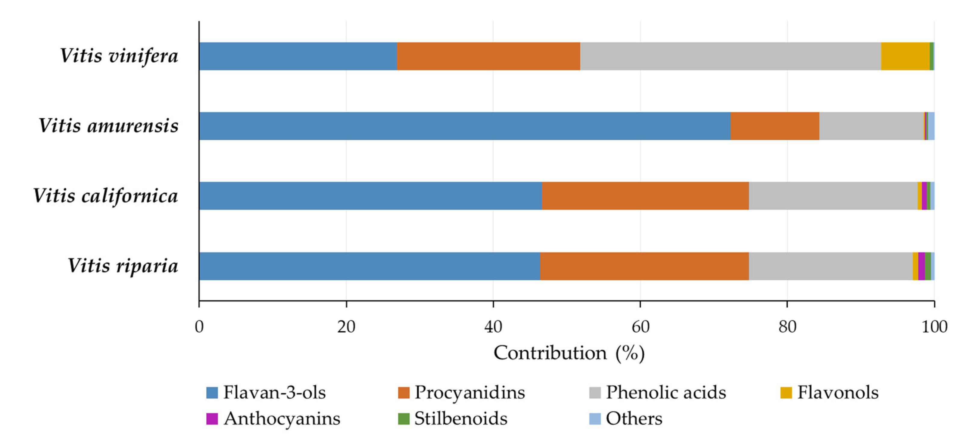
The contribution of various classes of phenolics in grape seed extracts to the total phenolics is shown in Figure 2. The proportions of phenolics in V. riparia and V. californica seed extracts were similar. For both species, flavan-3-ols, procyanidins, and phenolic acids accounted for approximately 46%, 28%, and 22%, respectively. The contributions of flavonols, anthocyanins, and stilbenoids were 0.59–0.71%, 0.65–0.94%, and 0.54–0.76%, respectively. The V. amurensis seed extract showed a higher prevalence of flavan-3-ols (72.32%) and lower contributions from other classes of phenolics, including procyanidins (12.06%) and phenolic acids (14.17%). Flavonols, anthocyanins, and stilbenoids occurred in a low percentage (below 0.3%). The extracts of the seeds of V. vinifera had a slightly different composition, with a relatively high proportion of phenolic acids (41.02%) and flavonols (6.55%) and a low contribution of flavan-3-ols (26.86%) compared to the other extracts.
The high contribution of flavan-3-ols and their dimers to the profile of phenolic compounds, as well as the dominant proportion of gallic acid among phenolic acids, was consistent with the literature data reported for seeds of V. vinifera varieties and their extracts. Peixoto and co-workers found that 8.3 mg/g of flavan-3-ols and procyanidins (81.4%), 1.3 mg/g of flavonols (12.7%), and 0.6 mg/g of gallic acid and gallic acid glucoside (5.9%) contributed to the total content of phenolic compounds at 10.2 mg/g in the seed extract [12]. An even lower share of gallic acid and its glucoside (1.8–3.9%) was determined by Krasteva and co-workers in the seeds of several V. vinifera cultivars [42]. Pantelić reported large disproportions between phenolic acids and flavan-3-ols in the seeds of Serbian V. vinifera cultivars; their contribution accounted for 6.02–59.9% and 36.6–92.3% of total phenolics, respectively, whereas 0.5–3.5% was noted for the share of flavonols [39]. The ratio of flavan-3-ols to procyanidins has usually been reported as approximately 1:1 [34,40,42], but a greater proportion of the former [3] as well as the latter [12] has also been shown. Liang and co-workers compared the phenolic compound profiles of seeds of 17 Vitis species and several accessions for each species and found that, on average, 46.9% of the phenolic compounds were flavan-3-ols, the contribution of procyanidin dimers and trimers was 35.1% and 14.4%, respectively, and the remaining compounds (gallic acid and its derivatives and flavonols) accounted for less than 4% [34]. The authors also showed that the total phenolic content was the main driver of variation among species (84.5% of the total variation); however, the variation among accessions within species was also significant [34]. Flavonols and stilbenoids were the classes of phenolics that significantly differentiated grapevines within species and accessions. In turn, while analyzing the metabolomic profiles of grapes of American Vitis species and V. vinifera, Narduzzi and co-workers concluded that flavonols differentiated the phenolics of grape skins [32]. In the seeds, V. vinifera varieties had a higher content of flavan-3-ols and oligomeric procyanidins than wild American grapes. The differentiating factor was also the content of gallic acid and its derivatives, the accumulation of which was higher in the seeds of American Vitis species, although some varieties of V. vinifera with a high content of compounds of this class were also found. Our study demonstrated a high contribution of flavan-3-ols to the phenolic profile of V. amurensis seed extracts, a high contribution of phenolic acids and flavonols to the profile of V. vinifera seed extract, as well as a high proportion of anthocyanins and stilbenoids in the profiles of extracts of V. riparia and V. californica seeds (Figure 2). These seem to be the main factors differentiating the Vitis species.
The antioxidant activity of the extracts from the seeds of the considered Vitis species can be attributed to the high contents of flavan-3-ols, procyanidins, and gallic acid, the more so that these compounds, especially procyanidins and proanthocyanidins, are known for their high antioxidant activity [44,45]. They are excellent free radical scavengers, and their antioxidant potential is higher than that of tocopherols and ascorbic acid. A previous study has shown that flavan-3-ols and procyanidins are responsible for the antioxidant activity of the seeds of V. vinifera and other Vitis species [35,37,38]. Flavan-3-ols and procyanidins also have high antimicrobial activity and, in addition, can act synergistically with other antibacterial agents [29,46]. Due to their ability to bind to proteins, they can inactivate bacterial enzymes, interact with cell wall proteins, transporter proteins, penicillin-binding proteins, and other surface-adhesion proteins, and consequently promote cell death [29,30]. Gallic acid may also be involved in the antimicrobial effects of grape seed extracts [30].

2.5. Overall Rate of Results with Principal Component Analysis

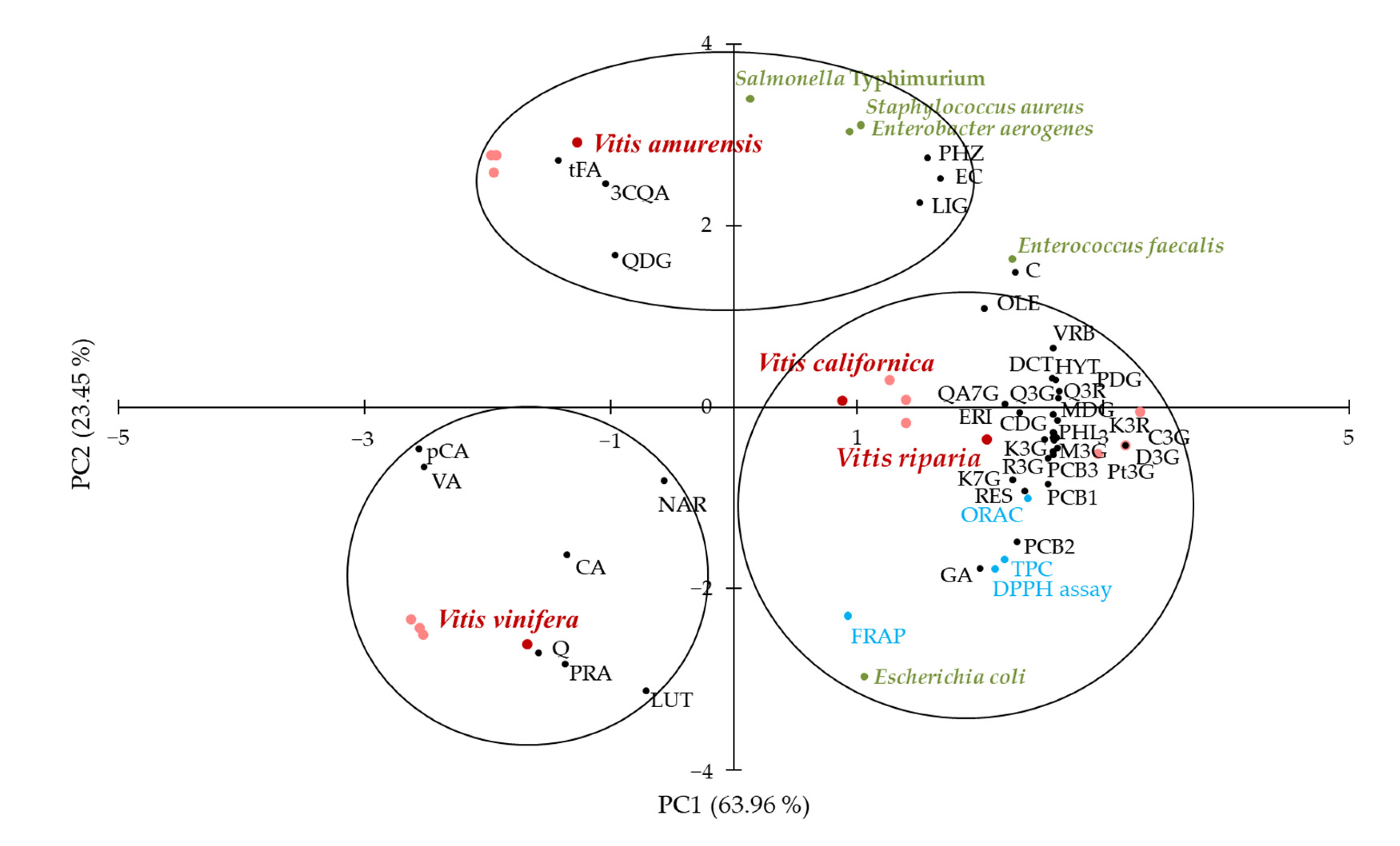
Principal component analysis (PCA) was performed to identify possible relationships between Vitis species and variables, i.e., TPC, antioxidant activity, antimicrobial activity, and contents of individual phenolics. The distribution of samples and variables in the PCA plot is shown in Figure 3.
The two first principal components (PC1 and PC2) explained 87.41% of the total variance. A coherent segregation between grape seed extracts was observed. There was discrimination along PC1 between clustered V. riparia, V. californica, and other Vitis species and along PC2 between V. vinifera and V. amurensis. The clustering of V. riparia and V. californica with TPC, antioxidant activity determined in all assays (FRAP, ORAC, DPPH), antimicrobial activity against E. coli, and most of the individual phenolic contents were evident. It was noteworthy that this group of variables included all detected procyanidins (B1–B3), anthocyanins (cyanidin 3-O-glucoside, cyanidin 3,5-O-diglucoside, delphinidin 3-O-glucoside, peonidin 3,5-O-diglucoside, malvidin 3-O-glucoside, malvidin 3,5-O-diglucoside, and petunidin 3-O-glucoside), stilbenoids (resveratrol and its glucoside), as well as most flavonols (quercetin 3-O-glucoside, quercetin 3-O-rutinoside, quercetagetin 7-O-glucoside, kaempferol 3-O-glucoside, kaempferol 7-O-glucoside, kaempferol 3-O-rutinoside). Gallic acid, the most abundant phenolic acid in Vitis species seed extracts, was also related to this group. However, other phenolic acids were associated with V. vinifera (protocatechuic, caffeic, vanillic, and p-coumaric acids) and V. amurensis (3-O-caffeoylquinic and trans-ferulic acids). The second of these was also clustered with antimicrobial activity against S. Typhimurium, E. aerogenes, and S. aureus. The antimicrobial activity against E. faecalis and content of (+)-catechin were between two clusters on the PCA plot; they were related to both groups but less so. Quercetin, whose content in the extracts was the highest among flavonols, was grouped with V. vinifera.
The clustering of TPC, ORAC, DPPH scavenging activity, and antimicrobial activity against E. coli in PCA is in line with the results of the Pearson’s correlation analysis, showing significant positive correlations among these variables (Table 2). Similarly, discrimination between antimicrobial activity against E. coli and against other bacteria along PC1 is consistent with significant negative Pearson correlations. In turn, the clustering of all anthocyanins and stilbenoids with V. californica and V. riparia, flavan-3-ols with V. amurensis, and most of the phenolic acids with V. vinifera confirmed our supposition mentioned above that the compounds of these classes were mainly responsible for the differentiation of the Vitis species phenolic profile.

3. Materials and Methods

3.1. Plant Material, Chemicals and Reagents

The experiments were conducted on Vitis riparia Michx., Vitis californica Benth., Vitis amurensis Rupr. and Vitis vinifera L. seeds supplied by Sandeman Seeds (Lalongue, France).
All standards and reagents were of analytical grade. Hexane, methanol, Folin-Ciocalteu’s reagent, gallic acid, sodium carbonate, tripyridyltriazine, Trolox, 2,2-diphenyl-1-picrylhydrazyl (DPPH) radical, 2,2′-azobis(2-amidinopropane) dihydrochloride (AAPH), phosphate buffered saline (PBS), and LC-MS/MS standards including gallic acid (GA), hydroxytyrosol (HYT), protocatechuic acid (PRA), cyanidin 3,5-O-diglucoside (CDG), delphinidin 3-O-glucoside (D3G), peonidin 3,5-O-diglucoside (PDG), malvidin 3,5-O-diglucoside (MDG), cyanidin 3-O-glucoside (C3G), procyanidin B1 (PCB1), petunidin 3-O-glucoside (Pt3G), caffeic acid (CA), procyanidin B3 (PCB3), vanillic acid (VA), malvidin 3-O-glucoside (M3G), procyanidin B2 (PCB2), quercetagetin 7-O-glucoside (QA7G), quercetin 3-O-glucoside (Q3G), verbascoside (VER), kaempferol 3-O-rutinoside (K3R), kaempferol 7-O-glucoside (K7G), resveratrol 3-O-glucoside (R3G), oleuropein (OLE), ligstroside (LIG), luteolin (LUT), eriodictyol (ERI), naringenin (NAR), and phloretin (PHL) were purchased from Sigma-Aldrich, Inc. (St. Louis, MO, USA). The other chromatographic standards, i.e., 3-O-caffeoylquinic acid (3CQA), (+)-catechin (C), (−)-epicatechin (EC), quercetin 3,4-O-diglucoside (QDG), quercetin 3-O-rutinoside (Q3R), p-coumaric acid (pCA), trans-ferulic acid (tFA), 2,3-dicaffeoyl-tartaric acid (DCT), kaempferol 3-O-glucoside (K3G), phloridzin (PHZ), resveratrol (RES), and quercetin (Q) were acquired from Extrasynthese (Genay, France). The bacterial media Mueller Hinton Broth (MHB) and Mueller Hinton Agar (MHA) were purchased from VWR (Radnor, PA, USA).

3.2. Extract Preparation

Seeds were ground in a coffee mill (Bosch & Siemens Hausgeräte GmbH, Munich, Germany) into powder with a particle size <0.8 mm and defatted with hexane in a Soxhlet apparatus for 6–8 h. Powdered defatted material was extracted using 80% (v/v) methanol at a solid to solvent ratio of 1:10 (w/v) at 65 °C for 15 min using an SW22 water bath (Julabo, Seelbach, Germany) [47,48]. The process was carried out three times, and the supernatants from each step were pooled. Then, the organic solvent was evaporated under vacuum at 40 °C, and the aqueous residue was freeze-dried using the Lyph Lock 6 freeze-dry system (Labconco, Kansas City, MO, USA). Three extracts were obtained for each seed type. The prepared extracts were stored at −20 °C until analysis.

3.3. Determination of the Total Phenolic Content

An assay with Folin-Ciocalteu’s reagent was performed to determine TPC according to the method described by Singleton and co-workers [49]. A UV/Vis Lambda 365 spectrophotometer (Perkin Elmer, Waltham, MA, USA) was used for absorbance measurement. TPC was expressed as mg of gallic acid equivalent (GAE) per g of extract.

3.4. In Vitro Antioxidant Activity Assays

The antioxidant activity of grape seed extracts was explored as FRAP, DPPH scavenging activity, and ORAC. The Benzie and Strain method was used for FRAP determination [50]. The FRAP reagent was prepared, and a ferric tripyridyltriazine complex was reduced to its ferrous form by aqueous solutions of grape seed extracts (1 mg/mL) exactly as in the original method. Absorbance was measured with a UV/Vis Lambda 365 spectrophotometer (Perkin Elmer) at 593 nm after 30 minutes of color development. The results were calculated based on the standard curve plotted for Trolox and expressed as mg of Trolox equivalent (TE) per g of extract.
The DPPH radical scavenging activity was determined according to the procedure reported by Boudjou and co-workers [51]. The DPPH radicals and the grape seed extracts were dissolved in methanol. After mixing both solutions at a ratio of 39:1 (v/v), the reaction mixture was left in the dark at ambient temperature for 60 min. In parallel, a mixture with methanol instead of the extract solution was prepared. Absorbance was measured at 517 nm with a UV/Vis Lambda 365 spectrophotometer (Perkin Elmer) using methanol as a blank. Trolox was used as a standard, and results were expressed as mg of TE per g of extract.
The ORAC assay was performed as previously detailed by Gabriele and co-workers [29]. Peroxyl radicals were generated by AAPH photolysis in a solution of 0.075 M phosphate buffer (pH 7.4) at 37 °C. Fluorescein sodium salt at a 0.04 mM concentration in the reaction mixture was used as a probe. Fluorescence was read using a VictorTM X3 multilabel plate reader (Perkin Elmer) at 485 nm excitation and 514 nm emission wavelengths. Results were expressed as mg of TE per g of extract.

3.5. Determination of the Antimicrobial Activity

The bacterial strains were supplied by the American Type Culture Collection (ATCC; Manassas, VA, USA). The antimicrobial activity of Vitis species seed extracts was analyzed against three Gram-negative bacteria, specifically Escherichia coli (ATCC 25922), Salmonella enterica ser. Typhimurium (ATCC 14028), and Enterobacter aerogenes (ATCC 13048), and two Gram-positive bacteria, i.e., Enterococcus faecalis (ATCC 29212) and Staphylococcus aureus (ATCC 25923). The effect of extracts on selected bacterial growth was determined according to the procedure detailed in a previous paper of ours [52], with some modifications. The bacteria were cultured in MHB at 37 °C for 16 h and diluted to match the turbidity of the 0.5 McFarland standard. Then, bacterial suspensions contained about 1–5 × 10⁵ CFU/mL (50 µL) MHB (100 µL), and grape seed extract solutions with concentrations of 0.063, 0.125, 0.25, 0.5, 1, and 2.5 mg/mL (100 µL) were pipetted into a 96-well plate. Parallel, a control with water instead of the extract solution was added to the plate. The optical density (O.D.) was measured at 630 nm using a microplate reader (Eti-System fast reader, Sorin Biomedica, Modena, Italy) after plate incubation at 37 °C for 24 h. Results were expressed as final growth (O.D.), which referred to the final density of the bacteria incubated with or without extracts. In addition, the inhibition rate was calculated according to Equation (1). This parameter was used in the Pearson’s correlation and the principal component analyses.
Inhibition rate (%) = (100 − O.D.sample/O.D.control) × 100,
where, O.D.sample is the optical density of the sample with the extract and O.D.control is the optical density of the control without the extract.

3.6. Determination of Phenolic Profile of the Extracts

Liquid chromatography (LC) separation and tandem mass spectrometry (MS/MS) detection of phenolic compounds of grape seed extracts were performed employing a 5500+ QTrap mass spectrometer with a Turbo V ion-spray source (AB Sciex, Framingham, MA, USA) connected with an Exion LC AC system consisting of two ExionLC AC pumps, an autosampler, controller, degasser, and tray (Shimadzu, Kyoto, Japan). The extract solutions were injected into a Kinetex Byphenyl column (2.1 × 100 mm, 2.6 µm particle size; Phenomenex, Torrance, CA, USA), and elution was performed in a gradient mode using (A) water containing 0.1% (v/v) formic acid and (B) acetonitrile containing 0.1% (v/v) formic acid. The gradient program was as follows: 0–10.0 min, 5–95% B; 10.0–12.0 min, 95% B; 12.0–12.1 min, 95–5% B; and 12.1–16.0 min, 5% B. The flow rate of the mobile phase was 0.4 mL/min. The MS/MS operation source parameters were as follows: nebulizer gas (GS1), 70 (arbitrary units); turbo gas (GS2), 50 (arbitrary units); curtain gas (CUR), 10 (arbitrary units); temperature, 500 °C; ion spray voltage (IS), −4500 V (for phenolics excluding anthocyanins, negative ion mode) or +5500 V (for anthocyanins, positive ion mode); entrance potential (EP), 10 V; and dwell time, 20 ms. Nitrogen was used as a collision gas. Compound-dependent parameters, including declustering potential (DP), collision energy (CE), and collision cell exit potential (CXP), were adjusted for the specific selected reaction monitoring (SRM) transition for each compound (Table S1, Supplementary Materials). Analyst 1.7.3 software and OS 1.7 software (AB Sciex) were used for data collection and processing, respectively. Qualitative confirmation was achieved using information-dependent acquisition (IDA) criteria, taking advantage of the ion trap functionalities of the 5500+ QTrap to switch from SRM to enhanced product ions (EPI), obtaining the MS/MS spectrum using a CE of 35 eV with a CE spread of 15 eV [53]. The spectra were compared with a custom-made spectral library. The data were normalized according to matrix effect and recovery percentage. The matrix effect was calculated as the peak area of the sample spiked after extraction per peak area of the standard, while recovery was calculated as the peak area of the sample spiked before extraction per peak area of the sample spiked after extraction. Calibration curves for quantitative analysis were built using a standard mix containing all the analytes at concentrations of 0.5, 1, 2, 4, 8, 16, 32, 64, 128, and 256 ng/mL.

3.7. Statistical Analysis

The data were reported as the mean and standard deviation of triplicates. The statistically significant (p < 0.05) differences between extracts were valued with the analysis of variance (ANOVA) and Tukey’s test. Pearson’s correlation analysis was conducted in order to evaluate the correlation between variables (TPC, antioxidant activity, and antimicrobial activity). In turn, the principal component analysis (PCA) was applied as the analysis of multivariate data to characterize and separate Vitis species in relation to the tested variables (TPC, antioxidant activity, antimicrobial activity, and contents of individual phenolics). Analyses were performed using XLSTAT software (version 2019).

4. Conclusions

The extracts from seeds of V. californica and V. riparia had 1.2–2.6 times higher TPC and 1.4–4.2 times higher antiradical activity than those from V. vinifera and V. amurensis seeds. All extracts showed antimicrobial activity against both Gram-negative and Gram-positive bacteria, and those from V. californica and V. riparia seeds exhibited, generally, a higher ability to inhibit bacteria growth than the extracts from the other Vitis species tested. It should be noted, however, that the species of bacteria significantly determined the effectiveness of the extracts as antimicrobial agents. Their particularly low concentrations were sufficient to inhibit the growth of S. aureus. PCA confirmed the effect of the species on TPC, antioxidant, and antibacterial activities, underscoring that V. riparia and V. californica seed extracts are especially interesting in this respect. Mainly flavan-3-ols, procyanidins, phenolic acids, flavonols, anthocyanins, and stilbenoids were detected in the phenolic profile of the extracts from seeds of Vitis species. Overall, the contents of (+)-catechin, (−)-epicatechin, gallic acid, and procyanidin B2 were the highest. The contribution of phenolics of various classes in the profiles of V. californica and V. riparia seed extracts was comparable to the high proportion of anthocyanins and stilbenoids compared to the other Vitis species. Other classes of phenolics differentiating Vitis species were flavan-3-ols (high contribution in the phenolic profile of V. amurensis seeds) and phenolic acids (dominating in V. vinifera seed extracts).
The study has shown the viability of not only V. vinifera seed extract but also extracts from other Vitis species, especially V. riparia and V. californica, in the food industry as functional food additives, food preservatives, and ingredients of innovative food packaging materials, as well as in the pharmaceutical and cosmetic industries due to their antioxidant and antimicrobial activity. The results of this study highlight the importance of conducting further research to screen the bioactive properties of different wild Vitis species and select the most suitable species for developing new products with antioxidant and antibacterial potential.

Supplementary Materials

The following supporting information can be downloaded at: https://www.mdpi.com/article/10.3390/molecules28134924/s1, Table S1: The retention times (tR), selected reaction monitoring (SRM) transitions, and relative MS/MS parameters of phenolic compounds identified in extracts from seeds of Vitis species.

Author Contributions

Conceptualization, L.P., S.W., R.A. and M.K.; methodology, L.P., A.R. and M.K.; validation, L.P. and A.R.; formal analysis, T.G.; investigation, T.G.; resources, S.W.; data curation, L.P.; writing—original draft preparation, L.P. and M.K.; writing—review and editing, L.P. and M.K.; visualization, L.P. and M.K.; supervision, R.A.; project administration, V.L.; funding acquisition, V.L. All authors have read and agreed to the published version of the manuscript.

Funding

This research received no external funding.

Institutional Review Board Statement

Not applicable.

Informed Consent Statement

Not applicable.

Data Availability Statement

Data is contained within the article.

Conflicts of Interest

The authors declare no conflict of interest.

Sample Availability

Not applicable.

References

  1. Rao, M.; Bast, A.; de Boer, A. Valorized Food Processing By-Products in the EU: Finding the Balance between Safety, Nutrition, and Sustainability. Sustainability 2021, 13, 4428. [Google Scholar] [CrossRef]
  2. Troilo, M.; Difonzo, G.; Paradiso, V.M.; Summo, C.; Caponio, F. Bioactive Compounds from Vine Shoots, Grape Stalks, and Wine Lees: Their Potential Use in Agro-Food Chains. Foods 2021, 10, 342. [Google Scholar] [CrossRef]
  3. Maier, T.; Schieber, A.; Kammerer, D.R.; Carle, R. Residues of Grape (Vitis vinifera L.) Seed Oil Production as a Valuable Source of Phenolic Antioxidants. Food Chem. 2009, 112, 551–559. [Google Scholar] [CrossRef]
  4. Beres, C.; Costa, G.N.S.; Cabezudo, I.; da Silva-James, N.K.; Teles, A.S.C.; Cruz, A.P.G.; Mellinger-Silva, C.; Tonon, R.V.; Cabral, L.M.C.; Freitas, S.P. Towards Integral Utilization of Grape Pomace from Winemaking Process: A Review. Waste Manag. 2017, 68, 581–594. [Google Scholar] [CrossRef]
  5. Heuzé, V.; Tran, G.; Grape Seeds and Grape Seed Oil Meal. Feedipedia, a Programme by INRAE, CIRAD, AFZ and FAO. Available online: https://www.feedipedia.org/node/692 (accessed on 21 March 2023).
  6. Lutterodt, H.; Slavin, M.; Whent, M.; Turner, E.; Yu, L. Fatty Acid Composition, Oxidative Stability, Antioxidant and Antiproliferative Properties of Selected Cold-Pressed Grape Seed Oils and Flours. Food Chem. 2011, 128, 391–399. [Google Scholar] [CrossRef]
  7. Kowalska, H.; Czajkowska, K.; Cichowska, J.; Lenart, A. What’s New in Biopotential of Fruit and Vegetable By-Products Applied in the Food Processing Industry. Trends Food Sci. Technol. 2017, 67, 150–159. [Google Scholar] [CrossRef]
  8. Kapcsándi, V.; Lakatos, E.H.; Sik, B.; Linka, L.Á.; Székelyhidi, R. Antioxidant and Polyphenol Content of Different Vitis vinifera Seed Cultivars and Two Facilities of Production of a Functional Bakery Product. Chem. Pap. 2021, 75, 5711–5717. [Google Scholar] [CrossRef]
  9. Kuchtová, V.; Kohajdová, Z.; Karovičová, J.; Lauková, M. Physical, Textural and Sensory Properties of Cookies Incorporated with Grape Skin and Seed Preparations. Pol. J. Food Nutr. Sci. 2018, 68, 309–317. [Google Scholar] [CrossRef]
  10. Kaynarca, G.B.; Kamer, D.D.A.; Yucel, E.; Gümüş, T. Proposed Use of a Polyvinyl Alcohol with Grape Pomace Extract as an Edible Coating for Strawberries. Pol. J. Food Nutr. Sci. 2023, 73, 151–162. [Google Scholar] [CrossRef]
  11. Vostrejs, P.; Adamcová, D.; Vaverková, M.D.; Enev, V.; Kalina, M.; Machovsky, M.; Šourková, M.; Marova, I.; Kovalcik, A. Active Biodegradable Packaging Films Modified with Grape Seeds Lignin. RSC Adv. 2020, 10, 29202–29213. [Google Scholar] [CrossRef]
  12. Peixoto, C.M.; Dias, M.I.; Alves, M.J.; Calhelha, R.C.; Barros, L.; Pinho, S.P.; Ferreira, I.C.F.R. Grape Pomace as a Source of Phenolic Compounds and Diverse Bioactive Properties. Food Chem. 2018, 253, 132–138. [Google Scholar] [CrossRef] [Green Version]
  13. Rockenbach, I.I.; Jungfer, E.; Ritter, C.; Santiago-Schübel, B.; Thiele, B.; Fett, R.; Galensa, R. Characterization of Flavan-3-ols in Seeds of Grape Pomace by CE, HPLC-DAD-MSn and LC-ESI-FTICR-MS. Food Res. Int. 2012, 48, 848–855. [Google Scholar] [CrossRef] [Green Version]
  14. Rodríguez-Pérez, C.; García-Villanova, B.; Guerra-Hernández, E.; Verardo, V. Grape Seeds Proanthocyanidins: An Overview of in Vivo Bioactivity in Animal Models. Nutrients 2019, 11, 2435. [Google Scholar] [CrossRef] [Green Version]
  15. Pugajeva, I.; Perkons, I.; Górnaś, P. Identification and Determination of Stilbenes by Q-TOF in Grape Skins, Seeds, Juice and Stems. J. Food Compos. Anal. 2018, 74, 44–52. [Google Scholar] [CrossRef]
  16. Silva, V.; Igrejas, G.; Falco, V.; Santos, T.P.; Torres, C.; Oliveira, A.M.P.; Pereira, J.E.; Amaral, J.S.; Poeta, P. Chemical Composition, Antioxidant and Antimicrobial Activity of Phenolic Compounds Extracted from Wine Industry By-Products. Food Control 2018, 92, 516–522. [Google Scholar] [CrossRef] [Green Version]
  17. Gengaihi, S.E.; Ella, F.M.A.; Emad, M.H.; Shalaby, E.; Doha, H. Antioxidant Activity of Phenolic Compounds from Different Grape Wastes. J. Food Process. Technol. 2014, 5, 296. [Google Scholar] [CrossRef] [Green Version]
  18. Tang, G.Y.; Zhao, C.N.; Liu, Q.; Feng, X.L.; Xu, X.Y.; Cao, S.Y.; Meng, X.; Li, S.; Gan, R.Y.; Li, H.B. Potential of Grape Wastes as a Natural Source of Bioactive Compounds. Molecules 2018, 23, 2598. [Google Scholar] [CrossRef] [Green Version]
  19. Terra, X.; Pallarés, V.; Ardèvol, A.; Bladé, C.; Fernández-Larrea, J.; Pujadas, G.; Salvadó, J.; Arola, L.; Blay, M. Modulatory Effect of Grape-Seed Procyanidins on Local and Systemic Inflammation in Diet-Induced Obesity Rats. J. Nutr. Biochem. 2011, 22, 380–387. [Google Scholar] [CrossRef]
  20. Pascual-Serrano, A.; Bladé, C.; Suárez, M.; Arola-Arnal, A. Grape Seed Proanthocyanidins Improve White Adipose Tissue Expansion during Diet-Induced Obesity Development in Rats. Int. J. Mol. Sci. 2018, 19, 2632. [Google Scholar] [CrossRef] [Green Version]
  21. Soto, M.U.R.; Brown, K.; Ross, C.F. Antioxidant Activity and Consumer Acceptance of Grape Seed Flour-Containing Food Products. Int. J. Food Sci. Technol. 2012, 47, 592–602. [Google Scholar] [CrossRef]
  22. Lorenzo, J.M.; Sineiro, J.; Amado, I.R.; Franco, D. Influence of Natural Extracts on the Shelf Life of Modified Atmosphere-Packaged Pork Patties. Meat Sci. 2014, 96, 526–534. [Google Scholar] [CrossRef]
  23. Libera, J.; Latoch, A.; Wójciak, K.M. Utilization of Grape Seed Extract as a Natural Antioxidant in the Technology of Meat Products Inoculated with a Probiotic Strain of LAB. Foods 2020, 9, 103. [Google Scholar] [CrossRef] [Green Version]
  24. Blakeney, M. Food Loss and Food Waste: Causes and Solutions; Edward Elgar Publishing: Cheltenham, UK, 2019; pp. 1–225. [Google Scholar]
  25. Gomomo, Z.; Fanadzo, M.; Mewa-Ngongang, M.; Hoff, J.W.; van der Rijst, M.; Okudoh, V.I.; Kriel, J.; du Plessis, H.W. Control of Mould Spoilage on Apples Using Yeasts as Biological Control Agents. Pol. J. Food Nutr. Sci. 2022, 72, 119–128. [Google Scholar] [CrossRef]
  26. Lianou, A.; Panagou, E.Z.; Nychas, G.J.E. Microbiological Spoilage of Foods and Beverages. In The Stability and Shelf Life of Food, 2nd ed.; Subramaniam, P., Ed.; Woodhead Publishing: Duxford, UK, 2016; pp. 3–42. [Google Scholar]
  27. Kowalska, J.; Maćkiw, E.; Korsak, D.; Postupolski, J. Characteristic and Antimicrobial Resistance of Bacillus Cereus Group Isolated from Food in Poland. Pol. J. Food Nutr. Sci. 2022, 72, 297–304. [Google Scholar] [CrossRef]
  28. Kirk, M.D.; Angulo, F.J.; Havelaar, A.H.; Black, R.E. Diarrhoeal Disease in Children due to Contaminated Food. Bull. World Health Organ. 2017, 95, 233–234. [Google Scholar] [CrossRef]
  29. Mattos, G.N.; Tonon, R.V.; Furtado, A.A.L.; Cabral, L.M.C. Grape By-Product Extracts against Microbial Proliferation and Lipid Oxidation: A Review. J. Sci. Food Agric. 2017, 97, 1055–1064. [Google Scholar] [CrossRef]
  30. Silva, A.; Silva, V.; Igrejas, G.; Gaivão, I.; Aires, A.; Klibi, N.; Dapkevicius, M.d.L.E.; Valentão, P.; Falco, V.; Poeta, P. Valorization of Winemaking By-Products as a Novel Source of Antibacterial Properties: New Strategies to Fight Antibiotic Resistance. Molecules 2021, 26, 2331. [Google Scholar] [CrossRef]
  31. Chen, Q.; Diao, L.; Song, H.; Zhu, X. Vitis amurensis Rupr: A Review of Chemistry and Pharmacology. Phytomedicine 2018, 49, 111–122. [Google Scholar] [CrossRef]
  32. Narduzzi, L.; Stanstrup, J.; Mattivi, F. Comparing Wild American Grapes with Vitis vinifera: A Metabolomics Study of Grape Composition. J. Agric. Food Chem. 2015, 63, 6823–6834. [Google Scholar] [CrossRef]
  33. Howard, J.L. Vitis californica. In Fire Effects Information System; U.S. Department of Agriculture, Forest Service, Rocky Mountain Research Station, Fire Sciences Laboratory (Producer): Missoula, MT, USA, 1993; Available online: https://www.fs.usda.gov/database/feis/plants/vine/vitcal/all.html (accessed on 15 June 2023).
  34. Liang, Z.; Yang, Y.; Cheng, L.; Zhong, G.Y. Characterization of Polyphenolic Metabolites in the Seeds of Vitis Germplasm. J. Agric. Food Chem. 2012, 60, 1291–1299. [Google Scholar] [CrossRef]
  35. Weidner, S.; Rybarczyk, A.; Karamać, M.; Król, A.; Mostek, A.; Grębosz, J.; Amarowicz, R. Differences in the Phenolic Composition and Antioxidant Properties between Vitis coignetiae and Vitis vinifera Seeds Extracts. Molecules 2013, 18, 3410–3426. [Google Scholar] [CrossRef]
  36. Weidner, S.; Powałka, A.; Karamać, M.; Amarowicz, R. Extracts of Phenolic Compounds from Seeds of Three Wild Grapevines—Comparison of Their Antioxidant Activities and the Content of Phenolic Compounds. Int. J. Mol. Sci. 2012, 13, 3444–3457. [Google Scholar] [CrossRef] [Green Version]
  37. Zhu, L.; Li, X.; Hu, X.; Wu, X.; Liu, Y.; Yang, Y.; Zang, Y.; Tang, H.; Wang, C.; Xu, J. Quality Characteristics and Anthocyanin Profiles of Different Vitis amurensis Grape Cultivars and Hybrids from Chinese Germplasm. Molecules 2021, 26, 6696. [Google Scholar] [CrossRef]
  38. Poudel, P.R.; Tamura, H.; Kataoka, I.; Mochioka, R. Phenolic Compounds and Antioxidant Activities of Skins and Seeds of Five Wild Grapes and Two Hybrids Native to Japan. J. Food Compos. Anal. 2008, 21, 622–625. [Google Scholar] [CrossRef]
  39. Pantelić, M.M.; Dabić Zagorac, D.; Davidović, S.M.; Todić, S.R.; Bešlić, Z.S.; Gašić, U.M.; Tešić, Ž.L.; Natić, M.M. Identification and Quantification of Phenolic Compounds in Berry Skin, Pulp, and Seeds in 13 Grapevine Varieties Grown in Serbia. Food Chem. 2016, 211, 243–252. [Google Scholar] [CrossRef]
  40. Jayaprakasha, G.K.; Selvi, T.; Sakariah, K.K. Antibacterial and Antioxidant Activities of Grape (Vitis vinifera) Seed Extracts. Food Res. Int. 2003, 36, 117–122. [Google Scholar] [CrossRef]
  41. Craft, B.D.; Kerrihard, A.L.; Amarowicz, R.; Pegg, R.B. Phenol-Based Antioxidants and the in Vitro Methods Used for Their Assessment. Compr. Rev. Food Sci. Food Saf. 2012, 11, 148–173. [Google Scholar] [CrossRef]
  42. Krasteva, D.; Ivanov, Y.; Chengolova, Z.; Godjevargova, T. Antimicrobial Potential, Antioxidant Activity, and Phenolic Content of Grape Seed Extracts from Four Grape Varieties. Microorganisms 2023, 11, 395. [Google Scholar] [CrossRef]
  43. Radulescu, C.; Buruleanu, L.C.; Nicolescu, C.M.; Olteanu, R.L.; Bumbac, M.; Holban, G.C.; Simal-Gandara, J. Phytochemical Profiles, Antioxidant and Antibacterial Activities of Grape (Vitis vinifera L.) Seeds and Skin from Organic and Conventional Vineyards. Plants 2020, 9, 1470. [Google Scholar] [CrossRef]
  44. Shi, J.; Yu, J.; Pohorly, J.E.; Kakuda, Y. Polyphenolics in Grape Seeds—Biochemistry and Functionality. J. Med. Food 2003, 6, 291–299. [Google Scholar] [CrossRef]
  45. Karamać, M. In-Vitro Study on the Efficacy of Tannin Fractions of Edible Nuts as Antioxidants. Eur. J. Lipid Sci. Technol. 2009, 111, 1063–1071. [Google Scholar] [CrossRef]
  46. Bauza-Kaszewska, J.; Żary-Sikorska, E.; Gugolek, A.; Ligocka, A.; Kosmala, M.; Karlińska, E.; Fotschki, B.; Juśkiewicz, J. Synergistic Antimicrobial Effect of Raspberry (Rubus idaeus L., Rosaceae) Preparations and Probiotic Bacteria on Enteric Pathogens. Pol. J. Food Nutr. Sci. 2021, 71, 51–59. [Google Scholar] [CrossRef]
  47. Orak, H.H.; Karamać, M.; Amarowicz, R.; Orak, A.; Janiak, M.A.; Tenikecier, H.S. Variations of Genotypes of Vicia Species as Influenced by Seed Phenolic Compounds and Antioxidant Activity. Zemdirb. Agric. 2022, 109, 35–42. [Google Scholar] [CrossRef]
  48. Gai, F.; Janiak, M.A.; Sulewska, K.; Peiretti, P.G.; Karamać, M. Phenolic Compound Profile and Antioxidant Capacity of Flax (Linum usitatissimum L.) Harvested at Different Growth Stages. Molecules 2023, 28, 1807. [Google Scholar] [CrossRef]
  49. Singleton, V.L.; Orthofer, R.; Lamuela-Raventós, R.M. Analysis of Total Phenols and Other Oxidation Substrates and Antioxidants by Means of Folin-Ciocalteu Reagent. In Oxidants and Antioxidants Part A; Packer, L., Ed.; Academic Press: Cambridge, MA, USA, 1999; Volume 299, pp. 152–178. [Google Scholar]
  50. Benzie, I.F.F.; Strain, J.J. The Ferric Reducing Ability of Plasma (FRAP) as a Measure of “Antioxidant Power”: The FRAP Assay. Anal. Biochem. 1996, 239, 70–76. [Google Scholar] [CrossRef] [Green Version]
  51. Boudjou, S.; Oomah, B.D.; Zaidi, F.; Hosseinian, F. Phenolics Content and Antioxidant and Anti-Inflammatory Activities of Legume Fractions. Food Chem. 2013, 138, 1543–1550. [Google Scholar] [CrossRef]
  52. Pozzo, L.; Russo, R.; Frassinetti, S.; Vizzarri, F.; Árvay, J.; Vornoli, A.; Casamassima, D.; Palazzo, M.; Della Croce, C.M.; Longo, V. Wild Italian Prunus spinosa L. Fruit Exerts In Vitro Antimicrobial Activity and Protects against In Vitro and In Vivo Oxidative Stress. Foods 2020, 9, 5. [Google Scholar] [CrossRef] [Green Version]
  53. Raffaelli, A.; Saba, A. Ion Scanning or Ion Trapping: Why Not Both? Mass Spectrom. Rev. 2021, 42, 1152–1173. [Google Scholar] [CrossRef]
Figure 1. Final growth expressed as optical density (O.D.) of Staphylococcus aureus (a), Enterococcus faecalis (b), Escherichia coli (c), Enterobacter aerogenes (d) and Salmonella enterica ser. Typhimurium (e) in the presence of extracts from seeds of Vitis riparia (V1), Vitis californica (V2), Vitis amurensis (V3) and Vitis vinifera (V4) at different concentrations. Results are reported as the mean and standard deviation (n = 3). Sings above bars indicate a significant difference compared to control:  p < 0.0001, & p < 0.001, $ p < 0.01 and * p < 0.05.
Molecules 28 04924 g001
Figure 2. Percentage contribution of different classes of phenolics to the total phenolics of seed extracts from different Vitis species.
Molecules 28 04924 g002
Figure 3. Principal component analysis (PCA) plot with distribution of the variables including total phenolic content (TPC), antioxidant activity (DPPH assay, FRAP, and ORAC), antimicrobial activity against Escherichia coli, Salmonella enterica ser. Typhimurium, Enterobacter aerogenes, Staphylococcus aureus, and Enterococcus faecalis, and contents of individual phenolic compounds of the Vitis species seed extracts. The acronyms at the black dots correspond to the names of the compounds given in Table 3. FRAP, ferric-reducing antioxidant power; ORAC, oxygen radical absorbance capacity; DPPH assay, assay with 2,2-diphenyl-1-picrylhydrazyl radical; PC1, first principal component; PC2, second principal component.
Molecules 28 04924 g003
Table 1. Total phenolic content (TPC), ferric-reducing antioxidant power (FRAP), DPPH scavenging activity, and oxygen radical absorbance capacity (ORAC) of extracts from seeds of Vitis species.
Grapevine SpeciesTPC
(mg GAE/g)
FRAP
(mg TE/g)
DPPH Assay
(mg TE/g)
ORAC
(mg TE/g)
Vitis riparia121 ± 12 a40.5 ± 2.7 a232 ± 21 a262 ± 20 a
Vitis californica97.2 ± 7.3 a,b35.5 ± 1.4 a,b221 ± 17 a252 ± 12 a
Vitis amurensis46.6 ± 3.0 c22.4 ± 2.0 b55.2 ± 5.6 c141.1 ± 6.2 b
Vitis vinifera78.7 ± 4.4 b,c35.4 ± 1.4 a,b154 ± 14 b174 ± 13 b
Results were expressed as mean ± standard deviation of three replicates. Values with different superscripts (a–c) by column differ significantly (p < 0.05). GAE, gallic acid equivalent; TE, Trolox equivalent; DPPH, 2,2-diphenyl-1-picrylhydrazyl radical.
Table 2. Coefficients of Pearson’s correlations between antimicrobial activity against selected bacteria, antioxidant activity and total phenolic content of extracts from seeds of different Vitis species.
E. coliS. aureusS. TyphimuriumE. aerogenesE. faecalisTPCFRAPDPPH Assay
S. aureus−0.617
S. Typhimurium−0.8380.926
E. aerogenes−0.6430.9630.895
E. faecalis−0.0210.7720.5150.711
TPC0.812−0.103−0.447−0.1350.518
FRAP0.641−0.492−0.652−0.460−0.0280.624
DPPH assay0.730−0.151−0.476−0.1470.3810.8670.568
ORAC0.6010.093−0.2610.0820.6370.9260.5540.853
Values in bold indicate significant correlations between variables (p < 0.05). Results of antimicrobial activity analyses subjected to correlation analysis were expressed as inhibition rate. TPC, total phenolic content; FRAP, ferric-reducing antioxidant power; ORAC, oxygen radical absorbance capacity; DPPH assay, assay with 2,2-diphenyl-1-picrylhydrazyl radical.
Table 3. Content of individual phenolic compounds in extracts from seeds of Vitis species (mg/100 g).
Compound NameAcronymVitis ripariaVitis californicaVitis amurensisVitis vinifera
Gallic acidGA166 ± 32 a137 ± 18 a44.6 ± 3.3 b105.8 ± 5.1 a,b
Protocatechuic acidPRA5.75 ± 0.38 b6.13 ± 0.13 b4.22 ± 0.18 b39.93 ± 0.83 a
3-O-Caffeoylquinic acid3CQA0.102 ± 0.034 b0.0574 ± 0.0071 b0.207 ± 0.024 a0.0838 ± 0.0079 b
Caffeic acidCA0.0864 ± 0.0063 a0.137 ± 0.026 a0.115 ± 0.023 a0.220 ± 0.090 a
Vanillic acidVA1.418 ± 0.074 c1.56 ± 0.16 c3.33 ± 0.27 b4.25 ± 0.21 a
p-Coumaric acidpCA0.495 ± 0.039 c0.537 ± 0.016 c0.835 ± 0.019 b0.960 ± 0.023 a
trans-Ferulic acidtFA0.225 ± 0.058 b0.239 ± 0.049 b0.607 ± 0.036 a0.267 ± 0.034 b
2,3-Dicaffeoyl-tartaric acidDCT0.0423 ± 0.0029 a0.0280 ± 0.0041 b0.0111 ± 0.0033 c0.0037 ± 0.0005 c
∑ Phenolic acids 174.45145.4053.89151.48
QuercetinQ3.76 ± 0.36 b2.03 ± 0.17 b0.215 ± 0.026 b23.8 ± 2.9 a
Quercetin 3-O-glucosideQ3G0.0752 ± 0.0023 a0.0600 ± 0.0036 b0.0383 ± 0.0037 c0.0355 ± 0.0036 c
Quercetin 3-O-rutinosideQ3R0.705 ± 0.020 a0.446 ± 0.011 b0.1710 ± 0.0080 c0.0865 ± 0.0047 d
Quercetin 3,4-O-diglucosideQDG0.0667 ± 0.0094 a0.0910 ± 0.0084 a0.0932 ± 0.0085 a0.0766 ± 0.0043 a
Quercetagetin 7-O-glucosideQA7G0.471 ± 0.010 b0.695 ± 0.022 a0.120 ± 0.017 c0.0785 ± 0.0033 c
Kaempferol 7-O-glucosideK7G0.1046 ± 0.0096 a0.1099 ± 0.0082 a0.0646 ± 0.0040 b0.0747 ± 0.0027 b
Kaempferol 3-O-glucosideK3G0.312 ± 0.011 a0.217 ± 0.014 b0.0414 ± 0.0026 c0.0539 ± 0.0025 c
Kaempferol 3-O-rutinosideK3R0.098 ± 0.013 a0.0679 ± 0.0044 a0.0057 ± 0.0012 b0.0054 ± 0.0011 b
∑ Flavonols 5.593.720.7524.20
Cyanidin 3-O-glucosideC3G0.1360 ± 0.0033 a0.0699 ± 0.0065 b0.0058 ± 0.0003 c0.0038 ± 0.0003 c
Cyanidin 3,5-O-diglucosideCDG0.809 ± 0.020 a0.381 ± 0.018 b0.0166 ± 0.0029 c0.0024 ± 0.0005 c
Delphinidin 3-O-glucosideD3G0.433 ± 0.027 a0.3411 ± 0.0090 b0.0163 ± 0.0025 c0.0242 ± 0.0033 c
Peonidin 3,5-O-diglucosidePDG1.128 ± 0.088 a0.719 ± 0.014 b0.184 ± 0.012 c0.0041 ± 0.0004 c
Malvidin 3-O-glucosideM3G0.350 ± 0.041 a0.2131 ± 0.0049 b0.0179 ± 0.0017 c0.0391 ± 0.0033 c
Malvidin 3,5-O-diglucosideMDG4.41 ± 0.17 a2.384 ± 0.083 b0.290 ± 0.025 c0.0170 ± 0.0033 c
Petunidin 3-O-glucosidePt3G0.1630 ± 0.0041 a0.0832 ±0.0041 b0.0022 ± 0.0005 c0.0044 ± 0.0005 c
∑ Anthocyanins 7.434.190.530.10
(+)-CatechinC246 ± 35 a206 ± 16 a168 ± 15 a69.8 ± 3.3 b
(−)-EpicatechinEC117 ± 15 a90.7 ± 3.4 a107.2 ± 8.4 a29.4 ± 2.5 b
∑ Flavan-3-ols 363.17296.24275.1399.18
Procyanidin B1PCB151.9 ± 2.4 a40.6 ± 2.5 b4.32 ± 0.35 c13.4 ± 2.2 c
Procyanidin B2PCB2124 ± 11 a104 ± 13 a,b35.8 ± 3.3 c69.3 ± 4.5 b,c
Procyanidin B3PCB346.3 ± 3.1 a34.0 ± 3.3 b5.73 ± 0.53 c9.42 ± 0.85 c
∑ Procyanidins 222.25178.8845.8592.12
Resveratrol 3-O-glucosideR3G2.72 ± 0.39 a1.87 ± 0.23 a0.437 ± 0.025 b0.652 ± 0.030 b
ResveratrolRES3.25 ± 0.21 a1.559 ± 0.065 b0.566 ± 0.030 c1.08 ± 0.17 b,c
∑ Stilbenoids 6.973.431.001.72
HydroxytyrosolHYT0.560 ± 0.033 a0.378 ± 0.022 b0.144 ± 0.011 c0.0395 ± 0.0037 d
VerbascosideVER0.219 ± 0.017 a0.1705 ± 0.0084 b0.0741 ± 0.0040 c0.0123 ± 0.0029 d
OleuropeinOLE0.1089 ± 0.0074 a0.0504 ± 0.0029 b,c0.0638 ± 0.0036 b0.0297 ± 0.0041 c
LigstrosideLIG0.0078 ± 0.0014 a0.0066 ± 0.0012 a0.0074 ± 0.0009 a0.0032 ± 0.0004 a
PhloridzinPHZ2.72 ± 0.17 a2.425 ± 0.062 a2.77 ± 0.16 a0.198 ± 0.025 b
PhloretinPHL0.0304 ± 0.0029 a0.0169 ± 0.0049 b0.0030 ± 0.0013 c0.0027 ± 0.0006 c
LuteolinLUT0.0351 ± 0.0048 b0.0165 ± 0.0029 c0.0051 ± 0.0010 c0.0741 ± 0.0033 a
EriodictyolERI0.0895 ± 0.0090 a0.101 ± 0.015 a0.0395 ± 0.0030 b0.0343 ± 0.0047 b
NaringeninNAR0.105 ± 0.016 a0.118 ± 0.033 a0.111 ± 0.031 a0.1274 ± 0.0090 a
∑ Others 3.873.283.210.52
Results were expressed as the mean ± standard deviation of three replicates. Values with different superscripts (a–d) by row differ significantly (p < 0.05).
Disclaimer/Publisher’s Note: The statements, opinions and data contained in all publications are solely those of the individual author(s) and contributor(s) and not of MDPI and/or the editor(s). MDPI and/or the editor(s) disclaim responsibility for any injury to people or property resulting from any ideas, methods, instructions or products referred to in the content.