Article

In Vivo, In Vitro and In Silico Study of Cucurbita moschata Flower Extract: A Promising Source of Natural Analgesic, Anti-Inflammatory, and Antibacterial Agents

1
Department of Pharmacy, University of Science and Technology, Foy’s Lake, Chittagong 4202, Bangladesh
2
Department of Biochemistry and Molecular Biology, Medical University of South Carolina, Charleston, SC 29425, USA
3
Gwanju Center, Korea Basic Science Institute, Gwanju 61715, Republic of Korea
4
Department of Pharmacy, University of Chittagong, Chittagong 4331, Bangladesh
5
Department of Biological Science, University at Buffalo, Buffalo, NY 14260, USA
6
Department of Biomedical Sciences, Institute for Medical Science, Jeonbuk National University Medical School, Jeonju 54907, Republic of Korea
7
Department of Pharmacy, East West University, Dhaka 1212, Bangladesh
*
Authors to whom correspondence should be addressed.
Molecules 2023, 28(18), 6573; https://doi.org/10.3390/molecules28186573
Submission received: 25 May 2023 / Revised: 2 September 2023 / Accepted: 3 September 2023 / Published: 11 September 2023

Abstract

:
For thousands of years, medicinal plants have played a pivotal role in maintaining human health and improving the quality of human life. This study was designed to analyze the analgesic, anti-inflammatory, and antibacterial potentials of a hydro-methanolic extract of Cucurbita moschata flowers, along with qualitative and quantitative phytochemical screening. The anti-inflammatory effect was tested using the in vitro membrane stabilizing method for human red blood cells (HRBC), the analgesic effect was tested using the in vivo acetic acid-induced writing method, and the antibacterial effect was tested using the disc diffusion method. In silico ADME/T and molecular docking studies were performed to assess the potential of the stated phytochemicals against Cyclooxygenase-II enzyme. Phytochemical screening confirmed the presence of flavonoids, alkaloids, glycosides, tannins, and carbohydrates. The flower extract demonstrated the maximum protection of human red blood cells at 1000 µg/mL, with a 65.73% reduction in hemolysis in a hypotonic solution. The extract also showed significant (p < 0.05) and dose-dependent analgesic effects at oral doses of 200 and 400 mg/kg on the tested animals. Furthermore, the flower extract exhibited potent antibacterial activity due to the disc diffusion method, which was compared with standard ciprofloxacin. In silico testing revealed that 42 phytochemicals exhibited notable pharmacokinetic properties and passed drug likeness screening tests. Among the six best-selected compounds, 3,4-dihydro-2H-pyran-2-yl)methanamine showed the highest binding affinity (−10.1) with significant non-bonding interactions with the target enzyme. In conclusion, the hydro-methanolic extract of Cucurbita moschata was found to be rich in various phytochemicals that may be associated with therapeutic potential, and this study supports the traditional use of Cucurbita moschata flowers in the management of inflammation and painful conditions.

1. Introduction

Pain is commonly understood to be an unpleasant sensory and emotional experience that can be linked to or explained in terms of actual or prospective tissue injuries [1]. It is often triggered by unpleasant stimuli and conveyed to the central nervous system (CNS) via specific neuronal networks where it is regarded intrinsically. It protects the body from harm that may otherwise come to it [2]. On the other hand, inflammation is the body’s protective response to unpleasant stimuli, such as infections and injuries, and it is mediated by both the adaptive and innate immune systems [3,4]. Pain and inflammation tend to be the most unpleasant and severe health concerns that affect 80% of individuals worldwide. This is true despite the fact that suitable drugs are available to treat both conditions [5]. Moreover, globally, pain is a clinical, social, and economic issue. It is estimated that the global cost of pain is as high as 3.0% of GDP (Gross Domestic Product), illustrating the substantial economic impact of pain. In terms of annual costs, pain is more expensive than cardiovascular illnesses or cancers are [6], and it has been shown in studies that physical pain is a reliable predictor of suicidal ideation and behavior [7].
The progression of serious inflammatory diseases, such as rheumatoid arthritis, glomerulonephritis, inflammatory bowel disease, hypersensitivities, hay fever, atherosclerosis, and asthma, is also caused by unresolved inflammation. If they are not treated and managed effectively, these debilitating conditions—which are the leading cause of disabilities—can be fatal [8].
Steroids and Nonsteroidal anti-inflammatory drugs (NSAIDs) are often used to relieve inflammation and pain. By inhibiting Cyclooxygenase (COX) enzymes, especially COX-II, NSAIDs relieve inflammation and pain. However, the prolonged use of NSAIDs has been linked to a variety of adverse liver and GIT (Gastrointestinal tract) complications. Additionally, they have effects on the kidney and the cardiovascular system [9,10]. Since NSAIDs are toxic to pathways, causing oxidative damage, researchers have highlighted the crucial role of antioxidants as natural medications for the remedy of NSAID-induced toxic effects [11,12]. This is why there has been a rise in the use of herbal drugs for the treatment of pain and inflammation. Hence, the approval of traditional South Asian therapeutic plants is gaining ground.
Cucurbita moschata is commonly called ‘MistiKumra’ in Bangla or Pumpkin in English, is an important horticultural crop that belongs to the family Cucurbitaceae [13]. The nutritional, health-protective value and presence of various phytochemicals make this plant interesting to many researchers. In the last few decades, many researchers have studied Cucurbita moschata and have found that Cucurbita moschata has many medicinal applications, such as anthelmintic, anti-diabetes, antibacterial, anticancer, and anti-obesity properties [14]. This plant has also been an integral part of rural as well as some urban diets around the world since antiquity [15]. All of its structural components are consumable, contain minimal calories, and are easily digested [16]. Phytoconstituents like alkaloids, flavonoids, palmitic, oleic, and linoleic acids are present in this plant [17]. In addition, the seeds of this plant are used in the formulation of cosmetic products, while the peel extract is reported to have burn wound healing activity, and the pulp has exhibited several activities, like antioxidant, anti-inflammatory, and anti-angiogenesis ones, along with anti-fatigue activities in mice models [18].
As there is no existing report on the analgesic, anti-inflammatory, and antibacterial effects of the hydro-methanolic extract of Bangladeshi pumpkin flowers, the aim of this study was to investigate its bioactive phytochemicals, analgesic, antibacterial, and anti-inflammatory potential through in vitro, in vivo, and in silico studies.

2. Results

2.1. Phytochemical Screening

2.1.1. Qualitative Screening

A variety of phytochemicals, including alkaloids, flavonoids, phenolic compounds, reducing sugars, glycosides, and tannins, were detected in the flower extracts upon phytochemical screening, as shown in Table 1.

2.1.2. Quantitative Screening

The results obtained reveal that the extract of Cucurbita moschata contains significant amounts of TPC and TFC. The Total Phenolic content was found 18.16 ± 0.28 mg/g gallic acid equivalent in reference to the standard curve (y = 0.001x + 0.013, R² = 0.998), and the total flavonoids content was found 10.42 ± 0.10 mg/g quercetin equivalent in reference to the standard curve (y = 0.001x + 0.045, R² = 0.997) (Table 2).

2.2. Anti-Inflammatory Activity

HRBC Membrane Stabilizing Assay

The hydro-methanolic extract of the Cucurbita moschata flower was investigated for in vitro anti-inflammatory activity. The extract showed maximum protection and minimum hemolysis of the HRBC, which equate to 65.73% and 34.27%, respectively, at a concentration of 1000 µg/mL in a hypotonic solution. Furthermore, at 125 µg/mL, the extract exhibited maximum hemolysis and minimum protection values of 80.35% and 19.65%, respectively. The results were compared with the standard drug diclofenac, which showed a value of 90.89% for protection and a value of 9.11% for hemolysis (Figure 1).

2.3. Analgesic Activity

Acetic Acid-Induced Writhing Test

Table 3 shows the effects of the hydro-methanolic extract of Cucurbita moschata flower on acetic acid-induced writhing among experimental mice. The extract exhibited a significant reduction (p < 0.05) of writhing induced by acetic acid after intraperitoneal administration in a dose-dependent manner. After the oral administration of two different doses, 200 and 400 mg/kg body weight, the percent inhibition was found to be 25.53% and 56.03%, respectively. Standard drug diclofenac sodium (80.85%) was found to be more potent than the plant extracts were.

2.4. Antibacterial Activity

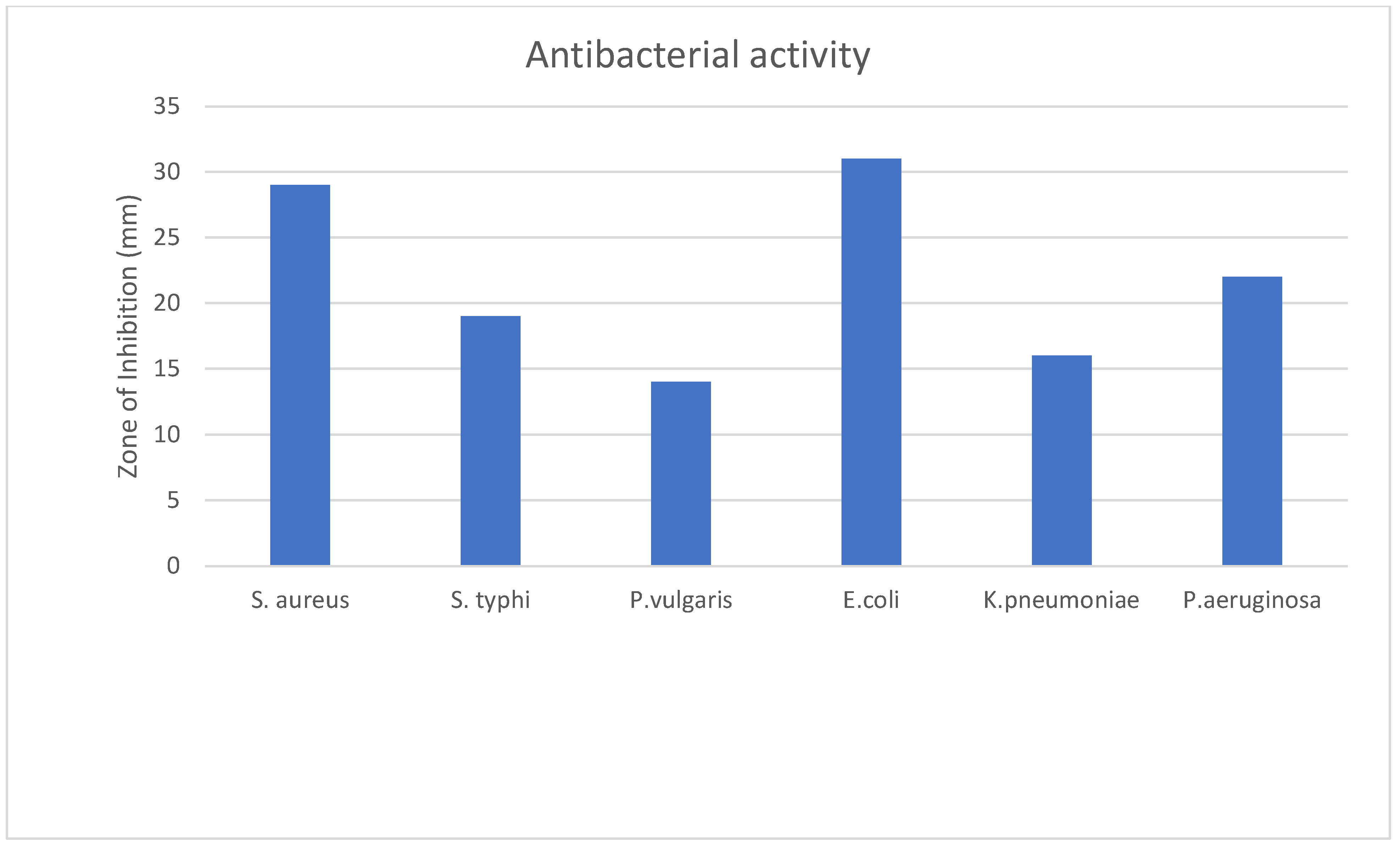
The test agents’ effectiveness as antibacterial substances was evaluated based on their ability to limit bacterial growth around the discs, creating a clear zone of inhibition. Following incubation, the diameter of the zone of inhibition resulting from using the test materials was measured in millimeters using a transparent scale. This provided information on their antimicrobial efficacy, which is recorded in Table 4 and Figure 2.
The Cucurbita moschata flower test sample exhibited antibacterial activity, with a 31 mm inhibition zone observed against E. coli and 29 mm and 21.67 mm inhibition zones against Staphylococcus aureus and Pseudomonas aeruginosa, respectively. Based on the data presented, it can be concluded that the effectiveness of the extract and the standard antimicrobial agent (Ciprofloxacin) varied depending on the bacterial strain tested, where the Cucurbita moschata extract was more effective against Staphylococcus aureus and E. coli than the standard antibiotic was.

2.5. In Silico Study

2.5.1. ADME/T and Drug-Likeness Analysis

In order to demonstrate their potential as therapeutic targets, reported phytochemicals were screened for their pharmacokinetic and drug likeliness qualities prior to docking analysis. The pharmacokinetic profiles of the reported phytochemicals are shown in Table 5, which depict that they do not violate any of the criteria for the Lipinski rule and have no mutagenic and carcinogenic properties. In silico ADMET and drug likeness studies of the compounds present in Cucurbita moschata using pKCSM and SwissADME online tools showed notable results. Of the eighty-four compounds examined, forty-two passed the Lipinski’s rule of five for drug likeness without exhibiting any toxicity. The other compounds which do not follow the Lipinski’s properties and exhibit a poor pharmacokinetic profile with organ toxicity were not considered for further docking studies.

2.5.2. Molecular Docking and Post-Docking Analysis

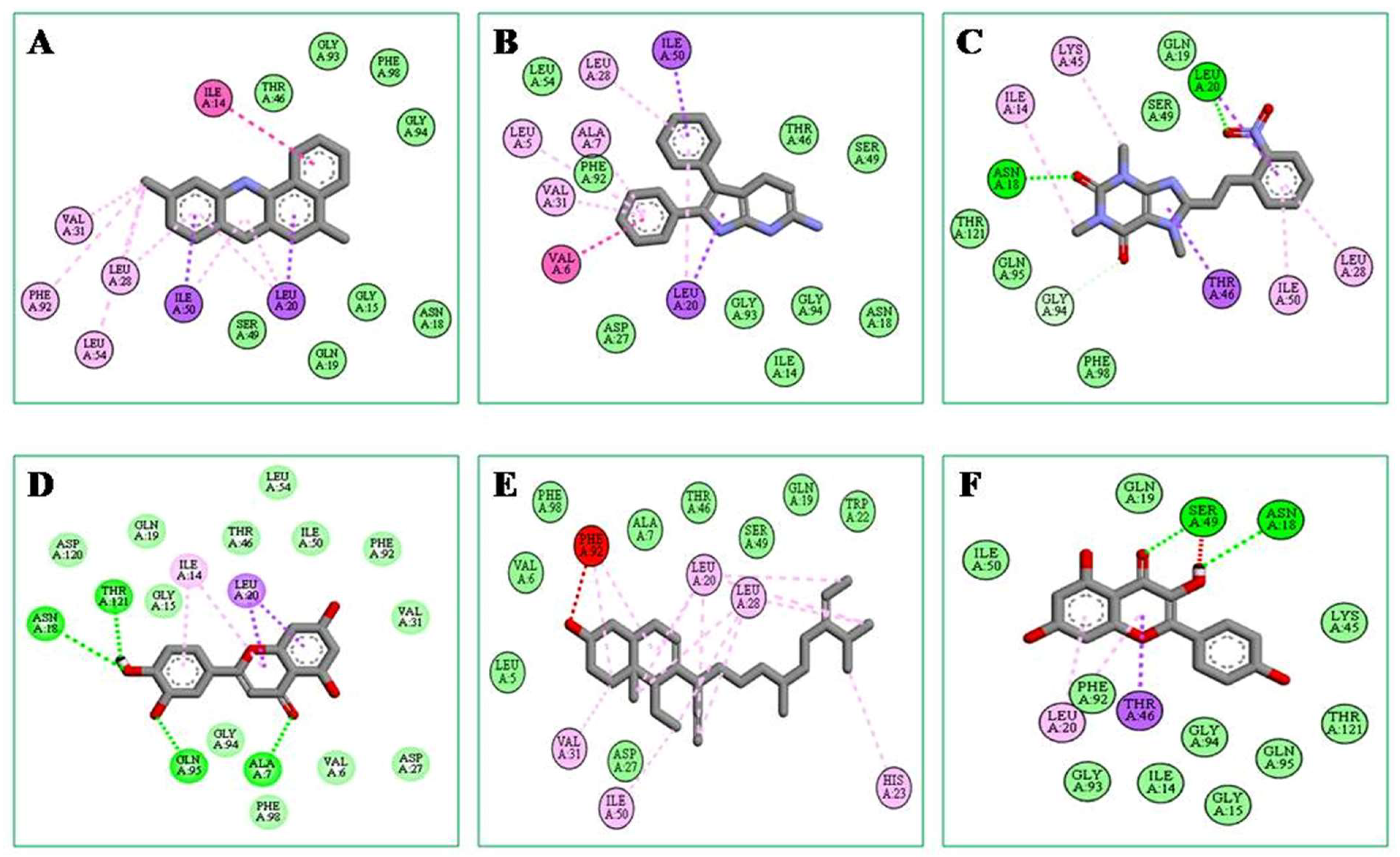
A docking score with non-bonding interactions was used as the parameter for the binding interactions and affinity of the compounds, along with those of standard drugs, Diclofenac and Celecoxib (Table 6). Of the total of forty-two selected compounds, 3,4-dihydro-2H-pyran-2-yl)methanamine (−10.1 kcal/mol) showed the highest binding affinity, which was higher than those of the standard drugs Celecoxib (−9.5 kcal/mol) and Diclofenac (−8.0 kcal/mol). The docking scores of five compounds, i.e., 2,3-diphenyl-1H-pyrrolo[2,3-b]pyridin-6-amine, Oxacyclotridecan-2-one (−9.1), Luteolin (−8.7); (E)-1,3,7-trimethyl-8-(2-nitrostyryl)-3,7-dihydro-1H-purine-2,6-dione (−8.6), and Kaempferol, (−8.4) also showed a promising binding affinity with Cyclooxygenase II enzyme.
Using the Biovia Discovery Studio Visualizer program, the binding interactions between the best-docked molecule and the amino acid residues of the target enzyme shown in Figure 3A–F were investigated further. This analysis showed that 2H-pyran, 3,4-dihydro-2-aminomethyl had a similar binding mode and interaction with COX-2 protein-like diclofenac (Table 5, Figure 2A). The critical evaluation of the docking process showed that 2H-Pyran, 3,4-dihydro-2-aminomethyl formed eight Van der Waals bonds with GLY A:15, ASN A:18, GLN A:19, THR A:46, SER A:49, GLY A:93, GLY A:94, and PHE A:98 and four hydrophobic interactions with LEU A:28, LEU A:54, VAL A:31, and PHE A:92, with short intermolecular distances, which is suggestive that it has a good binding affinity toward the active site of COX-2 enzyme. As hydrophobic interactions are one of the major driving forces of drug-receptor interactions, these bonds with shorter distances (<5 Å) cause strong binds; hence, they have a higher docking score.

3. Discussion

In recent years, natural medicines made from plants have gained popularity for their ability to effectively treat pain and inflammation [19]. In Bangladeshi traditional medicine, various natural substances and formulations have been used to alleviate pain and inflammation [20,21]. In the current study, a hydro-methanolic extract of Cucurbita moschata flowers exhibited a potential anti-inflammatory action during membrane stabilization in an HRBC test in a dose-dependent manner (Figure 1). The extract demonstrated membrane stabilization by inhibiting the hypotonicity-induced lysis of the RBC membrane [21,22], suggesting that the extract could stabilize lysosomal membranes. During an inflammatory reaction, lysosomal components of active neutrophils, such as bactericidal enzymes and proteases, are released, which can damage the tissue further. Therefore, lysosomal stability is crucial in limiting the inflammatory response [23,24]. The phytochemicals present in the Cucurbita moschata flower extract may inhibit these processes and improve the efflux of intracellular components [25].
The acetic acid-induced abdominal contraction response can be effectively studied to detect the peripheral analgesic impact, with specific peritoneal receptors playing a role in the abdominal contraction mechanism [26,27]. The acetic acid-induced writhing method draws out a pain sensation by activating a local inflammatory reaction, which is a consequence of free arachidonic acid being released from tissue phospholipids [28,29]. This reaction leads to an increase in the levels of prostaglandin E2 (PGE2) and prostaglandin F2α (PGF2α) in the peritoneal fluids, as well as lipoxygenase products [30]. The elevated prostaglandin level in the peritoneal cavity causes an increment of pain by expanding the capillary permeability [31].
An agent that reduces the amount of writhing among acetic acid-induced mice provides an analgesic effect, preferably by inhibiting prostaglandin synthesis, a peripheral mechanism of pain inhibition [29,32]. The significant pain reduction observed with the Cucurbita moschata flower extract may be due to the presence of analgesic principles acting on the prostaglandin pathways.
Again, various qualitative phytochemical screening tests can be used to identify the presence of phenols, flavonoids, alkaloids, glycosides, tannins, and carbohydrates. In quantitative phytochemical screening, significant amounts of phenolic and flavonoid contents were identified (Table 2). The analgesic and anti-inflammatory effects of plant-derived phytochemicals like phenols, flavonoids, alkaloids, glycosides, tannins, and carbohydrates have been reported [33].
The extract of the flower exhibited an antibacterial effect in tests conducted on six different strains of bacteria. The efficacy of the test agents in terms of their antibacterial properties were evaluated based on their ability to impede the growth of bacteria surrounding the discs, resulting in a clear zone of inhibition. The presence of phenols, flavonoids, alkaloids, glycosides, and tannins in the extract may account for its antibacterial effect, as these phytochemicals are known to possess antibacterial properties [34,35,36,37,38,39,40].
In silico ADME/T and molecular docking studies are a good way to come up with new drugs for a wide range of diseases. This technique has the advantage of consuming less time and requiring less money than traditional lab experiments do [41]. The phytochemicals reported in Cucurbita moschata [42,43] were evaluated for pharmacokinetic parameters and drug likeliness, and most of the compounds showed notable results (Table 5). Among them, forty-two compounds passed the drug-likeness study with no toxicity. In this research, we used a docking method making use of open software programs as well as virtualized interactions of the ligands 3,4-dihydro-2H-pyran-2-yl)methanamine, Oxacyclotridecan-2-one, Luteolin, and Kaempferol with COX-II enzyme. An important phase in ligand docking in favorable conformations is the formation of non-bonding interactions, including hydrogen bonds, hydrophobic interactions, and Van der Waals interactions with essential amino acids, as revealed by the docking score. According to our findings, each ligand–protein interaction is mediated by a large amount of amino acid residues in the hydrophobic and Van der Waals bond interactions. 3,4-dihydro-2H-pyran-2-yl)methanamine showed the highest docking score, which was higher than the standard drugs, Celecoxib and Diclofenac, with excellent non-bonding interactions with Cyclooxygenase-II enzyme (Figure 2). Additionally, it should be noted that this substance has excellent pharmacokinetic characteristics (Table 5) and also passed the drug-likeness screening test. These observations further confirm that 3,4-dihydro-2H-pyran-2-yl)methanamine may be a more effective anti-inflammatory and analgesic compound, especially with respect to COX-2 protein-mediated inflammation and pain, compared to other traditional NSAIDs.

4. Materials and Methods

4.1. Plant Material

Pumpkin flowers were obtained locally in Chattogram City, Bangladesh, and were authenticated by Dr. Shaikh Bokhtear Uddin, Professor, Department of Botany, University of Chittagong. A specified amount was stored at the Department of Pharmacy, University of Science and Technology Chittagong (USTC).

4.2. Preparation of Plant Extract

The flowers were cleaned properly with running tap water and dried in sunlight after being collected and identified. The flowers were finely powdered using a blender after drying. About 1000 g Cucurbita moschata flowers powder was then extracted with 80% methanol in water for 72 h while continuously stirring with a magnetic stirrer at room temperature. Following that, the flower extract was filtered through a clean cotton filter, and then through Whitman filter paper. The solvent was evaporated with a Rotary evaporator (Lab Tech EV311) at 40 °C under reduced pressure. The extract was then refrigerated (2–8 °C) until further usage.

4.3. Experimental Animals

Swiss albino mice of either sexes weighing 20–30 g were obtained from Animal Research Branches of BCSIR, Chittagong. In the animal house of the Department of Pharmacy, USTC, they were housed in dry and clean iron cages under a 12 h light and dark cycle at 25 ± 3 °C and 45–55% relative humidity. The mice were given free access to water and a typical laboratory diet supplied by the BCSIR laboratory. Food was withdrawn 12 h before the experiment and while it was going on. Given their acute sensitivity to environmental changes, these animals were acclimated to the laboratory for a full week before the trial began. All experiments were conducted in a quiet, soundproofed space. The investigation was permitted by the institutional ethics committee of USTC under ethical approval number USTMEBBC/20/02/05.

4.4. Phytochemical Screening

4.4.1. Qualitative Screening

The presence of different phytochemicals, such as an alkaloid, carbohydrate, flavonoid, glycoside, tannin, steroid, and saponin, was qualitatively assessed in the freshly prepared crude extract. Standard protocols were used to identify them based on distinctive color changes [44].

4.4.2. Quantitative Screening

Total Phenolic Content

The Folin–Ciocalteu technique was used to determine the Total Phenolic content of the extract [45]. Briefly, 200 µL of the crude extract (1 mg/mL) was diluted to 3 mL with distilled water, mixed well with 0.5 mL of Folin–Ciocalteu reagent (10%) for 3 min, and then 2 mL of 20% (w/v) sodium carbonate was added. After another 60 min in the dark, the mixture was checked for absorbance at 650 nm. The calibration curve was used to figure out the Total Phenolic content, which was written as mg of gallic acid equivalent per gram of dry weight.

Total Flavonoid Content

The aluminum chloride colorimetric technique was used to determine the total flavonoid content of crude extract [46]. Briefly, 50 µL of crude extract (1 mg/mL) was converted into 1 mL with methanol and mixed with 4 mL of distilled water and 0.3 mL of 5% NaNO2 solution; 0.3% of 10% AlCl3 solution was added after 5 min of incubation, and the mixture was allowed to stand for 6 min. Then, 2 mL of 1 mol/L NaOH solution was added, and the final volume of the mixture was brought to 10 mL with double distilled water. After allowing the mixture to sit for 5 min, the absorbance was measured at 510 nm. The total flavonoid content was determined using a calibration curve and is expressed as mg of quercetin per gram of dry weight.

4.5. Anti-Inflammatory Study

Human RBC Membrane Stabilization Assay

Human red blood cells membrane stabilization was used as a method to evaluate the anti-inflammatory activity [47]. The collected blood was mixed with an equal volume of sterilized Alsever medium (2%, (w/v) dextrose, 0.8% sodium citrate, 0.5% citric acid, and 0.42% sodium chloride in water). The blood was further centrifuged at 3000 rpm for 10 min, and the packed cells were washed with isosaline (0.85%, pH 7.2), and finally, 10% (v/v) suspension was made with isosaline. The assay mixture contained the extract, 1 mL phosphate buffer (0.15 M, pH 7.4), 2 mL hyposaline (0.36%), and 0.5 mL human RBC suspension. Diclofenac sodium was used as a standard drug. Instead of hyposaline, 2 mL distilled water was used as the control. The assay mixtures were incubated at 37 °C for 30 min and centrifuged at 3000 rpm for 10 min. The hemoglobin content in the supernatant was estimated using a UV–Visible spectrophotometer at 560 nm. The percentage protection was calculated using the following equation:
The percentage of hemolysis of HRBC membrane was calculated as follows:
%   H e m o l y s i s = O p t i c a l   d e n s i t y   o f   t e s t   s a m p l e O p t i c a l   d e n s i t y   o f   c o n t r o l   ×   100
The percentage of HRBC membrane stabilization (protection) was calculated as follows:
%   p r o t e c t i o n = 100 ( O p t i c a l   d e n s i t y   o f   t e s t   s a m p l e O p t i c a l   d e n s i t y   o f   c o n t r o l   ×   100 )

4.6. Analgesic Activity

Acetic Acid-Induced Writhing Test

This test was performed according to Zulfiker AH et al. [29]. The mice were randomly divided into 4 groups designed as group I, group II group III, and group IV, each containing 6 individuals for the control, diclofenac sodium, and extract treated groups, respectively. The analgesic effect of the hydro-methanolic extract of Cucurbita moschata was investigated by giving 200 and 400 mg/kg (10 mL/kg body weight) extract via oral administration to mice. Diclofenac (50 mg/kg; 10 mL/kg body weight) was used as a positive control, and it was dissolved in 1% tween 80. For the negative control animals, 10 mL/kg body weight of water was administered using the same protocol. Each mouse was injected intra-peritoneally with 0.7% (v/v) acetic acid at a dose of 10 mL/kg body weight after 30 min of each respective treatment. After 15 min of the acetic acid treatment, the number of writhing reactions by each animal was recorded over the course of 5 min. During writhing motions, the muscles in the abdomen region contract, while the muscles in the hind limbs stretch. The percentage of inhibition was calculated using the following ratio:
%   inhibition = ( Control   mean treated   mean Control   mean )   ×   100

4.7. Antibacterial Activity

Disc Diffusion Method

The conventional method employed in this study involves creating a concentration gradient through the diffusion of antibiotics from a contained source into nutrient agar gel [48]. As a positive and negative control, standard antibiotic (Ciprofloxacin 5 µg/mL) discs and blank discs are used, respectively. Test samples, in predetermined quantities, are placed on sterile and dried nutrient agar discs (6 mm diameter), which are then inoculated with the test microorganisms. This results in the formation of a zone of inhibition when the discs are exposed to compounds that restrict microbial growth in the surrounding media. For this study, pure cultures of six bacterial strains, namely Staphylococcus aureus, Salmonella typhi, Proteus vulgaris, E. coli, Klebsiella pneumoniae, and Pseudomonas aeruginosa, were obtained from the Department of Microbiology, University of Chittagong.
To ensure accuracy, antimicrobial screening was performed in a laminar flow hood with strict adherence to all necessary safety measures to eliminate the possibility of contamination and cross-contamination with the test organisms. Prior to entering the laminar hood, UV lighting was activated for an hour. Autoclaving at 121 degrees Celsius and 15 pounds per square inch pressure for 20 min are the recommended conditions for sterilizing glassware, such as Petri dishes, cotton, forceps, micropipette tips, and blank discs.
To perform the test, discs were soaked in a solution of 50 μL drawn from a 1 mg/mL sample solution and carefully placed in the designated areas of the agar plates that were inoculated with test bacteria. The plates were then inverted and stored in a 40 °C refrigerator for approximately 24 h to facilitate diffusion of the components from the discs to the surrounding agar medium. Subsequently, the plates were inverted and incubated for 24 h at 37 °C. The diameter of the zone of inhibition produced by the flower extract was then determined and compared to that produced by standard antibiotic ciprofloxacin (5 µg/mL).

4.8. In Silico Study

4.8.1. Selection of Ligands

A total of eighty-two compounds were retrieved after a thorough review of the literature and our previously completed GCMS analyses. Compounds, namely, 3,4-dihydro-2H-pyran-2-yl)methanamine-,2,3-diphenyl-1H-pyrrolo[2,3-b]pyridin-6-amine,Oxacyclotridecan-2-one, Luteolin, (E)-1,3,7-trimethyl-8-(2-nitrostyryl)-3,7-dihydro-1H-purine-2,6-dione, Kaempferol, etc. [42,43], were retrieved from the PubChem database (pubchem.ncbi.nlm.nih.gov).

4.8.2. Validation of the Ligands as Potential Therapeutic Agents

The physical, molecular features and pharmacokinetic parameters like ADME/T (absorption, distribution, metabolism, excretion, and toxicity) of compounds play a vital role in the selection of these agents as drug candidates. These properties of the listed compounds were analyzed by using the pKCSM online tool (http://biosig.unimelb.edu.au/pkcsm/) (accessed on 31 July 2021) to validate them as potential ligands against therapeutic targets [49]. The compounds were then filtered through Lipinski’s rule of five to predict their drug likeliness using the SwissADME online server [50]. Forty-two out of eighty-four compounds were suitable for further docking analysis, and forty-two compounds were rejected due to violation of Lipinski’s rule for more than one parameter.

4.8.3. Protein Preparation and Active Site Determination

The crystal structure of target protein COX2 with a selective inhibitor with a resolution of 2.80 Å (PDB ID: 6COX) was collected from the RCSB protein data bank [51]. The active site of the enzyme was identified using previously given information from Kurumbail et al. [52]. Necessary cleaning and preparations, like the deletion of heteroatoms, cofactor, and water, were achieved using BIOVIA Discovery Studio4.5 Client and Swiss-PdbViewer (v4.1). Hydrogen atoms were added to their geometry, and the target protein was minimized using MMFF94s force field with the PyRx-virtual screening tool. The target protein was saved in pdb format for docking investigations.

4.8.4. Molecular Docking and Post-Docking Analysis

Docking calculations were performed with AutoDock, version 4.2, using the PyRx 0.3 (http://pyrx.scripps.edu) (accessed on 31 July 2021) [53]. A grid box size of X: 46.0156; Y: 48.1357; Z: 43.0279 Å points with a grid spacing of 0.375 Å was generated using AutoGrid. Analysis of the docking results was performed using PyMOL. Such tools can help elucidate which type of interaction (e.g., hydrogen bond, π-π interaction, and cation-π interaction) contribute to ligand binding. PyMOL was used to provide complementary information on ligand–receptor interaction [54].

4.9. Statistical Analysis

All the results are expressed as mean ± standard error of mean (SEM). All the data were analyzed statistically by One-way ANOVA, followed by Dunnett’s t-test. The values were obtained were compared with the vehicle control group and were considered statistically significant when p < 0.05.

5. Conclusions

The hydro-methanolic extract of Cucurbita moschata (Pumpkin) flower has shown great promise in the area of natural medicine, according to the results of our study. We have found that this extract is rich in bioactive phytochemicals that exhibit anti-inflammatory, antibacterial, and analgesic properties both in vitro and in animal studies.
One compound of particular interest that we have identified in our experiment is 3,4-dihydro-2H-pyran-2-yl)methanamine, which may have the potential to serve as a selective COX-2 inhibitor for the treatment of pain and inflammation. However, further detailed studies are needed to determine the efficacy of this compound as an anti-inflammatory and analgesic molecule.
In conclusion, the hydro-methanolic extract of Cucurbita moschata (Pumpkin) flower has shown significant promise as a natural remedy for various health conditions. The bioactive phytochemicals found in this extract demonstrate anti-inflammatory, antibacterial, and analgesic properties that could potentially be harnessed to create effective treatments for a range of ailments. Future research is necessary to fully understand the therapeutic potential of these compounds and to develop safe and effective treatment options for patients.

Author Contributions

All authors conceived and designed the study. M.R.H.: Conceptualization, methodology, investigation, data curation, writing—original draft, and software. T.A.E.: Validation; writing—review and editing, and R.A., M.F.K., H.M. and M.A.H.: Software, visualization; writing—original draft. H.-J.C., S.-T.H. and S.M.M.H.: Validation, Investigation, Data curation. All authors contributed to manuscript revisions. All authors have read and agreed to the published version of the manuscript.

Funding

This research was funded by the Korea Basic Science Institute (KBSI) grants C380300, C320000 and C330340 and This research was supported by a grant of the Korea Health Technology R&D Project through the Korea Health Industry Development Institute (KHIDI), Funded by the Ministry of Health & Welfare, Republic of Korea (grant number: HV22C0171). This research was also supported by the Bio&Medical Technology Development Program of the National Research Foundation (NRF) funded by the Korean government (MSIT) (No. RS-2023-00224099).

Institutional Review Board Statement

The University of Science and Technology Medical Ethics, Biosafety and Biosecurity Committee (USTMEBBC) of the University of Science and Technology, Chittagong, Bangladesh, provided the rules and regulations that were used in the design and implementation of animal model experiments according to the United Kingdom Animals (Scientific Procedures) Act of 1986 (amended 2013, the USTMEBBC accepted the study protocol (reference number USTMEBBC/20/02/05).

Informed Consent Statement

Not applicable.

Data Availability Statement

Upon reasonable request, the corresponding author will provide the data that back up the study’s conclusions.

Acknowledgments

This research is supported by Pharmacy Research Group, Department of Pharmacy, University of Science and Technology Chittagong, Bangladesh.

Conflicts of Interest

The authors declare no conflict of interest.

References

  1. Kumar, K.H.; Elavarasi, P. Definition of pain and classification of pain disorders. J. Adv. Clin. Res. Insights 2016, 3, 87–90. [Google Scholar] [CrossRef]
  2. Stanton-Hicks, M.; Jänig, W.; Hassenbusch Sa Haddox, J.; Boas, R.; Wilson, P. Reflex sympathetic dystrophy: Changing concepts and taxonomy. Pain 1995, 63, 127–133. [Google Scholar] [CrossRef]
  3. Tadele, A.; Asres, K.; Melaku, D.; Mekonnen, W. In vivo anti-inflammatory and antinociceptive activities of the leaf extracts of Clematis simensis Fresen. Ethiop. Pharm. J. 2009, 27, 33–41. [Google Scholar] [CrossRef]
  4. Salzano, S. Redox Regulation of Inflammation and Immunity; University of Brighton: Boston, MA, USA, 2013. [Google Scholar]
  5. World Health Organization. Guidelines on the Pharmacological Treatment of Persisting Pain in Children with Medical Illnesses; World Health Organization: Geneva, Switzerland, 2012. [Google Scholar]
  6. Henschke, N.; Kamper, S.J.; Maher, C.G. The epidemiology and economic consequences of pain. Mayo Clin. Proc. 2015, 90, 139–147. [Google Scholar] [CrossRef]
  7. Calati, R.; Bakhiyi, C.L.; Artero, S.; Ilgen, M.; Courtet, P. The impact of physical pain on suicidal thoughts and behaviors: Meta-analyses. J. Psychiatr. Res. 2015, 71, 16–32. [Google Scholar] [CrossRef] [PubMed]
  8. Geremew, H.; Shibeshi, W.; Tamiru, W.; Engdawork, E. Experimental evaluation of analgesic and anti-inflammatory activity of 80% methanolic leaf extract of Moringa stenopetala Bak. F. in mice. Ethiop. Pharm. J. 2015, 31, 15–26. [Google Scholar] [CrossRef]
  9. Wehling, M. Non-steroidal anti-inflammatory drug use in chronic pain conditions with special emphasis on the elderly and patients with relevant comorbidities: Management and mitigation of risks and adverse effects. Eur. J. Clin. Pharmacol. 2014, 70, 1159–1172. [Google Scholar] [CrossRef] [PubMed]
  10. Huerta, C.; Castellsague, J.; Varas-Lorenzo, C.; Rodríguez, L.A.G. Nonsteroidal anti-inflammatory drugs and risk of ARF in the general population. Am. J. Kidney Dis. 2005, 45, 531–539. [Google Scholar] [CrossRef]
  11. Varışlı, B.; Caglayan, C.; Kandemir, F.M.; Gür, C.; Ayna, A.; Genç, A.; Taysı, S. Chrysin mitigates diclofenac-induced hepatotoxicity by modulating oxidative stress, apoptosis, autophagy and endoplasmic reticulum stress in rats. Mol. Biol. Rep. 2022, 50, 433–442. [Google Scholar] [CrossRef]
  12. Özbolat, S.N.; Ayna, A. Chrysin Suppresses HT-29 Cell Death Induced by Diclofenac through Apoptosis and Oxidative Damage. Nutr. Cancer 2020, 73, 1419–1428. [Google Scholar] [CrossRef]
  13. Andres, T. (Ed.) Diversity in tropical pumpkin (Cucurbita moschata): Cultivar Origin and History. In Progress in Cucurbit Genetics and Breeding Research, Proceedings of Cucurbitaceae; Palacký University in Olomouc: Olomouc, Czech Republic, 2004; pp. 113–118. [Google Scholar]
  14. Men, X.; Choi, S.-I.; Han, X.; Kwon, H.-Y.; Jang, G.-W.; Choi, Y.-E.; Park, S.-M.; Lee, O.-H. Physicochemical, nutritional and functional properties of Cucurbita moschata. Food Sci. Biotechnol. 2021, 30, 171–183. [Google Scholar] [CrossRef] [PubMed]
  15. Jacobo-Valenzuela, N.; Maróstica-Junior, M.R.; de Jesús Zazueta-Morales, J.; Gallegos-Infante, J.A. Physicochemical, technological properties, and health-benefits of Cucurbita moschata Duchense vs. Cehualca. Food Res. Int. 2011, 44, 2587–2593. [Google Scholar] [CrossRef]
  16. Kulczyński, B.; Gramza-Michałowska, A. The Profile of Secondary Metabolites and Other Bioactive Compounds in Cucurbita pepo L. and Cucurbita moschata Pumpkin Cultivars. Molecules 2019, 24, 2945. [Google Scholar] [CrossRef] [PubMed]
  17. Yadav, M.; Jain, S.; Tomar, R.; Prasad, G.B.K.S.; Yadav, H. Medicinal and biological potential of pumpkin: An updated review. Nutr. Res. Rev. 2010, 23, 184–190. [Google Scholar] [CrossRef]
  18. Prommaban, A.; Kuanchoom, R.; Seepuan, N.; Chaiyana, W. Evaluation of Fatty Acid Compositions, Antioxidant, and Pharmacological Activities of Pumpkin (Cucurbita moschata) Seed Oil from Aqueous Enzymatic Extraction. Plants 2021, 10, 1582. [Google Scholar] [CrossRef]
  19. de Oliveira, M.L.M.; Nunes-Pinheiro, D.C.S.; Bezerra, B.M.O.; Leite, L.O.; Tomé, A.R.; Girão, V.C.C. Topical anti-inflammatory potential of pumpkin (Cucurbita pepo L.) seed oil on acute and chronic skin inflammation in mice. Acta Sci. Vet. 2013, 41, 1–9. [Google Scholar]
  20. Apu, A.S.; Bhuyan, S.H.; Prova, S.S.; Muhit, M.A. Anti-inflammatory activity of medicinal plants native to Bangladesh: A review. J. Appl. Pharm. Sci. 2012, 2, 7–10. [Google Scholar]
  21. Seraj, S.; Jahan, F.; Chowdhury, A.; Monjur-Ekhuda, M.; Khan, M.; Aporna, S.; Jahan, R.; Samarrai, W.; Islam, F.; Khatun, Z.; et al. Tribal formulations for treatment of pain: A study of the Bede community traditional medicinal practitioners of Porabari Village in Dhaka District, Bangladesh. Afr. J. Tradit. Complement. Altern. Med. 2013, 10, 26–34. [Google Scholar] [CrossRef]
  22. Islam, R.; Kabir, M.F.; Alam, R.; Dhar, R.; Rana, M.N.; Islam, E.; Parvin, M.S.; Hossain, A. Sedative, membrane stability, cytotoxic and antioxidant properties of methanol extract of leaves of Protium serratum Wall. Asian Pac. J. Trop. Dis. 2014, 4, S928–S933. [Google Scholar] [CrossRef]
  23. Chou, C.T. The Anti-inflammatory Effect of an Extract of Tripterygium wilfordii Hook F on Adjuvant-induced Paw Oedema in Rats and Inflammatory Mediators Release. Phytother. Res. Int. J. Devoted Med. Sci. Res. Plants Plant Prod. 1997, 11, 152–154. [Google Scholar]
  24. Chakrabarty, N.; Chung, H.J.; Alam, R.; Emon, N.U.; Alam, S.; Kabir, M.F.; Islam, M.M.; Hong, S.T.; Sarkar, T.; Sarker, M.M.; et al. Chemico-Pharmacological Screening of the Methanol Extract of Gynura nepalensis D.C. Deciphered Promising Antioxidant and Hepatoprotective Potentials: Evidenced from in vitro, in vivo, and Computer-Aided Studies. Molecules 2022, 27, 3474. [Google Scholar] [CrossRef] [PubMed]
  25. Parameswari, P.; Devika, R.; Vijayaraghavan, P. In vitro anti-inflammatory and antimicrobial potential of leaf extract from Artemisia nilagirica (Clarke) Pamp. Saudi J. Biol. Sci. 2019, 26, 460–463. [Google Scholar] [CrossRef] [PubMed]
  26. Adeyemi, O.O.; Adeneye, A.A.; Alabi, T.E. Analgesic activity of the aqueous seed extract of Hunteria umbellata (K. Schum.) Hallier f. in rodents. Indian J. Exp. Biol. 2011, 49, 698–703. [Google Scholar]
  27. Bentley, G.; Newton, S.; Starr, J. Studies on the antinociceptive action of α-agonist drugs and their interactions with opioid mechanisms. Br. J. Pharmacol. 1983, 79, 125–134. [Google Scholar] [CrossRef]
  28. Ahmed, F.; Hossain, M.H.; Rahman, A.A.; Shahid, I.Z. Antinociceptive and sedative effects of the bark of Cerbera odollam Gaertn. Adv. Tradit. Med. 2006, 6, 344–348. [Google Scholar]
  29. Zulfiker, A.; Rahman, M.M.; Hossain, M.K.; Hamid, K.; Mazumder, M.; Rana, M.S. In vivo analgesic activity of ethanolic extracts of two medicinal plants-Scoparia dulcis L. and Ficus racemosa Linn. Biol. Med. 2010, 2, 42–48. [Google Scholar]
  30. Deraedt, R.; Jouquey, S.; Delevallée, F.; Flahaut, M. Release of prostaglandins E and F in an algogenic reaction and its inhibition. Eur. J. Pharmacol. 1980, 61, 17–24. [Google Scholar] [CrossRef]
  31. Zakaria, Z.A.; Ghani, Z.D.F.A.; Nor, R.N.S.R.M.; Gopalan, H.K.; Sulaiman, M.R.; Jais, A.M.M.; Somchit, M.N.; Kader, A.A.; Ripin, J. Antinociceptive, anti-inflammatory, and antipyretic properties of an aqueous extract of Dicranopteris linearis leaves in experimental animal models. J. Nat. Med. 2008, 62, 179–187. [Google Scholar] [CrossRef]
  32. Sultana, N.; Chung, H.-J.; Emon, N.U.; Alam, S.; Taki, T.I.; Rudra, S.; Tahamina, A.; Alam, R.; Ahmed, F.; Al Mamun, A. Biological Functions of Dillenia pentagyna Roxb. Against Pain, Inflammation, Fever, Diarrhea, and Thrombosis: Evidenced From in vitro, in vivo, and Molecular Docking Study. Front. Nutr. 2022, 9, 911274. [Google Scholar] [CrossRef]
  33. Sengupta, R.; Sheorey, S.D.; Hinge, M.A. Analgesic and anti-inflammatory plants: An updated review. Int. J. Pharm. Sci. Rev. Res. 2012, 12, 114–119. [Google Scholar]
  34. Nostro, A.; Cannatelli, M.A.; Crisafi, G.; Musolino, A.D.; Alonzo, V. The effect of essential oils on methicillin-resistant Staphylococcus aureus using a dressing model. Burns 2004, 30, 772–777. [Google Scholar]
  35. Cushnie, T.P.; Lamb, A.J. Antimicrobial activity of flavonoids. Int. J. Antimicrob. Agents 2005, 26, 343–356. [Google Scholar] [CrossRef] [PubMed]
  36. Mishra, B.B.; Tiwari, V.K.; Singh, A. Natural products: An evolving role in future drug discovery. Eur. J. Med. Chem. 2011, 46, 4769–4807. [Google Scholar] [CrossRef]
  37. Cowan, M.M. Plant products as antimicrobial agents. Clin. Microbiol. Rev. 1999, 12, 564–582. [Google Scholar] [CrossRef]
  38. Wang, W.; Wu, N.; Zu, Y.; Fu, Y. Antioxidative activity of Rosmarinus officinalis L. essential oil compared to its main components. Food Chem. 2005, 91, 519–524. [Google Scholar] [CrossRef]
  39. Mahady, G.B. Medicinal plants for the prevention and treatment of bacterial infections. Curr. Opin. Investig. Drugs 2005, 6, 223–225. [Google Scholar] [CrossRef]
  40. Ooi, L.S.; Li, Y.; Kam, S.L.; Wang, H.; Wong, E.Y.; Ooi, V.E. Antimicrobial activities of cinnamon oil and cinnamaldehyde from the Chinese medicinal herb Cinnamomum cassia Blume. Am. J. Chin. Med. 2006, 34, 511–522. [Google Scholar] [CrossRef]
  41. Lionta, E.; Spyrou, G.; KVassilatis, D.; Cournia, Z. Structure-based virtual screening for drug discovery: Principles, applications and recent advances. Curr. Top. Med. Chem. 2014, 14, 1923–1938. [Google Scholar] [CrossRef] [PubMed]
  42. Enneb, S.; Drine, S.; Bagues, M.; Triki, T.; Boussora, F.; Guasmi, F.; Nagaz, K.; Ferchichi, A. Phytochemical profiles and nutritional composition of squash (Cucurbita moschata D.) from Tunisia. S. Afr. J. Bot. 2020, 130, 165–171. [Google Scholar] [CrossRef]
  43. Batool, M.; Ranjha, M.M.A.N.; Roobab, U.; Manzoor, M.F.; Farooq, U.; Nadeem, H.R.; Nadeem, M.; Kanwal, R.; AbdElgawad, H.; Al Jaouni, S.K.; et al. Nutritional Value, Phytochemical Potential, and Therapeutic Benefits of Pumpkin (Cucurbita sp.). Plants 2022, 11, 1394. [Google Scholar] [CrossRef]
  44. Ghani, A. Medicinal Plants of Bangladesh; The Asiatic Society of Bangladesh: Dhaka, Bangladesh, 2003; pp. 1–16. [Google Scholar]
  45. Ainsworth, E.A.; Gillespie, K.M. Estimation of total phenolic content and other oxidation substrates in plant tissues using Folin–Ciocalteu reagent. Nat. Protoc. 2007, 2, 875–877. [Google Scholar] [CrossRef] [PubMed]
  46. Ghafar, F.; Nazrin, T.T.; Salleh, M.; Hadi, N.N.; Ahmad, N.; Hamzah, A.A.; Yusof, Z.A.; Azman, I.N. Total phenolic content and total flavonoid content in Moringa oleifera seed. Galeri Waris. Sains 2017, 1, 23–25. [Google Scholar] [CrossRef]
  47. Yesmin, S.; Paul, A.; Naz, T.; Rahman, A.B.M.A.; Akhter, S.F.; Wahed, M.I.I.; Bin Emran, T.; Siddiqui, S.A. Membrane stabilization as a mechanism of the anti-inflammatory activity of ethanolic root extract of Choi (Piper chaba). Clin. Phytoscience 2020, 6, 59. [Google Scholar] [CrossRef]
  48. Bauer, A.W.; Kirby, W.M.; Sherris, J.C.; Turck, M. Antibiotic susceptibility testing by a standardized single disk method. Am. J. Clin. Pathol. 1966, 45, 493–496. [Google Scholar] [CrossRef]
  49. Pires, D.E.; Blundell, T.L.; Ascher, D.B. pkCSM: Predicting small-molecule pharmacokinetic and toxicity properties using graph-based signatures. J. Med. Chem. 2015, 58, 4066–4072. [Google Scholar] [CrossRef]
  50. Daina, A.; Michielin, O.; Zoete, V. SwissADME: A free web tool to evaluate pharmacokinetics, drug-likeness and medicinal chemistry friendliness of small molecules. Sci. Rep. 2017, 7, 42717. [Google Scholar] [CrossRef]
  51. Gutmanas, A.; Alhroub, Y.; Battle, G.M.; Berrisford, J.M.; Bochet, E.; Conroy, M.J.; Dana, J.M.; Montecelo, M.A.F.; van Ginkel, G.; Gore, S.P.; et al. PDBe: Protein data bank in Europe. Nucleic Acids Res. 2014, 42, D285–D291. [Google Scholar] [CrossRef] [PubMed]
  52. Kurumbail, R.G.; Stevens, A.M.; Gierse, J.K.; McDonald, J.J.; Stegeman, R.A.; Pak, J.Y.; Gildehaus, D.; Iyashiro, J.M.; Penning, T.D.; Seibert, K.; et al. Structural basis for selective inhibition of cyclooxygenase-2 by anti-inflammatory agents. Nature 1996, 384, 644–648. [Google Scholar] [CrossRef]
  53. Wolf, L.K. PyRx. C&EN 2009, 87, 31. [Google Scholar]
  54. Sousa, S.F.; Fernandes, P.A.; Ramos, M.J. Protein–ligand docking: Current status and future challenges. Proteins Struct. Funct. Bioinform. 2006, 65, 15–26. [Google Scholar] [CrossRef]
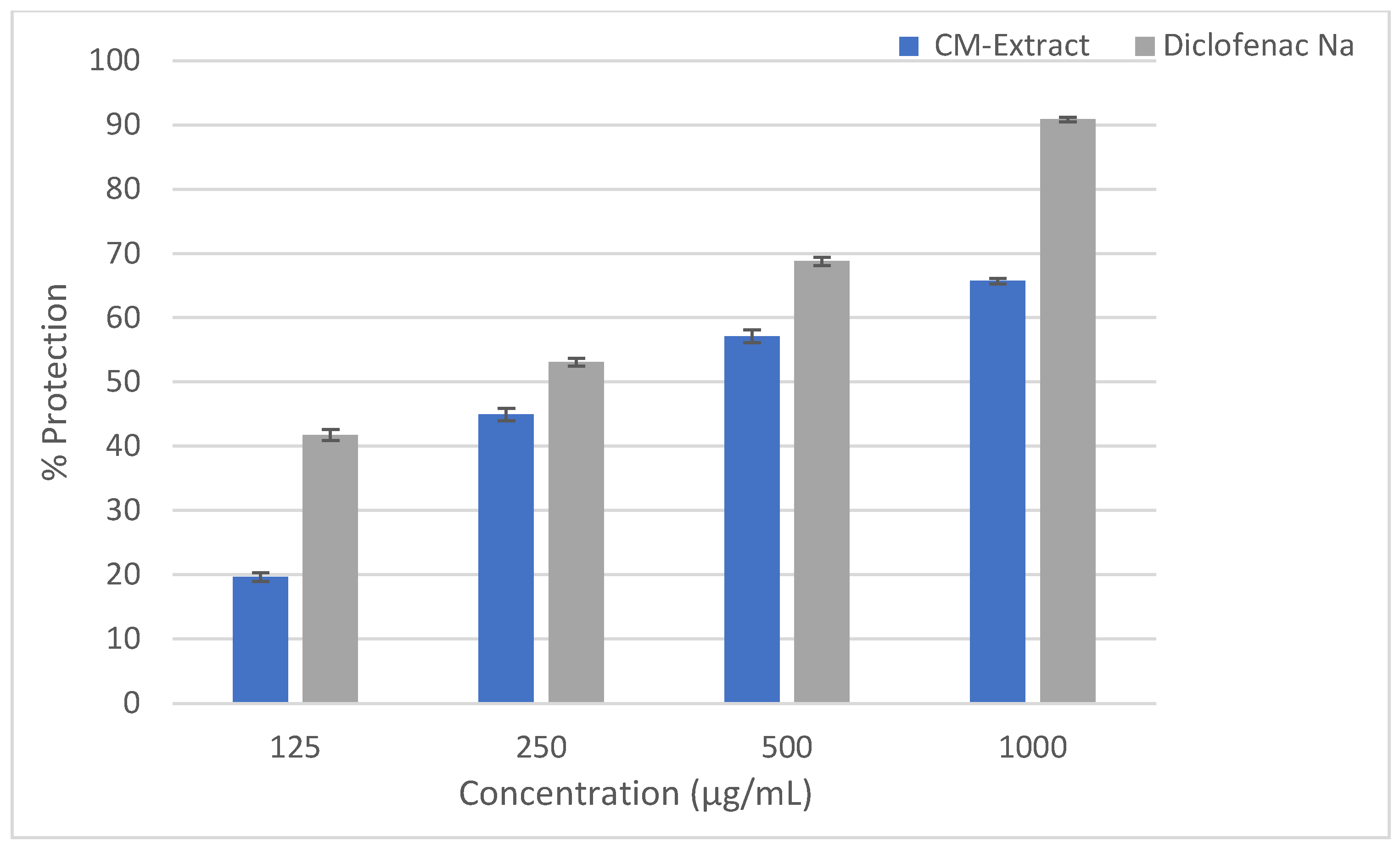
Figure 1. Effect of the hydro-methanolic extract of Cucurbita moschata flower on percent protection in HRBC membrane stabilizing method. Data are represented as mean ± SEM (n = 3).
Molecules 28 06573 g001
Figure 2. Antibacterial activity of Cucurbita moschata flower against six bacterial strains.
Molecules 28 06573 g002
Figure 3. In silico non-bonding interaction of six best selected ligands with Cyclooxygenase-II enzyme ((A) 2H-Pyran, 3,4-dihydro-2-aminomethyl- (−10.1); (B) 1H-Pyrrolo[2,3-B]pyridin-6-amine, 2,3-diphenyl- (−10), (C) Oxacyclotridecan-2-one (−9.1); (D) Luteolin (−8.7); (E) Purin-2,6-dione, 1,3,9-trimethyl-8-[2-nitrophenethenyl] (−8.6); (F) Kaempferol (−8.4)).
Molecules 28 06573 g003
Table 1. Qualitative phytochemical analysis of hydro-methanolic flower extract of Cucurbita moschata.
Phytochemical ConstituentsSpecific TestsInference
AlkaloidsMayer’s test+
Hager’s test+
Wagner test-
CarbohydratesMolisch’s test+
Benedict’s test-
Fehling’s test+
FlavanoidsAlkaline reagent test+
PhenolsFerric chloride test+
SaponinsFoam test-
TanninsGelatin test+
GlycosidesLiebermann’s test+
+: Presence; -: absence.
Table 2. TPC and TFC of hydro-methanolic flower extract of Cucurbita moschata.
ExtractsTotal Phenolic Content (TPC)
mg/g GAE
Total Flavonoids Content (TFC) mg/g GAE
CM extract18.16 ± 0.2810.42 ± 0.10
Note: Data are mean ± SEM (n = 3).
Table 3. Effect of the hydro-methanolic extract of Cucurbita moschata flower on acetic acid-induced writhing among mice.
GroupsTreatmentDose &RouteNo. of Writhing% Inhibition
G-I1% Tween 80 (Control)10 mL/kg; p.o23.5 ± 0.91NA
G-IIDiclofenac Na (Standard)50 mg/kg; p.o4.5 ± 0.6180.85
G-IIICM extract200 mg/kg; p.o17.5 ± 0.84 *25.53
G-IVCM extract400 mg/kg; p.o10.33 ± 0.56 *56.03
Each values represent the mean ± SEM of 6 experiments; * p < 0.05 Dunnett’s t-test as compared to control.
Table 4. Antibacterial activity of Cucurbita moschata flower against pathogenic bacterial strains.
SampleStephylococcus aureusSalmonella 88typhiProteus vulgarisE. coliKlebsiella pneumoniaePseudomonas aeruginosa
Zone of Inhibition (mm)
CM Extract (1 mg/mL)29 ± 0.5819.33 ± 0.8814 ± 1.1531 ± 1.1616.33 ± 0.3321.67 ± 1.45
Ciprofloxacin (5 µg/mL)21.33 ± 0.31.67 ± 0.6619.67 ± 1.7625.33 ± 0.3322.33 ± 0.8923.33 ± 0.88
Table 5. In silico ADME/T and drug likeliness study of reported phytochemicals of Cucurbita moschata flower.
Compounds NameAbsorptionDistributionMetabolismExcretionToxicityDrug LikelinessBioavailability
Water Solubility (log mol/L)Intestinal Absorption (Human) (% Absorbed)VDss (Human) (log L/kg)BBB Permeability(log BB)CYP3A4 SubstrateTotal Clearance (log ml/min/kg)AMES ToxicityHepato Toxicity
Protocatechuic acid−2.0771.17−1.29−0.683No0.551NoNoYes0.56
Vanillic acid−1.8478.15−1.74−0.38No0.719NoNoYes0.85
Caffeic acid−2.3369.41−1.09−0.647No0.508NoNoYes0.56
Syringic acid−2.2273.07−1.44−0.191No0.646NoNoYes0.56
Ferulic acid−2.8293.68−1.36−0.239No0.623NoNoYes0.85
trans-sinapic acid−2.8793.06−1.11−0.247No0.718NoNoYes0.85
Tyrosol−1.1585.26−0.11−0.218No0.283NoNoYes0.56
Luteolin−3.0981.131.15−0.907No0.495NoNoYes0.55
Kaempferol−3.0474.291.274−0.939No0.477NoNoYes0.56
3,4-dihydro-2H-pyran-2-yl)methanamine−0.0696.570.27−0.229No1.038NoNoYes0.56
5,10-dimethylbenzo[c]acridine-−5.6199.45 0.130.645Yes0.851YesYesYes0.56
2,3-diphenyl-1H-pyrrolo [2,3-b]pyridin-6-amine−3.4194.96 −0.4630.464Yes0.542YesYesYes0.85
Oxacyclotridecan-2-one−2.75995.30 0.1420.408No1.345NoNoYes0.56
(E)-1,3,7-trimethyl-8-(2-nitrostyryl)-3,7-dihydro-1H-purine-2,6-dione−3.20983.450.073−1.362Yes0.045YesYesYes0.55
Avenasterol−6.71594.64 0.1790.764Yes0.613NoNoYes0.55
Table 6. In silico binding affinity and non-bonding interaction of selected phytochemicals of Cucurbita moschata flowers.
Compounds NameDocking ScoreNon-Bonding Interaction
Hydrogen BondHydrophobic BondVan Der Waals
3,4-dihydro-2H-pyran-2-yl)methanamine-−10.1ALA A:7, ASN A:18LEU A:28, LEU A:54, VAL A:31, PHE A:92GLY A:15, ASN A:18, GLN A: 19, THR A:46, SER A:49, GLY A:93, GLY A:94, PHE A:98
2,3-diphenyl-1H-pyrrolo[2,3-b]pyridin-6-amine−10THR A:21, VAL A:31LEU A:5, ALA A:7, LEU A:28, VAL A:31ILE A: 14, ASN A:18, ASP A:27, THR A:46, SER A: 49, LEU A:54, PHE A:92, GLY A:93, GLY A:94
Oxacyclotridecan-2-one−9.1ASN A:18, LEU A:20ILE A:14, LEU A:28, LYS A:45, ILE A:50GLN A:19, SER A:49, GLN A:95, PHE A:98, THR A:121
Luteolin−8.7ALA A:7, ASN A:18, GLN A:95, THR A:21ILE A:14, LEU A:20VAL A:6, GLY A:15, GLN A:19, ASP A:27, VAL A:31, THR A:46, ILE A:50, LEU A:54, PHE A:92, GLY A:94, PHE A:98, ASP A:120
(E)-1,3,7-trimethyl-8-(2-nitrostyryl)-3,7-dihydro-1H-purine-2,6-dione−8.6 LEU A:20, LEU A:28, HIS A:23, VAL A:31, ILE A:50LEU A:5, VAL A:6, ALA A:7, GLN A:19, TRP A:22, ASP A:27, THR A:46, SER A:49, PHE A:98
Kaempferol−8.4SER A:49, ASN A:18LEU A:20, THR A:46ILE A:14, GLY A:15, GLN A:19, LYS A:45, ILE A:50, PHE A:92, GLY A:93, GLY A:94, GLN A:95, THR A:121
Avenasterol−7.8THR A:46LEU A:20, VAL A:31, LYS A:45, ILE A:50, PHE A:92LEU A:5, VAL A:6, ALA A:7, ILE A:14, GLY A:15, ASP A:27, LEU A:28, GLY A:93, GLY A:94, GLN S:95, THR A:96, THR A:121
Diclofenac−8.0ALA A:7, ILE A:50, VAL A:31LEU A:28, GLY A:93, PHE A:98, GLY A:15VAL A:6, PHE A:92, GLY A:94
Celecoxib−9.5ARG A:89, ARG A:482, SER A:322, GLN A:161VAL A:492, ALA A:496, GLY A:495, LEU A:321, TYR A:354, VAL A:318
Disclaimer/Publisher’s Note: The statements, opinions and data contained in all publications are solely those of the individual author(s) and contributor(s) and not of MDPI and/or the editor(s). MDPI and/or the editor(s) disclaim responsibility for any injury to people or property resulting from any ideas, methods, instructions or products referred to in the content.