Review

Absolute Stereochemistry Determination of Bioactive Marine-Derived Cyclopeptides by Liquid Chromatography Methods: An Update Review (2018–2022)

1
Laboratório de Química Orgânica e Farmacêutica, Departamento de Ciências Químicas, Faculdade de Farmácia, Universidade do Porto, Rua de Jorge Viterbo Ferreira 228, 4050-313 Porto, Portugal
2
Interdisciplinary Centre of Marine and Environmental Research (CIIMAR), Edifício do Terminal de Cruzeiros do Porto de Leixões, Avenida General Norton de Matos s/n, 4050-208 Matosinhos, Portugal
3
ICBAS-Instituto de Ciências Biomédicas Abel Salazar, Universidade do Porto, Rua de Jorge Viterbo Ferreira 228, 4050-313 Porto, Portugal
*
Author to whom correspondence should be addressed.
Molecules 2023, 28(2), 615; https://doi.org/10.3390/molecules28020615
Submission received: 4 December 2022 / Revised: 3 January 2023 / Accepted: 4 January 2023 / Published: 7 January 2023
(This article belongs to the Special Issue Innovative Marine Molecules: Chemistry, Biology and Analysis)

Abstract

:
Cyclopeptides are considered as one of the most important classes of compounds derived from marine sources, due to their structural diversity and a myriad of their biological and pharmacological activities. Since marine-derived cyclopeptides consist of different amino acids, many of which are non-proteinogenic, they possess various stereogenic centers. In this respect, the structure elucidation of new molecular scaffolds obtained from natural sources, including marine-derived cyclopeptides, can become a very challenging task. The determination of the absolute configurations of the amino acid residues is accomplished, in most cases, by performing acidic hydrolysis, followed by analyses by liquid chromatography (LC). In a continuation with the authors’ previous publication, and to analyze the current trends, the present review covers recently published works (from January 2018 to November 2022) regarding new cyclopeptides from marine organisms, with a special focus on their biological/pharmacological activities and the absolute stereochemical assignment of the amino acid residues. Ninety-one unreported marine-derived cyclopeptides were identified during this period, most of which displayed anticancer or antimicrobial activities. Marfey’s method, which involves LC, was found to be the most frequently used for this purpose.

1. Introduction

Since more than two-thirds of the global surface area are covered by oceans, marine organisms represent more than half of the total biodiversity [1]. Therefore, oceans constitute a rich source of many unique and novel compounds [2,3]. Long-term evolution of marine organisms promoted the fruitful development of a great number of natural products when exposed to extreme conditions, such as pressure, light, temperature, and salinity [4,5]. From 2015 to 2018, 17 clinically available drugs, based on marine natural products or their derivatives, were reported being 28 drugs in Phases I–III clinical trials [6]. Since 2018, great efforts have been made to find new therapeutically potent chemical and biological entities and, currently, 68 patents from marine organisms have been filed [7].
Among a large number of metabolites from marine-derived organisms, peptides are of pivotal relevance due to their unique structures, chemical properties, and interesting biological effects [8,9]. They are found in marine tunicates [10,11], sponges [12,13,14], algae [15,16], bacteria [17,18,19], cyanobacteria [20,21,22], fungi [23,24,25], and other invertebrates [26]. Several marine-derived peptides are obtained from symbionts [27], as well as non-symbiotic microorganisms such as sponge-associated fungi [28,29].
Marine-derived peptides display a broad spectrum of bioactivities [30,31,32], such as antibacterial, antifungal, antiviral, antitumor, antioxidant, antihypertensive, antiatherosclerotic, anticoagulant, antidiabetic, analgesic, immunomodulatory, neuroprotective, and appetite-suppressing activities, which are attracting the increasing attention of pharmaceutical [33,34], cosmeceutical [35,36], and nutraceutical [37,38,39] industries. Two marine peptide-derived drugs have reached the market: ziconotide [40], a synthetic version of the N-type calcium channel blocker ϖ-conotoxin MVIIA for the treatment of patients suffering from chronic pain, and brentuximab vedotin (Adcetris®), a synthetic derivative of dolastatin 10 linked to an antibody for the treatment of Hodgkin’s lymphoma [41]. Moreover, marine-derived peptides constitute one of the research topics that provides a very high output, with a huge increase in the number of publications (268 per year) from 2010 to 2020 (Figure 1A), with both Asia (37.2%) and Europe (33.1%) as the major contributors (Figure 1B), and this trend is expected to continue [42].
The remarkable properties of cyclopeptides, such as low inherent toxicity, good affinity, and high selectivity for protein targets [43], make them attractive molecules to be explored. Their structures may comprise diverse structural scaffolds, usually non-proteinogenic amino acids [44], or with some modifications such as methylation [45], sulfuration [46], lipidation, and acetylation [47]. These characteristics play a crucial role in the interactions with biotargets [48,49]. Nevertheless, not all groups of marine organisms produce cyclopeptides with all these characteristics, and some are more common in certain groups. For example, cyclopeptides found in marine sponges such as Lithistid and Theonella are characterized by a high proportion of d- and/or N-methylated amino acids originating from symbiotic microorganisms [50,51,52,53,54]. Bacteria incorporate non-proteinogenic amino acids to prevent the proteolysis of peptides through stabilization of the backbone conformation and/or by elimination of the enzyme recognition site [55,56,57,58]. Secondary structures, especially the α helix, are common in antimicrobial marine peptides [59,60,61,62] because the presence of the α helix allows their penetration through a microbe’s membrane [60,61].
Cyclodepsipeptides—molecules where one or more amide groups are replaced by the corresponding ester—are often found in marine organisms [63,64,65]. Depsipeptides are biosynthesized by non-ribosomal peptide synthetases in combination with either polyketide synthase [66,67,68] or fatty acid synthase enzyme systems. This type of bioactive peptide has also contributed to revolutionizing the peptide chemical space [69]. Proline-rich cyclopeptides also play a crucial role in drug discovery that may provide useful scaffolds for modulating more challenging biological targets, such as protein–protein interactions and allosteric binding sites [70]. Lipodepsipeptides, which contain a lipid moiety linked to a peptide portion, are also well distributed among marine organisms and have shown different biological activities, such as phytotoxic, antimicrobial, immunosuppressive, cytotoxic, hemolytic, among others. Their bioactivities seem to be related to the interactions between a hydrophobic segment with the plasma membrane because they mimic the architecture of the membranes [71].
As one of the richest sources of structurally diverse bioactive compounds, considerable attention has been drawn towards the production and bioactivity of marine cyclopeptides [42]. Moreover, in recent decades, advanced synthetic methods have been applied to overcome supply problems of marine peptides of interest, affording higher quantities of compounds required for large-scale biological assays [72,73,74,75,76]. In addition to classical solid-phase and solution-phase techniques of peptide synthesis [77], significant efforts have been carried out for the introduction of sustainable and innovative processes for synthesis and purification methodologies [78,79]. Marine cyclopeptides are also interesting models for molecular modifications to obtain more potent compounds with improved properties [80,81]. For example, non-proteinogenic amino acids can be incorporated to prevent the proteolysis of peptides [81]. Acetylation of the N-terminus of short peptides has shown to improve peptidase stability in serum, and hence, increases their half-life [82]. Protein glycosylation can increase protein–protein interaction and protein stability. Interestingly, the glycosylation of peptides is suggested to improve peptide permeability [83], increase metabolic stability, and lower clearance rate, thereby improving bioavailability [84]. Peptide half-life is expanded through lipidation, where a long-chain saturated lipid is acylated to an amino acid to facilitate its binding to a carrier serum protein [85,86].
Cyclopeptides isolated from marine sources can comprise a different number of amino acid subunits, and the assignment of their absolute configurations is an absolute requirement for complete characterization [87]. Their physical, chemical, and biological/pharmacological properties are closely related to their absolute configurations, and stereochemical assignments continue to be a great challenge for natural products and medicinal chemists [88].
Currently, different tools, such as X-ray crystallography [89,90], vibrational circular dichroism [91,92,93], optical rotatory dispersion [94], electronic circular dichroism [95,96,97], nuclear magnetic resonance (NMR) [98,99,100], Marfey’s method in combination with liquid chromatography (LC) [101,102,103,104], and chiral LC [105,106,107], can be used to determine the absolute configurations. In some cases, such as lipopeptides, additional stereogenic centers are present in the lipid chain that might require additional experiments, such as Mosher’s method, or others [108,109].
A literature survey from 1996 to 2017 covering all the reports on chromatographic methods for the absolute stereochemistry determination of marine peptides revealed that Marfey´s method and/or chiral LC are very efficient and widely used [110]. Moreover, 52% of the reports described the use of Marfey’s method, 21% employed chiral LC analysis, and 27% included the application of both methods. In some cases, a combination of both methods are found to be necessary for the absolute configurations assessment of all the amino acid residues [110]. In addition, LC methods are also widely used for the assignment of the absolute configurations of peptides isolated from natural sources other than marine organisms [111,112,113,114].
In a continuation of our previous publication [110] and to analyze current trends, herein, recent studies concerning new bioactive marine cyclopeptides are analyzed regarding the absolute stereochemistry determination of the amino acid residues by LC methodologies. The chemical structures and biological/pharmacological activities of the marine-derived peptides are also described to provide an update for this field of research. Structural issues relevant to the biological/pharmacological activities are also emphasized when considered pertinent.

2. Liquid Chromatography Methods for Determination of Absolute Configurations of Peptides

LC is an indispensable technique in several research laboratories all over the world, especially when combined with different detectors such as ultraviolet-visible (UV-Vis), mass spectrometry (MS), and fluorescence detection [115]. LC is a method of choice associated to both Marfey´s and chiral LC methods [110].
The first step, in both methods, to determine the absolute configurations of the amino acids of marine peptides is a total or partial hydrolysis of peptide bonds to obtain individual amino acid residues (Figure 2). Typically, the hydrolysis reaction is carried out in acidic condition using 6 N of HCl solution at 110–115 °C for 24 h [110].
In Marfey´s method, after the acidic hydrolysis of peptides, the amino acid residues are derivatized with suitable chiral derivatizing reagents (CDRs). The reagents 1-fluoro-2,4-dinitrophenyl-5-d,l-alanine amide (FDAA) and 1-fluoro-2,4-dinitrophenyl-5-d,l-leucine amide (FDLA) (Figure 3A) are the most widely used [110].
The CDRs react stoichiometrically, without racemization, under gentle heating at 40 °C within 1–2 h in alkaline condition, with the amino group of l- and d-amino acids residues yielding diastereomers (Figure 3B) [116]. It is important to mention that this method was found to be ineffective for some amino acid residues, especially those containing ring- and α-methyl-substituted phenylalanine and phenylalanine amides [117]. Nevertheless, a new approach, called the “O-Marfey method”, was described for the derivatization of α-hydroxy acids with l-FDAA by increasing the nucleophilicity of the hydroxyl group for the reaction to take place [118]. This method requires the addition of NaH (60% dispersed in oil) to a solution of α-hydroxy acids in tetrahydrofuran at room temperature. Then, the CDR (l-FDAA) is added to this solution and stirred under argon or nitrogen atmosphere [118]. In other cases, there is a need for optimization, such as by using a solution of FDAA in acetone, as well as a higher reagent concentration and longer reaction times [119]. Of note is that diastereomers can also be obtained by microwave-assisted synthesis [120].
The synthesis of FDAA as a CDR to determine the stereochemistry of amino acids was described for the first time by Peter Marfey in 1984 [121]. FDAA was obtained from difluoro dinitro benzene by substitution of one of its two fluorine atoms by l-alanine amide. Then, a nucleophilic substitution of the remaining reactive aromatic fluorine was performed with the free amino group on l- and d- alanine. From this reaction, diastereomeric pairs were obtained, which were separated by RP-LC. Amide was chosen because it is quite stable and neutral and does not undergo racemization [121]. Now, FDAA is universally known as Marfey’s reagent and continues to be widely used. Over the years, other structural variants of FDAA have been developed [122].
Generally, after the derivatization reaction, the diastereomers are analyzed by an RP-LC, using suitable standards for comparison to accurately determine the absolute configurations of the peptides [119]. Additionally, other methodologies have also emerged, including LC-mass spectrometry (LC-MS), thin-layer chromatography (TLC), and gas chromatography (GC) [123,124]. The key advantages of Marfey´s method include improved detection sensitivity compared to underivatized amino acids, the commercial availability of both enantiomers of FDAA, and the use of readily available LC technology.
Harada et al. [125] proposed a method named “advanced Marfey’s method”. This method uses LC-MS to determine the absolute configurations of the amino acid residues. Since this method does not necessarily require amino acid standards to detect and identify target amino acids, it can be used to determine the absolute configurations of unusual amino acids for which standards are not available. In this method, the absolute configuration of an amino acid is deduced from its LC retention times of the l-FDLA derivative of the original amino acid, and its enantiomer formed by racemization. Therefore, the racemization is essential for this method [125]. Later, the same group introduced “dl-FDLA derivatization”, a procedure that does not require the conventional chemical racemization process [126]. Due to this d,l-FDLA system, the advanced Marfey’s method has been improved to be simple, more rapid and reliable, and its applicability has been significantly extended [103,104].
In order to develop a highly sensitive analytical Marfey’s method capable of rapidly and unambiguously determining the absolute configurations and enantiomer regiochemistry for all commonly found amino acids, Vijayasarathy et al. [102] developed the C3 Marfey’s method. This method is a more refined variation on the existing Marfey’s method, using the same CDRs (l-FDAA and d-FDAA) but with a C3 LC column held at 50 °C, and a H2O/MeOH gradient elution modified by isocratic 1% HCO2H in CH3CN, and taking advantage of both UV (340 nm) and (±)-ESIMS detection. This method was shown to be very effective, allowing the stereochemical assignments of all amino acids, including isoleucine stereoisomers. Moreover, the residual Marfey’s reagent did not obscure any target amino acid [102].
The applicability and limitations of Marfey’s method and its optimized methods have been extensively examined in several reviews [116,119,122]. These methods were also found to be promising for applications in several other research fields and for other chiral molecules/drugs [122].
Regarding the chiral LC method, determination of the absolute configurations is developed by a direct analysis of the amino acid residues present in the acid hydrolysate without prior derivatization. First, enantioseparation of standards amino acids is performed on a chiral stationary phase (CSP). Then, comparison and/or co-injection of hydrolysate with the amino acids standards is performed, under the same chromatographic conditions, leading to the identification of the amino acid constituents [110].
The success of an efficient enantioseparation is mainly determined by the chiral discriminative power of the CSP. Over the years, various types of CSPs have been described [127,128,129]; nevertheless, the development of new CSPs for LC continued to be an evolutionary subject, encompassing the preparation of new chiral selectors [130], the introduction of new chromatographic supports [131], and the application of different synthetic approaches for the preparation of CSPs [132]. Macrocyclic antibiotic-based, crown ether-based, and ligand exchange-type (Figure 4) are the most-used CSPs for the enantiomeric separation of amino acids and primary amine-containing compounds [133,134,135,136]. Evidently, the same trend was observed for the stereochemical determination of the amino acids subunits of marine peptides [110].
A direct analysis of amino acid hydrolysates without further derivatization is the main advantage of chiral LC, since it is a faster and simpler procedure when compared to indirect methods. Its principal drawback is the high cost of the commercial chiral columns, which could be a hurdle for these analyses [137].
Although these methods are highly effective, one limitation of both is that racemization can occur for some amino acid residues during the acid hydrolysis [138,139]. For example, the analysis of the acidic hydrolysate of a cyclopeptide isolated from a marine sponge, Homophymia sp., showed that the racemization of lysine took place. Although this peptide contains only one lysine residue, both d- and l-enantiomers were detected in almost equal amounts. By performing an analysis of the hydrolysates with shorter reaction times of 4 and 8 h, it was found that racemization occurred during acidic hydrolysis (Figure 5). The longer the reaction time, the higher the content of l-lysine, indicating a formation of this enantiomer by epimerization [138].
Other difficulties, such as the absence of commercially available standard amino acids, demanding time and synthetic efforts to prepare them [140], the existence of identical amino acids in the peptide skeleton [141], or amino acids with more than one chiral stereocenters [142], make elucidation a more difficult task. One strategy to solve this kind of problem is the use of synthesis, which in some cases, resulted in a revision of the proposed structures [139,143,144].

3. New Marine-Derived Cyclopeptides Reported from January 2018 to November 2022

There are several reports describing the isolation and biological activity evaluation of new marine-derived peptides during the period of January 2018 to November 2022. Among them, chromatographic methods—specifically Marfey’s method and chiral LC analysis—were used to assess the absolute configurations of 91 new marine-derived cyclopeptides (191). Table 1 summarizes the names and types of cyclopeptides, compound producers, chromatographic methods, the biological activities of each compound and their references. Several marine sources were described, including marine-derived bacteria, cyanobacteria, marine sponges, and marine-derived fungi, among others. The potential of marine-derived cyclic peptides 191 as anticancer and antimicrobial agents is expected based on previous reviews [145,146,147], which reinforces the marine organisms as a rich source of bioactive compounds for these therapeutic classes.
In this section, the marine-derived cyclopeptides are organized according to the main biological activity.

3.1. Anticancer Activity

One of the most described biological activities associated to marine-derived cyclopeptides is anticancer activity [185]. Consistent with similar findings in previous works, several isolated marine peptides exhibited cytotoxicity. Among the 91 new marine-derived cyclopeptides, 43 showed promising results regarding anticancer activity and 11 (111) were isolated from marine-derived bacteria (Figure 6). Marine-derived bacteria have been shown to be a rich source of natural products, including cyclopeptides [71,186]. Bacterial natural products constitute 29% of Food and Drug Administration (FDA)-approved natural product-derived drugs, which mostly function as antitumor or antibacterial agents [187].
A new cyclic lipopeptide, bacilohydrin A (1), was isolated from a crude culture extract of Bacillus sp. SY27F, obtained from the Indian Ocean hydrothermal vent [148]. Compound 1 exhibited significant cytotoxicity against three cancer cell lines—specifically, DU-145, MCF-7, and HepG2—with IC50 values of 50.3–175.1 nM. The positive control, paclitaxel, showed IC50 values of 10.9–94.3 nM. The absolute configurations of the amino acids in 1 were determined by a modified Marfey’s method revealing the presence of seven mixed d/l-amino acids. It is important to highlight that it was the first report of a determination of the absolute configuration of a d-aspartic acid residue in the surfactin class [148].
The advanced Marfey’s method, in combination with 3JHH and rotating-frame Overhauser effect spectroscopy (ROESY) analysis, DP4 calculation, and genomic analysis, was applied to establish the absolute configuration of the amino acid units of epoxinnamide (2), a new cinnamoyl-containing nonribosomal bicyclic decadepsipeptide (Figure 6), which was isolated from the culture extract of an intertidal mudflat-derived Streptomyces sp. OID44 [149]. Cinnamoyl-containing nonribosomal peptides represent a unique family of actinobacterial metabolites associated with diverse bioactivities [188]. Compound 2 induced quinone reductase activity in murine Hepa-1c1c7 cells, at 5 µM, by 1.6-fold. Additionally, 2 displayed considerable antiangiogenesis activity in human umbilical vein endothelial cells, with an IC50 of 13.4 µM. The positive control, sunitinib, showed an IC50 value of 1.7 µM [149].
A new family of polypeptides, bathiapeptides A1 (3), A2 (4), and B-E (58), isolated from a marine biofilm-derived Bacillus sp. B19-2, exhibited high cytotoxicity against MGC-803, Hep G2, MCF-7, and HeLa cell lines. Compound 3 exhibited the highest cytotoxicity in all cell lines, with IC50 values ranging from 0.5 to 4.1 µM. The advanced Marfey’s method was able to establish the absolute configuration of isoleucine and phenylalanine of 38 [150]. Due to the presence of a rare polythiazole moiety in 38 (Figure 6), the thiazole-derived alanine moiety could not be directly determined by Marfey’s method (or chiral LC) since the epimerization of thiazole-based amino acids can occur during acid hydrolysis [189]. To solve this issue, ozonolysis was carried out to break the thiazole ring before derivatization by Marfey’s reagents [190]. Moreover, since Marfey’s method does not allow distinguishing l-isoleucine from l-allo-isoleucine [104], an X-ray analysis was performed to prove the structure of l-isoleucine [150].
Dentigerumycin E (9), a new cyclic hexapeptide containing three piperazic acids and a pyran-bearing polyketide acyl moiety (Figure 6), was isolated from the extract of a coculture of marine-derived Streptomyces and Bacillus strains, which were simultaneously isolated from an intertidal mudflat. The absolute configurations of the amino acid residues of 9 were established by a combination of advanced Marfey’s method with ROESY correlations. Compound 9 exhibited moderate antiproliferative and antimetastatic activities against several human cancer cell lines, including A549, HCT-116, MDA-MB-231, SK-HEP-1, and SNU638, with IC50 values ranging from 27 to 39 μM. The positive control, etoposide, showed IC50 values ranging from 0.42 to 6.21 µM. Compound 9 was also assayed against MCF-10A to determine its cancer cell-specific cytotoxicity and showed an IC50 value higher than 50 µM, indicating that 9 did not exhibit significant cytotoxicity against normal human epithelial cells. Additionally, 9 also showed antimetastatic potential against MDA-MB-231 in wound healing and cell invasion assays. In the wound healing assay, 9, at 20 and 40 µM, inhibited cell migration by 20 and 48%, respectively, while in the cell invasion assay, 9 exhibited inhibitory activity by 10 and 34% at 20 and 40 µM, respectively. A structure–activity relationship (SAR) study suggested that 2-N-OH, 16-N-OH, and 37-OH (carboxylic acid) in 9 are essential for its antiproliferative and antimetastatic activities [151].
Another promising compound with anticancer activity is a cyclic depsipeptide, nobilamide I (10), isolated from the saline cultivation of a marine-derived bacterium, Saccharomonospora sp. strain CNQ-490, obtained from a 45 m deep-sea sediment sample at 2 km west of the Scripps pier, La Jolla Canyon, in California. Nobilamide I (10) decreased cancer cell motility by inhibiting epithelial-mesenchymal transition (EMT) effectors. At a concentration of 5 μM, 10 inhibited migrations of AGS, A549, and Caco2 cells by ca. 70, 50, and 60%, respectively, and their invasions by ca. 60, 30, and 60%, respectively. It was suggested that 10 modulated the protein and mRNA expression levels of EMT N-cadherin and E-cadherin markers by downregulating the transcription factors, Snail, Slug, and Twist. In addition, 10 modulated the protein and mRNA expression levels of the matrix metalloproteinase family (MMP2 and MMP9) in the three cell lines. The absolute configurations of the amino acid residues were assigned by using the C3 Marfey’s analysis [152].
A new cyclic hexapeptide, nocardiotide A (11) (Figure 6), was isolated from a broth culture of an actinomycete, Nocardiopsis sp. UR67, associated with the marine sponge, Callyspongia sp., which was collected from the Red Sea, Ras Mohamed, Sinai, Egypt. The planar structure of the compound was elucidated by HRESIMS and a 1D and 2D NMR spectral analysis. The absolute configurations of the amino acid residues were solved by Marfey’s method. Compound 11 displayed significant cytotoxicity towards human MM.1S multiple myeloma, human HeLa cervix carcinoma, and murine CT26 colon carcinoma cell lines, with IC50 values of 8, 11, and 12 µM, respectively [153].
Cyanobacteria are also an important source of new marine-derived cyclopeptides [191,192]. Compounds 1223 (Figure 7) with anticancer activity were discovered in 2018.
Phyo et al. [154] described the isolation of a new cyclic decapeptide, trikoramide A (12) (Figure 7), isolated from samples of the marine cyanobacterium, Symploca hydnoides, which was collected from the intertidal shores of Trikora beach, Bintan Island, Indonesia. Compound 12 is a C-prenylated cyclotryptophan-containing cyanobactin whose absolute configuration was determined by Marfey’s method and a NOESY correlations analysis. Compound 12 displayed cytotoxicity against the MOLT-4 and AML2 cancer cell lines, with IC50 values of 4.8 and 8.2 μM, respectively [154].
Later, the same group isolated another three cyanobactins, trikoramides B–D (1315) (Figure 7), from the same samples. Compounds 1315 are analogues of 12. The main difference in their structures is in the C-prenylated cyclotryptophan unit. Marfey’s method, in combination with the ECD spectra and NOESY correlations, were applied to determine their absolute structures. Compounds 13 and 15 showed cytotoxicity against the MOLT-4 cell line, with IC50 values of 5.2 µM and 4.7 µM, respectively [155].
Very recently, the same group further isolated three new cyclic depsipeptides, triproamide (16), and pemukainalides A (17) and B (18) (Figure 7), from the same cyanobacterium, which was collected from the same location. Compound 16 features a unique structure containing a rare 4-phenylvaline and a β-amino acid, dolamethylleucine. Stereochemical analyses were carried out by utilizing a combination of Marfey’s method, J-based configuration, and chiral LC analyses. For the absolute configuration of the 2-hydroxyisovaleric acid unit in 16, a chiral column was used with a ligand exchange-type CSP to establish the R-configuration. Compound 17 exhibited cytotoxicity against the MOLT-4 leukemia cell line, with an IC50 value of 5.6 μM. The positive control, dolastatin, showed an IC50 value of 2.5 µM [156].
By using mass spectrometry-guided fractionation, together with molecular networking, Keller et al. [157] described the isolation of tutuilamides A (19) and B (20) (Figure 7), from the marine cyanobacterium, Schizothrix sp., which was collected from the main island of Tutuila in American Samoa, and tutuilamides C (21) (Figure 7), from a cyanobacterium, Coleofasciculus sp., which was collected from Palmyra Atoll. The structures of 1921 contain a novel vinyl chloride-containing residue and several unusual residues, including 3-amino-6-hydroxy-2-piperidone and 2-amino-2-butenoic acid. Marfey’s method was used to determine the absolute configurations of their amino acid residues. Compounds 1921 exhibited a moderate potency of cytotoxicity against H-460 lung cancer cell line. Interestingly, 1921 displayed potent elastase inhibitory activity. Additionally, the binding mode to elastase was analyzed by X-ray crystallography, which revealed a reversible binding mode. Moreover, the amino acid backbone of the flexible side chain of 19 can establish an additional hydrogen bond in the elastase binding pocket, allowing enhanced inhibitory activity [157].
Two new cyclic lipopeptides, laxaphycins B4 (22) and A2 (23) (Figure 7), were isolated, along with the known peptide laxaphycin A, from the marine cyanobacterium, Hormothamnion enteromorphoides, which was collected at Garden Key, Dry Tortugas National Park [158]. Compound 22 displayed growth inhibition against HCT-116 cell line with an IC50 of 1.7 µM, and a synergistic effect to inhibit the growth of this cancer cell line when 22 was used in combination with laxaphycin A. The absolute stereochemistry of the amino acid residues of 22 and 23 was determined by both chiral LC using a macrocyclic antibiotic-based CSP, and by advanced Marfey’s method. In addition to those methods, 1D and 2D ROESY correlations were used to determine the geometric configuration of α,β-dehydro-aminobutyric acid [158].
Cyclopeptides have also been isolated from a myriad of marine-derived fungi, mainly from genera Aspergillus, Acremonium, Fusarium, and Penicillium [193]. Marine-derived fungi represent a potential source for bioactive natural products, and an increasing number of new fungal metabolites, including peptides, have been discovered over the past years [194,195]. In addition, peptides isolated from the association of fungi with other macroorganisms produced a valuable and extra-large chemical database, exhibiting relevant biological activities [196]. From 2018, six marine-derived cyclopeptides (2429) with anticancer activity were isolated from marine-derived fungi (Figure 8).
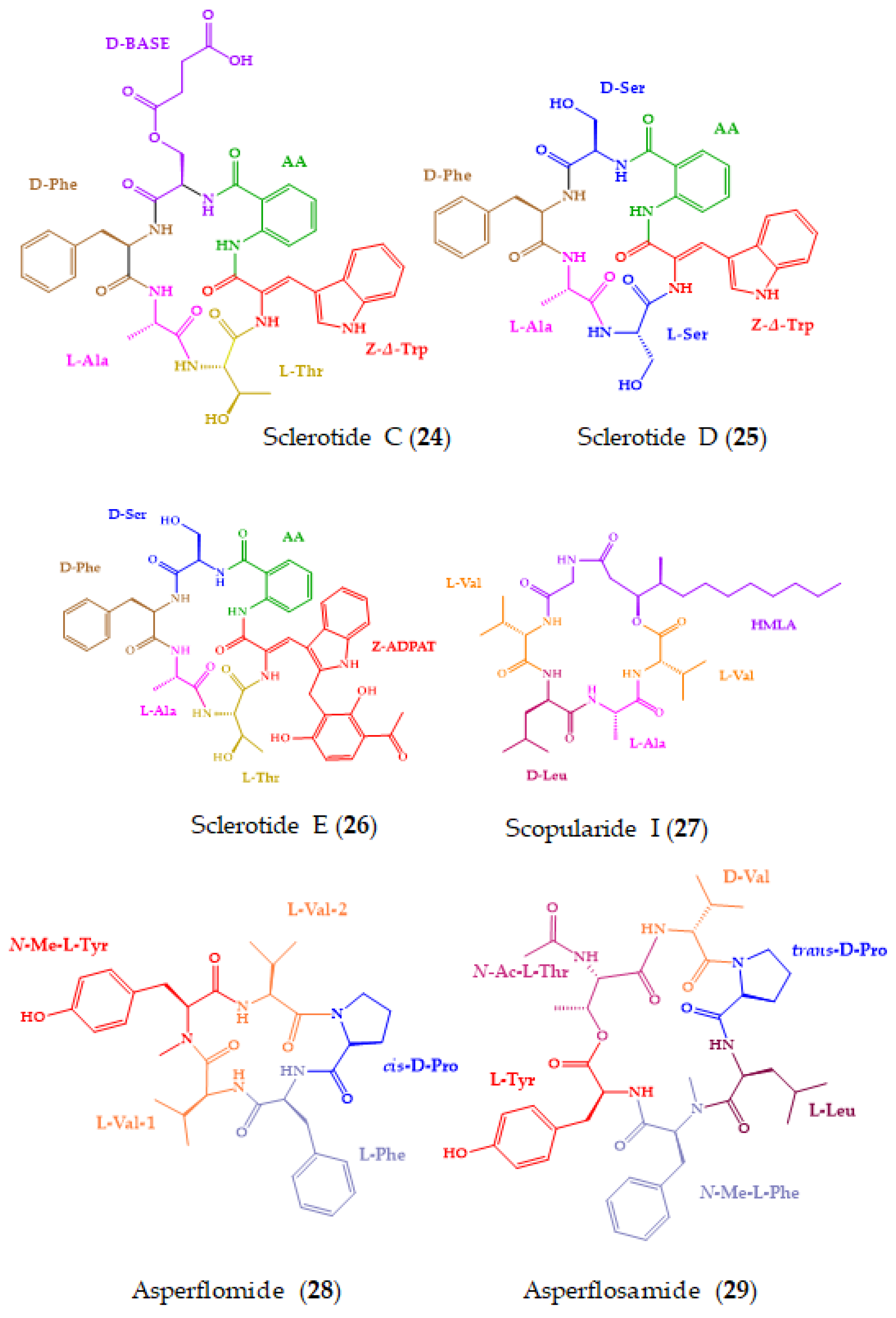
Sclerotides C–E (2426) (Figure 8), new cyclohexapeptides, and scopularide I (27), a new lipodepsipeptide, were discovered from the fermented rice cultures of Aspergillus sclerotiorum SCSIO 41031, isolated from a soft coral, which was collected in Beihai, Guangxi Province, China. Among the isolated compounds, only 27 showed cytotoxicity towards human nasopharyngealcarcinoma cell lines, HONE1 and HONE1-EBV, with IC50 values of 13.0 and 10.1 µM, respectively (sorafenib was used as a positive control). Compound 27 also displayed moderate inhibition of acetylcholinesterase, with an IC50 value of 15.6 µM (tacrine was used as a positive control). The absolute configurations of the amino acid residues in 25 and 26 were established by Marfey’s method, while those of 24 and 27 were determined by a single crystal X-ray diffraction analysis [159].
Two new N-methylated cyclopeptides, asperflomide (28) and asperflosamide (29) (Figure 8), were obtained from the culture extract of Aspergillus flocculosus 16D-1, isolated from the inner tissue of the marine sponge, Phakellia fusca, which was collected from Yongxing Island, China. The planar structures of 28 and 29 were elucidated by the interpretation of HRESIMS and extensive analysis of 1D and 2D NMR spectra. The absolute configurations of the amino acid constituents in 28 and 29 were established by Marfey’s method, using UPLC-HRMS for analysis. Although 28 and 29 did not exhibit cytotoxicity against A2780, HCT-8, PC-9, SW480, MDA-MB-231, and U251 cancer cell lines, and nonmalignant cells (human cardiomyoblast cell lines HaCaT and CCD-18Co), they showed weak tankyrase1/2 inhibitory activity at the centration of 40 µM (XAV939 was used as a positive control) [160]. Tankyrase is a member of the poly(ADP-ribose)polymerase family which mediates Wnt signal transduction and has emerged as a new molecular target for the therapy of different kinds of cancer. Consequently, tankyrase inhibitors are considered as promising therapeutics for cancer treatment [197].
A number of peptides and depsipeptides with intriguing structures and interesting biological activities have been discovered from marine sponges [198,199], some of which were shown to be produced by symbiotic microorganisms [27]. From 2018, 14 marine-derived cyclopeptides (3043) (Figure 9) were isolated from marine sponges and from sponge-associated microorganisms.
From a deep-sea marine sponge, Pachastrella sp., two new peptides, microsclerodermins N and O (30 and 31) (Figure 9), were isolated [161]. Compounds 30 and 31 have unique structures since they possess a p-ethoxyphenyl moiety instead of a typical p-methoxyphenyl moiety at the terminus of the ω-phenyl-polyhydroxylated β-amino acid unit. The absolute configurations of all the stereogenic centers were determined by Marfey’s method and were found to be identical with other microsclerodermins. For a stereochemical study, a naphthalene-bonded stationary phase was used. Relevant cytotoxic activity was observed for both compounds against HeLa cells, with an IC50 of 0.77 µM and 0.81 µM, respectively. The positive control, doxorubicin hydrochloride, showed an IC50 value of 0.27 µM [161].
Theonellamide J (32), 5-cis-Apoa-theopalauamide (33), and theonellamide K (34) (Figure 9) were isolated from a red sea sponge, Theonella swinhoei, which was collected in the southern part of the Gulf of Aqaba. Compounds 33 and 34 exhibited a growth inhibitory activity against HCT-116 cell line, with an IC50 of 21.8 and 3.5 µM, respectively. The positive control, cytochalasin D, showed an IC50 value of 0.8 µM. This finding suggested that the 3-amino-4-hydroxy-6-methyl-8-phenyl-5E,7E-octadienoic acid (Apoa) subunit is relevant for the activity. The absolute configurations of the amino acids were established by Marfey’s and advanced Marfey’s methods. The samples were first analyzed by Marfey’s method, which allowed the configurations of four amino acids to be elucidated, followed by LC-MS (advanced Marfey’s method) for the assignment of the stereochemistry of the remaining amino acids [162].
Fuscasins A–D (3538) (Figure 8) are new cyclic heptapeptides isolated from the marine sponge, Phakellia fusca, which was collected from the South China Sea. Compound 35 exhibited a remarkable cytotoxicity against HepG2 cells, with an IC50 value of 4.6 μM, while 3638 were inactive even at a concentration up to 20 μM. Cisplatin was used as the positive control with an IC50 value of 4.2 μM. Interestingly, 35 did not exhibit cytotoxicity against the nonmalignant rat cardiomyoblast H9C2 cell line even at concentrations up to 100 μM, suggesting that this compound may have a selective growth inhibitory effect against HepG2 cells. Moreover, none of the compounds displayed cytotoxicity against NCI-H460, MCF-7, HeLa, PC9, and SW480 cell lines. Compound 35 is a structurally unique cyclic heptapeptide with a pyrrolidine-2,5-dione-bearing backbone. The absolute configurations of amino acid residues were determined by the advanced Marfey’s method by derivatizing its acid hydrolysis products with l-FDLA, followed by analysis with UPLCESI-QTOF MS. Figure 10 shows the chromatograms obtained after appropriate selective ion monitoring channels of the amino acid standards (green peaks) and Marfey’s derivatives (red peaks) for comparison of the retention times [163].
Marfey’s method was also used to determine the absolute configurations of the amino acid residues of a new cyclic heptapeptide, ectyoplasin (39), which was isolated from the marine sponge, Ectyoplasia ferox, collected on Tuxpan reef, Veracruz, Mexico. Compound 39 possesses significant cytotoxic activity against DU-145, Jurkat, MM144, HeLa, and CADO-ES1 cell lines, with IC50 values ranging from 2.9 to 23.5 µM, being more remarkable against DU-145 cell line with an IC50 value of 2.91 µM (the positive control, doxorubicin, showed IC50 values of 10−7–10−8 M). Moreover, it was shown that 39 induced apoptosis in DU-145 cells [164].
Three new kynurenine-bearing cyclic heptapeptides, phakefustatins A–C (4042) (Figure 9), were isolated from the marine sponge, Phakellia fusca, which was collected off Yongxing Island in the South China Sea at a depth of 25 m, by using a neutral-loss scanning method based on LC-MS/MS. The structures of 4042 were elucidated by HRESIMS and extensive analysis of their 1D and 2D NMR spectra, and the absolute configurations of their amino acid residues were determined by the advanced Marfey’s method. Compound 40 was identified as a retinoic X receptor-α (RXRα) modulator, which is an important nuclear receptor that could control various biological processes through inhibition of the PI3K/Akt pathway [200,201]. These findings suggested that 40 might inhibit cancer cell growth and induce apoptosis through the RXRα-mediated PI3K/Akt signaling pathway, and its pharmacophores could be kynurenine and guanidine groups [165].
A new cyclic lipopeptide, aciculitin D (43), was isolated from the marine sponge, Poecillastra sp., which was collected by dredging at a depth of 245 m near the seamount Gonsone, Japan. The structure of 43 was elucidated by HRESIMS and 1D and 2D NMR spectral analysis as well as chemical degradation. The absolute configurations of the amino acid residues were elucidated by Marfey’s method, while the absolute configurations of the sugar (lyxose) and the fatty acid (2,3-dihydroxytetradeca-4,6-dienoicacid) were established by chemical degradation and chiral LC/MS analysis. In the case of 2,3-dihydroxytetradeca-4,6-dienoicacid (Dhtda) unit, a polysaccharide-based CSP was used, allowing the conclusion that the absolute configurations of C-2 and C-3 were 2S,3R [166]. Compound 43 showed cytotoxicity against HeLa and HCT-116 cells, with IC50 values of 4.5 µM and 1.4 µM, respectively. The IC50 values of the positive control, doxorubicin, were 0.57 and 0.51 µM against HeLa and HCT-116 cells, respectively [166].

3.2. Antimicrobial Activity

A number of marine-derived cyclopeptides exhibited a broad spectrum of antimicrobial activities, including bacteria, protozoa, fungi, and viruses [202,203,204]. Moreover, some of the marine cyclopeptides also showed antimicrobial activities towards several drug-resistant bacteria and fungi, making them a very promising source of novel antimicrobial agents to revert multidrug-resistance [203]. Among the 91 new marine-derived cyclopeptides isolated from 2018, 16 (4459) were tested for their antimicrobial activity (Figure 11) and showed promising results. They were isolated from diverse marine sources including bacteria, cyanobacteria, and fungi, among others.
Bacicyclin (44) (Figure 11), a new cyclic hexapeptide, was isolated from a culture broth of Bacillus sp. strain BC028, which is associated with Mytilus edulis, and this was collected from the Kiel Fjord (Baltic Sea, Germany). The planar structure of 44 was elucidated by an extensive analysis of 1D and 2D NMR and HRESIMS spectra. Marfey’s method was used to determine the absolute configurations of the amino acid constituents and revealed that, in all cases, the amino acids of the building block have the l-configuration, except for alanine and phenylalanine, which had a d-configuration. Compound 44 showed antibacterial activity against the clinically relevant strains Enterococcus faecalis JH212 and S. aureus NTCT8325, with minimal inhibitory concentration (MIC) values of 8 and 12 µM, respectively. Streptomycin was used as a positive control and exhibited an MIC value of 5.24 mM against E. faecalis and 2.09 mM against S. aureus, respectively [167].
A nonribosomal lipodecapeptide, taeanamide A (45), was isolated from the culture of Streptomyces sp. AMD43, which was obtained from an intertidal-mudflat sample collected in Anmyeondo, Republic of Korea. The planar structure of 45 was elucidated by an analysis of HRESIMS and 1D and 2D spectra. The absolute configurations of the amino acid residues were determined by Marfey’s method, except for two serine residues whose stereochemistry was determined by a bioinformatic analysis of the biosynthetic gene cluster of the taeanamides. Compound 45 showed antitubercular activity against Mycobacterium tuberculosis mc2 6230, with a concentration at which 50% of the strains were inhibited (MIC50) of 27 µM. Bedaquiline was used as the positive control with an MIC50 = 0.4 µM [168].
Ogipeptins A–D (4649) (Figure 11), isolated from the culture broth of the marine bacterium, Pseudoalteromonas sp. SANK 71,903, which was collected from Ogi-machi, Sado-shi, Niigata Pref., Japan, exhibited antimicrobial activity against Gram-negative bacteria. Compounds 4649 blocked lipopolysaccharide (LPS) binding to the cluster of differentiation 14 (CD14), with IC50 values of 4.8, 6.0, 4.1, and 5.6 nM, respectively [169,205]. LPS is a component of the cell membrane of Gram-negative bacteria and is known to induce a strong immune response [169]. LPS is well known as a bacterial endotoxin, and it is a key component for triggering sepsis or septic shock [205]. The absolute configurations of the amino acid building blocks of 4649 were elucidated by the advanced Marfey’s method. This method also allowed the presence of (2S,3S) and (2S,3R) β-hydroxy-α,γ-diaminobutyric acid isomers in a ratio of 2:1 to be proved [101]. This finding was very important because chiral natural product molecules are generally assumed to be biosynthesized in an enantiomerically pure form [206].
Trikoramides B (13) and D (15) (Figure 7), isolated from the marine cyanobacterium, Symploca hydnoides, were also evaluated for their quorum-sensing inhibitory activity using the Pseudomonas aeruginosa PAO1 lasB-gfp and rhlA-gfp bioreporter strains. Compound 13 not only exhibited moderate quorum-sensing inhibitory activity, but also did not show a dose-dependent response. On the contrary, 15 exhibited moderate to significant dose-dependent quorum-sensing inhibitory effects against PAO1 lasB-gpf and rhlA-gfp bioreporter strains, with IC50 values of 19.6 µM and 7.3 µM, respectively. The authors hypothesized that the higher potency of 15 could be due to the brominated indole ring at the hydroxylated prenyl-tryptophan residue [155].
Three new cyclic lipopeptides, maribasins C–E (5052) (Figure 11), were obtained from Aspergillus sp. SCSIO 41501, which was isolated from the Sea gorgonian, Melitodes squamata Nutting (Melithaidae), collected from the South China Sea, Sanya, Hainan Province. The absolute configurations of the amino acid residues were determined by Marfey’s method. Compounds 5052 showed significant antifungal activity against five phytopathogenic fungal strains, Alternaria solani, Curvularia australiensis, Colletotrichum gloeosporioiles, Fusarium oxysporum, and Pyricularia oryzae, with MIC values of 3.12–50 μg/disc. Actidione (50 μg/disc) was used as a positive control. Comparison of the structures of the tested lipopeptides revealed that the difference in the β-amino fatty acid side chain may considerably influence the potency of the antifungal activity of this type of cyclic lipopeptides [170].
Simplicilliumtides N (53) and O (54) (Figure 11) were obtained from the deep-sea-derived fungal strain Simplicillium obclavatum EIODSF 020, which was isolated from a marine sediment sample collected in the East Indian Ocean. The planar structures of both compounds were established based on an analysis of HRESIMS and 1D and 2D NMR spectra. The absolute configurations of the amino acid constituents were determined by Marfey’s method. Compounds 53 and 54 showed significant antifungal activity against two phytopathogenic fungi, Alternaria solani (MIC values of 6.250 and 1. 562 μg/disc, respectively) and Colletotricum asianum (MIC values of 3.125 and 0.195 μg/disc, respectively). The positive control, ketoconazole, displayed MIC values of 6.250 and 12.5 μg/disc, against A. solani and C. asianum, respectively, while amphotericin B showed MIC values of 0.195 and 25 μg/disc against A. solani and C. asianum, respectively [171].
Acremonpeptides A–D (5558) (Figure 11), new hydroxamate-containing cyclopeptides, were obtained from Acremonium persicinum SCSIO 115, which was isolated from a marine sediment sample collected in the South China Sea. The planar structures of 5558 were established based on the interpretation of HRESIMS and 1D and 2D spectra, and the absolute configurations of the amino acid residues were established using Marfey’s method. Compounds 5558 feature three 2-amino-5-(N-hydroxyacetamido)pentanoic acid (N5-hydroxy-N5-acetyl-L-ornithine) metal ion chelating moieties. Compounds 5557 exhibited antiviral activity against Herpes simplex virus 1 with effective concentration in 50% of the population (EC50) values of 16, 8.7, 27, and 24 μM, respectively. The EC50 value of ganciclovir, a positive control, was 0.025 μM [172].
Motobamide (59), a new cyclopeptide containing a C-prenylated cyclotryptophan residue (Figure 11), was isolated from a marine cyanobacterium, Leptolyngbya sp., which was collected at Bise, Okinawa Island, Okinawa Prefecture, Japan. The planar structure of the compound was established based on an extensive analysis of 1D and 2D NMR and HRESIMS spectra. The absolute configurations of all normal amino acids were determined by a chiral LC analysis (using a ligand exchange-type CSP) of the hydrolysate after acid hydrolysis. For a prenyl-tryptophan residue, the combination of NOESY correlations and comparison of the calculated and experimental ECD spectra were applied to determine the absolute configurations of its stereogenic carbons. Motobamide (59) was shown to inhibit the growth of bloodstream forms of Trypanosoma brucei rhodesiense strains IL-1501 (IC50 of 2.3 μM), a causative agent of human African sleeping sickness [173].

3.3. Other Activities

Among the 91 new marine-derived cyclopeptides isolated from various marine sources, 15 (6074) (Figure 12) were tested for other bioactivities, some of which showed promising results.
A cyclic tetrapeptide, violaceotide A (60), and an aspochracin-type cyclic tripeptide, sclerotiotide L (61) (Figure 12), were isolated from Aspergillus violaceofuscus, isolated from the inner part of the marine sponge, Reniochalina sp., which was collected from the Xisha Islands in the South China Sea. The planar structures of both compounds were established by an extensive analysis of HRESIMS, 1D and 2D NMR spectra, and the absolute configurations of their amino acid residues were determined by Marfey’s method. Flow cytometry using the Human Inflammation Cytometric Bead Array (CBA) revealed that 60 and 61, at a concentration of 10 µM, displayed anti-inflammatory activity against IL-10 expression of the LPS-induced THP-1 cells, with inhibitory rates of 84.3 and 78.1%, respectively [174].
Three new cyclic hexapeptides, petrosamides A–C (6264) (Figure 12), were obtained from Aspergillus sp. 151304, which was isolated from the inner tissue of a marine sponge, Petrosia sp., collected from Yongxing Island, China. The planar structures of the compounds were elucidated by HRESIMS, and 1D and 2D spectral analysis. The configurations of the amino acid constituents were determined by the advanced Marfey’s method. Compounds 6264 inhibited pancreatic lipase-mediated 4-methylumbelliferyl oleate (4-MUO) hydrolysis in a dose-dependent manner, with IC50 values of 7.6, 1.8, and 0.5 μM, respectively. The positive controls, isoginkgetin and orlistat, showed IC50 values of 2.90 μM and 0.75 nM, respectively. Further inhibition kinetics analyses showed that 64 inhibited pancreatic lipases in a non-competitive manner, while molecular dynamics simulation revealed that it could bind to pancreatic lipase at the entrance of the catalytic pocket [175].
A new proline-rich cyclopeptide containing prenylated tryptophan, croissamide (65) (Figure 12), was isolated from a marine cyanobacterium, Symploca sp., which was collected at Minna Island, Okinawa, Japan. The structures and sequence of amino acid residues were elucidated by interpretation of HRESIMS and 1D and 2D NMR spectra. The configurations of the amino acid residues were established by a chiral LC analysis, using a ligand exchange-based CSP column, of the hydrolysate resulting from the acid hydrolysis of 65. Compound 65 showed weak inhibitory activity against NO production in LPS-stimulated RAW 264.3 cells, with an inhibition of 41.5% at 30 mM [176].
Cystargamides C (66) and D (67) (Figure 12), new lipodepsipeptides bearing six amino acids with an epoxy fatty acid side chain, were isolated from a marine actinomycete strain Streptomyces sp. JMS132, isolated from tidal flat sediment samples which were collected at Beolgyo, South Korea. The planar structures of the compounds were established by an extensive analysis of 1D and 2D NMR and HRESIMS spectra. The absolute structures of 66 and 67 were determined by a comparison of their circular dichroism curves with that of the previously reported cystargamide B, whose absolute configurations of its amino acid residues were determined by advanced Marfey’s methods. The absolute configurations of the stereogenic carbons of the epoxide ring of the fatty acid side chains in 66 and 67 were determined by ROESY correlations and the value of coupling constant of the vicinal protons. Compounds 66 and 67 were evaluated for their antioxidant properties through their 2,2-diphenyl-1-picrylhydrazyl (DPPH) and 2,2-azino-bis (3-ethylbenzothiazoline-6-sulfonic acid) (ABTS) radical scavenging effects. Vitamin C was used as a positive control. Compounds 66 and 67 showed significant scavenging activity on DPPH free radicals in a dose-dependent manner, with 66 able to decrease DPPH free radicals by ca. 53% at a concentration of 200 mg/mL. Compound 67, at a concentration of 200 mg/mL, decreased ABTS free radicals by 100%, which is comparable to vitamin C [177].
By combining LC-MS/MS-dependent molecular networking and 1H NMR techniques, it was possible to identify seven new cyclohexadepsipeptides, chrysogeamides A–G (6874) (Figure 12), from a coral-derived fungus, Penicillium chrysogenum CHNSCLM-0003. Compounds 6874 exhibited angiogenesis towards zebrafish embryo at 1.0 μg/mL, with 68 and 69 promoting the cavity of blood vessels without toxicity in embryonic zebrafish at 100 μg/mL. Chrysogeamide D (71) comprises a rare 3-hydroxy-4-methylhexanoic acid moiety, which was first discovered to derive from marine-derived organisms. The absolute configurations of the amino acid residues were established by an HPLC-DAD and UPLC-MS analysis of the acid hydrolysate derivatized with Marfey’s reagent. Interestingly, isotope-labeling feeding experiments indicated that d-Leu could be isomerized from l-Leu [178].

3.4. No Demonstrated Biological Activity

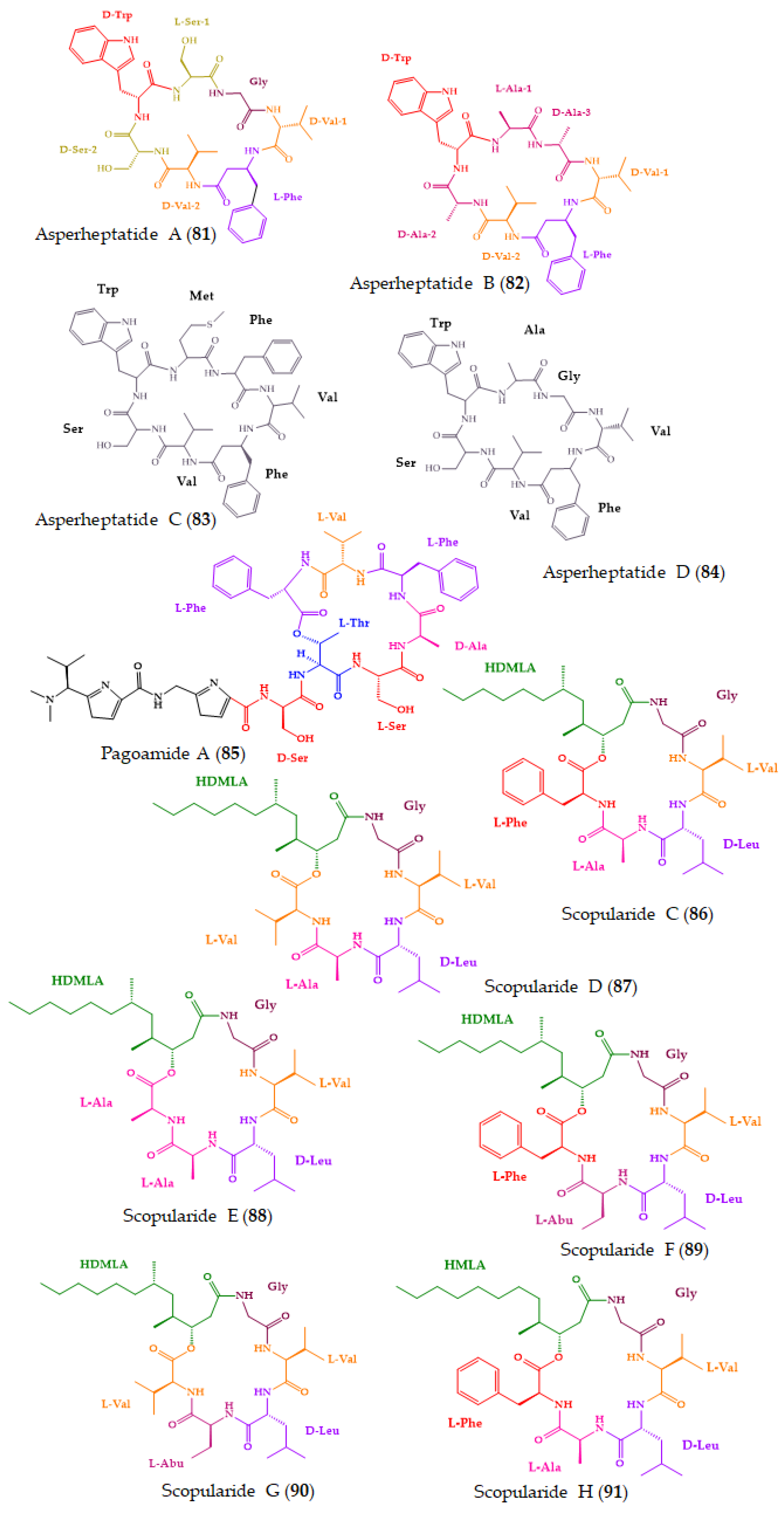
Among the 91 new marine-derived cyclopeptides reported herein, 17 (7591) (Figure 13) were devoid of any activities in the assays performed. Consequently, these compounds should be evaluated for their potential biological effects in other available assays.
A marine-derived fungus, Sesquicillium microsporum RKAG 186, obtained from a marine sediment collected at low tide from the intertidal zone of Frobisher Bay, Nunavut, Canada, furnished four new cyclic decapeptides, auyuittuqamides A–D (7578). The planar structure and the sequence of amino acids were established by HRESIMS and 1D and 2D NMR analysis. Marfey’s method was used to determine the absolute configurations of the amino acid units, except for the d-allo-threonine residue due to a lack of availability of a commercial standard of N-Me-d-allo-threonine. The auyuittuqamides represent a unique class of the decapeptide family due to the presence of multiple N-methylated amino acids. Compounds 7578 showed neither cytotoxic activity against MCF-7, HTB-26, and a human epithelial keratinocyte cell line (IC50 values higher than 10 μM), nor antimicrobial activity at concentrations up to 128 μM [179].
Haloirciniamide A (79) (Figure 13), a dibromopyrrole cyclopeptide containing a chlorohistidine ring, was isolated from a marine sponge, Ircinia sp., which was collected in the Thousand Islands of Indonesia. The planar structure of the compound and its amino acid sequence were elucidated by an extensive 1D and 2D NMR spectral analysis as well as by (+)-HRESI-TOFMS and QTOFMS. The absolute configurations of its amino acid residues were determined by Marfey’s methods, except for N-methyl-chlorohistidine due to the absence of the standard amino acid. Compound 79 neither showed cytotoxicity against four human tumor cell lines, A-549, HT-29, MDA-MB-231, and PSN-1 (concentration that was able to cause 50% cell growth inhibition (GI50) > 1.2 × 1p−5 M), nor inhibited the enzyme topoisomerase I (at a concentration of 1.0 × 10⁻⁵ M). Moreover, 79 was unable to impair the interaction between the programmed cell death protein PD-1 and its natural ligand PDL1 [180].
Unguisin G (80) (Figure 13) was obtained from the culture extract of a marine-derived fungus, Aspergillus candidus NF2412, which was isolated from sponge collected from the East China Sea at Ningbo, Zhejiang, Province, China. The planar structure of 80, established by comprehensive analyses of HRESIMS and 1D and 2D NMR spectra, revealed the presence of non-proteinogenic amino acid residues, kynurenine and γ-aminobutyric acid. The advanced Marfey’s method was used to determine the absolute configurations of the amino acids of the hydrolysate obtained by acid hydrolysis of 80, revealing not only the presence of d-alanine, d-valine, and l-phenylalanine, but also a pair of signals for l- and d-kynurenine. As shown in Figure 14, the LC-MS analysis of d/l-FDAA derivatives of acid hydrolysates of 80 with those of authentic samples showed a pair of ion signals for l- and d- kynurenine in a 1:1 ratio, indicating that this residue in 80 possesses two configurations. Even though 80 was isolated as a pair of diastereomers, attempts to separate them failed.
Compound 80 was evaluated for its antibacterial activity, by the agar diffusion method, against a series of pathogens, including Xanthomonas oryzae pv. oryzicola Swings, Acidovorax avenae subsp. citrulli, Pseudomonas syringae pv. Lachrymans, Bacillus subtilis CICC 10283, Escherichia coli ATCC 25922, Pseudomonas aeruginosa CICC 10351, Micrococcus luteus CMCC(B) 28,001, Candida albicans and S. aureus CMCC(B) 26003; however, it showed no activity against all the tested strains even at a concentration as high as 125 µg/disk [181].
By using MS/MS-based molecular networking guidance, four cyclic heptapeptides, named asperheptatides A–D (8184), were isolated from a culture extract of Aspergillus versicolor strain CHNSCLM-0063, which was isolated from the gorgonian coral, Rumphella aggregata, collected from the Nansha Islands in the South China Sea. The planar structures and amino acid sequences of 81 and 82, which were major compounds, were elucidated by an interpretation of 1D and 2D NMR and HRESIMS spectra. The absolute configurations of the amino acid residues of 81 and 82 were established by an LC analysis of their acid hydrolysates after derivatization with Marfey’s reagent. On the other hand, the planar structures of 83 and 84, which were isolated as trace compounds, were characterized only by ESI-MS/MS fragmentation experiments. Compounds 81 and 82 were tested for their antitubercular activity against M. tuberculosis H37Ra; however, none were active (MIC values ≥ 100 µM). Rifampin and isoniazid were used as positive controls, showing MIC values of 6.25 and 12.5 nM, respectively [182].
By using MS/MS-based molecular networking guidance, pagoamide A (85) (Figure 13), a thiazole-containing cyclodepsipeptide comprising 11 amino acids, was isolated from laboratory cultures of a marine green alga, Derbesia sp., which were collected from a water depth of 12 to 18 m in Fagatele Bay, America Samoa. The planar structure of the compound and the amino acid sequence were established by 1D and 2D NMR as well as HRESIMS spectral analysis. The absolute configurations of the amino acid constituents of 85 were resolved by the advanced Marfey’s method, chiral LC, and ROESY correlations. The configuration of the N,N-dimethyl valine residue was determined as l by chiral LC using a ligand exchange-type CSP [(d)-penicillamine stationary phase] by comparison of the hydrolyzed product with synthetic d/l-N,N-dimethyl valine standards. Interestingly, 85 has two serine and two phenylalanine residues, and each comprises one l- and one d-residues. Density functional theory (DFT) calculations in the Gaussian program to calculate the lowest energy conformers in combination with ROESY correlations allowed the positions of d- and l-serine, and d- and l-phenylalanine residues to be assigned. The cytotoxicity of 85 against H-460 cancer cell line was evaluated; however, it was inactive at the maximum concentration tested (30 μM) [183].
By a comparative global natural product social molecular networking analysis of ×63 co-isolated fungi, new scopularides were isolated and identified: scopularides C–G (8690) from Beauveria sp. CMB-F585, and scopularide H (91) from Scopulariopsis sp. CMB-F115. The chemical structures and absolute configurations of the amino acid residues were assigned by spectroscopic and C3 Marfey’s analysis, together with X-ray analyses of 86 and 91, and biosynthetic considerations. Compounds 8691 did not display significant growth inhibitory activity against a selection of Gram-positive and Gram-negative bacteria (E. coli ATCC 11775, S. aureus ATCC 9144, and three clinical isolates of methicillin-susceptible S. aureus, methicillin-resistant S. aureus, and vancomycin-resistant E. faecalis), a fungus (C. albicans ATCC 10231), and a panel of three human carcinoma cell lines (adherent SW620, NCI-H460, and HepG2) [184].

3.5. Final Remarks

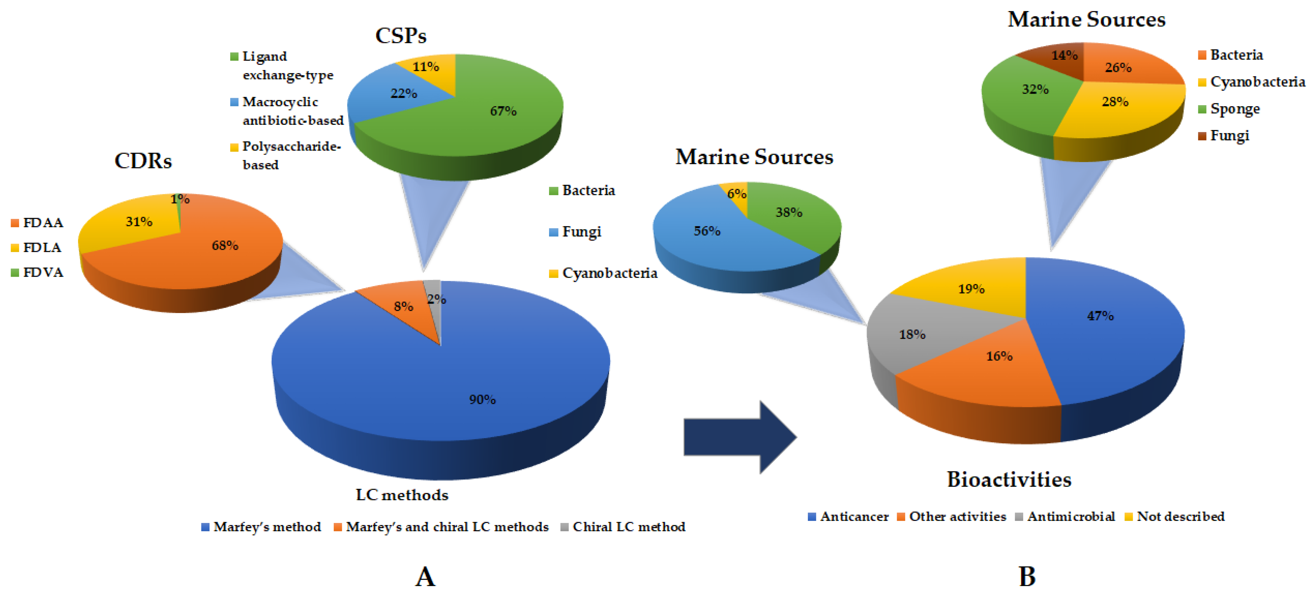
Regarding the stereochemical determination of cyclopeptides amino acid residues by LC methods, Marfey’s analysis was demonstrated to be the most widely used method (Figure 15), allowing the determination of the absolute configurations of 89 cyclopeptides, seven of which were associated with chiral LC. Intriguingly, there were only two marine cyclopeptides whose absolute configurations of their amino acid residues were determined only by chiral LC. As expected, the most used CDRs were FDAA, accounting for 68% of the reported analyses, while FDLA accounted for only 31% of the reported analyses. Considering the chiral LC method, the ligand exchange-type CSPs were used in most cases. There are a few reasons that may justify a massive application of Marfey’s method. First, it is a simple method which offers a better resolution when compared with a chiral LC analysis. Moreover, several derivatizing agents, such as FDAA and FDLA, are commercially available.
It is important to highlight that from January 2018 to November 2022, there were several studies on the structural revision of marine-derived cyclopeptides discovered before 2018, reporting the reinvestigation of the stereochemistry by LC and other methods, for the establishment of complete absolute configurations [138,207,208]. The focus of this review is on marine cyclopeptides, even though LC methods were chosen for the determination of the absolute configurations of the amino acid residues of the recently isolated linear peptides [209,210,211]. Surprisingly, there are still a number of reports describing the identification, structure characterization, and biological activity evaluation of novel marine cyclopeptides without stereochemistry assignments [212], or referring to only the relative configurations [213,214].
As shown in Figure 15, interesting bioactivities have been described for the marine-derived cyclopeptides, mainly anticancer (47%) and antimicrobial (18%). Nevertheless, other activities were found to account for 16% of the reported marine-derived cyclopeptides.
Regarding anticancer activity, it is important to highlight bacilohydrin A (1), which displayed potent cytotoxicity against three cancer cell lines with IC50 values of nM scale. Other relevant examples are bathiapeptides (38), trikoramide A (12), laxaphycin B4 (22), and microsclerodermins N (30) and O (31), with IC50 values ranging from 0.8 to 4.8 µM. Moreover, in addition to cytotoxicity assays, different mechanisms of action were also described. For example, epoxinnamide (2) exhibited antiangiogenesis activity, dentigerumycin E (9) showed antiproliferative and antimetastatic potential, nobilamide I (10) decreased cancer cell motility by inhibiting EMT effectors, ectyoplasin (39) was responsible for apoptotic cell death, phakefustatin A (40) was identified as a RXRα modulator, and asperflomide (28) and asperflosamide (29) showed inhibition of tankyrase1/2. Concerning antimicrobial activity, bacicyclin (44) and ogipeptins A–D (4649) showed the most promising results of antibacterial activity, while trikoramide B (15) displayed quorum-sensing inhibitory effects against PAO1 lasB-gpf and rhlA-gfp bioreporter strains. Moreover, the antiparasitic activity of motobamide (59), antiviral activity of acremonpeptide B (56), and antifungal activity of simplicilliumtides N (53) and O (54) also reflected the biological versatility of marine-derived cyclopeptides.
Fungi in association with macroorganisms and other marine sources (56%) and bacteria (38%) were the main marine sources to uncover marine-derived cyclopeptides with antimicrobial activities. For those exhibiting anticancer activity, sponges (32%), bacteria (26%), and cyanobacteria (28%) were the principal marine-derived cyclopeptides suppliers (Figure 15).
Great structural diversity was found, including cyclopeptides, cyclodepsipeptides, and cyclolipopeptides, with both l- and d-configured amino acid residues. Nevertheless, for some peptides, such as tutuilamides A–C (1921), petrosamide A (62), and croissamide (65), all the amino acids were in the l-configuration. In addition, some interesting stereochemical features were observed. For example, pagoamide A (85) and unguisin G (80) contain amino acids that appear twice in the molecule, but with opposite configuration.
Some of the peptides contained remarkable structural elements, such as non-proteinogenic amino acids. For example, a cinnamoyl moiety in epoxinnamide (2), polythiazole moiety in bathiapeptides (38), kynurenine and γ-aminobutyric acid units in unguisin G (80), C-prenylated tryptophan in trikoramide A (12) and motobamide (59), 4-phenylvaline and dolamethylleucine in triproamide (16), 2-amino-2-butenoic acid and 3-amino-6-hydroxy-2-piperidone in tutuilamides A–C (1921), a p-ethoxyphenyl moiety at the terminus of the ω-phenyl-polyhydroxylated β-amino acid unit in microsclerodermins N (30) and O (31), Apoa residue in theonellamide J (32), 5-cis-Apoa-theopalauamide (33), and theonellamide K (34), β-OH Dabs in ogipeptins A–D (4649), and chlorohistidine in haloirciniamide A (79), among others.

4. Conclusions

Ninety-one cyclopeptides have been isolated from marine sources over the last five years (January 2018–November 2022), and Marfey’s analysis demonstrated to be the most widely used method for the stereochemical determination of their amino acid residues. This review also describes several promising marine-derived cyclopeptides, some of which possess unique structural features, and with relevant biological activities, resulting from diverse marine sources. Despite being a promising area, the application of marine bioactive cyclopeptides is still lacking. Therefore, further research in bioactive cyclopeptides application is necessary to better understand their potential and applicability.

Author Contributions

Conceptualization: C.F.; Data collection and analysis: C.F.; Writing—Original draft preparation: C.F. and R.R.; Writing—Reviewing and Editing: C.F., M.P. and A.K. All authors have read and agreed to the published version of the manuscript.

Funding

This research was supported by national funds through FCT—Foundation for Science and Technology within the scope of UIDB/04423/2020, UIDP/04423/2020 (Group of Natural Products and Medicinal Chemistry—CIIMAR); and under the project PTDC/CTA-AMB/0853/2021, co-financed by COMPETE 2020, Portugal 2020, and the European Union through the ERDF; and by FCT through national funds and the structured program of R&D&I ATLANTIDA (NORTE-01-0145-FEDER-000040), supported by NORTE2020, through ERDF, and CHIRALSINTESE_APSFCT_IINFACTS_2021.

Institutional Review Board Statement

Not applicable.

Informed Consent Statement

Not applicable.

Data Availability Statement

Not applicable.

Acknowledgments

R.R. acknowledges BYTPhD program of CIIMAR and FCT for his grant (UI/BD/150912/2021).

Conflicts of Interest

The authors declare no conflict of interest.

References

  1. Agarwal, R.; Chhabra, P.; Goyal, A.P.; Srivastava, S. Predictive modelling to assess groundwater pollution and integration with water quality index. Int. J. Eng. Adv. Technol. 2019, 8, 1076–1084. [Google Scholar]
  2. Xing, L.; Wang, Z.; Hao, Y.; Zhang, W. Marine Products As a Promising Resource of Bioactive Peptides: Update of Extraction Strategies and Their Physiological Regulatory Effects. J. Agric. Food Chem. 2022, 70, 3081–3095. [Google Scholar] [CrossRef] [PubMed]
  3. Blunt, J.W.; Carroll, A.R.; Copp, B.R.; Davis, R.A.; Keyzers, R.A.; Prinsep, M.R. Marine natural products. Nat. Prod. Rep. 2018, 35, 8–53. [Google Scholar] [CrossRef] [Green Version]
  4. Cheung, R.C.F.; Ng, T.B.; Wong, J.H. Marine peptides: Bioactivities and applications. Mar. Drugs 2015, 13, 4006–4043. [Google Scholar] [CrossRef] [PubMed]
  5. Karthikeyan, A.; Joseph, A.; Nair, B.G. Promising bioactive compounds from the marine environment and their potential effects on various diseases. J. Genet. Eng. Biotechnol. 2022, 20, 14. [Google Scholar] [CrossRef] [PubMed]
  6. Shinde, P.; Banerjee, P.; Mandhare, A. Marine natural products as source of new drugs: A patent review (2015–2018). Expert Opin. Ther. Pat. 2019, 29, 283–309. [Google Scholar] [CrossRef] [PubMed]
  7. Banerjee, P.; Mandhare, A.; Bagalkote, V. Marine natural products as source of new drugs: An updated patent review (July 2018–July 2021). Expert Opin. Ther. Pat. 2022, 32, 317–363. [Google Scholar] [CrossRef]
  8. Macedo, M.W.F.S.; Cunha, N.B.d.; Carneiro, J.A.; Costa, R.A.d.; Alencar, S.A.d.; Cardoso, M.H.; Franco, O.L.; Dias, S.C. Marine organisms as a rich source of biologically active peptides. Front. Mar. Sci. 2021, 889, 667764. [Google Scholar] [CrossRef]
  9. Bhandari, D.; Rafiq, S.; Gat, Y.; Gat, P.; Waghmare, R.; Kumar, V. A Review on Bioactive Peptides: Physiological Functions, Bioavailability and Safety. Int. J. Pept. Res. Ther. 2020, 26, 139–150. [Google Scholar] [CrossRef]
  10. White, K.M.; Rosales, R.; Yildiz, S.; Kehrer, T.; Miorin, L.; Moreno, E.; Jangra, S.; Uccellini, M.B.; Rathnasinghe, R.; Coughlan, L.; et al. Plitidepsin has potent preclinical efficacy against SARS-CoV-2 by targeting the host protein eEF1A. Science 2021, 371, 926–931. [Google Scholar] [CrossRef]
  11. Agrawal, S.; Adholeya, A.; Deshmukh, S.K. The pharmacological potential of non-ribosomal peptides from marine sponge and tunicates. Front. Pharmacol. 2016, 7, 333. [Google Scholar] [CrossRef] [PubMed]
  12. Hassan, H.M.; Degen, D.; Jang, K.H.; Ebright, R.H.; Fenical, W. Correction to: Salinamide F, new depsipeptide antibiotic and inhibitor of bacterial RNA polymerase from a marine-derived Streptomyces sp. (The Journal of Antibiotics, (2015), 68, 3, (206-209), 10.1038/ja.2014.122). J. Antibiot. 2022, 75, 415. [Google Scholar] [CrossRef] [PubMed]
  13. Daletos, G.; Kalscheuer, R.; Koliwer-Brandl, H.; Hartmann, R.; De Voogd, N.J.; Wray, V.; Lin, W.; Proksch, P. Callyaerins from the Marine Sponge Callyspongia aerizusa: Cyclic Peptides with Antitubercular Activity. J. Nat. Prod. 2015, 78, 1910–1925. [Google Scholar] [CrossRef] [PubMed]
  14. Shin, H.J.; Rashid, M.A.; Cartner, L.K.; Bokesch, H.R.; Wilson, J.A.; McMahon, J.B.; Gustafson, K.R. Corrigendum to ‘Stellettapeptins A and B, HIV-inhibitory cyclic depsipeptides from the marine sponge Stelletta sp.’ [Tetrahedron Lett. 56(28) (2015) 4215–4219] (S0040403915008679) (10.1016/j.tetlet.2015.05.058)). Tetrahedron Lett. 2015, 56, 5482. [Google Scholar] [CrossRef]
  15. Bleakley, S.; Hayes, M. Algal proteins: Extraction, application, and challenges concerning production. Foods 2017, 6, 33. [Google Scholar] [CrossRef] [Green Version]
  16. O’connor, J.; Garcia-Vaquero, M.; Meaney, S.; Tiwari, B.K. Bioactive Peptides from Algae: Traditional and Novel Generation Strategies, Structure-Function Relationships, and Bioinformatics as Predictive Tools for Bioactivity. Mar. Drugs 2022, 20, 317. [Google Scholar] [CrossRef]
  17. Pesic, A.; Baumann, H.I.; Kleinschmidt, K.; Ensle, P.; Wiese, J.; Süssmuth, R.D.; Imhoff, J.F. Champacyclin, a new cyclic octapeptide from Streptomyces strain C42 isolated from the Baltic Sea. Mar. Drugs 2013, 11, 4834–4857. [Google Scholar] [CrossRef] [Green Version]
  18. Song, Y.; Li, Q.; Liu, X.; Chen, Y.; Zhang, Y.; Sun, A.; Zhang, W.; Zhang, J.; Ju, J. Cyclic hexapeptides from the deep South China sea-derived Streptomyces scopuliridis SCSIO ZJ46 active against pathogenic gram-positive bacteria. J. Nat. Prod. 2014, 77, 1937–1941. [Google Scholar] [CrossRef]
  19. Sun, P.; Maloney, K.N.; Nam, S.J.; Haste, N.M.; Raju, R.; Aalbersberg, W.; Jensen, P.R.; Nizet, V.; Hensler, M.E.; Fenical, W. Fijimycins A-C, three antibacterial etamycin-class depsipeptides from a marine-derived Streptomyces sp. Bioorg. Med. Chem. 2011, 19, 6557–6562. [Google Scholar] [CrossRef] [Green Version]
  20. Linington, R.G.; González, J.; Ureña, L.D.; Romero, L.I.; Ortega-Barría, E.; Gerwick, W.H. Venturamides A and B: Antimalarial constituents of the Panamanian marine cyanobacterium Oscillatoria sp. J. Nat. Prod. 2007, 70, 397–401. [Google Scholar] [CrossRef]
  21. Linington, R.G.; Edwards, D.J.; Shuman, C.F.; McPhail, K.L.; Matainaho, T.; Gerwick, W.H. Symplocamide A, a potent cytotoxin and chymotrypsin inhibitor from the marine cyanobacterium Symploca sp. J. Nat. Prod. 2008, 71, 22–27. [Google Scholar] [CrossRef] [PubMed]
  22. Milligan, K.E.; Marquez, B.L.; Williamson, R.T.; Gerwick, W.H. Lyngbyabellin B, a toxic and antifungal secondary metabolite from the marine cyanobacterium Lyngbya mojuscula. J. Nat. Prod. 2000, 63, 1440–1443. [Google Scholar] [CrossRef] [PubMed]
  23. De Sá, J.D.M.; Kumla, D.; Dethoup, T.; Kijjoa, A. Bioactive Compounds from Terrestrial and Marine-Derived Fungi of the Genus Neosartorya†. Molecules 2022, 27, 2351. [Google Scholar] [CrossRef]
  24. He, F.; Bao, J.; Zhang, X.Y.; Tu, Z.C.; Shi, Y.M.; Qi, S.H. Asperterrestide A, a cytotoxic cyclic tetrapeptide from the marine-derived fungus Aspergillus terreus SCSGAF0162. J. Nat. Prod. 2013, 76, 1182–1186. [Google Scholar] [CrossRef] [PubMed]
  25. Du, F.Y.; Zhang, P.; Li, X.M.; Li, C.S.; Cui, C.M.; Wang, B.G. Cyclohexadepsipeptides of the isaridin class from the marine-derived fungus Beauveria felina EN-135. J. Nat. Prod. 2014, 77, 1164–1169. [Google Scholar] [CrossRef] [PubMed]
  26. Wyche, T.P.; Hou, Y.; Vazquez-Rivera, E.; Braun, D.; Bugni, T.S. Peptidolipins B-F, antibacterial lipopeptides from an ascidian-derived Nocardia sp. J. Nat. Prod. 2012, 75, 735–740. [Google Scholar] [CrossRef] [Green Version]
  27. Amelia, T.S.M.; Suaberon, F.A.C.; Vad, J.; Fahmi, A.D.M.; Saludes, J.P.; Bhubalan, K. Recent Advances of Marine Sponge-Associated Microorganisms as a Source of Commercially Viable Natural Products. Mar. Biotechnol. 2022, 24, 492–512. [Google Scholar] [CrossRef] [PubMed]
  28. Prompanya, C.; Fernandes, C.; Cravo, S.; Pinto, M.M.M.; Dethoup, T.; Silva, A.M.S.; Kijjoa, A. A new cyclic hexapeptide and a new isocoumarin derivative from the marine sponge-associated fungus Aspergillus similanensis KUFA 0013. Mar. Drugs 2015, 13, 1432–1450. [Google Scholar] [CrossRef] [Green Version]
  29. Zin, W.W.M.; Buttachon, S.; Dethoup, T.; Fernandes, C.; Cravo, S.; Pinto, M.M.M.; Gales, L.; Pereira, J.A.; Silva, A.M.S.; Sekeroglu, N.; et al. New cyclotetrapeptides and a new diketopiperzine derivative from the marine sponge-associated fungus Neosartorya glabra KUFA 0702. Mar. Drugs 2016, 14, 136. [Google Scholar] [CrossRef] [Green Version]
  30. Mayer, A.M.; Rodríguez, A.D.; Taglialatela-Scafati, O.; Fusetani, N. Marine pharmacology in 2009–2011: Marine compounds with antibacterial, antidiabetic, antifungal, anti-inflammatory, antiprotozoal, antituberculosis, and antiviral activities; affecting the immune and nervous systems, and other miscellaneous mechanisms of action. Mar. Drugs 2013, 11, 2510–2573. [Google Scholar]
  31. Ghanbari, R. Review on the Bioactive Peptides from Marine Sources: Indication for Health Effects. Int. J. Pept. Res. Ther. 2019, 25, 1187–1199. [Google Scholar] [CrossRef]
  32. Cunha, S.A.; Pintado, M.E. Bioactive peptides derived from marine sources: Biological and functional properties. Trends Food Sci. Technol. 2022, 119, 348–370. [Google Scholar] [CrossRef]
  33. Rangel, M.; José Correia de Santana, C.; Pinheiro, A.; Dos Anjos, L.; Barth, T.; Rodrigues Pires Júnior, O.; Fontes, W.; Castro, M.S. Marine depsipeptides as promising pharmacotherapeutic agents. Curr.t Protein Pep. Sci. 2017, 18, 72–91. [Google Scholar] [CrossRef] [PubMed]
  34. Anjum, K.; Abbas, S.Q.; Akhter, N.; Shagufta, B.I.; Shah, S.A.A.; Hassan, S.S.U. Emerging biopharmaceuticals from bioactive peptides derived from marine organisms. Curr. Protein Pept. Sci. 2017, 90, 12–30. [Google Scholar] [CrossRef] [PubMed]
  35. Venkatesan, J.; Anil, S.; Kim, S.-K.; Shim, M.S. Marine fish proteins and peptides for cosmeceuticals: A review. Mar. Drugs 2017, 15, 143. [Google Scholar] [CrossRef] [PubMed]
  36. Corinaldesi, C.; Barone, G.; Marcellini, F.; Dell’Anno, A.; Danovaro, R. Marine microbial-derived molecules and their potential use in cosmeceutical and cosmetic products. Mar. Drugs 2017, 15, 118. [Google Scholar] [CrossRef]
  37. Kim, S.-K.; Wijesekara, I. Development and biological activities of marine-derived bioactive peptides: A review. J. Funct. Foods 2010, 2, 1–9. [Google Scholar] [CrossRef]
  38. Charoensiddhi, S.; Conlon, M.A.; Franco, C.M.; Zhang, W. The development of seaweed-derived bioactive compounds for use as prebiotics and nutraceuticals using enzyme technologies. Trends Food Sci. Technol. 2017, 70, 20–33. [Google Scholar] [CrossRef] [Green Version]
  39. Ghosh, S.; Sarkar, T.; Pati, S.; Kari, Z.A.; Edinur, H.A.; Chakraborty, R. Novel Bioactive Compounds From Marine Sources as a Tool for Functional Food Development. Front. Mar. Sci. 2022, 9, 832957. [Google Scholar] [CrossRef]
  40. McIntosh, M.; Cruz, L.J.; Hunkapiller, M.W.; Gray, W.R.; Olivera, B.M. Isolation and structure of a peptide toxin from the marine snail Conus magus. Arch. Biochem. Biophys. 1982, 218, 329–334. [Google Scholar] [CrossRef]
  41. Pettit, G.R.; Kamano, Y.; Herald, C.L.; Tuinman, A.A.; Boettner, F.E.; Kizu, H.; Schmidt, J.M.; Baczynskyi, L.; Tomer, K.B.; Bontems, R.J. The Isolation and Structure of a Remarkable Marine Animal Antineoplastic Constituent: Dolastatin 10. J. Am. Chem. Soc. 1987, 109, 6883–6885. [Google Scholar] [CrossRef]
  42. Sridhar, K.; Inbaraj, B.S.; Chen, B.H. Recent developments on production, purification and biological activity of marine peptides. Food Res. Int. 2021, 147, 468. [Google Scholar] [CrossRef] [PubMed]
  43. Zorzi, A.; Deyle, K.; Heinis, C. Cyclic peptide therapeutics: Past, present and future. Curr. Opin. Chem. Biol. 2017, 38, 24–29. [Google Scholar] [CrossRef] [PubMed] [Green Version]
  44. Pfeifer, V.; Nicholson, G.J.; Ries, J.; Recktenwald, J.r.; Schefer, A.B.; Shawky, R.M.; Schröder, J.; Wohlleben, W.; Pelzer, S. A polyketide synthase in glycopeptide biosynthesis: The biosynthesis of the non-proteinogenic amino acid (S)-3, 5-dihydroxyphenylglycine. J. Biol. Chem. 2001, 276, 38370–38377. [Google Scholar] [CrossRef] [Green Version]
  45. Jang, J.P.; Nogawa, T.; Futamura, Y.; Shimizu, T.; Hashizume, D.; Takahashi, S.; Jang, J.H.; Ahn, J.S.; Osada, H. Octaminomycins A and B, cyclic octadepsipeptides active against Plasmodium falciparum. J. Nat. Prod. 2017, 80, 134–140. [Google Scholar] [CrossRef]
  46. Fukuhara, K.; Takada, K.; Okada, S.; Matsunaga, S. Nazumazoles A-C, cyclic pentapeptides dimerized through a disulfide bond from the marine sponge Theonella swinhoei. Org. Lett. 2015, 17, 2646–2648. [Google Scholar] [CrossRef]
  47. Luo, S.; Krunic, A.; Kang, H.S.; Chen, W.L.; Woodard, J.L.; Fuchs, J.R.; Swanson, S.M.; Orjala, J. Trichormamides A and B with antiproliferative activity from the cultured freshwater cyanobacterium Trichormus sp. UIC 10339. J. Nat. Prod. 2014, 77, 1871–1880. [Google Scholar] [CrossRef] [Green Version]
  48. Raaijmakers, J.M.; de Bruijn, I.; Nybroe, O.; Ongena, M. Natural functions of lipopeptides from Bacillus and Pseudomonas: More than surfactants and antibiotics. FEMS Microbiol. Rev. 2010, 34, 1037–1062. [Google Scholar] [CrossRef] [Green Version]
  49. Sieber, S.A.; Marahiel, M.A. Molecular mechanisms underlying nonribosomal peptide synthesis: Approaches to new antibiotics. Chem. Rev. 2005, 105, 715–738. [Google Scholar] [CrossRef]
  50. Sable, R.; Parajuli, P.; Jois, S. Peptides, peptidomimetics, and polypeptides from marine sources: A wealth of natural sources for pharmaceutical applications. Mar. Drugs 2017, 15, 124. [Google Scholar] [CrossRef] [Green Version]
  51. van der Pijl, P.C.; Kies, A.K.; Ten Have, G.A.; Duchateau, G.S.; Deutz, N.E. Pharmacokinetics of proline-rich tripeptides in the pig. Peptides 2008, 29, 2196–2202. [Google Scholar] [CrossRef] [PubMed]
  52. Sparidans, R.W.; Stokvis, E.; Jimeno, J.M.; López-Lázaro, L.; Schellens, J.H.; Beijnen, J.H. Chemical and enzymatic stability of a cyclic depsipeptide, the novel, marine-derived, anti-cancer agent kahalalide F. Anti-Cancer Drugs 2001, 12, 575–582. [Google Scholar] [CrossRef]
  53. Sera, Y.; Adachi, K.; Nishida, F.; Shizuri, Y. Isolation and evaluation of nonsiderophore cyclic peptides from marine sponges. Biochem. Biophys. Res. Commun. 2001, 283, 976–981. [Google Scholar] [CrossRef]
  54. Liu, L.; Zheng, Y.-Y.; Shao, C.-L.; Wang, C.-Y. Metabolites from marine invertebrates and their symbiotic microorganisms: Molecular diversity discovery, mining, and application. Mar. Life Sci. Technol. 2019, 1, 60–94. [Google Scholar] [CrossRef] [Green Version]
  55. Vining, O.B.; Medina, R.A.; Mitchell, E.A.; Videau, P.; Li, D.; Serrill, J.D.; Kelly, J.X.; Gerwick, W.H.; Proteau, P.J.; Ishmael, J.E. Depsipeptide companeramides from a panamanian marine cyanobacterium associated with the coibamide producer. J. Nat. Prod. 2015, 78, 413–420. [Google Scholar] [CrossRef] [PubMed] [Green Version]
  56. Ding, C.Y.G.; Ong, J.F.M.; Goh, H.C.; Coffill, C.R.; Tan, L.T. Benderamide A, a Cyclic Depsipeptide from a Singapore Collection of Marine Cyanobacterium cf. Lyngbya sp. Mar. Drugs 2018, 16, 409. [Google Scholar] [CrossRef] [PubMed] [Green Version]
  57. Oku, N.; Kawabata, K.; Adachi, K.; Katsuta, A.; Shizuri, Y. Unnarmicins A and C, new antibacterial depsipeptides produced by marine bacterium Photobacterium sp. MBIC06485. J. Antibiot. 2008, 61, 11–17. [Google Scholar] [CrossRef] [Green Version]
  58. Ratnayake, A.S.; Bugni, T.S.; Feng, X.; Harper, M.K.; Skalicky, J.J.; Mohammed, K.A.; Andjelic, C.D.; Barrows, L.R.; Ireland, C.M. Theopapuamide, a cyclic depsipeptide from a Papua New Guinea lithistid sponge Theonella swinhoei. J. Nat. Prod. 2006, 69, 1582–1586. [Google Scholar] [CrossRef] [Green Version]
  59. Zhang, S.-K.; Song, J.-w.; Gong, F.; Li, S.-B.; Chang, H.-Y.; Xie, H.-M.; Gao, H.-W.; Tan, Y.-X.; Ji, S.-P. Design of an α-helical antimicrobial peptide with improved cell-selective and potent anti-biofilm activity. Sci. Rep. 2016, 6, 1–13. [Google Scholar] [CrossRef] [Green Version]
  60. Ravichandran, S.; Kumaravel, K.; Rameshkumar, G.; Ajithkumar, T. Antimicrobial peptides from the marine fishes. Res. J. Immunol. 2010, 3, 146–156. [Google Scholar] [CrossRef] [Green Version]
  61. Partridge, A.W.; Kaan, H.Y.K.; Juang, Y.-C.; Sadruddin, A.; Lim, S.; Brown, C.J.; Ng, S.; Thean, D.; Ferrer, F.; Johannes, C. Incorporation of putative helix-breaking amino acids in the design of novel stapled peptides: Exploring biophysical and cellular permeability properties. Molecules 2019, 24, 2292. [Google Scholar] [CrossRef] [PubMed] [Green Version]
  62. Falanga, A.; Lombardi, L.; Franci, G.; Vitiello, M.; Iovene, M.R.; Morelli, G.; Galdiero, M.; Galdiero, S. Marine antimicrobial peptides: Nature provides templates for the design of novel compounds against pathogenic bacteria. Int. J. Mol. Sci. 2016, 17, 785. [Google Scholar] [CrossRef] [PubMed]
  63. Gerwick, W.H.; Fenner, A.M. Drug discovery from marine microbes. Microb. Ecol. 2013, 65, 800–806. [Google Scholar] [CrossRef]
  64. Rocha-Martin, J.; Harrington, C.; Dobson, A.D.; O’Gara, F. Emerging strategies and integrated systems microbiology technologies for biodiscovery of marine bioactive compounds. Mar. Drugs 2014, 12, 3516–3559. [Google Scholar] [CrossRef] [PubMed]
  65. Kang, H.K.; Seo, C.H.; Park, Y. Marine peptides and their anti-infective activities. Mar. Drugs 2015, 13, 618–654. [Google Scholar] [CrossRef] [PubMed] [Green Version]
  66. Brito, A.; Gaifem, J.; Ramos, V.; Glukhov, E.; Dorrestein, P.C.; Gerwick, W.H.; Vasconcelos, V.M.; Mendes, M.V.; Tamagnini, P. Bioprospecting Portuguese Atlantic coast cyanobacteria for bioactive secondary metabolites reveals untapped chemodiversity. Algal Res. 2015, 9, 218–226. [Google Scholar] [CrossRef] [Green Version]
  67. Desriac, F.; Jégou, C.; Balnois, E.; Brillet, B.; Le Chevalier, P.; Fleury, Y. Antimicrobial peptides from marine proteobacteria. Mar. Drugs 2013, 11, 3632–3660. [Google Scholar] [CrossRef] [Green Version]
  68. Fisch, K.M. Biosynthesis of natural products by microbial iterative hybrid PKS–NRPS. RSC advances 2013, 3, 18228–18247. [Google Scholar] [CrossRef] [Green Version]
  69. Muttenthaler, M.; King, G.F.; Adams, D.J.; Alewood, P.F. Trends in peptide drug discovery. Nat. Rev. Drug Discov. 2021, 20, 309–325. [Google Scholar] [CrossRef]
  70. Fang, W.Y.; Dahiya, R.; Qin, H.L.; Mourya, R.; Maharaj, S. Natural proline-rich cyclopolypeptides from marine organisms: Chemistry, synthetic methodologies and biological status. Mar. Drugs 2016, 14, 194. [Google Scholar] [CrossRef] [Green Version]
  71. Evidente, A. Bioactive Lipodepsipeptides Produced by Bacteria and Fungi †. Int. J. Mol. Sci. 2022, 23, 12342. [Google Scholar] [CrossRef] [PubMed]
  72. Kohli, R.M.; Walsh, C.T.; Burkart, M.D. Biomimetic synthesis and optimization of cyclic peptide antibiotics. Nature 2002, 418, 658–661. [Google Scholar] [CrossRef] [PubMed]
  73. Albericio, F. Developments in peptide and amide synthesis. Curr. Opin. Chem. Biol. 2004, 8, 211–221. [Google Scholar] [CrossRef] [PubMed]
  74. Jiang, S.; Li, Z.; Ding, K.; Roller, P.P. Recent progress of synthetic studies to peptide and peptidomimetic cyclization. Curr. Org. Chem. 2008, 12, 1502–1542. [Google Scholar] [CrossRef]
  75. Lambert, J.N.; Mitchell, J.P.; Roberts, K.D. The synthesis of cyclic peptides. J. Chem. Soc. Perkin Trans. 1 2001, 5, 471–484. [Google Scholar] [CrossRef]
  76. Nielsen, D.S.; Shepherd, N.E.; Xu, W.; Lucke, A.J.; Stoermer, M.J.; Fairlie, D.P. Orally absorbed cyclic peptides. Chem. Rev. 2017, 117, 8094–8128. [Google Scholar] [CrossRef] [PubMed] [Green Version]
  77. Hamada, Y.; Shioiri, T. Recent progress of the synthetic studies of biologically active marine cyclic peptides and depsipeptides. Chem. Rev. 2005, 105, 4441–4482. [Google Scholar] [CrossRef]
  78. Ferrazzano, L.; Catani, M.; Cavazzini, A.; Martelli, G.; Corbisiero, D.; Cantelmi, P.; Fantoni, T.; Mattellone, A.; De Luca, C.; Felletti, S.; et al. Sustainability in peptide chemistry: Current synthesis and purification technologies and future challenges. Green Chem. 2022, 24, 975–1020. [Google Scholar] [CrossRef]
  79. Al Musaimi, O.; De La Torre, B.G.; Albericio, F. Greening Fmoc/tBu solid-phase peptide synthesis. Green Chem. 2020, 22, 996–1018. [Google Scholar] [CrossRef]
  80. Jing, X.; Jin, K. A gold mine for drug discovery: Strategies to develop cyclic peptides into therapies. Med. Res. Rev. 2020, 40, 753–810. [Google Scholar] [CrossRef]
  81. Gentilucci, L.; de Marco, R.; Cerisoli, L. Chemical modifications designed to improve peptide stability: Incorporation of non-natural amino acids, pseudo-peptide bonds, and cyclization. Curr. Pharm. Des. 2010, 16, 3185–3203. [Google Scholar] [CrossRef] [PubMed]
  82. Wallace, R.J. Acetylation of peptides inhibits their degradation by rumen micro-organisms. Br. J. Nutr. 1992, 68, 365–372. [Google Scholar] [CrossRef] [PubMed]
  83. Moradi, S.V.; Hussein, W.M.; Varamini, P.; Simerska, P.; Toth, I. Glycosylation, an effective synthetic strategy to improve the bioavailability of therapeutic peptides. Chem. Sci. 2016, 7, 2492–2500. [Google Scholar] [CrossRef] [PubMed] [Green Version]
  84. Javed, S.; Kohli, K.; Ali, M. Reassessing bioavailability of silymarin. Altern. Med. Rev. 2011, 16, 239. [Google Scholar] [PubMed]
  85. Lau, J.; Bloch, P.; Schäffer, L.; Pettersson, I.; Spetzler, J.; Kofoed, J.; Madsen, K.; Knudsen, L.B.; McGuire, J.; Steensgaard, D.B. Discovery of the once-weekly glucagon-like peptide-1 (GLP-1) analogue semaglutide. J. Med. Chem. 2015, 58, 7370–7380. [Google Scholar] [CrossRef] [PubMed]
  86. Trier, S.; Linderoth, L.; Bjerregaard, S.; Strauss, H.M.; Rahbek, U.L.; Andresen, T.L. Acylation of salmon calcitonin modulates in vitro intestinal peptide flux through membrane permeability enhancement. Eur. J. Pharm. Biopharm. 2015, 96, 329–337. [Google Scholar] [CrossRef] [Green Version]
  87. Kong, L.Y.; Wang, P. Determination of the absolute configuration of natural products. Chin. J. Nat. Med. 2013, 11, 193–198. [Google Scholar] [CrossRef]
  88. Zhu, T.H.; Ma, H.G.; Wang, C.; Zhao, C.Y.; Mei, X.G.; Zhu, W.M. Determination methods for absolute configuration of natural products. J. Int. Pharm. Res. 2015, 42, 773–785. [Google Scholar] [CrossRef]
  89. Harada, N. Determination of absolute configurations by X-ray crystallography and 1H NMR anisotropy. Chirality 2008, 20, 691–723. [Google Scholar] [CrossRef]
  90. Flack, H.D.; Bernardinelli, G. The use of X-ray crystallography to determine absolute configuration. Chirality 2008, 20, 681–690. [Google Scholar] [CrossRef]
  91. Freedman, T.B.; Cao, X.; Dukor, R.; Nafie, L.A. Absolute Configuration Determination of Chiral Molecules in the Solution State Using Vibrational Circular Dichroism. Chirality 2003, 15, 743–758. [Google Scholar] [CrossRef] [PubMed]
  92. Joseph-Nathan, P.; Gordillo-Román, B. Vibrational circular dichroism absolute configuration determination of natural products. Prog. Chem. Org. Nat. Prod. 2015, 100, 311–452. [Google Scholar] [CrossRef] [PubMed]
  93. Batista, J.M.; Da Silva Bolzani, V. Determination of the absolute configuration of natural product molecules using vibrational circular dichroism. Stud. Nat. Prod. Chem. 2014, 41, 383–417. [Google Scholar] [CrossRef]
  94. Zhao, S.D.; Shen, L.; Luo, D.Q.; Zhu, H.J. Progression of absolute configuration determination in natural product chemistry using optical rotation (dispersion), matrix determinant and electronic circular dichroism methods. Curr. Org. Chem. 2011, 15, 1843–1862. [Google Scholar] [CrossRef] [Green Version]
  95. Li, X.C.; Ferreira, D.; Ding, Y. Determination of absolute configuration of natural products: Theoretical calculation of electronic circular dichroism as a tool. Curr. Org. Chem. 2010, 14, 1678–1697. [Google Scholar] [CrossRef] [PubMed]
  96. Zhang, A.H.; Tan, R.X. Theoretical calculation of electronic circular dichroism: A promising tool for the determination of absolute configuration of natural products. J. Int. Pharm. Res. 2015, 42, 734–737. [Google Scholar] [CrossRef]
  97. Berova, N.; Di Bari, L.; Pescitelli, G. Application of electronic circular dichroism in configurational and conformational analysis of organic compounds. Chem. Soc. Rev. 2007, 36, 914–931. [Google Scholar] [CrossRef]
  98. Wenzel, T.J.; Chisholm, C.D. Assignment of absolute configuration using chiral reagents and NMR spectroscopy. Chirality 2011, 23, 190–214. [Google Scholar] [CrossRef]
  99. Mishra, S.K.; Suryaprakash, N. Some new protocols for the assignment of absolute configuration by NMR spectroscopy using chiral solvating agents and CDAs. Tetrahedron Asymmetry 2017, 28, 1220–1232. [Google Scholar] [CrossRef]
  100. Seco, J.M.; Quiñoá, E.; Riguera, R. The Assignment of Absolute Configuration by NMR. Chem. Rev. 2004, 104, 17–117. [Google Scholar] [CrossRef]
  101. Takiguchi, S.; Hirota-Takahata, Y.; Nishi, T. Application of the advanced Marfey’s method for the determination of the absolute configuration of ogipeptins. Tetrahedron Lett. 2022, 96, 153760. [Google Scholar] [CrossRef]
  102. Vijayasarathy, S.; Prasad, P.; Fremlin, L.J.; Ratnayake, R.; Salim, A.A.; Khalil, Z.; Capon, R.J. C3 and 2D C3 Marfey’s Methods for Amino Acid Analysis in Natural Products. J. Nat. Prod. 2016, 79, 421–427. [Google Scholar] [CrossRef] [PubMed]
  103. Fujii, K.; Ikai, Y.; Mayumi, T.; Oka, H.; Suzuki, M.; Harada, K.I. A nonempirical method using LC/MS for determination of the absolute configuration of constituent amino acids in a peptide: Elucidation of limitations of Marfey’s method and of its separation mechanism. Anal. Chem. 1997, 69, 3346–3352. [Google Scholar] [CrossRef]
  104. Fujii, K.; Harada, K.I. A nonempirical method using LC/MS for determination of the absolute configuration of constituent amino acids in a peptide: Combination of Marfey’s method with mass spectrometry and its practical application. Anal. Chem. 1997, 69, 5146–5151. [Google Scholar] [CrossRef]
  105. Montaser, R.; Abboud, K.A.; Paul, V.J.; Luesch, H. Pitiprolamide, a proline-rich dolastatin 16 analogue from the marine cyanobacterium Lyngbya majuscula from guam. J. Nat. Prod. 2011, 74, 109–112. [Google Scholar] [CrossRef] [Green Version]
  106. Montaser, R.; Paul, V.J.; Luesch, H. Pitipeptolides C-F, antimycobacterial cyclodepsipeptides from the marine cyanobacterium Lyngbya majuscula from Guam. Phytochemistry 2011, 72, 2068–2074. [Google Scholar] [CrossRef] [Green Version]
  107. Huang, H.; She, Z.; Lin, Y.; Vrijmoed, L.L.P.; Lin, W. Cyclic peptides from an endophytic fungus obtained from a mangrove leaf (Kandelia candel). J. Nat. Prod. 2007, 70, 1696–1699. [Google Scholar] [CrossRef]
  108. Iwasaki, A.; Ohno, O.; Sumimoto, S.; Matsubara, T.; Shimada, S.; Sato, T.; Suenaga, K. Mebamamides A and B, Cyclic Lipopeptides Isolated from the Green Alga Derbesia marina. J. Nat. Prod. 2015, 78, 901–908. [Google Scholar] [CrossRef]
  109. Tareq, F.S.; Lee, M.A.; Lee, H.S.; Lee, J.S.; Lee, Y.J.; Shin, H.J. Gageostatins A-C, antimicrobial linear lipopeptides from a marine Bacillus subtilis. Mar. Drugs 2014, 12, 871–885. [Google Scholar] [CrossRef] [Green Version]
  110. Phyo, Y.Z.; Ribeiro, J.; Fernandes, C.; Kijjoa, A.; Pinto, M.M. Marine natural peptides: Determination of absolute configuration using liquid chromatography methods and evaluation of bioactivities. Molecules 2018, 23, 306. [Google Scholar] [CrossRef] [Green Version]
  111. Phan, C.S.; Mehjabin, J.J.; Anas, A.R.J.; Hayasaka, M.; Onoki, R.; Wang, J.; Umezawa, T.; Washio, K.; Morikawa, M.; Okino, T. Nostosin G and Spiroidesin B from the Cyanobacterium Dolichospermum sp. NIES-1697. J. Nat. Prod. 2022, 85, 2000–2005. [Google Scholar] [CrossRef] [PubMed]
  112. Shin, Y.H.; Ban, Y.H.; Shin, J.; Park, I.W.; Yoon, S.; Ko, K.; Shin, J.; Nam, S.J.; Winter, J.M.; Kim, Y.; et al. Azetidine-Bearing Non-Ribosomal Peptides, Bonnevillamides D and E, Isolated from a Carrion Beetle-Associated Actinomycete. J. Org. Chem. 2021, 86, 11149–11159. [Google Scholar] [CrossRef]
  113. Ekanayake, D.I.; Perlatti, B.; Swenson, D.C.; Põldmaa, K.; Bills, G.F.; Gloer, J.B. Broomeanamides: Cyclic Octapeptides from an Isolate of the Fungicolous Ascomycete Sphaerostilbella broomeanafrom India. J. Nat. Prod. 2021, 84, 2028–2034. [Google Scholar] [CrossRef] [PubMed]
  114. Fernández-Pastor, I.; González-Menéndez, V.; Annang, F.; Toro, C.; Mackenzie, T.A.; Bosch-Navarrete, C.; Genilloud, O.; Reyes, F. Pipecolisporin, a novel cyclic peptide with antimalarial and antitrypanosome activities from a wheat endophytic Nigrospora oryzae. Pharmaceuticals 2021, 14, 268. [Google Scholar] [CrossRef] [PubMed]
  115. Parys, W.; Dołowy, M.; Pyka-Pająk, A. Significance of Chromatographic Techniques in Pharmaceutical Analysis. Processes 2022, 10, 172. [Google Scholar] [CrossRef]
  116. Bhushan, R.; Brückner, H. Use of Marfey’s reagent and analogs for chiral amino acid analysis: Assessment and applications to natural products and biological systems. J. Chromatogr. B Anal. Technol. Biomed. Life Sci. 2011, 879, 3148–3161. [Google Scholar] [CrossRef]
  117. Péter, A.; Olajos, E.; Casimir, R.; Tourwé, D.; Broxterman, Q.B.; Kaptein, B.; Armstrong, D.W. High-performance liquid chromatographic separation of the enantiomers of unusual α-amino acid analogues. J. Chromatogr. A 2000, 871, 105–113. [Google Scholar] [CrossRef]
  118. Moon, K.; Lim, C.; Kim, S.; Oh, D.C. Facile determination of the absolute configurations of α-hydroxy acids by chiral derivatization coupled with liquid chromatography-mass spectrometry analysis. J. Chromatogr. A 2013, 1272, 141–144. [Google Scholar] [CrossRef]
  119. Bhushan, R.; Brückner, H. Marfey’s reagent for chiral amino acid analysis: A review. Amino Acids 2004, 27, 231–247. [Google Scholar] [CrossRef]
  120. Bhushan, R.; Kumar, R. Reversed-phase high performance liquid chromatographic separation of diastereomers of β-amino alcohols and microwave assisted synthesis of Marfey’s reagent, its chiral variants and diastereomers. J. Chromatogr. A 2009, 1216, 2592–2596. [Google Scholar] [CrossRef]
  121. Marfey, P. Determination of D-amino acids. II. Use of a bifunctional reagent, 1,5-difluoro-2,4-dinitrobenzene. Carlsberg Res. Commun. 1984, 49, 591–596. [Google Scholar] [CrossRef] [Green Version]
  122. Sethi, S.; Martens, J.; Bhushan, R. Assessment and application of Marfey’s reagent and analogs in enantioseparation: A decade’s perspective. Biomed. Chromatogr. 2021, 35, 4990. [Google Scholar] [CrossRef] [PubMed]
  123. Hess, S. A universal HPLC-MS method to determine the stereochemistry of common and unusual amino acids. Methods Mol. Biol. 2012, 828, 63–75. [Google Scholar] [CrossRef] [PubMed]
  124. Dolowy, M.; Pyka, A. Application of TLC, HPLC and GC methods to the study of amino acid and peptide enantiomers: A review. Biomed. Chromatogr. 2014, 28, 84–101. [Google Scholar] [CrossRef]
  125. Harada, K.i.; Fujii, K.; Mayumi, T.; Hibino, Y.; Suzuki, M.; Ikai, Y.; Oka, H. A method usingL/CMS for determination of absolute configuration of constituent amino acids in peptide–advanced Marfey’s method. Tetrahedron Lett. 1995, 36, 1515–1518. [Google Scholar] [CrossRef]
  126. Harada, K.I.; Fujii, K.; Hayashi, K.; Suzuki, M.; Ikai, Y.; Oka, H. Application of D,L-FDLA derivatization to determination of absolute configuration of constituent amino acids in peptide by advanced Marfey’s method. Tetrahedron Lett. 1996, 37, 3001–3004. [Google Scholar] [CrossRef]
  127. Zhang, J.H.; Xie, S.M.; Yuan, L.M. Recent progress in the development of chiral stationary phases for high-performance liquid chromatography. J. Sep. Sci. 2022, 45, 51–77. [Google Scholar] [CrossRef]
  128. Fernandes, C.; Phyo, Y.Z.; Silva, A.S.; Tiritan, M.E.; Kijjoa, A.; Pinto, M.M.M. Chiral Stationary Phases Based on Small Molecules: An Update of the Last 17 Years. Sep. Purif. Rev. 2018, 47, 89–123. [Google Scholar] [CrossRef]
  129. Ribeiro, J.; Tiritan, M.E.; Pinto, M.M.M.; Fernandes, C. Chiral stationary phases for liquid chromatography based on chitin- and chitosan-derived marine polysaccharides. Symmetry 2017, 9, 190. [Google Scholar] [CrossRef]
  130. Teixeira, J.; Tiritan, M.E.; Pinto, M.M.M.; Fernandes, C. Chiral stationary phases for liquid chromatography: Recent developments. Molecules 2019, 24, 865. [Google Scholar] [CrossRef] [Green Version]
  131. Fernandes, C.; Lima, R.; Pinto, M.M.M.; Tiritan, M.E. Chromatographic supports for enantioselective liquid chromatography: Evolution and innovative trends. J. Chromatogr. A 2022, 1684, 463555. [Google Scholar] [CrossRef] [PubMed]
  132. Fernandes, C.; Teixeira, J.; Pinto, M.M.M.; Tiritan, M.E. Strategies for preparation of chiral stationary phases: Progress on coating and immobilization methods. Molecules 2021, 26, 5477. [Google Scholar] [CrossRef] [PubMed]
  133. Ilisz, I.; Pataj, Z.; Aranyi, A.; Péter, A. Macrocyclic antibiotic selectors in direct HPLC enantioseparations. Sep. Purif. Rev. 2012, 41, 207–249. [Google Scholar] [CrossRef]
  134. Berkecz, R.; Németi, G.; Péter, A.; Ilisz, I. Liquid chromatographic enantioseparations utilizing chiral stationary phases based on crown ethers and cyclofructans. Molecules 2021, 26, 4648. [Google Scholar] [CrossRef]
  135. Carenzi, G.; Sacchi, S.; Abbondi, M.; Pollegioni, L. Direct chromatographic methods for enantioresolution of amino acids: Recent developments. Amino Acids 2020, 52, 849–862. [Google Scholar] [CrossRef]
  136. Ianni, F.; Pucciarini, L.; Carotti, A.; Natalini, S.; Raskildina, G.Z.; Sardella, R.; Natalini, B. Last ten years (2008–2018) of chiral ligand-exchange chromatography in HPLC: An updated review. J. Sep. Sci. 2019, 42, 21–37. [Google Scholar] [CrossRef] [Green Version]
  137. Tiritan, M.E.; Ribeiro, A.R.; Fernandes, C.; Pinto, M. Chiral Pharmaceuticals. In Kirk-Othmer Encyclopedia of Chemicl Technology; Wiley: New York, NY, USA, 2016. [Google Scholar]
  138. Kanki, D.; Nakamukai, S.; Ogura, Y.; Takikawa, H.; Ise, Y.; Morii, Y.; Yamawaki, N.; Takatani, T.; Arakawa, O.; Okada, S.; et al. Homophymamide A, Heterodetic Cyclic Tetrapeptide from a Homophymia sp. Marine Sponge: A Cautionary Note on Configurational Assignment of Peptides That Contain a Ureido Linkage. J. Nat. Prod. 2021, 84, 1848–1853. [Google Scholar] [CrossRef]
  139. Martin, M.J.; Rodriguez-Acebes, R.; Garcia-Ramos, Y.; Martinez, V.; Murcia, C.; Digon, I.; Marco, I.; Pelay-Gimeno, M.; Fernández, R.; Reyes, F.; et al. Stellatolides, a new cyclodepsipeptide family from the sponge Ecionemia acervus: Isolation, solid-phase total synthesis, and full structural assignment of stellatolide a. J. Am. Chem. Soc. 2014, 136, 6754–6762. [Google Scholar] [CrossRef]
  140. Coello, L.; Reyes, F.; Martín, M.J.; Cuevas, C.; Fernández, R. Isolation and structures of pipecolidepsins A and B, cytotoxic cyclic depsipeptides from the madagascan sponge Homophymia lamellosa. J. Nat. Prod. 2014, 77, 298–303. [Google Scholar] [CrossRef]
  141. Gao, J.; Caballero-George, C.; Wang, B.; Rao, K.V.; Shilabin, A.G.; Hamann, M.T. 5-OHKF and NorKA, Depsipeptides from a hawaiian collection of Bryopsis pennata: Binding properties for NorKA to the human neuropeptide Y Y1 receptor. J. Nat. Prod. 2009, 72, 2172–2176. [Google Scholar] [CrossRef] [Green Version]
  142. Urda, C.; Pérez, M.; Rodríguez, J.; Jiménez, C.; Cuevas, C.; Fernández, R. Pembamide, a N-methylated linear peptide from a sponge Cribrochalina sp. Tetrahedron Lett. 2016, 57, 3239–3242. [Google Scholar] [CrossRef]
  143. Junk, L.; Kazmaier, U. Total Synthesis and Configurational Revision of Mozamide A, a Hydroxy-Brunsvicamide. J. Org. Chem. 2019, 84, 2489–2500. [Google Scholar] [CrossRef] [PubMed]
  144. Junk, L.; Kazmaier, U. Total Synthesis of Keramamides A and L from a Common Precursor by Late-Stage Indole Synthesis and Configurational Revision. Angew. Chem. Int. Ed. 2018, 57, 11432–11435. [Google Scholar] [CrossRef] [PubMed]
  145. Lee, Y.; Phat, C.; Hong, S.C. Structural diversity of marine cyclic peptides and their molecular mechanisms for anticancer, antibacterial, antifungal, and other clinical applications. Peptides 2017, 95, 94–105. [Google Scholar] [CrossRef] [PubMed]
  146. Suarez-Jimenez, G.M.; Burgos-Hernandez, A.; Ezquerra-Brauer, J.M. Bioactive peptides and depsipeptides with anticancer potential: Sources from marine animals. Mar. Drugs 2012, 10, 963–986. [Google Scholar] [CrossRef] [PubMed]
  147. Aneiros, A.; Garateix, A. Bioactive peptides from marine sources: Pharmacological properties and isolation procedures. J. Chromatogr. B Anal. Technol. Biomed. Life Sci. 2004, 803, 41–53. [Google Scholar] [CrossRef]
  148. Zhou, H.; He, Y.; Tian, Y.; Cong, B.; Yang, H. Bacilohydrin A, a new cytotoxic cyclic lipopeptide of surfactins class produced by Bacillus sp. sy27f from the indian ocean hydrothermal vent. Nat. Pro. Comm. 2019, 14, 141–146. [Google Scholar] [CrossRef] [Green Version]
  149. Kang, S.; Han, J.; Jang, S.C.; An, J.S.; Kang, I.; Kwon, Y.; Nam, S.J.; Shim, S.H.; Cho, J.C.; Lee, S.K.; et al. Epoxinnamide: An Epoxy Cinnamoyl-Containing Nonribosomal Peptide from an Intertidal Mudflat-Derived Streptomyces sp. Mar. Drugs 2022, 20, 455. [Google Scholar] [CrossRef]
  150. Wu, C.; Tang, J.; Limlingan Malit, J.J.; Wang, R.; Sung, H.H.Y.; Williams, I.D.; Qian, P.Y. Bathiapeptides: Polythiazole-Containing Peptides from a Marine Biofilm-Derived Bacillus sp. J. Nat. Prod. 2022, 85, 1751–1762. [Google Scholar] [CrossRef]
  151. Shin, D.; Byun, W.S.; Moon, K.; Kwon, Y.; Bae, M.; Um, S.; Lee, S.K.; Oh, D.C. Coculture of marine Streptomyces sp. with Bacillus sp. produces a new piperazic acid-bearing cyclic peptide. Front. Chem. 2018, 6, 498. [Google Scholar] [CrossRef] [Green Version]
  152. Le, T.C.; Pulat, S.; Lee, J.; Kim, G.J.; Kim, H.; Lee, E.Y.; Hillman, P.F.; Choi, H.; Yang, I.; Oh, D.C.; et al. Marine Depsipeptide Nobilamide i Inhibits Cancer Cell Motility and Tumorigenicity via Suppressing Epithelial-Mesenchymal Transition and MMP2/9 Expression. ACS Omega 2022, 7, 1722–1732. [Google Scholar] [CrossRef] [PubMed]
  153. Ibrahim, A.H.; Attia, E.Z.; Hajjar, D.; Anany, M.A.; Desoukey, S.Y.; Fouad, M.A.; Kamel, M.S.; Wajant, H.; Gulder, T.A.M.; Abdelmohsen, U.R. New cytotoxic cyclic peptide from the marine sponge-associated Nocardiopsis sp. Ur67. Mar. Drugs 2018, 16, 290. [Google Scholar] [CrossRef] [PubMed]
  154. Phyo, M.Y.; Ding, C.Y.G.; Goh, H.C.; Goh, J.X.; Ong, J.F.M.; Chan, S.H.; Yung, P.Y.M.; Candra, H.; Tan, L.T. Trikoramide A, a Prenylated Cyanobactin from the Marine Cyanobacterium Symploca hydnoides. J. Nat. Prod. 2019, 82, 3482–3488. [Google Scholar] [CrossRef] [PubMed]
  155. Phyo, M.Y.; Goh, T.M.B.; Goh, J.X.; Tan, L.T. Trikoramides b–d, bioactive cyanobactins from the marine cyanobacterium Symploca hydnoides. Mar. Drugs 2021, 19, 548. [Google Scholar] [CrossRef]
  156. Phyo, M.Y.; Goh, J.X.; Tan, L.T. Triproamide and Pemukainalides, Cyclic Depsipeptides from the Marine Cyanobacterium Symploca hydnoides. J. Nat. Prod. 2022, 85, 485–492. [Google Scholar] [CrossRef] [PubMed]
  157. Keller, L.; Canuto, K.M.; Liu, C.; Suzuki, B.M.; Almaliti, J.; Sikandar, A.; Naman, C.B.; Glukhov, E.; Luo, D.; Duggan, B.M.; et al. Tutuilamides A-C: Vinyl-Chloride-Containing Cyclodepsipeptides from Marine Cyanobacteria with Potent Elastase Inhibitory Properties. ACS Chem. Biol. 2020, 15, 751–757. [Google Scholar] [CrossRef] [PubMed]
  158. Cai, W.; Matthew, S.; Chen, Q.Y.; Paul, V.J.; Luesch, H. Discovery of new A- and B-type laxaphycins with synergistic anticancer activity. Bioorg. Med. Chem. 2018, 26, 2310–2319. [Google Scholar] [CrossRef]
  159. Long, J.; Chen, Y.; Chen, W.; Wang, J.; Zhou, X.; Yang, B.; Liu, Y. Cyclic peptides from the soft coral-derived fungus Aspergillus sclerotiorum scsio 41031. Mar. Drugs 2021, 19, 701. [Google Scholar] [CrossRef]
  160. Jiao, F.R.; Zhu, H.R.; Gu, B.B.; Wu, Y.; Sun, F.; Wang, S.P.; Zhang, A.; Jiao, W.H.; Xu, S.H.; Lin, H.W. Asperflomide and asperflosamide, new N-methylated cyclopeptides from the marine sponge-derived fungus Aspergillus flocculosus 16D-1. Tetrahedron 2022, 109, 132579. [Google Scholar] [CrossRef]
  161. Tian, T.; Takada, K.; Ise, Y.; Ohtsuka, S.; Okada, S.; Matsunaga, S. Microsclerodermins N and O, cytotoxic cyclic peptides containing a p-ethoxyphenyl moiety from a deep-sea marine sponge Pachastrella sp. Tetrahedron 2020, 76, 130997. [Google Scholar] [CrossRef]
  162. Hasin, O.; Shoham, S.; Kashman, Y.; Ilan, M.; Carmeli, S. Theonellamides j and k and 5-cis-apoa-theopalauamide, bicyclic glycopeptides of the red sea sponge Theonella swinhoei. Mar. Drugs 2022, 20, 31. [Google Scholar] [CrossRef] [PubMed]
  163. Wu, Y.; Liu, L.; Chen, H.F.; Jiao, W.H.; Sun, F.; Liu, L.Y.; Zhu, H.R.; Wang, S.P.; Lin, H.W. Fuscasins A-D, Cycloheptapeptides from the Marine Sponge Phakellia fusca. J. Nat. Prod. 2019, 82, 970–979. [Google Scholar] [CrossRef]
  164. Ortiz-Celiseo, A.; Valerio-Alfaro, G.; Sosa-Rueda, J.; López-Fentanes, F.C.; Domínguez-Melendez, V.; Cen-Pacheco, F. Ectyoplasin, a novel cytotoxic cyclic peptide from Ectyoplasia ferox sponge. Nat. Prod. Res. 2022, 36, 3957–3964. [Google Scholar] [CrossRef] [PubMed]
  165. Wu, Y.; Liao, H.; Liu, L.Y.; Sun, F.; Chen, H.F.; Jiao, W.H.; Zhu, H.R.; Yang, F.; Huang, G.; Zeng, D.Q.; et al. Phakefustatins A-C: Kynurenine-bearing cycloheptapeptides as RXRα modulators from the marine sponge Phakellia fusca. Org. Lett. 2020, 22, 6703–6708. [Google Scholar] [CrossRef] [PubMed]
  166. Sugawara, K.; Kanki, D.; Watanabe, R.; Matsushima, R.; Ise, Y.; Yokose, H.; Morii, Y.; Yamawaki, N.; Ninomiya, A.; Okada, S.; et al. Aciculitin D, a cytotoxic heterodetic cyclic peptide from a Poecillastra sp. marine sponge. Tetrahedron 2022, 119, 132859. [Google Scholar] [CrossRef]
  167. Wiese, J.; Abdelmohsen, U.R.; Motiei, A.; Humeida, U.H.; Imhoff, J.F. Bacicyclin, a new antibacterial cyclic hexapeptide from Bacillus sp. strain BC028 isolated from Mytilus edulis. Bioorg. Med. Chem. Lett. 2018, 28, 558–561. [Google Scholar] [CrossRef]
  168. Cui, J.; Kim, E.; Moon, D.H.; Kim, T.H.; Kang, I.; Lim, Y.; Shin, D.; Hwang, S.; Du, Y.E.; Song, M.C.; et al. Taeanamides A and B, Nonribosomal Lipo-Decapeptides Isolated from an Intertidal-Mudflat-Derived Streptomyces sp. Mar. Drugs 2022, 20, 400. [Google Scholar] [CrossRef]
  169. Kozuma, S.; Hirota-Takahata, Y.; Fukuda, D.; Kuraya, N.; Nakajima, M.; Ando, O. Identification and biological activity of ogipeptins, novel LPS inhibitors produced by marine bacterium. J. Antibiot. 2017, 70, 79–83. [Google Scholar] [CrossRef]
  170. Yao, F.H.; Liang, X.; Cheng, X.; Ling, J.; Dong, J.D.; Qi, S.H. Antifungal peptides from the marine gorgonian-associated fungus Aspergillus sp. SCSIO41501. Phytochemistry 2021, 192, 112967. [Google Scholar] [CrossRef]
  171. Karim, F.; Liang, X.; Qi, S.H. Bioassay-guided isolation of antifungal cyclopeptides from the deep-sea-derived fungus Simplicillium obclavatum EIODSF 020. Phytochem. Lett. 2022, 48, 68–71. [Google Scholar] [CrossRef]
  172. Luo, M.; Zang, R.; Wang, X.; Chen, Z.; Song, X.; Ju, J.; Huang, H. Natural Hydroxamate-Containing Siderophore Acremonpeptides A-D and an Aluminum Complex of Acremonpeptide D from the Marine-Derived Acremonium persicinum SCSIO 115. J. Nat. Prod. 2019, 82, 2594–2600. [Google Scholar] [CrossRef] [PubMed]
  173. Takahashi, H.; Iwasaki, A.; Kurisawa, N.; Suzuki, R.; Jeelani, G.; Matsubara, T.; Sato, T.; Nozaki, T.; Suenaga, K. Motobamide, an Antitrypanosomal Cyclic Peptide from a Leptolyngbya sp. Marine Cyanobacterium. J. Nat. Prod. 2021, 84, 1649–1655. [Google Scholar] [CrossRef] [PubMed]
  174. Liu, J.; Gu, B.; Yang, L.; Yang, F.; Lin, H. New anti-inflammatory cyclopeptides from a sponge-derived fungus Aspergillus violaceofuscus. Front. Chem. 2018, 6, 226. [Google Scholar] [CrossRef] [PubMed]
  175. Tang, W.Z.; Liu, J.T.; Hu, Q.; He, R.J.; Guan, X.Q.; Ge, G.B.; Han, H.; Yang, F.; Lin, H.W. Pancreatic Lipase Inhibitory Cyclohexapeptides from the Marine Sponge-Derived Fungus Aspergillus sp. 151304. J. Nat. Prod. 2020, 83, 2287–2293. [Google Scholar] [CrossRef]
  176. Iwasaki, K.; Iwasaki, A.; Sumimoto, S.; Sano, T.; Hitomi, Y.; Ohno, O.; Suenaga, K. Croissamide, a proline-rich cyclic peptide with an N-prenylated tryptophan from a marine cyanobacterium Symploca sp. Tetrahedron Lett. 2018, 59, 3806–3809. [Google Scholar] [CrossRef]
  177. Seo, J.; Shin, Y.H.; Jo, S.J.; Du, Y.E.; Um, S.; Kim, Y.R.; Moon, K. Cystargamides C and D, New Cyclic Lipopeptides From a Tidal Mudflat-Derived Streptomyces sp. JMS132. Front. Microbiol. 2022, 13, 904954. [Google Scholar] [CrossRef]
  178. Hou, X.M.; Li, Y.Y.; Shi, Y.W.; Fang, Y.W.; Chao, R.; Gu, Y.C.; Wang, C.Y.; Shao, C.L. Integrating Molecular Networking and 1H NMR to Target the Isolation of Chrysogeamides from a Library of Marine-Derived Penicillium Fungi. J. Org. Chem. 2019, 84, 1228–1237. [Google Scholar] [CrossRef]
  179. Grunwald, A.L.; Cartmell, C.; Kerr, R.G. Auyuittuqamides A-D, Cyclic Decapeptides from Sesquicillium microsporum RKAG 186 Isolated from Frobisher Bay Sediment. J. Nat. Prod. 2021, 84, 56–60. [Google Scholar] [CrossRef]
  180. Fernández, R.; Bayu, A.; Hadi, T.A.; Bueno, S.; Pérez, M.; Cuevas, C.; Putra, M.Y. Unique polyhalogenated peptides from the marine sponge Ircinia sp. Mar. Drugs 2020, 18, 396. [Google Scholar] [CrossRef]
  181. Li, W.; Jiao, F.W.; Wang, J.Q.; Shi, J.; Wang, T.T.; Khan, S.; Jiao, R.H.; Tan, R.X.; Ge, H.M. Unguisin G, a new kynurenine-containing cyclic heptapeptide from the sponge-associated fungus Aspergillus candidus NF2412. Tetrahedron Lett. 2020, 61, 152322. [Google Scholar] [CrossRef]
  182. Chao, R.; Hou, X.M.; Xu, W.F.; Hai, Y.; Wei, M.Y.; Wang, C.Y.; Gu, Y.C.; Shao, C.L. Targeted Isolation of Asperheptatides from a Coral-Derived Fungus Using LC-MS/MS-Based Molecular Networking and Antitubercular Activities of Modified Cinnamate Derivatives. J. Nat. Prod. 2021, 84, 11–19. [Google Scholar] [CrossRef] [PubMed]
  183. Li, Y.; Yu, H.B.; Zhang, Y.; Leao, T.; Glukhov, E.; Pierce, M.L.; Zhang, C.; Kim, H.; Mao, H.H.; Fang, F.; et al. Pagoamide A, a Cyclic Depsipeptide Isolated from a Cultured Marine Chlorophyte, Derbesia sp., Using MS/MS-Based Molecular Networking. J. Nat. Prod. 2020, 83, 617–625. [Google Scholar] [CrossRef] [PubMed]
  184. Elbanna, A.H.; Khalil, Z.G.; Bernhardt, P.V.; Capon, R.J. Scopularides revisited: Molecular networking guided exploration of lipodepsipeptides in Australian marine fish gastrointestinal tract-derived fungi. Mar. Drugs 2019, 17, 475. [Google Scholar] [CrossRef] [Green Version]
  185. Kang, H.K.; Choi, M.C.; Seo, C.H.; Park, Y. Therapeutic properties and biological benefits of marine-derived anticancer peptides. Int. J. Mol. Sci. 2018, 19, 919. [Google Scholar] [CrossRef] [Green Version]
  186. Ahmed, I.; Asgher, M.; Sher, F.; Hussain, S.M.; Nazish, N.; Joshi, N.; Sharma, A.; Parra-Saldívar, R.; Bilal, M.; Iqbal, H.M.N. Exploring Marine as a Rich Source of Bioactive Peptides: Challenges and Opportunities from Marine Pharmacology. Mar. Drugs 2022, 20, 208. [Google Scholar] [CrossRef] [PubMed]
  187. Patridge, E.; Gareiss, P.; Kinch, M.S.; Hoyer, D. An analysis of FDA-approved drugs: Natural products and their derivatives. Drug Discov. Today 2016, 21, 204–207. [Google Scholar] [CrossRef] [PubMed]
  188. Shi, J.; Liu, C.L.; Zhang, B.; Guo, W.J.; Zhu, J.; Chang, C.Y.; Zhao, E.J.; Jiao, R.H.; Tan, R.X.; Ge, H.M. Genome mining and biosynthesis of kitacinnamycins as a STING activator. Chem. Sci. 2019, 10, 4839–4846. [Google Scholar] [CrossRef] [Green Version]
  189. Hao, X.; Yu, J.; Wang, Y.; Connolly, J.A.; Liu, Y.; Zhang, Y.; Yu, L.; Cen, S.; Goss, R.J.M.; Gan, M. Zelkovamycins B-E, Cyclic Octapeptides Containing Rare Amino Acid Residues from an Endophytic Kitasatospora sp. Org. Lett. 2020, 22, 9346–9350. [Google Scholar] [CrossRef]
  190. Portmann, C.; Sieber, S.; Wirthensohn, S.; Blom, J.F.; Da Silva, L.; Baudat, E.; Kaiser, M.; Brun, R.; Gademann, K. Balgacyclamides, antiplasmodial heterocyclic peptides from Microcystis aeruguinosa EAWAG 251. J. Nat. Prod. 2014, 77, 557–562. [Google Scholar] [CrossRef] [Green Version]
  191. Uzair, B.; Tabassum, S.; Rasheed, M.; Rehman, S.F. Exploring marine cyanobacteria for lead compounds of pharmaceutical importance. Sci. World J. 2012, 2012, 179782. [Google Scholar] [CrossRef] [Green Version]
  192. Mi, Y.; Zhang, J.; He, S.; Yan, X. New peptides isolated from marine cyanobacteria, an overview over the past decade. Mar. Drugs 2017, 15, 132. [Google Scholar] [CrossRef] [PubMed] [Green Version]
  193. Wang, X.; Lin, M.; Xu, D.; Lai, D.; Zhou, L. Structural diversity and biological activities of fungal cyclic peptides, excluding cyclodipeptides. Molecules 2017, 22, 2069. [Google Scholar] [CrossRef] [PubMed]
  194. Imhoff, J.F. Natural products from marine fungi—Still an underrepresented resource. Mar. Drugs 2016, 14, 19. [Google Scholar] [CrossRef] [Green Version]
  195. Shin, H.J. Natural products from marine fungi. Mar. Drugs 2020, 18, 230. [Google Scholar] [CrossRef] [PubMed]
  196. Chen, Y.; Pang, X.; He, Y.; Lin, X.; Zhou, X.; Liu, Y.; Yang, B. Secondary Metabolites from Coral-Associated Fungi: Source, Chemistry and Bioactivities. J. Fungi 2022, 8, 1043. [Google Scholar] [CrossRef]
  197. Verma, A.; Kumar, A.; Chugh, A.; Kumar, S.; Kumar, P. Tankyrase inhibitors: Emerging and promising therapeutics for cancer treatment. Med. Chem. Res. 2021, 30, 50–73. [Google Scholar] [CrossRef]
  198. Gogineni, V.; Hamann, M.T. Marine natural product peptides with therapeutic potential: Chemistry, biosynthesis, and pharmacology. Biochim. Biophys. Acta Gen. Subj. 2018, 1862, 81–196. [Google Scholar] [CrossRef] [PubMed]
  199. Andavan, G.S.B.; Lemmens-Gruber, R. Cyclodepsipeptides from marine sponges: Natural agents for drug research. Mar. Drugs 2010, 8, 810–834. [Google Scholar] [CrossRef] [PubMed] [Green Version]
  200. Zhou, H.; Liu, W.; Su, Y.; Wei, Z.; Liu, J.; Kolluri, S.K.; Wu, H.; Cao, Y.; Chen, J.; Wu, Y.; et al. NSAID Sulindac and Its Analog Bind RXRα and Inhibit RXRα-Dependent AKT Signaling. Cancer Cell 2010, 17, 560–573. [Google Scholar] [CrossRef] [Green Version]
  201. Zhang, R.; Li, H.; Zhang, S.; Zhang, Y.; Wang, N.; Zhou, H.; He, H.; Hu, G.; Zhang, T.C.; Ma, W. RXRα provokes tumor suppression through p53/p21/p16 and PI3K-AKT signaling pathways during stem cell differentiation and in cancer cells article. Cell Death Dis. 2018, 9, 532. [Google Scholar] [CrossRef] [Green Version]
  202. Datta, S.; Roy, A. Antimicrobial Peptides as Potential Therapeutic Agents: A Review. Int. J. Pept. Res. Ther. 2021, 27, 555–577. [Google Scholar] [CrossRef]
  203. Ribeiro, R.; Pinto, E.; Fernandes, C.; Sousa, E. Marine Cyclic Peptides: Antimicrobial Activity and Synthetic Strategies. Mar. Drugs 2022, 20, 397. [Google Scholar] [CrossRef] [PubMed]
  204. Vilas Boas, L.C.P.; Campos, M.L.; Berlanda, R.L.A.; de Carvalho Neves, N.; Franco, O.L. Antiviral peptides as promising therapeutic drugs. Cell. Mol. Life Sci. 2019, 76, 3525–3542. [Google Scholar] [CrossRef] [PubMed]
  205. Hirota-Takahata, Y.; Kozuma, S.; Kuraya, N.; Fukuda, D.; Nakajima, M.; Takatsu, T.; Ando, O. Ogipeptins, novel inhibitors of LPS: Physicochemical properties and structural elucidation. J. Antibiot. 2017, 70, 84–89. [Google Scholar] [CrossRef]
  206. Batista, A.N.L.; dos Santos, F.M.; Batista, J.M.; Cass, Q.B. Enantiomeric mixtures in natural product chemistry: Separation and absolute configuration assignment. Molecules 2018, 23, 492. [Google Scholar] [CrossRef] [Green Version]
  207. Hwang, S.; Shin, D.; Kim, T.H.; An, J.S.; Jo, S.I.; Jang, J.; Hong, S.; Shin, J.; Oh, D.C. Structural Revision of Lydiamycin A by Reinvestigation of the Stereochemistry. Org. Lett. 2020, 22, 3855–3859. [Google Scholar] [CrossRef]
  208. Tarazona, G.; Fernández, R.; Cruz, P.G.; Pérez, M.; Rodríguez, J.; Jiménez, C.; Cuevas, C. Combining JBCA and Marfey’s methodology to determine the absolute configuration of threonines: The case of gunungamide A, a new cyclic depsipeptide containing chloropyrrole from the sponge Discodermia sp. Org. Chem. Front. 2019, 6, 15–21. [Google Scholar] [CrossRef]
  209. Dewapriya, P.; Khalil, Z.G.; Prasad, P.; Salim, A.A.; Cruz-Morales, P.; Marcellin, E.; Capon, R.J. Talaropeptides A-D: Structure and Biosynthesis of Extensively N-methylated Linear Peptides From an Australian Marine Tunicate-Derived Talaromyces sp. Front. Chem. 2018, 6, 394. [Google Scholar] [CrossRef]
  210. Youssef, D.T.A.; Shaala, L.A.; Genta-Jouve, G. Asperopiperazines A and B: Antimicrobial and Cytotoxic Dipeptides from a Tunicate-Derived Fungus Aspergillus sp. DY001. Mar. Drugs 2022, 20, 451. [Google Scholar] [CrossRef]
  211. Hao, X.; Li, S.; Ni, J.; Wang, G.; Li, F.; Li, Q.; Chen, S.; Shu, J.; Gan, M. Acremopeptaibols A-F, 16-Residue Peptaibols from the Sponge-Derived Acremonium sp. IMB18-086 Cultivated with Heat-Killed Pseudomonas aeruginosa. J. Nat. Prod. 2021, 84, 2990–3000. [Google Scholar] [CrossRef]
  212. Routhu, S.R.; Ragi, N.C.; Yedla, P.; Shaik, A.B.; Venkataraman, G.; Cheemalamarri, C.; Chityala, G.K.; Amanchy, R.; Sripadi, P.; Kamal, A. Identification, characterization and evaluation of novel antifungal cyclic peptides from Neobacillus drentensis. Bioorg. Chem. 2021, 115, 105180. [Google Scholar] [CrossRef] [PubMed]
  213. Liao, Y.; Hong, X.; Yang, J.; Qin, J.J.; Zhang, B.; Lin, J.; Shao, Z.; Wang, W. Structure elucidation of a novel cyclic tripeptide from the marine-derived fungus Aspergillus ochraceopetaliformis DSW-2. Nat. Prod. Res. 2022, 36, 3572–3578. [Google Scholar] [CrossRef] [PubMed]
  214. Jiang, L.; Huang, P.; Ren, B.; Song, Z.; Zhu, G.; He, W.; Zhang, J.; Oyeleye, A.; Dai, H.; Zhang, L.; et al. Antibacterial polyene-polyol macrolides and cyclic peptides from the marine-derived Streptomyces sp. MS110128. Appl. Microbiol. Biotechnol. 2021, 105, 4975–4986. [Google Scholar] [CrossRef] [PubMed]
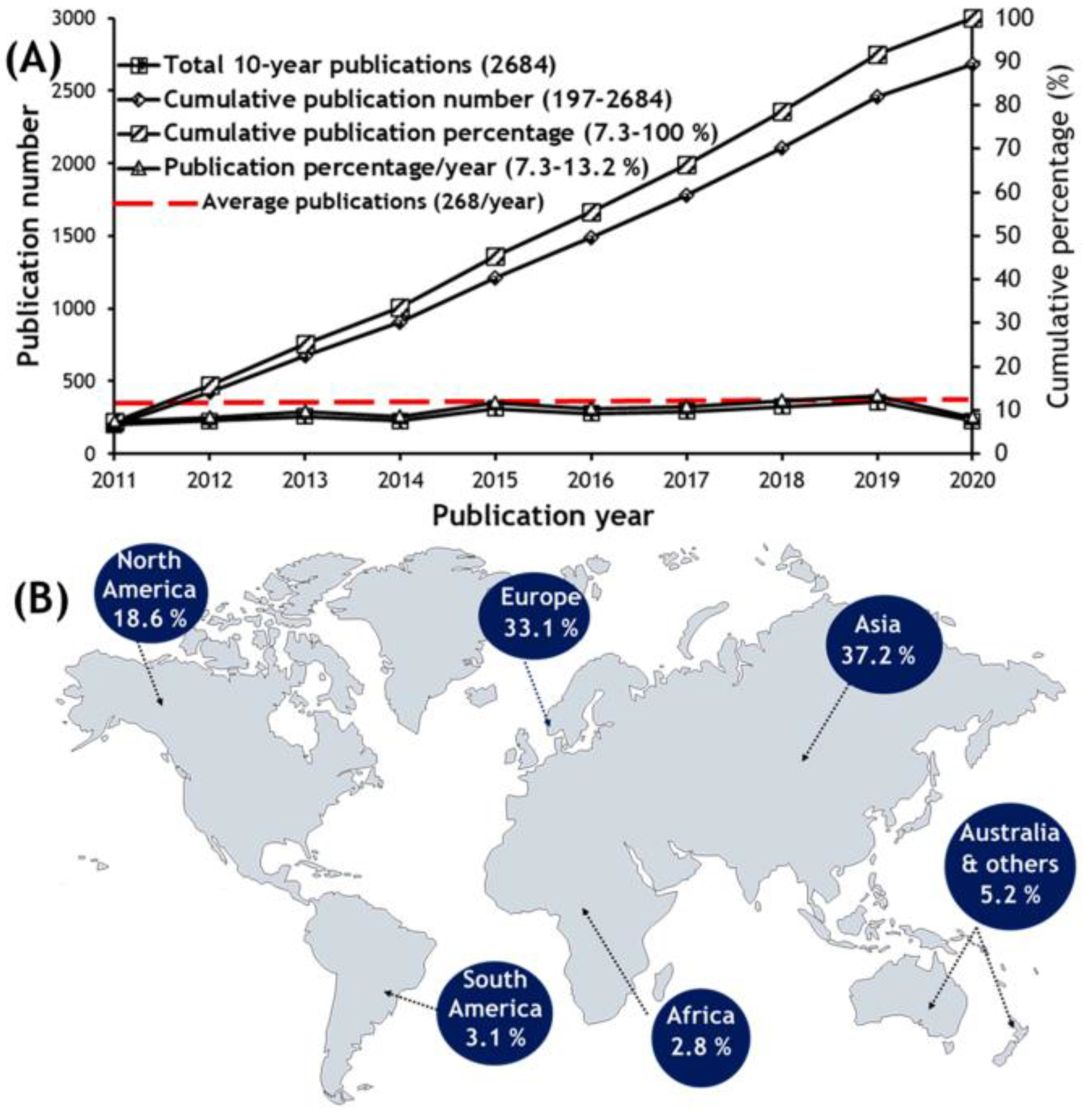
Figure 1. (A) Research trend on marine derived peptides from 2011 to 2020, and (B) the global distribution in terms of publication number and percentage. (Reprint with permission from [42], Copyright (2021) ELSEVIER).
Molecules 28 00615 g001
Figure 2. The steps generally used to determine the configurations of the amino acid residues of peptides by LC methods. RP-LC: reversed-phase liquid chromatography; HPLC: high-performance liquid chromatography; CSP: chiral stationary phase.
Molecules 28 00615 g002
Figure 3. Chemical structures of both enantiomers of 1-fluoro-2,4-dinitrophenyl-5-d,l-alanine amide (FDAA) and 1-fluoro-2,4-dinitrophenyl-5-d,l-leucine amide (FDLA) derivatizing agents (A); reaction of d/l-amino acids (dl-aa) with l-FDAA to give a pair of diastereomers (B).
Molecules 28 00615 g003
Figure 4. Chemical structures of macrocyclic antibiotic-based (A), crown ether-based (B), ligand exchange-type (C) chiral stationary phases.
Molecules 28 00615 g004
Figure 5. Time course of the racemization of lysine. Acid hydrolysate LDAA lysine derivatives: (a) 4 h hydrolysis; (b) 8 h hydrolysis; (c) 16 h hydrolysis. (Reprint with permission from [138], Copyright (2021) American Chemical Society).
Molecules 28 00615 g005
Figure 6. Chemical structures of marine-derived cyclopeptides with anticancer activity isolated from marine bacteria (111).
Molecules 28 00615 g006
Figure 7. Chemical structures of marine-derived cyclopeptides with anticancer activity isolated from marine cyanobacteria (1223).
Molecules 28 00615 g007aMolecules 28 00615 g007b
Figure 8. Chemical structures of marine-derived cyclopeptides with anticancer activity isolated from marine-derived fungi (2429).
Molecules 28 00615 g008
Figure 9. Chemical structures of marine-derived cyclopeptides with anticancer activity isolated from marine sponges and marine sponge-associated microorganisms (3043).
Molecules 28 00615 g009aMolecules 28 00615 g009b
Figure 10. Advanced Marfey’s method coupled with a UPLC-HRMS analysis of fuscasin A (35), using the amino acid standards l-Pro, l-Ala, l-Val, l-Phe, l-Leu, l-Tyr, l-Arg, and l-Asp, which were treated with l-FDLA and d-FDLA. (Reprint with permission from [163], Copyright (2019) American Chemical Society).
Molecules 28 00615 g010
Figure 11. Chemical structures of marine-derived cyclopeptides with antimicrobial activity (4459).
Molecules 28 00615 g011aMolecules 28 00615 g011b
Figure 12. Chemical structures of marine-derived cyclopeptides with other bioactivities (6074).
Molecules 28 00615 g012
Figure 13. Chemical structures of marine-derived cyclopeptides that demonstrated no biological activity (7591).
Molecules 28 00615 g013aMolecules 28 00615 g013b
Figure 14. Advanced Marfey’s method using HRESIMS of unguisin G (80), using suitable amino acid standards: l-Kyn, l-Val, l-Ala, l-Phe. FDAA: 1-fluoro-2,4-dinitrophenyl-5-d,l-alanine amide; FDLA: 1-fluoro-2,4-dinitrophenyl-5-d,l-leucine amide (Reprint with permission from [181], Copyright (2020) ELSEVIER).
Molecules 28 00615 g014
Figure 15. Distribution concerning: (A) the LC methods used for determination of the stereochemistry of marine-derived cyclopeptides, including the CSPs for chiral LC and CDRs for Marfey’s analysis; (B) the biological activities evaluated, highlighting the marine sources.
Molecules 28 00615 g015
Table 1. Structure, LC method for absolute configurations assignment to amino acid residues, and biological activities of new cyclopeptides (191), isolated from marine sources.
Marine
Peptide
Type of
Cyclopeptide
Marine
Source
Chromatographic
Method
Biological
Activities
Ref.
Bacilohydrin A (1)Cyclic lipoheptapeptide Bacillus sp. SY27F from the Indian ocean Hydrothermal VentModified Marfey’s method combined with UPLC-ESIMS: CDR: l-FDAA; Column: C18 (1.7 µm, 2.1 × 100 mm); MP: 0.05% NH4OH: CH3CN:0.01% HCO2H in elution gradient; Flow rate: 0.3 mL/min; Detection: ESIMSSignificant cytotoxicity against DU-145, MCF-7, and HepG2 cancer cell lines. [148]
Epoxinnamide (2)Cyclic decapeptideIntertidal Mudflat-Derived Streptomyces sp. OID44Advanced Marfey’s method combined with LC-MS: CDR: l-FDAA and D-FDAA; Column: C18 (5 µm, 4.6 × 100 mm): MP: CH3CN:H2O: 0.1% HCO2H in elution gradient; Flow rate: 0.7 mL/min; Detection: UV at 340 nm and MS Induced QR activity in murine Hepa-1c1c7 cells. Antiangiogenesis activity in HUVECs.[149]
Bathiapeptides A1 (3), A2 (4) and B-E (58)Cyclic hexapeptides Marine biofilm-derived Bacillus sp. B19-2Advanced Marfey’s method combined with UPLC-MS: CDR: l-FDLA or d-FDLA; Column: C18; MP: CH3CN:0.05% TFA in elution gradient; Detection: MSCytotoxicity against Hep G2, HeLa, MCF-7, and MGC-803 cell lines.[150]
Dentigerumycin E (9)Cyclic hexapeptide Coculture of marine Streptomyces and Bacillus strains isolated together from an intertidal mudflatAdvanced Marfey’s method combined with LC-MS: CDR: l-FDAA and d-FDAA; Column: Phenomenex Luna C18(2), (5 µm 4.6 × 100 mm); MP: CH3CN:H2O:0.1% HCO2H in elution gradient; Flow rate: 0.7 mL/min; Detection: UV at 340 nm and MS Moderate cytotoxicity against A549, HCT-116, MDA-MB-231, and SK-HEP-1 cancer cell lines.
Inhibition of MDA-MB-231 cell migration and cell invasion inhibition.
[151]
Nobilamide I (10)Cyclic depsiheptapeptideMarine-derived bacterium Saccharomonospora sp., strain CNQ-490C3 Marfey’s analysis combined with LC-ESI-MS: CDR: l-FDAA; Column: Agilent Zorbax SB-C3 (5 μm, 4.6 × 150 mm); MP: H2O:MeOH:0.02% HCO2H in elution gradient; Flow rate: 1 mL/min; Detection: UV at 340 nm and ESI-MS Inhibition of A549, AGS, and Caco2 cancer cell lines motility and tumorigenicity via suppressing EMT effectors and MMP2/9 expression.[152]
Nocardiotide A (11)Cyclic hexapeptideNocardiopsis sp. UR67 strain associated with the marine sponge Callyspongia sp.Marfey’s method combined with LC: CDR: l-FDAA; Column: Gemini-NX RP-C18; MP: H2O:CH3CN in elution gradient; Flow rate: 1 mL/min; Detection: UV at 340 nmCytotoxicity towards MM.1S, HeLa, and CT26 cell lines.[153]
Trikoramide A (12)Cyclic decapeptideMarine cyanobacterium Symploca hydnoidesMarfey’s method combined with LC-MS: CDR: l-FDVA; Column: Phenomenex Kinetex C18 (2.6 μm, 4.6 × 100 mm); MP: H2O:CH3CN:0.1% HCO2H in gradient elution; Flow rate: 0.2 mL/min; Detection: MSCytotoxicity against MOLT-4 and AML2 cancer cell lines.[154]
Trikoramides B–D (1315)Cyclic decapeptidesMarine cyanobacterium Symploca hydnoidesMarfey’s method combined with LC:
CDR: l-FDAA; Column: Phenomenex Kinetex C18 (2.6 µm, 4.6 × 250 mm,); MP: CH3CN: 0.05 M TFA (40:60); Flow rate: 0.5 mL/min; Detection: UV at 340 nm
Cytotoxicity against MOLT-4 cell line. QSI activity for 15 against PAO1 lasB-gpf and rhlA-gfp bioreporter strains.[155]
Triproamide (16) and pemukainalides A (17) and B (18)Cyclic depsiocta and hexapeptides Marine cyanobacterium Symploca hydnoidesMarfey’s method combined with HR-LCMS: CDR: l-FDAA; Column; Phenomenex Kinetex C18 (2.6 μm, 4.6 × 250 mm); MP: CH3CN:0.05 M TFA (40:60); Flow rate: 1.0 mL/min flow rate; Detection: MS
Chiral LC: Column: Phenomenex Chirex 3126 (d)-penicillamine (4.6 × 50 mm); CSP: ligand exchange-based; MP: 1 mM CuSO4:IPA (85:15); Flow rate: 1.0 mL/min; Detection: UV at 254 nm
17 exhibited cytotoxicity against the MOLT-4 cell line.[156]
Tutuilamides A–C (1921)Cyclic hexadepsipeptidesMarine cyanobacteria Schizothrix sp. (1920) and Coleofasciculus sp. (21)Marfey’s method combined with LC-MS:
CDR: l-FDAA or d-FDAA; Column: Phenomenex Kinetex C18 (5 μm, 4.6 × 100 mm); MP: H2O: CH3CN:0.1% HCO2H in elution gradient; Flow rate: 0.6 mL/min; Detection: UV at 220, 254 and 280 nm, and HRESIMS
Cytotoxicity in H-460 cell line. Potent elastase inhibitory activity.[157]
Laxaphycins B4 (22) and A2 (23)Cyclic lipoundeca-peptidesMarine cyanobacterium Hormothamnion enteromorphoidesChiral LC-MS: Column: Chirobiotic TAG (4.6 × 250 mm); CSP: Macrocyclic antibiotic-based; MP: MeOH:10 mM NH4OAc (40:60, pH 5.12) or (90:10, pH 6.0); Flow rate: 0.5 mL/min; Detection: ESIMS
Advanced Marfey’s method combined with LC-MS:
CDR: l-FDLA or dl-FDLA; Column: Phenomenex Kinetex C18 (2.6 μm, 2.1 × 100 mm) or Alltech Alltima C18 (5 μm, 4.6 × 250 mm), MP: H2O: CH3CN:0.1% HCO2H or MeOH:0.1% HCO2H, in elution gradient; Flow rate: 0.2 or 1.0 mL/min; Detection: MS
Antiproliferative effects of 22 against HCT-116 cell line, while 23 exhibited weak activity. [158]
Sclerotides C–E (2426) and scopularide I (27)Cyclic hexapeptides (2426) and lipodepsipeptide (27)Soft coral-derived fungus Aspergillus sclerotiorum SCSIO 41031Marfey’s method combined with LC-ESIMS/MS: CDR: l-FDAA; Column: YMC-Pack ODS-A (5 µm 4.6 × 250 mm); Mobile phase: CH3CN:H2O:0.03% TFA in elution gradient; Flow rate: 1 mL/min; Detection: UV at 340 nm and MS 27 showed cytotoxicity against HONE-EBV cancer cell line and AChE inhibitory activity.[159]
Asperflomide (28) and asperflosamide (29)Cyclic pentapeptide (28) and depsihexapeptide (29) Marine sponge-derived fungus Aspergillus flocculosus 16D-1Marfey’s analysis combined with UPLC-HRMS: CDR: d-/l-FDLA; Column: Acquity UPLC HSS T3 (1.8 µm; 2.1 × 100 mm); MP: CH3CN:H2O in elution gradient; Flow rate: 0.4 mL/min; Detection: HRMS Weak tankyrase1/2 inhibitory activity.[160]
Microsclerodermins N (30) and O (31)Cyclic hexapeptides Deep-sea marine sponge Pachastrella sp.Marfey’s analysis combined with LC-MS:
CDR: d-FDAA or l-FDAA; Column: COSMOSIL 2.5 π NAP (2.1 mm); MP: CH3CN:0.45% CH3CO2H in elution gradient; Flow rate of 0.5 mL/min; Detection: MS
Cytotoxic against HeLa cells.[161]
Theonellamides J (32) and K (33), and 5-cis-Apoa-theopalauamide (34)Bicyclic glycoundecapeptidesRed sea sponge
Theonella swinhoei
Advanced Marfey’s method combined with UPLC-MS: CDR: l,d-FDAA; Column: C18 (1.7 µm, 2.1 × 100 mm); MP: (A) H2O:CH3CN: 0.1% HCO2H in elution gradient; Flow rate: 0.5 mL/min; Detection: UV at 340 nm and ESIMS
Marfey’s method combined with HPLC:
CDR: FDAA; Column: LiChroCART RP-18 (5 µm, 4.6 × 250 mm); MP: aq. TFA buffer (pH 3):CH3CN in elution gradient; Flow rate: 1 mL/min; Detection: UV at 340 nm
Significant cytotoxicity against the HTC-116 cell line.[162]
Fuscasins A–D (3538)Cyclic heptapeptidesMarine sponge Phakellia fusca Advanced Marfey’s method combined with UPLC-HRMS: CDR: l-FDLA; Column: Acquity UPLC HSS T3 (2.1 × 100 mm, 1.8 μm); MP: CH3CN:H2O: 0.1% HCO2H in gradient elution; Flow rate: 0.4 mL/min; Detection: HRMS35 displayed growth-inhibitory activity against HepG2 cells.[163]
Ectyoplasin
(39)
Cyclic heptapeptideMarine sponge
Ectyoplasia ferox
Marfey’s method combined with LC:
CDR: l-FDLA; Column: BridgeVR C-18 (5 µm, 4.6 × 100 mm); MP: CH3CN:H2O:0.05% HCO2H in elution gradient; Flow rate: 0.8 mL/min; Detection: 340 nm
Cytotoxicity against DU-145, Jurkat, MM144, HeLa and CADO-ES1 cancer cell lines. Apoptotic cell death of DU-145 cell line. [164]
Phakefutatins A–C (4042)Cyclic heptapeptidesMarine sponge Phakellia fuscaAdvanced Marfey’s method combined with UPLC-HRMS: CDR: l-FDLA and d-FDLA; Column: Acquity UPLC HSS T3 (2.1 × 100 mm, 1.8 μm); Flow rate: 0.4 mL/min; Detection: MS40 is a RXRα modulator to inhibit cancer cell growth.[165]
Aciculitin D (43)Cyclic
lipopeptide
Marine sponge
Poecillastra sp. collected in the deep-sea
Marfey’s method combined with LC-MS:
CDR: d- or l-FDAA; Column: C18; MP: CH3CN:0.45% CH3CO2H in gradient elution; Detection: MS
Chiral LC: Column: CHIRALCEL OJ-RH; CSP: polysaccharide-based; MP: CH3CN:0.45% CH3CO2H in gradient elution
Cytotoxicity against HeLa and HCT-116 cells.[166]
Bacicyclin (44)Cyclic hexapeptideMarine Bacillus sp. strain associated with Mytilus edulisMarfey’s analysis combined with LC: CDR: l-FDAA; Column: Gemini-NX RP-C18; MP: H2O:CH3CN in elution gradient; Flow rate: 1 mL/min; Detection: UV at 340 nmAntibacterial activity against the E. faecalis JH212 and S. aureus NCTC 8325. [167]
Taeanamide A (45)Cyclic lipo-decapeptideIntertidal-mudflat-derived Streptomyces sp. AMD43Advanced Marfey’s method combined with LC-MS: CDR: FDAA; Column: Phenomenex Luna C18 (2) (5 µm, 4.6 × 100 mm); MP: H2O:CH3CN:0.1% HCO2H in gradient elution; Flow rate: 0.7 mL/min; Detection: UV at 340 nm and MSAnti-tuberculosis activity.[168]
Ogipeptins A–D (4649)Cyclic heptapeptidesMarine bacterium Pseudoalteromonas sp. SANK 71,903 by Daiichi SankyoAdvanced Marfey’s method combined with LC-MS: CDR: FDLA; Column: C30 (5 µm, 4.6 × 50 mm); MP: H2O:CH3CN:0.1% HCO2H in gradient elution; Flow rate: 2.5 mL/min; Detection: UV at 254 nm and MSAntimicrobial activity against E. coli. 4649 blocked LPS binding to CD14. [101,169]
Maribasins C–E (5052)Cyclic lipopeptidesMarine gorgonian-associated fungus Aspergillus sp. SCSIO41501Marfey’s method combined with LC:
CDR: FDAA; Column: YMC-Pack ODS-A
(5 μm, 250 × 4.6 mm); MP: CH3CN:H2O: 0.03% TFA in gradient elution; Flow rate: 1 mL/min; Detection: UV at 340 nm
Antifungal activity against phytopathogenic fungi A. solani, P. oryzae, C. australiensis, C. gloeosporioiles, F. oxysporum.[170]
Simplicilliumtides N (53) and O (54)Cyclic hexapeptides Deep-sea-derived fungal strain Simplicillium obclavatum EIODSF 020Marfey’s method combined with LC:
CDR: FDAA; Column: YMC-Pack ODS-A (250 × 4.6 mm, S-5 mm, 12 nm); MP: CH3CN/H2O/TFA in gradient elution; Flow rate: 1 mL/min; Detection: UV at 340 nm
Antifungal activity against phytopathogenic fungi A. solani and C. asianum.[171]
Acremonpeptides A–D (5558)Cyclic
hexapeptides
Marine fungus Acremonium persicinum SCSIO 115Marfey’s method combined with LC:
CDR: l-FDAA; Column: Prodigy ODS (2) (5 μm, 4.6 × 150 mm); MP: H2O:CH3CN:0.1% TFA in gradient elution; Flow rate: 1 mL/min; Detection: UV at 340
Antiviral activity against H. simplex virus 1, for 55 and 56. [172]
Motobamide (59)Cyclic decapeptide Marine cyanobacterium Leptolyngbya sp. Chiral LC: Column: CHIRALPAK MA (+) (4.6 × 50 mm); CSP: ligand exchange-based; MP: 2 mM CuSO4, and CH3CN:2 mM CuSO4 (15:85); Flow rate: 1.0 mL/min; Detection: UV at 254 nm Inhibition of the growth of bloodstream forms of T. brucei. [173]
Violaceotide A (60), and sclerotiotide L (61)Cyclic tetrapeptide (60)
and lipotripeptide (61)
Marine sponge-derived fungus Aspergillus violaceofuscusMarfey’s method combined with LC-MS: CDR: l-FDLA; Column: Waters XBridge C18 (5 µm, 4.6 × 250 mm); MP: H2O:CH3CN: 0.1% HCO2H in gradient elution; Flow rate: 1.0 mL/min; Detection: MSAnti-inflammatory activity against IL-10 expression of the LPS-induced THP-1 cells.[174]
Petrosamides A–C (6264)Cyclic hexapeptidesSponge-derived
Aspergillus sp. 151304
Advanced Marfey’s method combined with UPLC-MS: CDR: l-FDLA; Column: Waters HSS T3 (1.8 μm, 2.1 × 100 mm); MP: CH3CN:H2O: 0.1% HCO2H in gradient elution; Flow rate: 0.4 mL/min); Detection: MSPancreatic lipase inhibitory activity.[175]
Croissamide (65)Cyclic decapeptide Marine cyanobacterium Symploca sp.Chiral LC: Column: CHIRALPAK MA(+) (4.6 × 50 mm); CSP: ligand exchange-based; Mobile phase: different proportions of CH3CN:2 mM CuSO4; Flow rate: 1.0 mL/min; Detection: UV at 254 nmInhibitory activity against NO production in LPS-stimulated RAW 264.3 cells.[176]
Cystargamides C and D (6667)Cyclic lipohexadepsipeptides Marine actinomycete strain Streptomyces sp. JMS132Advanced Marfey’s method combined with LC-MS: CDR: l-FDLA or d-FDLA; Column: Phenomenex C18 (5 µm, 4.6 × 100 mm); MP: CH3CN:H2O: 0.1% HCO2H in gradient elution; Flow rate: 0.4 mL/min; Detection: ESIMSAntioxidant activity. 66 decreased DPPH free radicals. 67 decreased ABTS free radicals.[177]
Chrysogeamides A–G (6874)Cyclic hexadepsipeptidesCoral-derived fungus Penicillium chrysogenum CHNSCLM-0003Marfey’s method combined with HPLC-DAD and UPLC-MS: CDR: l-FDAA; Column: YMC C18 (5 µm, 2.1 × 250 mm) or ACQUITY UPLC BEH C18 (1.7 µm, 2.1 × 50 mm); MP: H2O:CH3CN: 0.1% HCO2H in gradient elution; Flow rate: 0.5 or 1.0 mL/min; Detection: DAD and MSPro-angiogenic activity towards Tg(kdrl:EGFP) transgenic zebrafish line.[178]
Auyuittuqamides A–D (7578)Cyclic decapeptidesSesquicillium microsporum RKAG 186 Marfey’s method combined with LC-HRMS: CDR: l-FDAA; Column: C18 (1.9 μm, 2.1 × 50 mm); MP: H2O:CH3CN:0.1% HCO2H in elution gradient; Flow rate: 0.4 mL/min; Detection: HRMSInactive against MCF-7 and HTB-26 cancer cell lines as well as against a human epithelial keratinocyte cell line. No antimicrobial activity was observed.[179]
Haloirciniamide A (79)Cyclic pentapeptide Indonesian marine sponge of the genus IrciniaMarfey’s method combined with LC/MS:
CDR: l-FDAA; Column: Waters Symmetry (3.5 µm, 4.6 × 150 mm); MP: H2O:CH3CN: 0.04% HCO2H in gradient elution; Flow rate: 0.8 mL/min; Detection: LC/MS
Low cytotoxicity against A-549, HT-29, MDA-MB-231, and PSN-1 tumor cell lines.[180]
Unguisin G
(80)
Cyclic heptapeptide Sponge-derived fungus Aspergillus candidus NF2412Advanced Marfey’s method combined with LC-HRESIMS: CDR: l- and d-FDAA; Column: Agilent Poroshell 120 EC-C18 (2.7 μm, 3.0 × 50 mm); MP: CH3OH:H2:O:0.1% TFA in gradient elution; Flow rate: 0.5 mL/min; Detection: HRESIMS No antimicrobial activity against a series of pathogens. [181]
Asperheptatides A–D (8184)Cyclic heptapeptidesCoral-derived fungus Aspergillus versicolorAdvanced Marfey’s method combined with LC: CDR: l-FDAA; Column: C18; MP: CH3CN:H2O in gradient elution; Flow rate: 1 mL/min; Detection: UV at 340 nm No antitubercular activity against M. tuberculosis H37Ra.[182]
Pagoamide A (85)Cyclic depsiundecapeptideCultured Marine Chlorophyte, Derbesia sp.Advanced Marfey’s method combined with LC-MS: CDR: d-FDAA; Column: YMC-Triart C18 (5 μm, 10 × 250 mm);
Detection: MS
Chiral LC: Column: Phenomenex Chirex 3126 (d)-penicillamine (5 μm, 4.6 × 250 mm); CSP: ligand-exchange based; MP: 2 M CuSO4; Flow rate: 2 mL/min; Detection: MS
No cytotoxicity against
H-460 cancer cell line.
[183]
Scopularides C–G (8690) and H (91)Cyclic lipopentadepsipeptides Marine sponge-derived fungus Beauveria sp. CMB-F585, and Scopulariopsis sp. CMB-F115C3 Marfey’s method combined with LC-DAD and MS: CDR: l and d-FDAA; Column: Agilent Zorbax SB-C3 (5 µm, 4.6 × 150 mm,); MP: H2O:MeOH: CH3CN: 0.1% HCO2H in gradient elution; Flow rate: 1.0 mL/min; Detection: DAD and ESIMSNo antimicrobial activity against a series of pathogens. No cytotoxicity against a panel of human carcinoma cell lines.[184]
A. solani: Alternaria solani; A549: lung cancer cell line; ABTS: 2,2-azino-bis(3-ethylbenzothiazoline-6-sulfonic acid; AChE: acetylcholinesterase; AGS: gastric cancer cell line; AML2: acute myeloid leukemia cell line; C. asianum: Colletotricum asianum; C. gloeosporioiles: Colletotrichum gloeosporioiles; C. australiensis: Curvularia australiensis; CADO-ES1: Ewing’s sarcoma cell line; Caco2: colorectal cancer cell line; CD14: cluster of differentiation 14; CDR: chiral derivatizing reagent; CSP: chiral stationary phase; CT26: colon carcinoma cell line; DAD: diode-array detector; DPPH: 2,2-diphenyl-1-picrylhydrazyl; DU-145: prostate cancer cell line; E. coli: Escherichia coli; E. faecalis: Enterococcus faecalis; EMT: epithelial-mesenchymal transition; ESI: electrospray ionization; F. oxysporum: Fusarium oxysporum; FDAA: 1-fluoro-2-4-dinitrophenyl-5-d,l-alanine amide; FDLA: 1-fluoro-2-4-dinitrophenyl-5-d,l-leucine amide; FDVA: 1-fluoro-2-4-dinitrophenyl-5-d,l-valine amide; H. simplex: Herpes simplex; H-460: lung cancer cell line; HCT-116: colorectal cancer cell line; HeLa: human cervical carcinoma cell line; Hepa-1c1c7: murine hepatoma cell line; HepG2: hepatocellular carcinoma cell line; HT-29: colon cancer cell line; HTC-116: colon cancer cell line; HUVEC: human umbilical vein endothelial cells; IPA: 2-propanol; Jurkat: T-cell acute leukemia cell line; LC: liquid chromatography; LPS: lipopolysaccharide; M. tuberculosis: Mycobacterium tuberculosis; MCF-7: breast cancer cell line; MDA-MB-231: breast cancer cell line; MGC-803: gastric carcinoma cell line; MM144: human multiple myeloma cell line; MM.1S: multiple myeloma cell line; MMP: matrix metalloproteinase; MOLT-4: acute lymphoblastic leukemia cell line; MP: mobile phase; MS: mass spectrometry; P. oryzae: Pyricularia oryzae; PSN-1: pancreas cancer cell line; QR: quinone reductase; QSI: quorum-sensing inhibitory; RXRα: retinoic X receptor-α; S. aureus: Staphylococcus aureus; SK-HEP-1: liver cancer cell line; SNU638: stomach cancer cell line; T. brucei: Trypanosoma brucei; TEAP: triethylammonium phosphate solution; TFA: trifluoroacetic acid; THP-1: acute monocytic leukemia cell line; UPLC: ultra-performance liquid chromatography; UV: ultra violet.
Disclaimer/Publisher’s Note: The statements, opinions and data contained in all publications are solely those of the individual author(s) and contributor(s) and not of MDPI and/or the editor(s). MDPI and/or the editor(s) disclaim responsibility for any injury to people or property resulting from any ideas, methods, instructions or products referred to in the content.